Molecular Characterization and Detection of Infectious Bronchitis Virus
←
→
Page content transcription
If your browser does not render page correctly, please read the page content below
Molecular Characterization and
Detection of Infectious Bronchitis Virus

                        Shahid Abro
        Faculty of Veterinary Medicine and Animal Science
  Department of Biomedical Sciences and Veterinary Public Health
                         Uppsala, Sweden

                 Doctoral Thesis
     Swedish University of Agricultural Sciences
                  Uppsala 2013
Acta Universitatis agriculturae Sueciae
2013:2
Cover: Illustration by Mikhayil Hakhverdyan (Misha)
(photo: Bengt Ekberg)
ISSN 1652-6880
ISBN 978-91-576-7759-4
© 2013 Shahid Abro, Uppsala
Print: SLU Service/Repro, Uppsala 2013Molecular Characterization and Detection of Infectious Bronchitis
Virus
Abstract
This thesis deals with the molecular characterization and detection of infectious
bronchitis virus (IBV), an important pathogen that causes heavy losses in the poultry
populations worldwide. The aim of the research was to better understand the molecular
characteristics of the virus and to investigate the factors behind the continuous
emergence of new genetic variants and the occurrence of outbreaks. The studies
showed that the viral genome is under a continuous evolution, due to mutations, strong
selective pressure and recombination events. These forces lead to a wide genetic
diversity and the generation of new variants of this virus. The viral genes encoding the
spike, replicase and nucleocapsid proteins can be considered the main genomic regions,
which are indicating the evolution processes of IBV. The various strains contain
specific structural and functional motifs in their genes and the alterations in these
motifs may affect the infection biology of the virus. The constant emergence of new
variants in Sweden is likely due to the introduction of novel IBV strains from other
European countries through the import of poultry products, or by the continuous
migration of wild birds.
The in silico investigations of the spike glycoprotein coding regions of the
Massachusetts and QX-like genotypes demonstrated molecular differences between
these genotypic variants. It is hypothesized that the genetic diversity in the spike gene
of IBV and of other avian coronaviruses, as well as of human, bat, and other animal
coronaviruses could be associated with the adaptation and host specificity of these
infectious agents.
The data obtained by molecular characterization approach was also used for the
development of a new molecular method for the improved detection and genotyping of
this virus. A microarray platform was developed for the simultaneous detection and
rapid typing of IBV variants. This assay provides a practical tool for better diagnosis,
for studying the effectiveness of vaccination and for performing large-scale
epidemiological studies.
Keywords: Infectious bronchitis virus, IBV, coronavirus, spike glycoprotein,
mutation, selective pressure, recombination, genetic diversity, bioinformatics,
multiplex, diagnosis, VOCMA, microarray
Author’s address: Shahid Abro, SLU, Department of Biomedical Sciences and
Veterinary Public health, Faculty of Veterinary Medicine and Animal Science,
P.O. Box 7028, SE-750 07, Uppsala, Sweden E-mail: shahid.abro@slu.seDedication
To my Parents, Wife Rani
and Little Princess Samreen
for their
Endless LoveContents
List of Publications 7
Abbreviations 8
1 Introduction 11
1.1 History of infectious bronchitis 11
1.2 Clinical features of infectious bronchitis 11
1.3 Host specificity 12
1.4 Mode of transmission 12
1.5 Infectious Bronchitis Virus 13
1.5.1 Morphology and genome organization 13
1.5.2 Untranslated regions 13
1.5.3 Replicase proteins 14
1.5.4 Spike glycoprotein 14
1.5.5 The 3a and 3b proteins 14
1.5.6 Envelope protein 14
1.5.7 Membrane protein 15
1.5.8 The 5a and 5b proteins 15
1.5.9 Nucleocapsid protein 15
1.6 Post-transcriptional and post-translational modifications and structural
motifs 15
1.6.1 Cleavage site motif 15
1.6.2 Palmitoylation 15
1.6.3 N-glycosylation 16
1.6.4 Phosphorylation 16
1.6.5 Leucine-rich repeat 16
1.7 Genetic forces 17
1.8 Serotypes and genotypes of Infectious Bronchitis Virus 17
1.9 Detection of Infectious Bronchitis Virus 19
2 Aims of the studies 21
3 Materials and methods 23
3.1 Samples and screening for Infectious Bronchitis Viruses 23
3.2 Isolation of Infectious Bronchitis Viruses 23
3.3 RNA extraction, synthesis of cDNA,PCR amplification and sequencing 23
3.4 Recombination analysis 243.5 Selection pressure 24
3.6 Analysis of functional and structural motifs 24
3.7 Phylogenetic analysis 25
3.8 Variation tolerant capture multiplex assay (VOCMA) 25
4 Results and discussion 27
4.1 Characterization and genetic diversity of the S gene of IBV (Papers I
and III) 27
4.2 Analysis of the full-length sequence of an emerging QX-like isolate of
IBV (Paper II) 29
4.3 Development of a VOCMA for broad detection and typing of IBV
(Paper IV) 30
5 Conclusions 33
6 Future prospects 35
7 Populärvetenskaplig sammanfattning 37
References 39
Acknowledgements 51List of Publications
This thesis is based on the work contained in the following papers, referred to
by Roman numerals in the text:
I Abro S. H., Renström, L. H. M., Ullman, K., Isaksson, M., Zohari, S.,
Jansson, D. S., Belák, S., and Baule C. (2012). Emergence of novel strains
of avian infectious bronchitis virus in Sweden. Veterinary Microbiology
155: 237–246.
II Abro, S. H., Renström, L. H. M., Ullman, K., Belák, S. and Baule, C.
(2012). Characterization and analysis of the full-length genome of a strain
of the European QX-like genotype of infectious bronchitis virus. Archives
of Virology 157: 1211–1215.
III Abro, S. H., Ullman, K., Belák, S. and Baule, C. (2012). Bioinformatics
and evolutionary insight on the spike glycoprotein gene of QX-like and
Massachusetts strains of infectious bronchitis virus. Virology Journal
9:211.
IV Öhrmalm, C., Abro, S. H., Baule, C., Zohari, S., Bálint, A., Renström, L.
H. M., Blomberg, J. and Belák, S. Variation-tolerant capture multiplex
assay (VOCMA) for the simultaneous detection of avian infectious
bronchitis virus genotypes using multiplex RT-PCR and Luminex
technology (Manuscript).
Papers are reproduced with the permission of the publishers.
7Abbreviations
AGPT Agar gel precipitation test
AI Aliphatic index
Conn Connecticut
DNA Deoxyribonucleic acid
cDNA Complementary deoxyribonucleic acid
dNS Non-synonymous substitution
dS Synonymous substitution
E Envelope
ELISA Enzyme-linked immunosorbent assay
HI Haemagglutination inhibition
IBV Infectious bronchitis virus
IFA Immunofluorescence assay
IPA Immunoperoxidase assay
LRR Leucine-rich repeat
M Membrane
Mass Massachusetts
mRNA Messenger RNA
MFI Median fluorescence intensity
N Nucleocapsid
NJ Neighbor-Joining
Nsp Non-structural protein
nt Nucleotide
ORFs Open reading frames
PLpro Papain-like protease
RNA Ribonucleic acid
RDP Recombination detection program
RdRp RNA-dependent RNA polymerase
RT-PCR Reverse transcriptase polymerase chain reaction
8S Spike
SPF Specific pathogen-free
sgRNA Subgenomic RNA
UTRs Untranslated regions
VNT Virus neutralization test
VOCMA Variation-tolerant capture multiplex assay
VT Variation-tolerant
910
1     Introduction

1.1 History of infectious bronchitis
Infectious bronchitis (IB) is a highly contagious disease of serious economic
importance in the poultry industry worldwide. The first report of IB by Schalk
and Hawn referred to a highly contagious disease in young chicks with
respiratory symptoms in North Dakota, USA in 1931 (Schalk & Hawn, 1931).
Pathogenic alterations in the upper respiratory tract of the birds were
prominent; hence the disease was named “infectious bronchitis of young
chicks”. Five years later, it was demonstrated that the causative agent of this
disease is a virus, which was named Infectious Bronchitis Virus (IBV) (Beach
& Schalm, 1936). After the initial description of infectious bronchitis, many
cases of the disease were reported in the United States (Jungherr et al., 1956;
Hitchner et al., 1966; Johanson et al., 1973; Snyder & Marquardt, 1984;
Fabricant, 1998; Hitchner, 2004). Thereafter and to date, a wide range of
different IBV serotypes and genotypes have been detected around the world
(Jackwood et al., 1997; de Wit et al., 2011).

1.2 Clinical features of infectious bronchitis
Clinical cases of IB are associated with respiratory, reproductive, digestive and
renal infections in domestic poultry and in various other avian species
(Cavanagh, 2005). The disease is clinically manifested by coughing, sneezing,
tracheal coarse crackles, nasal discharge, decrease of feed intake and
conversion, loss of body weight, swollen sinuses, increased water intake, wet
droppings, depression, lethargy and poor growth in broilers. In layers the
disease, “false layer syndrome”, affects egg quality (thin, rough, fragile,
misshapen egg shells and thin watery egg) and causes decrease in egg
production. In some cases, the virus infection may cause severe damage to the

                                                                              11
oviduct and result in decreased or permanent loss of egg production (Otsuki et al., 1990; Fabricant, 1998; Cavanagh, 2003; Cavanagh & Naqi, 2003; Cavanagh & Gelb, 2008; Worthington et al., 2008). IB infections may lead to mortality up to 20-30% or higher at five to six weeks of age in chicken flocks (Ignjatovic et al., 2002; Seifi et al., 2010). The mortality can increase due to immunosuppression, mycoplasma and other secondary bacterial infections caused by various accompanying infectious agents, e.g. Escherichia coli, Ornithobacterium rhinotracheale and Bordetella avium (Hopkins & Yoder, 1984; Matthijs et al., 2003; Cavanagh & Gelb, 2008). The mortality rate can be as low as 1% and chickens may recover rapidly, if the infections are produced by mildly virulent strains and are not associated with secondary bacterial infections (Cavanagh & Gelb, 2008). 1.3 Host specificity IBV infects a wide range of avian species, especially those reared close to domesticated poultry, for example domestic fowl, partridge, geese, pigeon, guinea fowl, teal, duck and peafowl (Cavanagh, 2005; Cavanagh, 2007). In different hosts, the virus exhibits considerable similarities in its genome. For example, a virus that was isolated from teal and peafowl shared 90-99% sequence related to IBV (Liu et al., 2005). Evidence based on the nucleotide sequences of viruses isolated from samples of ducks, whooper swans, turkeys and pheasants have also shown high similarity to IBV (Breslin et al., 1999; Guy, 2000; Cavanagh et al., 2001; Jonassen et al., 2005; Hughes et al., 2009). 1.4 Mode of transmission IBV is a highly infectious pathogen, and the infected birds usually develop clinical signs very rapidly, within 36-48 hours. The virus replicates primarily in the upper respiratory tract, leading to viraemia, and then spreads to other organs (Raj & Jones, 1997). Usually, the virus is present in high concentrations in the upper respiratory tract during the first 3-5 days post infection (Cook, 1968; El-Houadfi et al., 1986). In general, a large amount of virus is detected in tracheal mucus and faeces during the acute and recovery phases of the disease. In some cases IBV persists as a latent infection, and the carrier birds continue to shed virus particles via faeces. The virus is transmitted horizontally by the contaminated feed, water or faeces. Infected birds shed the virus continuously in the environment and contaminate their surroundings, such as equipment, eggs, also working personnel and trucks, among others, which are the major sources of indirect transmission to different regions (Ignjatović & 12
Sapats, 2000). Wild birds may play a crucial role as reservoirs and long-
distance carriers of IBV (Chen et al., 2009; Hughes et al., 2009).

1.5 Infectious Bronchitis Virus

1.5.1 Morphology and genome organization
IBV belongs to the order Nidovirales, family Coronaviridae, genus
Gammacoronavirus (Gonzalez et al., 2003). The enveloped viral particles are
round and pleomorphic in shape. The virions are approximately 120 nm in
diameter and contain club-shaped surface projection called spikes, which are
20 nm in size (Cavanagh & Gelb, 2008). The positive sense RNA genome is
approximately 27.6 kb in size and is encompassing 5′ and 3′ untranslated
regions (UTRs) with a poly(A) tail (Figure 1) (Boursnell et al., 1987; Ziebuhr
et al., 2000; Mo et al., 2012). A major part of the genome is organized as two
overlapping open reading frames (ORFs), 1a and 1b, which are translated into
large polyproteins 1a and 1ab through a ribosomal frame shift mechanism. The
remaining part of the genome consists of regions coding for the main structural
proteins spike (S), envelope (E), membrane (M) and nucleocapsid (N). Two
accessory genes have been described, ORF3 and ORF5, that express accessory
proteins 3a & 3b and 5a & 5b, respectively (Lai & Cavanagh, 1997; Pasternak
et al., 2006).

Figure 1. A schematic genome organization of the infectious bronchitis virus

1.5.2 Untranslated regions
The small structural motifs of the IBV genome comprise 5 and 3 untranslated
regions (UTRs), which mediate physical interactions between UTRs, viral
encoded replicase proteins and host cellular proteins (Li et al., 2008).

                                                                               13
1.5.3 Replicase proteins Two-thirds of the genome consists of ORF1a and ORF1b that encode polyproteins 1a and 1ab, respectively, and contribute to formation of the replication and transcription complex (Imbert et al., 2008). These polyproteins are post-transitionally cleaved to generate 15 non-structural proteins (Nsp2- 16), comprising a main protease called 3C-like protease, a papain-like protease (PLpro), an RNA-dependent RNA-polymerase (RdRp) and other non-structural proteins (van Hemert et al., 2008). 1.5.4 Spike glycoprotein The spike glycoprotein of all coronaviruses contains four domains that are involved in anchoring of the S protein into the lipid bilayer of the virion. The IBV S gene consists of 1162 amino acids, and is cleaved into two sub-units, the N-terminal S1 subunit (535 amino acids) and the C-terminal S2 subunit (627 amino acids). The S1 subunit contains virus neutralization and serotype- specific antigenic determinants that are responsible for binding to the host cell, neutralization and immune response (Koch et al., 1990; Schultze et al., 1992; Ignjatovic & Galli, 1994; Johnson et al., 2003). The nucleotide sequence variation in the spike gene may result in a lower cross protection between serotypes. The high variation in the nucleotide sequences of spike gene can change the protection ability of a vaccine or immunity (Cavanagh, 2003; Cavanagh & Gelb, 2008). The S2 sub-unit contains a fusion peptide-like region and two heptade regions approximately 100 to 130 Å in length (771-879 amino acid in IBV) that are involved in oligomerisation of the protein and entry into susceptible host cells (Tripet et al., 2004; Guo et al., 2009; Shulla & Gallagher, 2009). 1.5.5 The 3a and 3b proteins Gene 3 contains two ORFs (ORF3a and ORF3b) that are functionally tricistronic in nature (Liu et al., 1991). ORF3a and ORF3b contain highly conserved nucleotide sequence regions (Mo et al., 2012), not only in IBV but also in other gammacoronaviruses (Cavanagh et al., 2001; Cavanagh et al., 2002). 1.5.6 Envelope protein The envelope protein is a small integral membrane protein associated with the envelope of the virions (Smith et al., 1990; Liu & Inglis, 1991). It has been demonstrated that the E protein is essential for virus assembly (Maeda et al., 1999). Mutations in the amino acid sequence of the E protein in coronaviruses 14
may considerably affect the assembling of the virus in cells (Fischer et al.,
1998).

1.5.7 Membrane protein
The largest portion of the integral membrane protein is embedded within the
lipid bilayer, which maintains the structural integrity (Godet et al., 1992). The
M protein is responsible for the organization and assembling of the virus
particle by interactions with other structural proteins (Vennema et al., 1996;
Hogue & Machamer, 2008).

1.5.8 The 5a and 5b proteins
Gene 5 is encompassing two ORFs (ORF5a and ORF5b) that encode the 5a
and 5b proteins, respectively (de Vries et al., 1997). It has been demonstrated
that these proteins are functionally bicistronic (Pendleton & Machamer, 2005).

1.5.9 Nucleocapsid protein
The N protein consists of 409 amino acids. This protein is involved in a variety
of functions such as viral packing, assembly, viral core formation, signal
transduction and modulating host cell processes (Drees et al., 2001; He et al.,
2004; You et al., 2007).

1.6 Post-transcriptional and post-translational modifications and
    structural motifs
The post-transcriptional and post-translational modifications such as cleavage,
palmitoylation, N-glycosylation, phosphorylation and structural motifs
Leucine-rich repeat have important role in virus biology. Therefore, there is
need to explore these modifications and structural motifs especially by
application bioinformatics, to identify the differences in the spike glycoprotein
of the virus variants, in order to bridge a platform for the in-vivo and in-vitro
studies, which is part of scope of the present thesis.

1.6.1 Cleavage site motif
The spike glycoprotein contains a protease cleavage site motif that is involved
in cleavage of the S1 and S2 sub-units during viral maturation (Cavanagh et al.,
1986). The cleavage site motif of IBV comprises one or two pairs of basic
amino acids that is cleaved by host cell serine proteases (Cavanagh et al.,
1992). The serine proteases of host cell catalyze hydrolyses reactions that
cause cleavage of peptide bonds (Voet & Voet, 1990).

                                                                              15
1.6.2 Palmitoylation Palmitoylation is the binding of organic molecules, e.g., palmitic acid, to the cysteine residues of membrane proteins (Linder, 2000). Palmitoylation plays an important role in localization and transport at the sub-cellular level, in protein- protein interactions, and in various physiological characteristics of proteins in the virus (Dunphy & Linder, 1998; Dietrich & Ungermann, 2004). The carboxyl- terminal cysteine peptides of viral membrane glycoproteins act as crucial palmitoylation sites (Ponimaskin & Schmidt, 1995; Petit et al., 2007). In coronaviruses, palmitoylation of viral glycoproteins affects the fusion to cellular membranes, viral assembly and infection of cells (Bos et al., 1995; Petit et al., 2007). 1.6.3 N-glycosylation The N-glycosylation characteristics of viral glycoprotein are associated with changes of virulence and cellular tropism (Li et al., 2000). Glycosylation in the spike and membrane glycoproteins of coronaviruses is involved in fusion, receptor binding and antigenic characteristics (Alexander & Elder, 1984; Braakman & van Anken, 2000; de Haan et al., 2003; Wissink et al., 2004). Variation in N-glycosylation sites can affect the interaction with receptors, and thus render a virus more susceptible to host innate immune responses and lower recognition by antibodies, affecting virus replication and infectivity (Meunier et al., 1999; Land & Braakman, 2001; Slater-Handshy et al., 2004; Vigerust & Shepherd, 2007). 1.6.4 Phosphorylation Protein phosphorylation plays a crucial role in the regulation of functional activities in microorganisms (Ingrell et al., 2007). For IBV, two phosphorylated clusters (Ser190 & Ser192 and Thr378 &Ser379) that were identified by a mass spectroscopic method, are located in the middle and C- terminal regions of the N protein (Chen et al., 2005). The C-terminal phosphorylation clusters are associated with the differentiation of viral RNA from non-viral RNA (Spencer et al., 2008). 1.6.5 Leucine-rich repeat The leucine-rich repeat (LRR) domain is contained in microbial proteins and is associated with innate immunity (Huang et al., 2008). Beyond innate immunity, LRR-containing proteins are associated with various cellular processes including apoptosis, ubiquitin-related processes and nuclear mRNA transport (Kobe & Kajava, 2001; Wei et al., 2008). 16
1.7 Genetic forces
The genetic forces such as mutations, recombination and selective pressure
play significance role in the viral genome evolution (Zhang et al., 2006; Liu et
al., 2007). In order to better understand the contribution of these forces in
genetic diversity of IBV, these were outlined as subjects of thesis.
Recombination commonly occurs between two or more viruses infecting
the same cell. It is believed that a high rate of recombination events occurs in
the genomes of non-segmented RNA viruses (Makino et al., 1986), such as in
IBV and in other coronaviruses (Lim et al., 2011; Thor et al., 2011; Jackwood
et al., 2012). In RNA viruses, a unique copy choice mechanism during
polymerase activity allows a high efficiency of RNA recombination (Liao &
Lai, 1992). Moreover, recombination, along with the polymerase error rate, is
involved in molecular mechanisms that are likely to be responsible for
changing tissue and host species tropism of the virus (Graham & Baric, 2010).
In the case of coronaviruses, mutations and selective pressure in genes,
especially in hypervariable regions, enable the virus to cross the species barrier
and adapt to new host species, hence contributing to viral evolution (Zhang et
al., 2006; Liu et al., 2007). It has been demonstrated that during the multiple
passages of IBVs in embryonated eggs, mutations and recombination result in
strong selective pressure in the S1 gene, leading to the generation of attenuated
viruses (Liu et al., 2007).
1.8 Serotypes and genotypes of Infectious Bronchitis Virus
A number of IBV serotypes, e.g. Arkansas, Connecticut (Conn), Massachusetts
(Mass), 4/91 or 793B, D274, H120, Italy-02, Baudette, California, Georgia 98
and QX have been identified and reported around the globe (Lee & Jackwood,
2001; Jackwood et al., 2003; Sjaak de Wit et al., 2011). Serotyping of IBV
strains is usually carried out by test systems based on chicken-induced IBV
serotype-specific epitope(s) (Koch et al., 1990; Keeler et al., 1998). The
appearance of a large number of IBV variants in different regions is a major
constraint for the practical application of serotyping (Sjaak de Wit et al., 2011).
It is important to note that serotyping of IBV is becoming less and less
common today, since the serological tests require a panel of serotype specific
antibodies, which is not easy to obtain. Thus, recently the typing of the virus is
mostly performed by various molecular methods as many types of the virus are
diagnosed by molecular methods.
Genotyping of IBV is based on the amplification of the highly variable
sequence region of the S1 gene by reverse transcriptase polymerase chain
reaction (RT-PCR), followed by sequencing (Jackwood et al., 1992; de Wit,
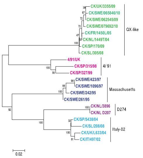
172000). The molecular diagnostic assays revealed that different genotypes, such as Massachusetts, 4/91, D274, Italy-02 and European QX-like, are circulating in European countries (Figure 2) such as Belgium, Denmark, France, Hungary, Italy, Germany, The Netherlands, Poland, Russia, Slovenia, Spain, Sweden and UK (Davelaar et al., 1984; Parsons et al., 1992; Farsang et al., 2002; Jones et al., 2005; Bochkov et al., 2006; Domanska-Blicharz et al., 2006; Worthington & Jones, 2006; Dolz et al., 2008; Worthington et al., 2008; Benyeda et al., 2009; Handberg et al., 2009; Valastro et al., 2010; Krapez et al., 2011; Abro et al., 2012a; Abro et al., 2012b). Figure 2. A phylogenetic tree, based on the comparative analysis of S1 gene sequences of different strains obtained from various countries, is showing the occurrence of different genotypes of IBV in Europe. 18
1.9 Detection of Infectious Bronchitis Virus
Due to constant evolution, there is continuous emergence of new IBV variants
all over the world. For example, different genotypes such as Massachusetts,
4/91, D274, Italy-02 and European QX-like are currently circulating in Europe.
Various methods are used for the detection and identification of IBV. The most
common assays for routine diagnosis are virus isolation, haemagglutination
inhibition    (HI),   enzyme-linked       immunosorbent      assay    (ELISA),
immunoperoxidase assay (IPA), virus neutralization test (VNT),
immunofluorescence assay (IFA), agar gel precipitation test (AGPT), RT-PCR
and real-time RT-PCR (de Wit, 2000). The majority of the conventional
diagnostic assays in fact are time- and material-consuming, laborious, costly
and provide relatively low specificity and sensitivity (King & Hopkins, 1983;
Mockett & Cook, 1986; Karaca & Naqi, 1993; Naqi et al., 1993; De Wit et al.,
1995; de Wit et al., 1997; de Wit, 2000). Various real-time RT-PCR assays
have been recently elaborated in veterinary medicine for the diagnosis of a
variety of infectious pathogens (Belák, 2007). Accordingly, a range of real-
time RT-PCR assays has been developed for the specific detection of IBV
using various approaches, such as the TaqMan technology. In the general real-
time RT-PCR technique is highly sensitive and it can be applied not only for
the rapid detection and identification of IBV, and even to quantify genomic
RNA in clinical samples (Callison et al., 2006). On the other hand, sometimes
even the real-time RT-PCR assay may lead to poor results due to limitations in
detecting the divergent or the new virus variants. In addition, being based on
the conserved 5’ UTR or the nucleocapsid genes, the amplicons obtained by
diagnostic PCR assays are not suitable for determining the types of the viruses.
The virus typing, in majority of the cases is based on the comparative analysis
of nucleotide sequences of the variable S gene.
       Considering that there are no reliable and practical assays available
today, that can simultaneously detect and type IBV variants; the development
of such an assay was contemplated in the present work. By applying variation-
tolerant detection chemistry and the Luminex technology, we have developed a
new tool for the rapid detection and accurate identification of various
genotypes of IBV, including newly emerging variants of this virus.

                                                                             19
20
2 Aims of the studies
The overall objectives of the present studies were to characterize various
strains of IBV, to determine genetic diversity and to investigate the factors
driving the evolution of this virus. A further task was, to develop a molecular
diagnostic assay, in order to improve the detection and typing of IBV variants
causing disease outbreaks in Sweden and in other European countries.
The specific aims were:
To perform molecular characterization of IBV isolates from Sweden
based on the comparative sequence analysis of the complete spike
gene, in order to better understand the epidemiology and the factors
behind the occurrence of new outbreaks (Paper I).
To study the full-length genome of the European QX-like isolate of
IBV, detected in Sweden, by sequencing and phylogenetic analysis
(Paper II).
To compare the molecular characteristics of isolates belonging to the
classical Massachusetts and emerging QX-like genotypes of IBV in
order to determine differences in potential functional and structural
motifs of the spike gene that may relate to the biological properties of
the virus. In addition, a comparative analysis of the complete spike
gene was performed based on sequences of avian and mammalian
coronaviruses (Paper III).
To develop a specific assay for the simultaneous detection and typing
of the QX-like, Italy-02, M41, 4/91 and D274 genotypes of IBV,
which are currently circulating in Europe (Paper IV).
2122
3     Materials and methods

3.1 Samples and screening for Infectious Bronchitis Viruses
Clinical samples (trachea, bronchi, ceaca) were obtained from different
outbreaks in Sweden (Papers I, II, III and IV). The D274, Italy-02 and 4/91
isolates kindly provided from the Netherlands, Slovenia and France (Merial)
were used in the study (Paper IV). The samples were screened by real-time
RT-PCR assay as described by Callison et al. (2006) for the presence of IBVs
(Papers I and IV).

3.2 Isolation of Infectious Bronchitis Viruses
The real-time RT-PCR positive IBV samples were propagated in specific
pathogen-free (SPF) embryonated hen’s eggs. Virus isolation was performed
by inoculation of 9–11 day old eggs with 200 µl of 10% tissue homogenates
and incubation at 37 oC for 72 hours. The allantoic fluid was harvested, and
isolates were stored at -20 oC for the further use.

3.3 RNA extraction, synthesis of cDNA, PCR amplification and
    sequencing
The viral RNA was extracted using the TRIzol reagent according to the
manufacturer’s instructions (Invitrogen, Carlsbad, USA) (Papers I, II, and IV).
Synthesis of cDNA was performed with gene specific primers using
SuperScript II reverse transcriptase (InvitrogenTM Life technologies, Carlsbad,
USA) as recommended by the manufacturer (Papers I and II). PCR
amplification and sequencing were performed for the S gene and also for
fragments covering the whole genome (Papers I, II and IV). Different pairs of
primers were used for amplification and sequencing of the full-length genome

                                                                            23
as reported by Liu et al. (2009) (Paper II). The sequencing reactions were performed by using Big Dye terminator sequencing kit (Applied Biosystems, Foster City, CA), as recommended by the manufacturer (Papers I, II, and IV). For sequence analysis, the sequence data set was pair wise edited and aligned with the software Lasergene DNASTAR (Madison, USA) (Papers I, II and IV). 3.4 Recombination analysis The recombination analyses were performed on the complete gene sequences using the Recombination Detection Program (RDP v3.44) (Martin et al., 2010). Different methods available in the program were applied in order to compare and accurately determine the positions of the recombination hot spots and to differentiate closest fragment of non-recombinant sequences (Ohshima et al., 2007). The marker positions were indexed for the determination of recombinant clusters (Papers I and II). 3.5 Selection pressure Evidence of selective pressure in individual genes was examined by using the SNAP (Korber et al., 2000) services available at web server http://hcv.lanl.gov/content/sequence/SNAP/SNAP.html (Papers I and II). The differences between synonymous (dS) and non-synonymous (dNS) amino acid substitutions were calculated in order to determine the substitution rate in the analyzed gene(s) individually (Papers I and II). 3.6 Analysis of functional and structural motifs As reported in Paper III, N-glycosylation sites were predicted using services available at web server http://www.cbs.dtu.dk/services/NetNGlyc. The potential phosphorylation sites were determined by using the website http://www.cbs.dtu.dk/services/NetPhos. The cleavage site motif analysis was performed by the traditional approach using the amino acid position in the sequence of the spike glycoprotein, as previously described by (Parker & Masters, 1990; Jackwood et al., 2001). LRR regions were identified by LRR finder, available at http://www.lrrfinder.com/result.php. The primary structures of the spike glycoprotein were predicted by http://expasy.org/tools. The secondary structures were predicted by the GOR4 method using services at http://npsa-pbil.ibcp.fr/cgi-bin/npsa_automat.pl?page=npsa_gor4.html. The palmitoylation sites were determined with the medium threshold frequency using web domain http://csspalm.biocuckoo.org/prediction.php. 24
3.7 Phylogenetic analysis
As described in Papers I, II and III, phylogenetic analysis was performed using
sequences generated in these studies, as well as sequence data downloaded
from the GenBank database. Phylogenetic relationship was determined using
both parsimony and nucleotide distance methods. The nucleotide distance
matrix between sequences was used to construct a phylogeny by Neighbor-
Joining (NJ) using MEGA 4 (Tamura et al., 2007). The NJ method was applied
because it is relatively fast and can be suitable for large data sets (Tamura et
al., 2004). The sequences were compared by using the gamma Tamura–Nei
model (Tamura & Nei, 1993). Bayesian analyses were performed by using
MrBayes 3.1 software (Ronquist & Huelsenbeck, 2003).

3.8 Variation-tolerant capture multiplex assay (VOCMA)
As reported in Paper IV, synthetic targets were designed as an alternative
positive control for each genotype: M41, D274, 4/91, Italy-02, QX-like,
IBV_Capsid (consensus). The hybridization events of variation tolerant (VT)
were designed using the NucZip algorithm (Öhrmalm et al., 2010; Öhrmalm et
al., 2012).
       The amplification of target nucleic acid by VOCMA, executed as single-
tube one-step multiplex amplification, was performed using long specific VT
primer-probes and short generic primers, with the biotinylated generic second
primer. The extracted nucleic acid or synthetic ssDNA target was added
directly to a mastermix one-step RT-PCR mixture. Specific synthetic 5 amino-
C12 modified detection probes for the different IBV genotypes were designed
and coupled to xMAP carboxylated colour-coded microspheres (Luminex
Corp., Austin TX, USA) according to the manufacturer’s instructions
(Luminex Corporation, Austin TX, USA). The biotin-labeled amplified
template was mixed with hybridization buffer of each probe-coupled xMAP
bead. The IBV VOCMA has six unique beads, each coupled with either six
specific VT detection probes of 48-62 nt (long probes) or of 33-36 nt (short
probes). The mixture was treated at 95 ºC for 2 min, followed by hybridization
at 50 ºC for 30 min with shaking at 600 rpm on a Thermostar (BMG LabTech;
Offenburg, Germany) microplate incubator. After a short centrifugation, 40 µL
of supernatant was discarded and a mixture of 38µL 3M TMAC-TE
hybridization buffers + 2 µL of Streptavidin-R-phycoerythrin (QIAGEN,
Hilden, Germany) was added. The tubes were further incubated at 50 ºC for 15
min, before analysis for internal bead and R-phycoerythrin reporter
fluorescence on the Luminex®200™ flow meter (Luminex corporation, Austin

                                                                             25
Tx). The quantity of biotinylated target that hybridized to the probe-linked beads was measured as Median Fluorescence Intensity (MFI). 26
4     Results and discussion

4.1 Characterization and genetic diversity of the S gene of IBV
    (Papers I and III)
Outbreaks of infectious bronchitis have been detected in Sweden between 1995
and 2010. Twenty samples originating from these outbreaks were selected for
molecular characterization based on analysis of sequences of the complete S
gene (Paper I). The viruses collected in 1995-1999 showed nucleotide
sequence difference of varying degree 18.7-21% in comparison to the isolates
from 2009-2010. The isolates from the 1990s shared lower identities to the
viruses collected in the 2000s, reflecting the genetic signature of new variants.
Considerable sequence variation was found in the region encompassing the S1
part of the S gene. In the S2 subunit, regions of sequence variation were found
interspaced with regions of high conservation, also contributing to overall
diversity of the S gene. The variation in nucleotide sequences of the S1 region
may lead to alter the virus characteristics such as epitopes and receptor binding
abilities. It has been reported that comparative analysis of the hypervariable
region of the S1 nucleotide sequences is a suitable tool for the discrimination
of various IBV field variants (Kusters et al., 1987; Kwon et al., 1993; Wang &
Huang, 2000). There were nucleotide insertions and deletions at different
locations in the spike gene of isolates from the 2000s in comparison to isolates
from the 1990s. The nucleotide insertions, deletions and point mutations in the
S gene contribute to evolution of IBV (Kusters et al., 1990; Lai, 1992; Kottier
et al., 1995). The significant substitutions in the S1 region warrant further
investigation to ascertain their relevance, if any, in the virus biology.
        Substitutions in the coding sequences are likely attributable to strong
positive selection pressures in the spike gene. Strong selective constraints
could affect the primary and secondary structures of the gene, which may lead
to alteration of genetic and molecular features of the virus. It has been reported

                                                                               27
that positive selection in IBV can lead to emergence of new strains that are
capable of escaping the immune system (Dolz et al., 2008).
Recombination events were detected in the spike gene sequences of
isolates from 2009-2010 (Paper I). This is an important information,
considering the fact that genetic recombination among heterologous IBVs
could lead to the emergence of new variants, and result in genetic diversity
(Kottier et al., 1995; Lee & Jackwood, 2000; Lim et al., 2011). Recombination
is believed to decrease the mutation rate and associated constraints which are
responsible for the genetic diversification that results in emergence of new
variants of coronaviruses (Worobey & Holmes, 1999).
Taken together, point mutations, strong selective pressure and to some
extent recombination events in the spike gene are contributing factors to the
generation of new emerging variants and genetic diversity that leads to
constant evolution of the virus.
Phylogenetic analysis based on partial S1 and complete S gene
sequences revealed four distinct clusters: Massachusetts, QX-like, Italy-02 and
4/91 genotypes. Swedish isolates from 1995 to 1999 clustered together with
strains from China, Korea, Spain and Thailand, represented as Massachusetts-
type. The Swedish isolates from 2009-2010 formed a group with sequences
from France, Italy, the Netherlands, Spain and UK, which belonged to the QX-
like genotype (Paper I). The analysis revealed new branches within the QX-
like genotype, indicative of the emergence of new strains. Furthermore, this
signifies a shift from predominantly classical Massachusetts viruses in 1990s to
emerging QX-like viruses dominating in Sweden. QX-like emerging viruses
showed close relatedness to QX-like types present in other European countries.
Hence, the emergence of these QX-like viruses could be attributed to trade and
to importation of chickens and poultry products from other European countries.
Alternatively, indirect routes may have contributed to the introduction of the
QX-like strains in Sweden. As has been reported, QX-like viruses were
detected in France in 2004 close to the border regions of Belgium and in The
Netherlands where these viruses were already prevalent (Worthington et al.,
2008). The introduction of viruses by wild or migratory birds as further hosts
and carriers of IBV still remains to be investigated, although it has been
reported that migratory birds play an important role as reservoirs, in
transmission and in long-distance carriage of coronaviruses (Chen et al., 2009;
Hughes et al., 2009; Muradrasoli et al., 2010).
As reported in Paper III, phylogenetic analysis of the complete S gene
revealed variable relationships among the investigated avian and mammalian
coronaviruses. The high genetic divergence observed in the coronaviral
genomes is likely due to host specificity and continuous mutations in this gene.
28Furthermore, this genetic diversity in the spike gene may affect the biology of
viruses, as changes favour interspecies transmission and tissue tropism that
leads to adaptation to new host species. Previously, a shift of tissue tropism in
IBV has been described (Zhou et al., 2004; Liu et al., 2006), resulting in
infection of a wide range of avian host species (Cavanagh, 2005).
As described in Paper III, with the help of an in silico approach,
comparative differences were observed between QX-like and Massachusetts
strains, notably, in N-glycosylation sites, palmitoylation domains,
phosphorylation peptides, cleavage sites, LRR motifs and primary and
secondary structures of the spike glycoprotein. The changes in these molecular
and structural characteristics may influence the infection biology of the virus.
However, the predicted characteristics need to be further investigated in
biological assays to determine if they relate to differences in the behavior of
these viruses in vivo.
4.2 Analysis of full-length genome sequence of an emerging
QX-like isolate of IBV (Paper II)
Recently, QX-like viruses are taking relevance in infections in chicken flocks
in most countries of Europe, posing problems for control of avian infectious
bronchitis. Thus, Paper II provides novel information about the genome of an
European QX-like virus responsible for one such outbreaks. The complete
genome of isolate CK/SWE/0658946/10 consists of six genes (27,664 nt) with
ten open reading frames (ORFs) flanked by 5 and 3 UTRs. In the comparison
of full-length genomes, the isolate was closely related to the sequence of the
strain ITA/90254/2005, with lower sequence similarity shown to the classical
Massachusetts strain. The sequences of the individual ORF1a, ORF1b, S and
M genes shared lower similarities to the corresponding sequences of the
classical Massachusetts strain. The lower similarities in the two viral genomes
may be the result of high nucleotide substitution rates or to strong selective
constraints. The evolutionary pressure analysis of the genome showed strong
selective constraints especially in the replicase and spike genes, with a high
substitutions rate across the genes. Also, recombination events were observed
in the N gene of the isolate. A high rate of mutation and strong selective
constraints across the genome contribute to high genetic divergence of the
virus.
In addition to the clinical features in flocks infected with QX-like
viruses, high sequence similarities and close phylogenetic relatedness was
found to the viruses isolated from other European countries. Taken together,
these characteristics explain the high genetic relatedness of QX-like viruses
29circulating in Europe. Currently, the wide spread of the QX-like viruses in
Europe may be associated with intensified poultry husbandry and less-strict
border controls on the continent.
The generated sequence data and information will be useful for potential
application of generation of an infectious clone, determination of pathogenic
nature of the virus, and has relevance for diagnostics and vaccines.
4.3 Development of VOCMA for broad detection and typing of
IBV (Paper IV)
The studies described in Papers I, II and III have demonstrated that high
sequence variability, strong selective pressure, recombinant events, and
variation in molecular motifs, especially in the spike gene are responsible for
genetic divergence within IBV. This high genetic diversity poses a problem for
diagnostics and especially for the accurate typing of strains involved in new
outbreaks. The majority of the currently used diagnostic methods are not able
to differentiate and type the different variants of IBV. This may lead to
diagnostic problems, especially when multiple genotypes are co-circulating in
a region, such as with the co-existence of Mass 41, Italy-02, 4/91, QX-like and
D274 recently in Europe.
Considering these problems, there is a need for an assay that could
simultaneously detect and type different IBV variants. For this reason, we
have designed and developed an assay for specific and simultaneous detection
of IBV QX-like, Italy-02, M41, 4/91 and D274 using a new technique, termed
VOCMA (Öhrmalm et al., 2012).
In order to provide broad-range detection, the assay targets the capsid
and hypervariable sequence region of the S gene that facilitates the
simultaneous typing of the various viral variants. Being based on the variation
tolerance and multiple detection principle, the assay has great potential to
target divergent sequences in the genomes of the different variants of IBV.
Two sets of genotype specific detection probes (long and short probes) were
designed to facilitate the detection of the specific genotype: long probes to
create a broader variation tolerance (48-62 nucleotides) and short probes to
gain higher specificity (33-36 nucleotides). Both probe-sets revealed high
specificity that could distinguish the targeted IBV genotypes from each other.
The analytical specificity of the assay was evaluated with the selected set of
different genotypic variants. The strong signals detected were specific to their
respective targets. The possible cross-reactions in the IBV assay were
evaluated and no cross-reactivity or non-specific signals were detected when
tested on heterologous avian and bovine coronaviruses. So far, the results
30demonstrated a low possibility of false positive reactions that could result from
the presence of other coronaviruses in the field samples.
The performance of the assay was evaluated by testing 46 samples for
the five targeted IBV genotypes. The detected signals were specific to their
respective targets. The positive samples were amplified and sequenced by
targeting in the spike gene, and the sequence analysis confirmed detection of
the correct genotype by the assay.
The strength of this assay is that it is able to detect and simultaneously
genotype a wide range of virus variants of IBV. A weakness is that, the
sensitivity of VOCMA is lower, compared to the real-time RT-PCR. Therefore,
a considerable amount of virus in the sample is needed for the detection by
VOCMA. Further work is required to increase the sensitivity of the assay.
In summary, the developed VOCMA method was found to be useful for
the simultaneous detection and typing of IBV strains representing different
genotypes. The assay is relatively fast and specific, and it provides a new tool
for surveillance and monitoring of IBV infections and for large-scale
epidemiological studies.
3132
5 Conclusions
The Massachusetts-type strains, which were predominant in Sweden in
the 1990s, have been replaced by QX-like variants. The emergence of
these QX-like viruses is likely the consequence of their introduction
from other European countries.
The complete genome sequence data and the phylogenetic analysis
have revealed that the genome of IBV is under a continuous process of
evolution, due to mutations, strong selective pressure and
recombination events. The data will be useful in further evolutionary
studies and for a better understanding of the infection biology of the
virus. Furthermore, the information will be helpful for the
development of novel diagnostic assays and vaccine candidates.
This study provided insights showing that functional and structural
motifs in the spike glycoprotein are different between the classical
Massachusetts and emerging QX-like genotypes. The predicted
characteristics have to be further investigated in biological assays, in
order to determine if they relate to differences in the behaviour of the
viruses under in vivo conditions.
The multiplex VOCMA assay provides a method for the molecular
detection and typing of the diverse IBV genotypes circulating in
Europe. Furthermore, this assay is considered to be a practical new
tool for the monitoring of IBV infections and for large-scale
epidemiological studies.
The observations concerning the genome construction and
evolutionary aspects of IBV variants, as well as the development of a
new detection and typing assay, provide information and new
possibilities to combat infectious bronchitis, a viral disease of global
importance.
3334
6 Future prospects
This thesis has dealt with the characterization and detection of IBV variants
currently circulating in Sweden and other European countries. The
characterization was intended to provide a better understanding of the
molecular biology of the virus and to investigate the factors behind the
continuous emergence of new genetic variants of this virus. The possibility
detect the various genotypes present in Europe was improved by the
development of a new genotyping assay. While the present research will be
helpful for diagnosis, epidemiology, disease monitoring and adoption of
effective control measures, further research questions have arisen that need
follow-up studies:
The continuous variation of IBV makes it very difficult to control
infectious bronchitis by using live attenuated vaccines for
immunization (Cavanagh, 2003). The knowledge gained from this
thesis can be useful for the development new vaccine candidates that
would provide adequate immunity, and protect poultry against
multiple IBV variants.
The biological characteristics of different strains of IBV, particularly
with regard to virulence, need to be further investigated. In addition,
the pathogenesis of IB induced by the currently circulating viral
variants needs to be further studied.
As the egg inoculation studies are time consuming and cumbersome it
would be of great value to establish susceptible cell lines and powerful
systems of virus production, in order to facilitate studies on the virus
and on the infection it causes.
Further investigations are needed to study tissue tropism, particularly
of the new virus variants, in order to better understand the changes in
virus behavior and the development of various disease manifestations,
35ranging from classical respiratory disease to symptoms associated with
the new and emerging strains.
Continued research is needed on reverse genetics for better
understanding of the molecular interactions and for the development
of novel IBV vaccines.
The role of various animal species for transmission of IB or IBV-
related coronaviruses, such as wild birds, bats etc., requires further
study.
This research would be helpful for improved prevention and for more
effective control of this disease.
367 Populärvetenskaplig sammanfattning
Infektiöst bronkitvirus (IBV) drabbar höns men även andra fågelarter som
lever i nära anslutning till tamhöns. Det har påvisats hos fasaner, pärlhöns,
kricka och påfågel. Sjukdomen infektiös bronkit (IB) är en för slaktkyckling-
och äggproduktion ekonomiskt över hela världen viktig sjukdom förutom att
den negativt påverkar fåglarnas välbefinnande.
Virushöljets glykoprotein (spike, S) binder viruspartikeln till värdcellen,
är målstruktur vid neutralisation av virus och har betydelse för induktion av
skyddande immunsvar. Ett antal studier har fokuserat på att karaktärisera en
del av gensekvensen för spikegenen, S1. Vilka andra regioner i genomet som
har betydelse för den genetiska diversiteten och variationen är relativt
outforskade, inte identifierade eller differentierade. Av virusets egenskap att
förändras och utvecklas för att undgå värdcellens försvar följer att det ständigt
uppstår nya IBV-stammar genom mutationer i och rekombination av
arvsmassan. I synnerhet rekombinationer kan inte påvisas om endast en kort
sekvens av S1 analyseras. Den genetiska variationen hos IBV innebär
utmaningar för molekylär epidemiologi, diagnostik och sjukdomskontroll
genom vaccination. Därför behövs lämpliga verktyg för att ställa en snabb
diagnos och samtidigt typa det påvisade viruset för epidemiologiska studier.
Avhandlingens syfte är att förstå den funktionella genetiska variationen
hos IBV och att hitta de specifika sekvenserna (sekvensmotiven) bakom denna
variation. Dessutom har en metod för molekylär detektion och typning av IBV-
varianter som nu cirkulerar i Europa utvecklats. De inledande studierna
påvisade förekomst av sekvensmotiv i spike-genen såväl som andra faktorer
bakom bildandet av nya IBV-stammar (Arbete I). För att ytterligare utforska de
molekylära särdragen hos de nya virus som nu förekommer i många europeiska
länder helgenomsekvenserades ett svenskt isolat av typen QX-liknande.
Studien visar att detta genom har uppstått genom mutationer och
selektionstryck som även omfattar N-genen, som antagits vara konserverad.
37Det är det första IBV QX-liknande virus från Europa som helgenomsekvenserats, och är nära besläktat med de virus som har orsakat IB i flera europeiska länder (Arbete II). IBV har inte tidigare karaktäriserats med avseende på förändringar i funktionella sekvensmotiv som kan relateras till virusets biologiska funktioner såsom positioner för N-glykosylering, palmitoylering, fosforylering av peptider, klyvningsställen, primär- och sekundärstruktur. Avhandlingen jämför dessa sekvensmotiv i spike-genen mellan Massachusettsstammen och QX-liknande stammar och visar att avgörande sekvensmotiv skiljer sig mellan stammarna Skillnaderna i sekvensmotiv mellan olika stammar behöver undersökas också med biologiska metoder för att avgöra om de är förknippade med biologiska skillnader in vivo. Den genetiska diversiteten hos stammar av coronavirus som infekterar fåglar, människor, fladdermöss och andra djurarter kan vara associerad med specificitet för värddjuret (Arbete III). För att förbättra en molekylär detektion av de olika IBV-stammar som cirkulerar i Västeuropa utvecklades en multiplex PCR-baserad analys. Analysen har optimerats för simultan detektion och typning av IBV baserad på amplifiering av kapsid- respektive S-genen. Den har genom att vara både multipel och tolerant mot genetisk variabilitet möjlighet att påvisa olika varierande sekvenser och är ett värdefullt verktyg för både övervakning av IBV-infektioner och för storskaliga epidemiologiska studier (Arbete IV), och är ett värdefullt verktyg för både övervakning av IBV- infektioner och för storskaliga epidemiologiska studier. 38
References
Abro, S.H., Renström, L.H., Ullman, K., Belák, S. & Baule, C. (2012a).
        Characterization and analysis of the full-length genome of a strain of the
        European QX-like genotype of infectious bronchitis virus. Arch Virol
        157(6), 1211-5.
Abro, S.H., Renström, L.H., Ullman, K., Isaksson, M., Zohari, S., Jansson, D.S.,
        Belák, S. & Baule, C. (2012b). Emergence of novel strains of avian
        infectious bronchitis virus in Sweden. Vet Microbiol 155(2-4), 237-46.
Alexander, S. & Elder, J.H. (1984). Carbohydrate dramatically influences immune
        reactivity of antisera to viral glycoprotein antigens. Science 226(4680),
        1328-30.
Beach, J.R. & Schalm, O.W. (1936). A filterable virus, distinct from that of
        laryngorracheitis, the cause of a respiratory disease of chicks. Poultry
        Science 1, 199-206.
Belák, S. (2007). Molecular diagnosis of viral diseases, present trends and future
        aspects: A view from the OIE Collaborating Centre for the Application of
        Polymerase Chain Reaction Methods for Diagnosis of Viral Diseases in
        Veterinary Medicine. Vaccine 25(30), 5444-52.
Benyeda, Z., Mató, T., Süveges, T., Szabó, E., Kardi, V., Abonyi-Tóth, Z., Rusvai,
        M. & Palya, V. (2009). Comparison of the pathogenicity of QX-like, M41
        and 793/B infectious bronchitis strains from different pathological
        conditions. Avian Pathol 38(6), 449-56.
Bochkov, Y.A., Batchenko, G.V., Shcherbakova, L.O., Borisov, A.V. & Drygin,
        V.V. (2006). Molecular epizootiology of avian infectious bronchitis in
        Russia. Avian Pathol 35(5), 379-93.
Bos, E.C., Heijnen, L., Luytjes, W. & Spaan, W.J. (1995). Mutational analysis of
        the murine coronavirus spike protein: effect on cell-to-cell fusion.
        Virology 214(2), 453-63.
Boursnell, M.E., Brown, T.D., Foulds, I.J., Green, P.F., Tomley, F.M. & Binns,
        M.M. (1987). Completion of the sequence of the genome of the
        coronavirus avian infectious bronchitis virus. J Gen Virol 68, 57-77.
Braakman, I. & van Anken, E. (2000). Folding of viral envelope glycoproteins in
        the endoplasmic reticulum. Traffic 1(7), 533-9.

                                                                               39
Breslin, J.J., Smith, L.G., Fuller, F.J. & Guy, J.S. (1999). Sequence analysis of the
turkey coronavirus nucleocapsid protein gene and 3' untranslated region
identifies the virus as a close relative of infectious bronchitis virus. Virus
Res 65(2), 187-93.
Callison, S.A., Hilt, D.A., Boynton, T.O., Sample, B.F., Robison, R., Swayne, D.E.
& Jackwood, M.W. (2006). Development and evaluation of a real-time
Taqman RT-PCR assay for the detection of infectious bronchitis virus
from infected chickens. J Virol Methods 138(1-2), 60-5.
Cavanagh, D. (2003). Severe acute respiratory syndrome vaccine development:
experiences of vaccination against avian infectious bronchitis coronavirus.
Avian Pathol 32(6), 567-82.
Cavanagh, D. (2005). Coronaviruses in poultry and other birds. Avian Pathol
34(6), 439-48.
Cavanagh, D. (2007). Coronavirus avian infectious bronchitis virus. Vet Res 38(2),
281-97.
Cavanagh, D., Davis, P.J., Cook, J.K., Li, D., Kant, A. & Koch, G. (1992).
Location of the amino acid differences in the S1 spike glycoprotein
subunit of closely related serotypes of infectious bronchitis virus. Avian
Pathol 21(1), 33-43.
Cavanagh, D., Davis, P.J., Pappin, D.J., Binns, M.M., Boursnell, M.E. & Brown,
T.D. (1986). Coronavirus IBV: partial amino terminal sequencing of spike
polypeptide S2 identifies the sequence Arg-Arg-Phe-Arg-Arg at the
cleavage site of the spike precursor propolypeptide of IBV strains
Beaudette and M41. Virus Res 4(2), 133-43.
Cavanagh, D. & Gelb, J. (2008). Infectious Bronchitis. In: Saif, Y.M., et al. (Eds.)
Diseases of Poultry 12 ed Iowa State Press, 117-135.
Cavanagh, D., Mawditt, K., Sharma, M., Drury, S.E., Ainsworth, H.L., Britton, P.
& Gough, R.E. (2001). Detection of a coronavirus from turkey poults in
Europe genetically related to infectious bronchitis virus of chickens.
Avian Pathol 30(4), 355-68.
Cavanagh, D., Mawditt, K., Welchman Dde, B., Britton, P. & Gough, R.E. (2002).
Coronaviruses from pheasants (Phasianus colchicus) are genetically
closely related to coronaviruses of domestic fowl (infectious bronchitis
virus) and turkeys. Avian Pathol 31(1), 81-93.
Cavanagh, D. & Naqi, S.A. (2003). Infectious bronchitis. In: Saif, Y.M. (Ed.)
Diseases of Poultry. 11th ed. Iowa: Iowa State Press, 101-119.
Chen, H., Gill, A., Dove, B.K., Emmett, S.R., Kemp, C.F., Ritchie, M.A., Dee, M.
& Hiscox, J.A. (2005). Mass spectroscopic characterization of the
coronavirus infectious bronchitis virus nucleoprotein and elucidation of
the role of phosphorylation in RNA binding by using surface plasmon
resonance. J Virol 79(2), 1164-79.
Chen, H.W., Huang, Y.P. & Wang, C.H. (2009). Identification of Taiwan and
China-like recombinant avian infectious bronchitis viruses in Taiwan.
Virus Res 140(1-2), 121-9.
40You can also read