Evolutionary recruitment of flexible Esrp-dependent splicing programs into diverse embryonic morphogenetic processes - PRBB
←
→
Page content transcription
If your browser does not render page correctly, please read the page content below
ARTICLE
             DOI: 10.1038/s41467-017-01961-y                OPEN

             Evolutionary recruitment of flexible Esrp-dependent
             splicing programs into diverse embryonic
             morphogenetic processes
             Demian Burguera1,2,3, Yamile Marquez1,3, Claudia Racioppi4,5, Jon Permanyer1,3, Antonio Torres-Méndez                                                1,3,

             Rosaria Esposito4, Beatriz Albuixech-Crespo2, Lucía Fanlo6, Ylenia D’Agostino4, Andre Gohr1,3,
             Enrique Navas-Perez2, Ana Riesgo7, Claudia Cuomo4, Giovanna Benvenuto 4, Lionel A. Christiaen 5,
             Elisa Martí6, Salvatore D’Aniello4, Antonietta Spagnuolo4, Filomena Ristoratore 4,
1234567890

             Maria Ina Arnone4, Jordi Garcia-Fernàndez 2 & Manuel Irimia 1,3

             Epithelial-mesenchymal interactions are crucial for the development of numerous animal
             structures. Thus, unraveling how molecular tools are recruited in different lineages to control
             interplays between these tissues is key to understanding morphogenetic evolution. Here, we
             study Esrp genes, which regulate extensive splicing programs and are essential for
             mammalian organogenesis. We find that Esrp homologs have been independently recruited
             for the development of multiple structures across deuterostomes. Although Esrp is involved in
             a wide variety of ontogenetic processes, our results suggest ancient roles in non-neural
             ectoderm and regulating specific mesenchymal-to-epithelial transitions in deuterostome
             ancestors. However, consistent with the extensive rewiring of Esrp-dependent splicing
             programs between phyla, most developmental defects observed in vertebrate mutants are
             related to other types of morphogenetic processes. This is likely connected to the origin of an
             event in Fgfr, which was recruited as an Esrp target in stem chordates and subsequently co-
             opted into the development of many novel traits in vertebrates.

             1 Centre for Genomic Regulation (CRG), Barcelona Institute of Science and Technology (BIST), Dr Aiguader 88, Barcelona 08003, Spain. 2 Department of

             Genetics, School of Biology, and Institut de Biomedicina (IBUB), University of Barcelona, Diagonal 643, Barcelona 08028, Spain. 3 Universitat Pompeu Fabra
             (UPF), Barcelona 08003, Spain. 4 Stazione Zoologica Anton Dohrn, Villa Comunale, 80121 Napoli, Italy. 5 Center for Developmental Genetics, Department of
             Biology, New York University, New York, NY 1003, USA. 6 Instituto de Biología Molecular de Barcelona, CSIC, Parc Científic de Barcelona, Baldiri Reixac 20,
             Barcelona 08028, Spain. 7 Department of Life Sciences, Natural History Museum of London, Cromwell Road, SW7 5BD London, UK. Yamile Marquez and
             Claudia Racioppi contributed equally to this work. Correspondence and requests for materials should be addressed to M.I.A. (email: miarnone@szn.it)
             or to J.G.-F. (email: jordigarcia@ub.edu) or to M.I. (email: mirimia@gmail.com)

             NATURE COMMUNICATIONS | 8: 1799      | DOI: 10.1038/s41467-017-01961-y | www.nature.com/naturecommunications                                            1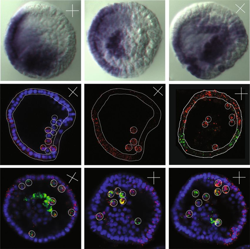
ARTICLE                                                                        NATURE COMMUNICATIONS | DOI: 10.1038/s41467-017-01961-y

D
         uring embryo development, tissues proliferate and dif-        different phyla. Exemplifying this split, we show that the Fgfr
         ferentiate in a coordinated manner to build a whole           event affecting the IgIII domain originated from a Bilateria hot-
         organism through a genome-guided process. Different cell      spot of recurrent AS evolution that was co-opted as an Esrp target
types express distinct transcriptomes to control cellular identity     at the base of chordates.
and physiology, and to establish differential interaction cap-
abilities between embryonic tissues. Final morphology is thus
achieved by cell-specific transcriptomic responses to external and      Results
internal stimuli within each tissue. Therefore, changes in the         esrp1 and esrp2 are involved in morphogenesis in zebrafish. A
genetic networks involved in morphogenesis are ultimately              broad phylogenetic analysis showed that the Esrp family predates
responsible for both the modification of organs and, at a mac-          the origin of metazoans and that a single copy of Esrp has been
roevolutionary scale, the origin of new structures1,2.                 maintained in most metazoan groups with the exception of the
   In particular, epithelial-mesenchymal interplays are essential to   vertebrate lineage, in which two copies are present in all studied
many organogenetic processes in vertebrates3,4. These tissues          species (Supplementary Figs. 1, 2). To investigate the evolution of
often interact in morphogenetic interfaces through the exchange        Esrp roles across deuterostomes at various phylogenetic distances,
of cells and signaling molecules5,6. Despite the great diversity of    we first examined the expression and function of the two Esrp
cell types across the embryo, the majority can be classified as         paralogs (esrp1 and esrp2) in zebrafish. A highly dynamic
showing either mesenchymal or epithelial characteristics. This         expression pattern was observed for both genes during the
broad distinction, which is independent of tissue origin, has also     development of zebrafish using whole-mount in situ hybridiza-
been shown to be reflected in the patterns of gene expression and       tion (WMISH) (Fig. 1a, Supplementary Fig. 3). At early stages,
alternative splicing (AS)7. Those transcriptomic programs confer       both genes displayed different expression patterns. esrp1 tran-
partly antagonistic morphogenetic properties to epithelial and         scripts were detected in the whole epidermis at 14 h post-
mesenchymal tissues by modulating certain cellular features, such      fertilization (h.p.f.), but its expression was restricted to the pos-
as adhesion, motility and polarity.                                    terior part of the embryo at 16 h.p.f.. On the other hand, esrp2
   Of particular importance for mammalian morphogenesis is a           was found only in the hatching gland rudiment at these stages.
mutually exclusive exon skipping event found in members of the         However, from 24 h.p.f. to 5 days post-fertilization (d.p.f.), the
FGF receptor (Fgfr) gene family8. Exons IIIb and IIIc encode a         expression of both genes converged in most territories. Their
region of the third immunoglobulin domain (IgIII) of the FGFR1,        transcription was transiently activated during the development of
FGFR2, and FGFR3 proteins, and are differentially included in          multiple organs, including the olfactory epithelium, otic vesicle,
transcripts from epithelial or mesenchymal cells, respectively.        pharynx, epidermis and notochord. esrp2 showed expression in a
Importantly, their mutually exclusive inclusion has a dramatic         few additional tissues, such as pronephros, hatching gland, liver,
effect on the affinity of the receptors for FGF ligands9, providing     and heart.
epithelial cells specificity for FGF signals secreted by the               Next, we used the CRISPR-Cas9 system to generate loss-of-
mesenchyme, and vice versa. Consistent with the importance of          function zebrafish mutant lines for esrp1 and esrp2. We targeted
this regulatory system in development, disruption of the Fgfr2-        single guide-RNAs to the first and third exons of esrp1 and esrp2,
IIIb isoform leads to severe defects during mice organogenesis10.      respectively (Fig. 1b). For esrp1, we selected a mutant allele with a
   These and other morphogenesis-associated AS events are              168-bp deletion and 14-bp insertion that induced the usage of an
directly regulated by the Epithelial Splicing Regulatory Protein       upstream cryptic splice donor, resulting in skipping of the whole
(Esrp) genes in mammalian species11. Esrp1 and Esrp2 were ori-         coding sequence of exon 1, including the start codon. For esrp2,
ginally identified as positive regulators of IIIb exon inclusion of     we selected a mutant allele with a 17-bp frame-disrupting deletion
the Fgfr2 gene12. They encode RNA-binding proteins that are            that produced a premature termination codon before the
dynamically expressed mainly in a subset of epithelial tissues         translation of any RNA-binding domain and that was predicted
during mouse development13, although mesenchymal expression            to trigger non-sense mediated decay (Fig. 1b). Expression of the
has also been reported in chicken14. Recently, double knockout         mutant alleles was highly reduced in homozygous embryos, while
(DKO) mice for both Esrp genes were shown to display severe            transcript levels of the other paralog were not increased as a
organogenetic defects and a complete shift to exon IIIc inclusion      compensatory mechanism in the single mutants, as shown by
in Fgfr1, Fgfr2, and Fgfr315. In addition, many Esrp exon targets      quantitative PCR (Supplementary Fig. 3m). Furthermore, western
were identified in genes involved in cell–cell adhesion, cell           blot assays confirmed the loss of the full-length protein for both
polarity, and migration16. However, the origin and evolution of        mutant alleles, although faint bands that might correspond to
Esrp morphogenetic functions and its regulated AS programs             shorter truncated proteins or unspecific signal were detected for
remain largely unknown.                                                esrp1 (Supplementary Fig. 3n). Therefore, to further determine
   Here, to investigate the evolution of Esrp functions and asso-      the functional impact of the mutations, we cloned the mutant and
ciated transcriptomic programs, we performed Esrp loss-of-             wild-type (WT) esrp1 alleles in a pcDNA3.1 vector and transfect
function or gain-of-function experiments in several deuterostome       the construct into human 293T cells. Whereas expression of the
species. Within bony vertebrates, Esrp genes play conserved roles      WT allele of zebrafish esrp1 was able to modify the splicing
in the development of numerous homologous organs. Con-                 pattern of previously reported endogenous targets (EXOC7,
sistently, Esrp regulates a core set of homologous exons in the        ARHGAP17, and FGFR1) in the same way as its human
three studied vertebrate species, including the mutually exclusive     ortholog16, expression of the mutant esrp1 allele did not produce
exons in the Fgfr family. Study of three non-vertebrate deuter-        any measurable effect in exon inclusion levels (Supplementary
ostomes showed that Esrp is involved in a wide variety of mor-         Fig. 3o), although an effect on other regulatory processes cannot
phogenetic processes in multiple unrelated structures in these         be ruled out. We were unable to clone the full-length transcript of
species, and that, among others, it likely played an ancestral role    the mutant esrp2 allele due to its reduced expression. Altogether,
in regulating specific mesenchymal-to-epithelial transitions            these data support an efficient loss of function for both Esrp genes
(METs) in the deuterostome ancestor. However, transcriptomic           in our mutant lines, particularly with regards to splicing
analyses showed that most exons present clade-restricted differ-       regulation.
ential regulation. In particular, no Esrp-dependent alternative           Morphological examination of single esrp1 or esrp2 homo-
exons were found conserved between studied species belonging to        zygous zebrafish mutants showed no apparent gross defects

2                                            NATURE COMMUNICATIONS | 8: 1799    | DOI: 10.1038/s41467-017-01961-y | www.nature.com/naturecommunications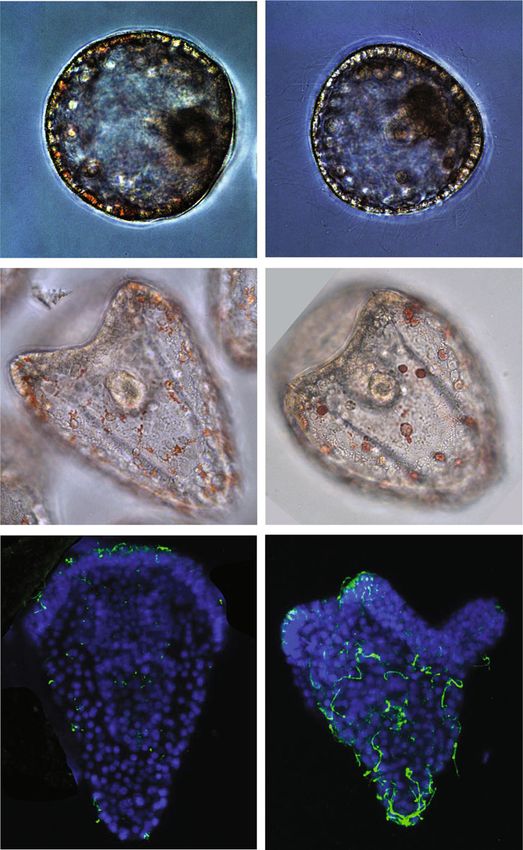
NATURE COMMUNICATIONS | DOI: 10.1038/s41467-017-01961-y ARTICLE
a esrp1 esrp2 esrp1 esrp2
ot
ot ep
ep ff
po
14 h.p.f.
24 h.p.f.
cl cl
hg ff
ol ol
ot ie ie
36 h.p.f.
16 h.p.f.
hr nt
ph ph
nt
he
tb
ot
ie
ol ie
48 h.p.f.
20 h.p.f.
ph
ph
tb ff
pn
b TSS
c esrp1 esrp2 Danio rerio 5 dpf
esrp1 WT
MUT WT MUT WT
1 ATG 2 3
esrp1 MUT
TSS
MUT WT MUT WT
esrp1MUT/MUT
1 ATG 2 3
esrp2 WT
MUT WT MUT WT
esrp2 MUT/MUT
2 3 4
esrp2 MUT
MUT WT MUT WT
esrp1MUT/MUT;esrp2 MUT/MUT(DMUT)
17 bp del PTC
2 3 4
*
d e f g h i
WT 6 dpf
dl
pr
DMUT 6 dpf
pr
dl
Fig. 1 Expression and developmental roles of esrp1 and esrp2 in zebrafish. a WMISH for esrp1 and esrp2 in Danio rerio WT embryos. At 14 h.p.f., esrp1
transcripts were observed in embryonic epidermis, while esrp2 expression was only detected in the polster (po). At 16 h.p.f., esrp1 was restricted to the
posterior and tailbud (tb) epidermis, whereas esrp2 persisted in the hatching gland rudiment (hr) and mild expression started to be detected in the otic
placode (ot). By 20 h.p.f., esrp1 was found in the tailbud epidermis and more subtly in the olfactory placode (ol), while esrp2 appeared in new territories,
such as pronephros (pn) and ectodermal cells of tailbud fin fold (ff). At 24 h.p.f., expression of both paralogs presented a similar pattern including olfactory
and otic placodes, cloaca (cl), and epidermis (ep), although esrp2 was also observed in the hatching gland (hg). By 36 h.p.f., both genes were detected in
the inner ear epithelium (ie), notochord (nt), and phanynx (ph), and esrp2 was also observed in the heart (he). At 48 h.p.f., expression was found
predominantly in inner ear and pharynx. b Schematic representation of the genomic and transcriptomic impact of the selected esrp1 and esrp2 mutations.
Blue boxes/lines represent genomic deletions in the mutants, while the red line depicts an altered splice junction in the esrp1 mutant allele. TSS,
transcription start site; PTC, premature termination codon; del, deletion. Standard and fluorescent (green dot) primers used during genotyping are
represented by arrows. c Left: genotyping of embryos by fluorescent PCR readily distinguished between WT and MUT alleles. Right: Representative 5 d.p.f.
larvae for wild type (WT), esrp1 mutant (esrp1MUT/MUT), esrp2 mutant (esrp2MUT/MUT), and double mutant (DMUT) genotypes. Deflated swim bladder in
the DMUT embryo is indicated by a red asterisk. d–i Phenotypic differences between 6 d.p.f. WT (top) and DMUT (bottom) embryos in different
embryonic structures. DMUT larvae showed impaired fin formation (arrows) and cleft palate (arrowhead) d, including malformation of the ethmoid bone,
as shown by Alcian blue staining e. f–i Transversal histological sections stained with hematoxylin and eosin showing structural differences in pectoral fin
f, esophagus g, inner ear h and olfactory epithelium i. Black arrowheads mark the dorso-lateral septum between semicircular canals in h. Proximal (pr) and
distal (dl) parts of the fin are indicated in f. Scale bars: 1 mm a, 2 mm c–e, 100 µm f–h, 50 µm i
NATURE COMMUNICATIONS | 8: 1799 | DOI: 10.1038/s41467-017-01961-y | www.nature.com/naturecommunications 3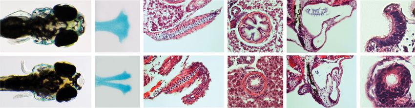
ARTICLE                                                                                                  NATURE COMMUNICATIONS | DOI: 10.1038/s41467-017-01961-y

     a                            10 h.p.f.                     d       9.5 h.p.f.   e               f                    m                                          18 h.p.f.     m′

                                              Middle tailbud
     Esrp    Twist>CD4::mCherry

     b                            13 h.p.f.                                                                                      Twist>GFP
                                                                                                                                 Twist>CD4::mCherry
                                                                                                     Merge
                                                                Esrp                 Twist-like2     DAPI                 n                                          18 h.p.f.     n′

                                                                g       14 h.p.f.    h               i
                 aTLC

                        pTLC                  Late tailbud
     Esrp
     Hand-r>LHG
     LexAOP>H2B::mCherry                                                                                                         Twist>Esrp
                                                                                                                                 Twist>CD4::mCherry
     c                            15 h.p.f.
                                                                                                     Merge                o                        p = 6.9 × 10⁻¹⁵
                                                                Esrp                 Twist-like2     DAPI
                aTLC                                                                                                                      100     4
                                                                                                                                           90                                      Abnormal
         pTLC
                                                                j       15 h.p.f.    k               l                                     80                   26                location of

                                                                                                                          % Individuals
                                                                                                                                           70                                    mCherry cells
                                              Late tailbud II

                                                                                                                                           60
                                                                                                                                           50                                     Trunk+Tail
                                                                                                                                                  51            26
                                                                                                                                           40                                     Trunk
                                                                                                                                           30                                     Tail
     Esrp                                                                                                                                  20
                                                                                                                                           10                   18                Normal
     Hand-r>LHG                                                                                     Esrp                                    0
     LexAOP>H2B::mCherry                                        Esrp                 Twist-like2    Twist-like2                                 Control    Twist>Esrp

Fig. 2 Esrp is expressed in a subpopulation of TLCs in Ciona and is able to modulate cell motility in the mesenchymal lineage. a Esrp expression (green) is
detected at 10 h.p.f. by fluorescent WMISH in the epidermis and in some cells within the mesenchymal lineage, as shown by co-staining with mChe driven
by the Twist promoter, which labels the Twist-like1-derived cell lineage. b, c Esrp (green) is expressed in the TLC lineage as shown by co-staining with Hand-
r > LHG/LexAOP > H2B::mCherry (red) in 13 h.p.f. and 15 h.p.f. embryos in lateral b and dorso-lateral c views, respectively. aTLC: Anterior TLCs, pTLC:
posterior TLCs. d–l Double fluorescent WMISH for Esrp (red) and Twist-like2 (green), with the exception of j, which corresponds to colorimetric Esrp mRNA
staining (purple). At middle tailbud stage, Esrp expression is detected in both epidermis and mesenchymal cell lineage (the latter is marked by arrows).
m-m’ Mesenchymal lineage in WT larvae at 18 h.p.f. stained in red using the Twist > CD4::mCherry construct. Twist > GFP was used as co-electroporation
control plasmid, showing full co-electroporation (green channel not shown for clarity). n–n’ Mesenchymal lineage stained in larvae co-electroporated with
Twist > CD4::mCherry and Twist > Esrp constructs. The trunk region is shown in m–n, while tail segments are shown in m’–n’. o Quantification of the
different phenotypes of mesenchymal cell lineage motility observed in Twist > Esrp and control larvae. These included individuals with abnormal migration
in the trunk (‘Trunk’), with ectopic mChe-positive cells in the tail (‘Tail’) or both phenotypes (‘Trunk + Tail’). P-values correspond to a two-sided Fisher
Exact tests (‘Normal’ vs rest). Scale bars correspond to 25 µm

during development (Fig. 1c). Although esrp2 mutant females                                        also required during the development of structures homologous
were unable to produce eggs, both mutant lines grew normally in                                    to some of these organs15, including several that exhibit distinct
homozygosity and became seemingly healthy adults. However,                                         morphologies and functions compared to zebrafish, such as the
double mutants (DMUT) presented multiple developmental                                             lungs/swim bladder, palate skeleton and pectoral limbs/fins.
abnormalities. Most larvae (28/37, 75.7%) died between 8 and                                          To characterize the phenotype of the DMUT embryos at the
10 d.p.f., and no fish survived beyond 14 d.p.f.. Phenotypic                                        molecular level, we performed RNA-seq of two replicates of
analysis of 6 d.p.f. DMUT larvae showed multiple fully penetrant                                   DMUT 5 d.p.f. and age-matched WT larvae (Methods section).
morphogenetic defects. All DMUT larvae presented cleft palate                                      This analysis confirmed the reduced expression for both mutant
(Fig. 1d), with the medial population of cartilage cells being                                     alleles in DMUT embryos (Supplementary Fig. 3r). Differential
absent in the ethmoid plate, as revealed by alcian blue staining                                   gene expression analysis identified 248 and 609 downregulated
(Fig. 1e). Fin development was also impaired, with evident                                         and upregulated genes, respectively, (Supplementary Data 1).
dysgenesis of its distal endoskeletal part (Fig. 1f). Reduction in                                 Gene Ontology (GO) enrichment analysis showed functional
esophagus diameter and loss of its villous shape was observed                                      categories that were highly consistent with some of the observed
(Fig. 1g). The volume of the inner ear was smaller compared to                                     phenotypes, such as skeletal system development and sensory
WT larvae, and the dorso-lateral septum that separates the rostral                                 perception (Supplementary Fig. 4). Interestingly, other signifi-
and caudal semicircular canals was abnormally invaded by                                           cantly enriched categories pointed to specific impaired processes
cellular and extracellular material (Fig. 1h). The posterior part of                               at the cellular level such as cell–cell adhesion, cell-matrix
the olfactory epithelium formed a spherical internal lumen,                                        adhesion and cell component morphogenesis.
instead of being open toward the embryo surface (Fig. 1i).
Mutants also showed an abnormal arrangement of basibranchial
pharyngeal cartilage (Supplementary Fig. 3p), in addition to a                                     Esrp expression and function during Ciona development. To
smaller and thicker swim bladder epithelium (Supplementary                                         examine the diversity of roles that Esrp genes play during
Fig. 3q), which failed to inflate in 24/35 (68.5%) of examined                                      embryogenesis beyond the vertebrate clade, we next studied the
DMUT embryos (Fig. 1c). Interestingly, Esrp genes in mouse are                                     ascidian Ciona robusta, a species belonging to the sister group of

4                                                                   NATURE COMMUNICATIONS | 8: 1799          | DOI: 10.1038/s41467-017-01961-y | www.nature.com/naturecommunications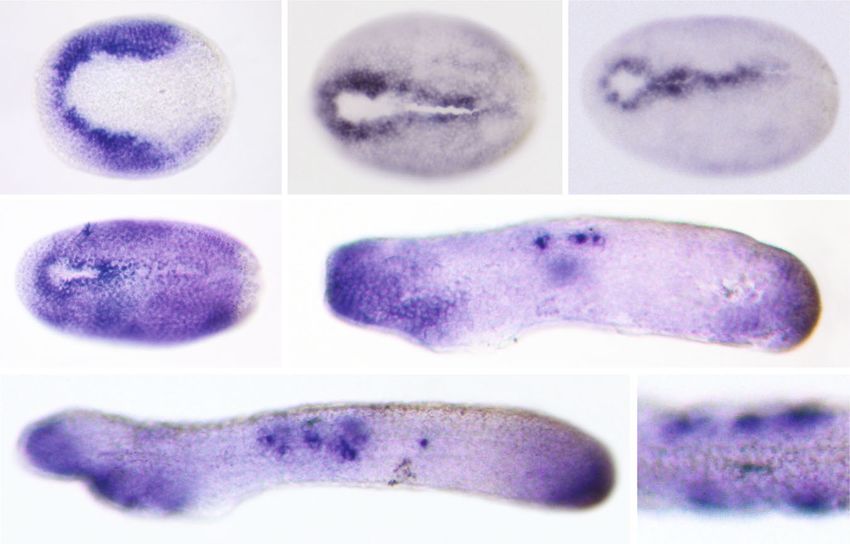
NATURE COMMUNICATIONS | DOI: 10.1038/s41467-017-01961-y                                                                                     ARTICLE

                             a                                    b                                  c
                                                                                                                   hp
                                                                                 hp                   npo
                                           npl

                                   br
                             14 h.p.f.                             16 h.p.f.                         18 h.p.f.

                             d                                    e
                                                 ep
                                 npo

                              21 h.p.f.                            30 h.p.f.

                             f                                                                               g

                                                                                                             36 h.p.f.
                             36 h.p.f. (Lateral view)                                                        (D view)

Fig. 3 Esrp is expressed dynamically in the non-neural ectoderm during amphioxus embryo development. WMISH of Esrp in Branchiostoma lanceolatum
embryos. Anterior is to the left in all cases. a 14 h.p.f. early neurula (dorsal view) showing expression in the ectodermal cells located in the border region
(br) next to the neural plate (npl). b, c 16 h.p.f. and 18 h.p.f. mid-neurula embryos (dorsal view) stained most strongly in the ectodermal cells next to the
neural plate border and that form the hinge points (hp) during neural tube closure. The location of the neuropore (npo) is indicated. d In 21 h.p.f. late
neurula (dorsal view), Esrp expression is extended throughout the whole epidermis (ep). e, f Early (30 h.p.f.) and late (36 h.p.f.) pre-mouth stages (lateral
views) showing Esrp-positive cells in anterior ectoderm, tailbud epithelia and in a few cells that likely corresponding to migrating sensory cells during
epidermal incorporation or already integrated into the epithelium (black arrows). g Dorsal view of a 36 h.p.f. embryo shows the epidermal location those
Esrp-positive cells (black arrows). Scale bars: 50 µm a–f, 25 µm g

vertebrates, the tunicates17. Expression of the single Esrp ortholog                Twist-like2 was restricted to anterior mesenchymal domains,
present in the Ciona genome was detected in the embryonic                           while Esrp transcripts were only detected in the posterior-most
epidermis after neurulation, as in the case of vertebrates (Fig. 2).                part of the trunk (Fig. 2j–l).
In addition, Esrp expression was also observed in a bilateral                          The common developmental origin of these two cell popula-
domain located within the mesenchymal lineage from early to late                    tions, the function of Twist-like2 as a key inducer of cellular
tailbud stage. Ciona mesenchymal cells derive from the Twist-                       migration23, and the described antagonistic roles of Esrp and
like1-expressing A7.6 [trunk lateral cells; TLCs], B7.7, and B8.5                   Twist genes in mammalian cells24 led us to hypothesize that Esrp
blastomeres of the 110-cell stage embryo18. As development                          may confer particular morphogenetic properties to the subset of
proceeds, those cells divide and give rise to three mesenchymal                     cells within the mesenchymal lineage in which it is expressed and
sub-lineages that are located in a region of the trunk adjacent to                  that, therefore, Esrp expression needs to be turned off in the other
the tail during mid-tailbud stages. Twist-like1 enhances the                        lineages for their correct ontogenesis. To test this possibility, we
transcription of several mesenchymal genes, including                               ectopically expressed Esrp in all mesenchymal cells (Twist > Esrp)
Twist-like2, before being downregulated around mid-tailbud                          during early stages, together with Twist > CD4::mChe to visualize
stage18,19. In subsequent stages, a number of cells coming from                     them. We fixed embryos at larval stage, when a large part of the
the Twist-like2-positive lineage migrate toward the anterior part                   cell migration towards the anterior part of the trunk has occurred.
of the trunk to contribute to the formation of mesodermal                           We observed two main phenotypes associated with an abnormal
organs20.                                                                           mesenchymal distribution in 75% of individuals (n = 70). In all
   We confirmed the expression of Esrp within the mesenchymal                        affected embryos, migrating mChe-positive cells were found only
lineage with a double-staining assay. Fertilized eggs were                          in the lateral sides adjacent to the epidermis, but not through the
electroporated with a construct (Twist > CD4::mChe) carrying                        middle part of the trunk (Fig. 2m, n, o). In addition, 35% of co-
the Twist-like1 promoter driving the expression of the                              electroporated larvae also showed ectopic mChe-positive cells
membrane-targeted Cherry (mChe) protein as a reporter21. An                         distributed along the tail, which were only very rarely observed in
anti-mChe antibody was used to track the mesenchymal cell                           control individuals (Fig. 2o). Interestingly, a previous study found
lineage, while endogenous Esrp expression was visualized by                         that some cells derived from B7.7 and B8.5 mesenchymal
fluorescent WMISH (Fig. 2a). A similar assay using a LexA/                           lineages, which normally do not express Esrp, localized in the
LexAop system driven by the Hand-r proximal enhancer22                              tail of Twist-like1 knockdown of Ciona larvae, integrating
narrowed the identity of this Esrp-expressing domain down to a                      muscular and endodermal tissues20. Altogether, our results thus
subset of posterior TLCs (Fig. 2b, c). This mesenchymal lineage                     suggest that Esrp may modulate motility properties of the
contributes to the formation of specific organs like the oral siphon                 mesenchymal cell lineage in ascidians, as well as compromise
muscle and the epithelium of the 1st/2nd gill slits20.                              topological cellular fate when ectopically expressed.
   To unravel the dynamics of these Twist-like1-derived Esrp-
positive cells, we next performed a double WMISH with probes
for Esrp and Twist-like2. This showed that the expression of both                   Dynamic Esrp expression during amphioxus embryogenesis.
genes is rapidly regulated during development and that they soon                    We next investigated Esrp expression in the amphioxus Bran-
acquire a mutually exclusive expression pattern, which becomes                      chiostoma lanceolatum, a cephalochordate species that shares a
evident by late tailbud stage (Fig. 2g–i). At late tailbud II stage,                general chordate bodyplan with vertebrates, although it lacks

NATURE COMMUNICATIONS | 8: 1799         | DOI: 10.1038/s41467-017-01961-y | www.nature.com/naturecommunications                                             5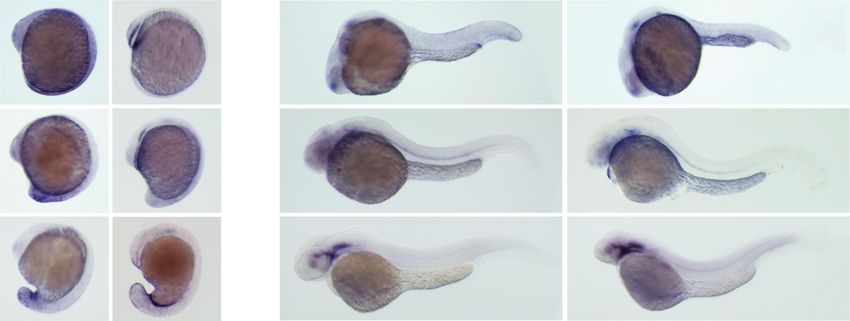
ARTICLE NATURE COMMUNICATIONS | DOI: 10.1038/s41467-017-01961-y
a vv a
a b c j Control k Esrp trMO
v
v
ec
ec
ns
ns
ns
42 h.p.f. 42 h.p.f.
24 h.p.f. Esrp 24 h.p.f. Esrp 36 h.p.f. Esrp
a a ab l Control m Esrp trMO
d e f r l
v v
nne o
ns
ns ns ne
ne
ec ec
68 h.p.f. Anal view 68 h.p.f. Anal view
Esrp Esrp
30 h.p.f. DAPI 30 h.p.f. Esrp 36 h.p.f. Hnf6 n o
Control Esrp trMO
ae ab a a cb
g r
h o ab i o cb
l
o ae v v
ae ae
ns
oe
Esrp Esrp Esrp
36 h.p.f. Gcm 36 h.p.f. Gcm 36 h.p.f. Gcm 96 h.p.f. Abanal view 96 h.p.f. Abanal/lat view
p q p = 1.5 × 10⁻³⁴ r p = 1.5 × 10⁻⁷
p = 3.6 × 10 –59
p = 9.6 × 10⁻⁸
Fw1 Fw2 Rv 100 100
30 2
* 90 90
% Pigment cells (68 h.p.f.)
2 3
% Individuals (68 h.p.f.)
80 80
70 Pigment 70 11
Hairy
178 82 cells phenotype
e
60 60
om
l
tro
O
lM
en
on
50 Round 50 14
sp
12 Strong
G
C
273
40 Stellate 40 Mild
30 30 Normal
20 6
20
54 32
10 10
0 1
%IR 2.3 74.8 0
Control trMO splMO Control trMO splMO
(n = 31) (n = 27) (n = 12)
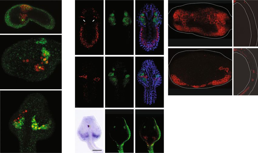
Fig. 4 Esrp represses cilia formation in aboral ectoderm and is necessary for complete MET of pigment cells. a, b Colorimetric WMISH of Esrp in sea urchin
embryos at 24 h.p.f., in lateral a and vegetal b views, showed expression in one side of the ectodermal territory (ec) and in some cells of the non-
skeletogenic mesoderm (ns). vv, vegetal view. c Lateral view at 36 h.p.f. confirmed Esrp’s asymmetric expression in one side of the ns mesoderm.
d, e Fluorescent WMISH at 30 h.p.f. (lateral view) confirmed ectodermal and mesodermal expression of Esrp. f Double fluorescent WMISH showed that
Esrp expression (red) at 36 h.p.f. (animal pole view) did not overlap with the neurogenic ectoderm (ne) marker Hnf6 (green). nne, non-neural ectoderm. g
Double fluorescent WMISH of Esrp (red) and Gcm (green) at 36 h.p.f. (animal pole view) revealed that Esrp was expressed in the aboral ectoderm (ae) and
in the pigment cell precursors. Pigment cell precursors that are already in contact with the ectodermal epithelium are marked by arrowheads. oe, oral
ectoderm. h, i Esrp (green) and Gcm (red) at 36 h.p.f. in two different stacks from same embryo (lateral view). The arrow indicates a representative
migratory path of pigment cells from mesoderm to ectoderm. At the top right corner of each panel a–i, the orientation of the depicted embryo along the
animal [a] - vegetal [v], oral [o] – aboral [ab], left [l] –right [r] axes are reported, when possible. j, k Early gastrula (42 h.p.f.) from uninjected control and
Esrp trMO embryos showed differences in pigment cell location. Red/white arrowheads indicate pigment cells already integrated into the ectoderm or in
the sub-ectodermal space, respectively. l, m Early pluteus larvae (68 h.p.f.) in abanal view show differences in pigment cell morphology upon trMO
treatment (roundish instead of stellate/dendritic). n, o Ectopic embryonic cilia stained with acTubulin in the aboral ectoderm (ae) of 96 h.p.f. Esrp-trMO
injected embryos. Apex is marked by a white arrowhead. cb, ciliary band. p RT-PCRs showing the levels of intron 2 retention in Esrp transcripts from control
and Esrp splMO embryos; genomic DNA was used as a reference for intron inclusion. The asterisk marks the position of the first in-frame termination
codon in intron-retained transcripts. q Quantification of pigment cell morphology in control, Esrp trMO and splMO knockdown 68 h.p.f. embryos (sum of
three independent experiments). r Quantification of the ‘hairy’ phenotype in control, Esrp trMO and spMO knockdown 68 h.p.f. embryos (sum of two
independent experiments). P-values correspond to 2-way q or 3-way r two-sided Fisher Exact tests. Hairy phenotype was considered “mild” when aboral
ectoderm cells show long cilia (of similar or longer length than ciliary band cells) and “strong” when, in addition to the latter, larvae show particularly long
cilia at the apex, as indicated by an arrowhead in panel o. Scale bars correspond to 20 µm
6 NATURE COMMUNICATIONS | 8: 1799 | DOI: 10.1038/s41467-017-01961-y | www.nature.com/naturecommunications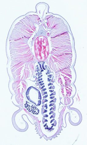
NATURE COMMUNICATIONS | DOI: 10.1038/s41467-017-01961-y                                                                                         ARTICLE

    a                                                                                       b                    Esrp-dependent CDS exon clusters
                                    183                                          114
                                                                                                      NH: 53                                          NH: 7
                                                                                                      NC: 72                                          NC: 60
                                                      114
                                    159                                          148
        13                                                  26        20
                 24        15
        30        7        16                         41    16        16
        IR      Alt5       Alt3    AltEx              IR    Alt5      Alt3      AltEx                                  73          79           70

                                                                                   76
                                    268                                                                           1                22
                                                                                                                            21            4
                           110                                                                            17
                                                            17                   123
        57       86                 229               42               27
        94                 125
                 55                                   17    27         26
                                                                                                                                   95                NH: 179
        IR      Alt5       Alt3    AltEx              IR    Alt5      Alt3      AltEx                                                                NC: 116
                                                                   Esrp-enhanced                NH: 142
    c                                                              Esrp-silenced                NC: 21
                       Human Esrp-dependent:
                          321 CDS clusters

                                           56 (17%)                                              45 (40%)                                       20 (90%)

                                           45 (14%)                                              10 (9%)                                        1 (5%)

                                           220 (69%)                                             57 (51%)                                       1 (5%)

                         Mouse Esrp-                                         Zebrafish Esrp-                                 Sea urchin Esrp-
                                           112/188*                                              22/48*                                       0/1*
                         dependent:                                            dependent:                                      dependent:

Fig. 5 Evolution of Esrp-dependent splicing programs. a Number Esrp-dependent AS events by type detected in human, mouse, zebrafish and sea urchin, RNA-
seq samples. Red/blue bars correspond to Esrp-enhanced/silenced inclusion of the alternative sequence. IR, intron retention; Alt5, alternative 5′ splice site
choice; Alt3, alternative 3′ splice site choice; AltEx, alternative cassette exons. b Venn diagram showing the overlap among homologous Esrp-dependent
cassette exons in coding regions detected as regulated in the same direction in the studied species. Only Esrp-dependent exons with homologs in at least two
species and sufficient read coverage in all the species in which they have homologs are included in the comparison. Esrp-dependent exons that lack homologous
counterparts in all the other species are indicated for each species (NH). Similarly, the number of Esrp-dependent exons that do not have sufficient read
coverage in at least one of the species with a homologous exon is displayed for each species (NC). c Summary of conservation at the level of genomic presence,
alternative splicing and Esrp-dependency for all 321 clusters of human Esrp-dependent coding exons in other species. Shared Esrp-dependent exons between the
previous phylogenetic group are classified in the test species into three categories: (i) the exon is not detected in the genome (top row, discontinuous line); (ii)
the homologous exon is detected in the genome, but it is constitutively spliced (middle row, gray exon); and (iii) the homologous exon is alternatively spliced
(bottom row, yellow exon, numbers in bold). Below this classification, the number of similarly regulated Esrp-dependent exons shared by the two species/
lineages over the number of alternatively spliced exons with sufficient read coverage from (iii) (indicated by asterisks), is shown

many of their key traits and presents several developmental                             Esrp is required for MET of pigment cells in sea urchin. We
particularities25. Amphioxus Esrp showed a highly dynamic                               next investigated the functions of Esrp in the purple sea urchin
expression pattern during embryo development, generally more                            Strongylocentrotus purpuratus. This organism belongs to Ambu-
restricted than its vertebrate counterparts (Fig. 3). In early                          lacraria, sister group of chordates, with a transcriptome-based
neurula embryos (14 h.p.f.), Esrp transcripts were observed in                          annotated genome and available genetic tools. Colorimetric
the border region, an ectodermal tissue adjacent to the                                 WMISHs revealed Esrp expression in the ectoderm and meso-
neural plate (Fig. 3a). During neurulation, Esrp was strongly                           derm in one half of the embryo at blastula stages (24–36 h.p.f.)
expressed along the ectodermal hinge points of neural tube                              (Fig. 4a–c). Expression was no longer detected at gastrula (48 h.p.
closure (Fig. 3b, c). Right after neurulation (21 h.p.f.), tran-                        f.) or early pluteus stages (72 h.p.f.). Fluorescent WMISH con-
scription of Esrp was extended to the epidermis of the whole                            firmed Esrp expression in the ectoderm and non-skeletogenic
embryo (Fig. 3d). Finally, in pre-mouth larvae stages, the                              mesodermal cells at 30 h.p.f. (Fig. 4d, e). To investigate the nature
expression was restricted to the tailbud region, anterior ectoderm,                     of the ectodermal region with Esrp expression, we performed
and, strikingly, in a few cells near and within the dorsal epidermis                    double fluorescent WMISH with Esrp and Hnf6, a marker for the
(arrows in Fig. 3e–g). Because of their shape and location, the                         ciliary band29, the region where ciliary cells and neurons differ-
latter group of cells may correspond to a previously described                          entiate (Fig. 4f). No signal overlap was observed between the two
population of epidermal sensory neurons26–28. These cells have                          genes, indicating that Esrp expression is restricted to the non-
been reported to delaminate from the ventral ectoderm, migrate                          neural ectoderm. Next, double WMISH with Gcm, a marker for
underneath the epithelium toward the dorsal part of the embryo                          aboral non-skeletogenic mesoderm at the mesenchyme from
and re-integrate into the ectoderm, where they become sensory                           blastula stage onward30, showed that Esrp expression in the
cells. Based on its dorsal expression, we speculate that Esrp might                     ectodermal tissue corresponds to the aboral side (Fig. 4g–i).
contribute to the process of epithelial integration at the final                         Interestingly, we observed co-expression of Gcm and Esrp in
migratory stages.                                                                       pigment cells precursors, a population of non-skeletogenic

NATURE COMMUNICATIONS | 8: 1799        | DOI: 10.1038/s41467-017-01961-y | www.nature.com/naturecommunications                                                   7
ARTICLE                                                                            NATURE COMMUNICATIONS | DOI: 10.1038/s41467-017-01961-y

mesodermal cells that delaminate from the aboral side of the tip             (R2 = 0.89, n = 12) (Supplementary Fig. 7). RNA regulatory maps
of the invaginating archenteron and migrate during gastrulation              for the top twelve hexamers bound by Esrp identified by SELEX-
from the mesoderm toward the ectoderm, where they insert                     seq33 showed clear enrichment above background at expected
between epithelial cells becoming immunocytes31,32.                          regions for Esrp-dependent exons for all species (Supplementary
   To explore the functions of Esrp in this organism, we generated           Fig. 8a), although results from sea urchin were less clear, likely
gene knockdowns by injecting morpholinos (MO) into sea urchin                reflecting a higher fraction of indirect targets detected using a
zygotes. To ensure that the knockdowns produced specific effects,             knockdown strategy in whole embryos.
we used two different MOs in independent experiments: one                       To understand the evolution of Esrp-dependent programs at the
blocking translation and another impairing splicing of intron 2,             exon level, we next built clusters of high confidence homologous
whose retention generates a premature termination codon four                 coding cassette exons within conserved gene intron-exon
aminoacids downstream of exon 2. Efficiency of the splicing MO                structures (Supplementary Data 4 and see Methods section).
was assessed by RT-PCR (Fig. 4p). We observed two main                       Comparisons between closely and distantly related organisms
phenotypic defects in both MO injections, supporting knockdown               uncovered different scenarios with regards to the sources for exon
specificity (Fig. 4q, r). First, whereas in control embryos most              recruitment. On the one hand, most Esrp-dependent exons among
pigment cells were already located in the ectoderm at gastrula stage         vertebrates presented a homologous counterpart in another
(42 h.p.f.) (Fig. 4j, k, Supplementary Fig. 5a) and acquired a               species, ranging from 97% in mouse to 59% in zebrafish
dendritic conformation by prism/early pluteus stages (68 h.p.f.)             (Supplementary Fig. 8b). On the other hand, we only detected
(Fig. 4l, m, q), in knockdown embryos these cells were usually               homologous counterparts in any of the studied genomes for 21%
observed in the sub-ectodermal space at gastrula stage and                   of sea urchin differentially regulated exons. When taking only the
maintained their roundish shape at prism stages (p < 10−4 for all            clusters with homologous exons in at least two species and with
comparisons, Fisher Exact test). This failure in the complete                sufficient read coverage in all the organisms in which the exon
integration of pigment cells into the ectoderm likely constitutes an         exists, both mammals shared most of their Esrp-dependent exon
impaired MET. Second, knockdown embryos showed a “hairy”                     sets (Fig. 5b, Supplementary Fig. 8c). On the contrary, we observed
phenotype at gastrula and pluteus stages, consisting of ectopic long         a large fraction of lineage-specific Esrp-dependent regulation in
cilia on the aboral ectoderm, especially at the apex (Fig. 4j, k, n, o, r,   the case of zebrafish (67%) and sea urchin (94%). Alternative
Supplementary Fig 4b; p < 10−6 for all comparisons, 3-way Fisher             exons with Esrp-like motifs in expected exonic or nearby intronic
Exact test). In summary, these results indicate that Esrp in sea             regions in vertebrates generally showed higher levels of shared
urchin is necessary for a complete integration of pigment cells into         Esrp-dependent regulation (Supplementary Fig. 9a), and presence
its destination epithelium and to avoid ciliogenesis in aboral               of Esrp-like motifs in equivalent positions in orthologous exons
ectodermal cells.                                                            was often associated with increased shared regulation
   To further characterize these developmental defects, we                   (Supplementary Fig. 9b). Consistently, human exons with shared
generated RNA-seq data for two replicates of SplMO-injected                  Esrp-dependent regulation between mammals or among verte-
embryos at 24 h.p.f. and age-matched controls. These data                    brates showed higher conservation of their flanking intronic
confirmed our RT-PCRs results showing a high level of retention               sequences than human-specific and non-Esrp-dependent alter-
of Esrp intron 2 due to the morpholino effect (Supplementary                 native exons (Supplementary Fig. 8d).
Fig. 5c). Differential gene expression analysis identified 360 and               When focusing on the full set of 321 clusters with human Esrp-
712 downregulated and upregulated genes, respectively (Supple-               dependent exons, 83% had a homologous exon in mouse, most of
mentary Data 2). Interestingly, some enriched GO categories for              which were also alternatively spliced in the rodent species
these gene sets were similar to those found for the zebrafish                 (Fig. 5c). Among the homologous alternative exons with sufficient
DMUT analysis, including cell–cell adhesion, cell component                  read coverage in mouse, 112/188 (60%) were identified as Esrp-
morphogenesis and ectoderm development (Supplementary                        dependent (in the same direction) also in this species. These
Fig. 5d). We also observed a significant enrichment for functions             events include numerous previously described Esrp-dependent
related to nervous system development among the upregulated                  exons in genes, such as Scrib, Nf2, Enah, and Grhl115. From this
genes, consistent with a possible failure in proper non-neural               set of shared mammalian targets, we found homologous exons in
ectoderm specification.                                                       zebrafish in 67 cases (60%), most of which were also alternatively
                                                                             spliced in this species (Fig. 5c). Interestingly, we detected a core
                                                                             set of 22 homologous exons classified as Esrp-dependent in the
Evolutionary comparison of Esrp-dependent splicing pro-                      three vertebrate species, including some in genes previously
grams. Next, we investigated the origin and evolution of alter-              associated with morphogenetic processes35 (e.g., Numb, p120-
native exon programs regulated by Esrp across multiple                       catenin, Arhgap17 or Itga6; Supplementary Table 1). However,
phylogenetic distances using our zebrafish and sea urchin RNA-                most of these exons (90%) did not have a detected homologous
seq data, as well as previously published RNA-seq data for Esrp              counterpart in sea urchin, and the two exons identified in the
perturbations in mouse epidermis15 and three human cell cul-                 echinoderm genome did not exhibit Esrp-dependent differential
tures33,34. Importantly, this approach detects both direct and               regulation (Fig. 5c).
indirect Esrp-regulated events, hereafter referred together as Esrp-            We also identified 49 orthologous genes whose AS was
dependent. We identified 342 differentially spliced cassette exons            dependent on Esrp in at least two species, but in which the
in human, 262 in mouse, 497 in zebrafish, and 199 in sea urchin               specific regulated exons were different (i.e., non-homologous).
(Supplementary File 1; see Methods section for details). In all              Remarkably, 21 of these cases involved sea urchin and at least one
species, these exons were found enriched in genes associated with            vertebrate. Moreover, we further observed a significant fraction of
certain GO terms, such as vesicle-mediated transport, GEF                    target genes with more than one Esrp-dependent exon, ranging
activity, and actin cytoskeleton cell components (Supplementary              from 5.2% in sea urchin to 19.1% in zebrafish. These observations
Fig. 6). Additionally, we detected lower numbers of other types of           highlight the evolutionary plasticity of some genes for multiple
AS regulatory changes, such as alternative 5′ or 3′ splice site              acquisition of Esrp-dependent AS regulation. An interesting case
choice and intron retention (Fig. 5a, Supplementary Data 3). RT-             is Cd44, which acquired at least nine Esrp-dependent novel exons
PCR assays validated all tested differentially regulated exons in            within the mammalian clade, and is involved in ureteric
zebrafish (ΔPSI correlation R2 = 0.90, n = 15) and in sea urchin              branching in mouse36, a process affected in Esrp1 KO mice37.

8                                                NATURE COMMUNICATIONS | 8: 1799    | DOI: 10.1038/s41467-017-01961-y | www.nature.com/naturecommunications
NATURE COMMUNICATIONS | DOI: 10.1038/s41467-017-01961-y ARTICLE
a c pcDNA3.1-BlaFgfr pcDNA3.1-BlaFgfr-ΔIIIx
Zebrafish IIIb IIIc
500 469
Conrtol
Conrtol
fgfr1a fgfr1b fgfr2 fgfr3
WT DMUT WT DMUT WT DMUT WT DMUT
500 469
BlaEsrp
BlaEsrp
b
DreEsrp1
DreEsrp1
500 469
Amphioxus Fgfr
IIIx IIIb IIIc
sk
nc d EXOC7 SLK OSBPL3
us ord
ms
c
Sk ut
H ic
e
ve
at
cl
dg
25 35 100
% inclusion (PSI)
% inclusion (PSI)
% inclusion (PSI)
nt
ills
in
ep
er
in
20 30 90
M
G
N
H
25
15 20 80
10 15 70
Fgfr-III AS gl 10
5 5 60
0 0 50
hd
l
1
p
l
1
p
l
1
p
tro
tro
tro
rp
sr
rp
sr
rp
sr
Esrp
on
aE
on
aE
on
aE
Es
Es
Es
C
C
C
re
Bl
re
Bl
re
Bl
D
D
D
Gadph
Fig. 6 Fgfr AS is regulated by Esrp genes in vertebrates and amphioxus. a RT-PCR assays showing differential Fgfr exon IIIb and IIIc inclusion in WT versus
DMUT 5 d.p.f. zebrafish embryos. b RT-PCR assays for Fgfr AS in different amphioxus adult tissues, depicted in a transversal section. nc, nerve cord, ms,
muscle, gl, gills, hd, hepatic diverticulum, nt, notochord, sk, skin. Reverse primers were designed in both exons IIIb and IIIc (arrows) and used together in
the same PCR reaction. c Top: schematic representation of pcDNA3.1-based minigene constructs containing the genomic region spanning the Fgfr AS event
of Branchiostoma lanceolatum, with (pcDNA3.1-BlaFGFR) and without (pcDNA3.1-BlaFGFRΔIIIx) exon IIIx. Bottom: relative intensity of fluorescent RT-PCR
bands supporting differential inclusion of exons IIIb and IIIc when transfecting the minigenes alone (Control) or together with a plasmid containing either
amphioxus or zebrafish full-length Esrp transcripts (BlaEsrp and DreEsrp1, respectively). Despite significant mis-splicing of the minigene in all conditions,
only the amphioxus construct was able to induce a dramatic switch toward exon IIIb inclusion. Primers were designed in the neighboring constitutive exons
(arrows). d RT-PCR assays for endogenous human AS events in the same control, BlaEsrp or DreEsrp1 transfected 293T cells showing that the amphioxus
and zebrafish Esrp constructs are able to modulate endogenous Esrp-dependent events in a similar manner. Error bars correspond to standard errors of
three biological replicates. Esrp-enhanced isoforms are marked with an isoform cartoon
Finally, it should be noted that additional transcriptomic data In addition, a cephalochordate-specific exon was found in this
from other tissues or developmental stages may increase the genomic region (exon IIIx; Fig. 6b). RT-PCR assays on dissected
fraction of shared Esrp-dependent exons detected among species, adult tissues showed that Esrp expression was only strongly
whereas the use of techniques such as CLIP-Seq would allow detected in the amphioxus skin, where the highest inclusion of
actual discrimination between direct and indirect targets in future exon IIIb was also observed (Fig. 6b). An amphioxus-specific
studies. isoform including both exons IIIx and IIIc was also detected in
this tissue, while inclusion of exon IIIc alone was nearly absent.
On the other hand, the rest of the tissues express predominantly
Regulation of Fgfr AS by Esrp is conserved among chordates. the isoform containing only the exon IIIc.
We next focused on the AS event in the Fgfr family, which is the To gain further insights into the regulatory evolution of this
only one of the 22 Esrp-dependent homologous exon groups event, we made two minigene constructs, one comprising the
shared by the three vertebrate species that affects multiple para- whole genomic region spanning the amphioxus Fgfr AS event,
logs in each organism. Evolutionary conservation of the mutually and one lacking the amphioxus-specific exon IIIx (Fig. 6c). These
exclusive exons encoding the IgIII domain of fgfr2 had been minigenes were individually transfected into human 293T cells
previously reported for zebrafish38. As in mammals39, we found together with an expression vector containing the amphioxus or
homologous AS events also in fgfr1a, fgfr1b and fgfr3, but not in the zebrafish full-length Esrp transcripts. Remarkably, despite
fgfr4. RT-PCR assays for those Fgfr genes showed a complete considerable mis-splicing of the minigenes in 293T cells,
isoform switch toward the mesenchymal IIIc exons in zebrafish amphioxus Esrp acted as a major regulator of Fgfr AS, promoting
esrp1 and esrp2 DMUT embryos (Fig. 6a). Interestingly, the Fgfr inclusion of IIIb exon in both minigenes (Fig. 6c). On the other
ortholog of sea urchin harbored only one exon homologous to hand, zebrafish esrp1 produced only very mild changes compared
those alternatively spliced in vertebrates, which was constitutively to the control, despite the fact that both amphioxus and zebrafish
included in all transcripts (see below). Therefore, to assess when Esrp genes were able to modify the AS pattern of endogenous
the vertebrate Fgfr AS event originated during evolution, we then exon targets as the human ESRP1 (Fig. 6d, Supplementary
turned to the chordate amphioxus. We found that the sole Fig. 10). Therefore, altogether, these results provide two main
amphioxus Fgfr gene harbors exons homologous to IIIb and IIIc insights into the evolution of Fgfr AS regulation: (i) its mutually
that are also alternatively spliced in a mutually exclusive manner. exclusive regulation by Esrp originated before the last common
NATURE COMMUNICATIONS | 8: 1799 | DOI: 10.1038/s41467-017-01961-y | www.nature.com/naturecommunications 9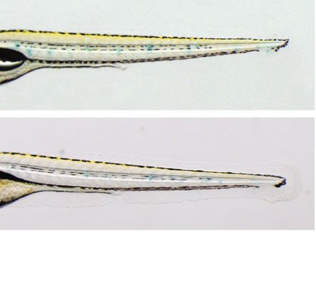
ARTICLE                                                                                  NATURE COMMUNICATIONS | DOI: 10.1038/s41467-017-01961-y

                                                                             FGFR Immunoglobulin domain III

                                                         Danio rerio              IIIa         IIIb    IIIc

                           IIIb-IIIc                  Ciona intestinalis
                             dupl.

                                           + IIIx
                                                       Branchiostoma
                                                        lanceolatum

                                                      Strongylocentrotus
                        Intron                            purpuratus
                         gain          + Amb

                                                        Saccoglossus
                                                         kowalevskii

                                  IIIa                  Apis mellifera
                                 dupl.

                                                          Tribolium
                                                         castaneum

                                                         Drosophila
                                       Retrotransp.     melanogaster

                                        + exons
                                                      Crassostrea gigas

                                                        Nematostella
                                                         vectensis

Fig. 7 Gene structure and AS at the IgIII domain of the Fgfr gene family in metazoans. Schematic representation of the AS diversity in the region encoding
the homolog of the Fgfr IgIII domain for zebrafish (Danio rerio), vase tunicate (Ciona intestinalis), amphioxus (Branchiostoma lanceolatum), purple sea urchin
(Strongylocentrotus purpuratus), acorn worm (Saccoglossus kowalevskii), honey bee (Apis mellifera), red flour beetle (Tribolium castaneum), fruit fly (Drosophila
melanogaster), pacific oyster (Crassostrea gigas), and starlet sea anemone (Nematostella vectensis). Boxes represent exons, horizontal lines are introns, and
diagonal lines connect splicing junctions. Homologous to vertebrate exons IIIa, IIIb, and IIIc are shown in purple, blue and red, respectively. The non-
chordate pro-orthologous exon of exons IIIb and IIIc is colored half blue and half red. The amphioxus- and ambulacrarian-specific alternative exons are
depicted in green and yellow, respectively. Light violet colors are used for the insect-specific mutually exclusive event involving exon IIIa, and brown and
light blue for oyster-specific exons. Gray exons are constitutive in all species. Fruit fly orthologs (heartless and branchless) are intronless genes. Dupl:
duplication; Amb: ambulacraria-specific exon; retrotransp.: retrotranscription

ancestor of chordates; and (ii) it exhibits lineage-specific                      overlapping expression of Fgfr and Esrp genes during sea urchin’s
regulatory requirements in trans in cephalochordates.                            development43. Remarkably, this exon is also present an alter-
                                                                                 natively spliced across the Ambulacraria clade, which comprises
                                                                                 echinoderms and hemichordates. In summary, our phylogenetic
Independent evolution of AS in Fgfr in Bilaterian lineages.                      survey shows that this genomic region is a hotspot for recurrent
Given its remarkable conservation across chordates for over 550                  AS evolution across Bilateria, likely due to its potential to mod-
million years of independent evolution, we next studied the                      ulate FGF signaling8.
evolution of Fgfr AS in further detail. A gene structure analysis for
Bilateria species showed that vertebrate exons IIIb and IIIc are the
result of a single tandem exon duplication that occurred in the                  Discussion
lineage to leading to chordates, before the split of its three main              Similar to recent reports in mouse13,15, we found that Esrp genes
sub-phyla (Fig. 7). In contrast, the single pro-orthologous exon                 are dynamically expressed in non-neural ectoderm territories
IIIb/IIIc is found as constitutively included in all observed tran-              during some developmental stages of zebrafish, vase tunicate,
scripts from all studied non-chordate species. However, previous                 amphioxus, and sea urchin. This suggests a putative ancestral
studies reported other AS events in the genomic locus encoding                   regulatory role for Esrp in embryonic non-neural ectoderm at
the region homologous to the IgIII domain in two non-vertebrate                  least since the last common ancestor of living deuterostomes.
species (the purple sea urchin40,41 and the red flour beetle42), and              Additionally, Esrp was also detected in these organisms in many
we further show that a diverse array of AS events independently                  other tissues involved in clade-specific morphogenetic processes.
evolved in most studied lineages (Fig. 7). In particular, the pre-               While this gene family is likely to play a wide variety of roles
viously reported alternative exon in sea urchin is located                       depending on the cell type and species during development, in
upstream of exon IIIb/IIIc and exhibits no sequence similarity                   some cases Esrp seems to influence epithelial integration and/or
with its neighboring exons. Moreover, its inclusion was not                      cell motility properties. For instance, in sea urchin, mesodermal
detected as Esrp-dependent, which is consistent with the non-                    Esrp-expressing cells undergo a MET, thereby becoming

10                                                     NATURE COMMUNICATIONS | 8: 1799    | DOI: 10.1038/s41467-017-01961-y | www.nature.com/naturecommunications
NATURE COMMUNICATIONS | DOI: 10.1038/s41467-017-01961-y ARTICLE
integrated into ectodermal epithelium, a process that was vertebrates. Strikingly, different AS events have independently
impaired upon Esrp knockdown. Moreover, a similar function evolved in other phyla in the orthologous genomic region of Fgfr
may explain Esrp expression in a group of amphioxus migrating genes encoding the IgIII domain. We suggest that most of these
cells. In the vase tunicate, our experiments suggest that co-option AS events in non-chordate species may also contribute to mod-
of Esrp into a subpopulation of the mesenchymal cell lineage may ulate the affinity of the receptors for different Fgf ligands,
have contributed to modulate their motility. And, in vertebrates, a although their specific developmental roles will be determined by
population of neural crest-derived migratory cells44 that integrate the splicing regulatory logic in each organism. Furthermore, this
into the palate are absent in this structure in both mouse and case exemplifies how a probably non-adaptive fixation at the
zebrafish DMUT embryos. Moreover, in human cell cultures and microevolutionary level had a long-term impact on animal evo-
cancer, ESRP1 has been shown to increase cell adhesion and lution: an intron gain mutation provided the Bilateria lineage
reduce cell motility24,35. Altogether, these results suggest that, with a hotspot of great macroevolutionary potential through post-
although they are usually involved in other types of ontogenetic transcriptional regulation.
functions in vertebrates, like branching morphogenesis15,37,45,
Esrp might also contribute to epithelial integration and/or cell Methods
motility during some specific developmental processes in this Domain and phylogenetic analyzes of Esrp genes. Putative Esrp orthologs for all
subphylum. Thus, given this phylogenetic distribution, it is organisms included in Supplementary Figs. 1, 2 were identified by combining
blastp and tblastn searches against NCBI databases and resources specific for
plausible that Esrp may have regulated specific MET processes in sponges54 and choanoflagellates (unpublished transcripts provided by Daniel
the ancestor of living deuterostomes. Richter). Domain detection was performed using the NCBI conserved domain
Despite these suggested ancestral roles, many of the detected search function55 (https://www.ncbi.nlm.nih.gov/Structure/cdd/wrpsb.cgi). To
exons showed clade-restricted regulation. Such diversification of reconstruct the phylogenetic relationships of Esrp genes among the Apoikozoa
transcriptomic targets may have allowed the refinement and clade (Metazoa + Choanoflagellata), we first aligned protein sequences with
MAFFT56. Neighbor-joining algorithm, as implemented by MEGA7 with default
expansion of Esrp ontogenetic functions in each lineage. Inter- parameters and a JTT substitution model, was employed for tree reconstruction
estingly, among studied vertebrates, non-overlapping programs using only conserved positions of the multi-sequence alignment, as identified by
have evolved mostly by recruiting pre-existing exons, which were MAFFT.
usually already alternatively spliced. On the other hand, only a
minor fraction of the echinoderm Esrp-dependent exons have Zebrafish experimental procedures. Breeding zebrafish (Danio rerio) were
detected homologous counterparts in any of the vertebrate gen- maintained at 28 °C on a 14 h light/10 h dark cycle as previously described. All
protocols used have been approved by the Institutional Animal Care and Use Ethic
omes (and vice versa). This indicates that origin of novel exons is Committee (PRBB–IACUEC), and implemented according to national and Eur-
a major factor contributing to re-assembly of lineage-specific Esrp opean regulations. All experiments were carried out in accordance with the prin-
regulatory programs among distantly related metazoan lineages, ciples of the 3Rs.
as it has been suggested for other AS factors46–48. However, we To investigate the expression of Esrp genes during development, embryos were
raised at 28 °C for staging57 and fixed overnight with 4% paraformaldehyde (PFA)
identified several gene functions that were enriched among Esrp- in PBS at 4 °C. RNA probes were labeled with digoxigenin, and WMISH was
dependent programs across the studied species, altogether indi- performed using Nitrobluetetrazolium/bromochloroindolyl phosphate (NBT/
cating that Esrp regulation may be impacting some common BCIP) as chromogenic substrate for the final alkaline phosphatase58. A minimum
molecular pathways. Moreover, we observed several cases of of 15 embryos of the same stage was used to evaluate expression patterns. To create
shared Esrp regulation at the gene level in which the specific loss-of-function zebrafish lines for esrp1 and esrp2, we used the CRISPR-Cas9
system. We first assessed the presence of multiple promoters in both genes looking
exons differ in each phyla, including genes involved in diverse at RNA-seq-based annotations and H3K4me3 ChIP-seq peaks at their respective
morphogenetic processes in metazoans49–53 (e.g., Exoc7, Slain2, loci in the UCSC browser. Since each gene had only one promoter, we designed
Epb41, Ift88, and Scrib). These could provide an explanation for single-guide RNAs (gRNAs) targeting the first and third exons of esrp1 and esrp2,
the progressive disentanglement of exon programs across phy- respectively. The genomic target sites were identified using a publicly available web
tool (http://crispr.mit.edu/). Selected targeted gRNA sequences corresponded to:
logeny while still having an impact on some related molecular 5′-GGAGCAAGTGGGGATAAGTTGGG-3′ for esrp1 and 5′-
functions. GGAGACCGGGCTCACTGCCGAGG-3′ for esrp2. The CRISPR-Cas9 approach
Our results also highlight multiple conserved roles of Esrp in was performed following the protocol from Chen and Wente laboratories59.
specific organogenetic processes within bony vertebrates, as Engineered vectors were obtained from Addgene. Volume of 1 nl of a mixed
solution containing gRNA (80 ng/µl) and purified Cas9 mRNA (150 ng/µl) was
similar phenotypes in various homologous structures are found in microinjected into one-cell stage zebrafish embryos. F0 founders were crossed with
both mouse and zebrafish mutants. This is reflected at the a WT AB strain, and F1 individuals genotyped by fin clipping to select the
molecular level by a core target set of at least 22 homologous appropriate mutations. We selected the following mutations for further analyzes: a
exons among the three studied vertebrate species, including 168-bp deletion together with a 14-bp insertion that induced the usage of a cryptic
splice donor upstream the start codon for esrp1, and a 17-bp frame-disrupting
members of the Fgfr family. In fact, a number of impaired deletion for esrp2 (Fig. 1b). Crosses between male and female heterozygous
structures reported in esrp1 and esrp2 DMUT embryos have also individuals carrying the same mutation were set to obtain single-mutant lines in
been described in conditional mutant mice for the Fgfr2-IIIb the F2 generation. In addition, individuals from these esrp1 and esrp2 F1 lines were
epithelial isoform10. This is consistent with our results that zeb- crossed to obtain a F2 generation of double heterozygous. DMUT embryos were
rafish Esrp genes, as well as their mammalian counterparts, are obtained in subsequent F3 generations from intercrosses between F2 double
heterozygous fish, at the expected Mendelian ratio. Expression of esrp1 and esrp2 in
essential for inclusion of IIIb exons in embryonic tissues. Thus, a WT, single and double mutant embryos was assessed by quantitative PCR using
major molecular cause for the phenotypes reported in mouse and Life Cycler 480 (Roche) with RNA extractions obtained from pools of 12 embryos.
zebrafish DMUTs is likely the disturbance of epithelial- Relative expression values were obtained by normalizing against Elfa housekeeping
mesenchymal FGF signaling during development of those gene60. Sequences of primers used for genotyping and quantitative PCR assays are
provided in Supplementary Table 2.
organs. This case illustrates how the recruitment of one or few For histological analysis, fixed embryos were progressively dehydrated in
key targets can both constrain and canalize the function of a ethanol and xylol, embedded in paraffin, sectioned in transversal orientation with a
regulatory factor within a certain clade. microtome at 7–10 µm thickness, stained with haematoxilyn and eosin, and
Finally, we determined that Esrp-dependent AS of Fgfr genes mounted with DPX (Eukitt). Alcian blue staining of chondrocytes at 6 d.p.f.
embryos was performed according to the Zebrafish Book61. At least 18 WT and
originated in stem chordates, after intragenic tandem duplication DMUT embryos were used for histological analyzes.
of the pro-homologous exon IIIb/IIIc. Thereby, chordates evolved
a unique way of using the FGF signaling to regulate epithelial- Protein extraction and western blot assays. Total cellular proteins were
mesenchymal crosstalk during development through a new extracted from a pool of embryos (~35 animals) in 0.25 ml of RIPA buffer (10 mM
molecular interaction that has been intensively exploited by Tris-HCl, pH 7.4; 1 mM EDTA; 150 mM, NaCl; 1% Triton X-100; 0.1% SDS;
NATURE COMMUNICATIONS | 8: 1799 | DOI: 10.1038/s41467-017-01961-y | www.nature.com/naturecommunications 11You can also read