Human Rev1 relies on insert-2 to promote selective binding and accurate replication of stabilized G-quadruplex motifs
←
→
Page content transcription
If your browser does not render page correctly, please read the page content below
Published online 8 February 2021                                                   Nucleic Acids Research, 2021, Vol. 49, No. 4 2065–2084
                                                                                                                  doi: 10.1093/nar/gkab041

Human Rev1 relies on insert-2 to promote selective
binding and accurate replication of stabilized
G-quadruplex motifs
Amit Ketkar1 , Lane Smith1 , Callie Johnson2 , Alyssa Richey2 , Makayla Berry1 ,
Jessica H. Hartman3 , Leena Maddukuri1 , Megan R. Reed1 , Julie E.C. Gunderson4 ,
Justin W.C. Leung 5 and Robert L. Eoff 1,*
1
  Department of Biochemistry and Molecular Biology, University of Arkansas for Medical Sciences, Little Rock, AR

                                                                                                                                                              Downloaded from https://academic.oup.com/nar/article/49/4/2065/6130841 by guest on 20 November 2021
72205, USA, 2 Arkansas School for Mathematics, Sciences, and the Arts, Hot Springs, AR 71901, USA, 3 Department
of Biochemistry and Molecular Biology, Medical University of South Carolina, Charleston, SC 29425, USA,
4
  Department of Physics, Hendrix College, Conway, AR 72032, USA and 5 Department of Radiation Oncology,
University of Arkansas for Medical Sciences, Little Rock, AR 72205, USA

Received November 03, 2020; Revised January 06, 2021; Editorial Decision January 07, 2021; Accepted January 15, 2021

ABSTRACT                                                                        INTRODUCTION
We previously reported that human Rev1 (hRev1)                                  The landscape of the genome contains information beyond
bound to a parallel-stranded G-quadruplex (G4) from                             DNA sequence that governs biological systems, includ-
the c-MYC promoter with high affinity. We have ex-                              ing the spatial arrangement of chromosomes, the compo-
tended those results to include other G4 motifs, find-                          sition and architecture of chromatin, and secondary struc-
                                                                                tures adopted by certain sequence motifs. These epigenetic
ing that hRev1 exhibited stronger affinity for parallel-
                                                                                features regulate important functional responses for the
stranded G4 than either anti-parallel or hybrid folds.
                                                                                cell. G-quadruplexes (G4) represent an important type of
Amino acids in the ␣E helix of insert-2 were identi-                            non-canonical DNA structure with ramifications for DNA
fied as being important for G4 binding. Mutating E466                           replication programs and gene expression profiles, as well
and Y470 to alanine selectively perturbed G4 bind-                              as post-transcriptional events. Sequence motifs capable of
ing affinity. The E466K mutant restored wild-type G4                            adopting G4 structures occur non-randomly throughout
binding properties. Using a forward mutagenesis as-                             the genome of all organisms, often at key regions like
say, we discovered that loss of hRev1 increased G4                              replication origins, promoters, and telomeres (1). They are
mutation frequency >200-fold compared to the con-                               formed by tandem tracts of 2–4 guanine bases, such that
trol sequence. Base substitutions and deletions oc-                             four guanines interact through the non-conventional Hoog-
curred around and within the G4 motif. Pyridostatin                             steen hydrogen bonds to form a planar ring, sometimes re-
                                                                                ferred to as a G-tetrad or G-quartet (2). G-quartets stack
(PDS) exacerbated this effect, as the mutation fre-
                                                                                on top of each other, associating via inter-planar pi bonds,
quency increased >700-fold over control and dele-                               to stabilize the four-stranded quadruplex structure. Further
tions upstream of the G4 site more than doubled.                                stabilization is achieved by the coordination of monova-
Mutagenic replication of G4 DNA (±PDS) was par-                                 lent cations like Na+ or K+ with the O6 atoms in the cen-
tially rescued by wild-type and E466K hRev1. The                                tral channel of the G-quartet. G4-DNA structures, once
E466A or Y470A mutants failed to suppress the PDS-                              formed, can be very stable in vitro, often with significantly
induced increase in G4 mutation frequency. These                                higher melting temperatures as compared to B-form DNA
findings have implications for the role of insert-2,                            (3).
a motif conserved in vertebrates but not yeast or                                  The existence of G4 structures in cells and in vivo is sup-
plants, in Rev1-mediated suppression of mutagen-                                ported by orthogonal studies in a variety of organisms (4).
esis during G4 replication.                                                     Maximally formed during S-phase of the cell cycle, the four-
                                                                                stranded secondary structure of G4 DNA represents an en-
                                                                                dogenous barrier to the replisome (5). Consequently, the
                                                                                coordinated interplay of multiple proteins equipped to re-
                                                                                solve these structures is required for effective G4 mainte-

* To   whom correspondence should be addressed. Tel: +1 501 686 8343; Fax: +1 501 686 8169; Email: RLEoff@uams.edu


C The Author(s) 2021. Published by Oxford University Press on behalf of Nucleic Acids Research.
This is an Open Access article distributed under the terms of the Creative Commons Attribution License (http://creativecommons.org/licenses/by/4.0/), which
permits unrestricted reuse, distribution, and reproduction in any medium, provided the original work is properly cited.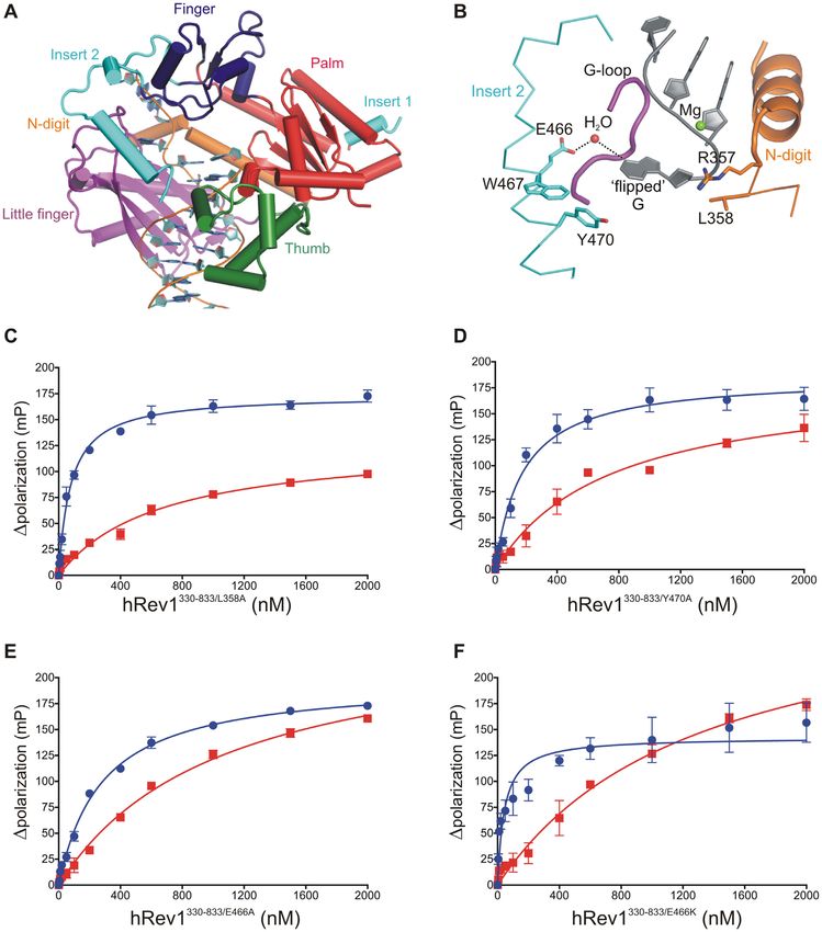
2066 Nucleic Acids Research, 2021, Vol. 49, No. 4
nance. Many studies have investigated the G4 unwinding by MATERIALS AND METHODS
helicases, such as Pif1, FANCJ, BLM, and WRN (among
Reagents and services
others)––revealing key features important for G4-selective
activity (2). The consequence of defective G4 unwinding is All chemicals and reagents used in this study were molec-
apparent from the increased genomic instability and epige- ular biology grade, or better. The dNTP solutions were
netic defects observed when the action of these helicases is purchased from GE Healthcare Life Sciences (Piscat-
lost. Additional studies have implicated other DNA repli- away, NJ, USA) and Thermo-Fisher Scientific (Waltham,
cation and repair factors, such as the repair polymerase pol MA, USA). All synthetic oligonucleotides were pur-
and translesion DNA synthesis polymerases (TLS pols), chased from Integrated DNA Technologies (Coralville, IA,
in the suppression of genomic and epigenomic instability USA) desalted and validated by mass spectrometry. p-
near G4 motifs (6,7). The exact nature of TLS enzyme par- Hydroxyphenylglyoxal (HPG) was purchased from Pro-
ticipation in G4 replication remains unclear, but a number teoChem (Hurricane, UT, USA). The HAP-1 cell lines
of studies have implicated the Y-family member, Rev1, as a (parental hRev1-proficient and REV1KO ) were obtained
Downloaded from https://academic.oup.com/nar/article/49/4/2065/6130841 by guest on 20 November 2021
central player in effecting the bypass of G4 motifs. from Horizon Discovery (Cambridge, UK), while the
As part of the replication stress response, Rev1 helps pro- HEK293T cells was obtained from American Type Culture
mote continuous synthesis past a variety of blocks to repli- Collection (ATCC; Manassas, VA, USA). Knock-out of the
cation and in response to nucleotide depletion (8). The role REV1 gene was confirmed by the company using Sanger se-
of Rev1 in G4 replication is likely multifaceted. Rev1 is atyp- quencing. We confirmed deletion of hRev1 protein expres-
ical among polymerases in that it uses a protein-template sion by immunoblotting (data not shown). A mouse mon-
guided mechanism of nucleotide selection, which almost ex- oclonal anti-Rev1 antibody was used (Santa Cruz Biotech-
clusively inserts dCMP and severely limits processive DNA nology, Dallas, TX, USA; Catalog #sc-393022). DNA se-
synthesis. Avian cells lacking Rev1 exhibit deficiencies in quencing to confirm mutations in plasmids used for expres-
G4 replication and maintenance of the epigenetic land- sion of recombinant hRev1 (wild-type and mutants) and
scape surrounding quadruplex-forming sequences (9,10). to determine the mutagenic profile of the samples from the
Like tolerance of DNA adducts, the replisome adapts to en- supF forward mutagenic assay was performed at the DNA
dogenous replication blocks, and Rev1 is part of a complex Sequencing Core facility at UAMS. The tryptic digestion of
of proteins that promotes G4 bypass. A key role for Rev1 HPG-reacted hRev1 and subsequent analysis of the HPG-
is related to a motif in the C-terminal domain that aids in labeled samples by mass spectrometry was performed at the
the recruitment of multiple TLS pols to sites of replication UAMS Proteomics Core facility.
stress (11–14). Deletion of the Rev1 C-terminus reduced the
efficiency of G4 bypass, with complete loss of Rev1 leading
to defective histone cycling and loss of epigenetic regulation
of gene expression. Both avian and human Rev1 (hRev1) DNA substrate preparation
could recover G4 replication efficiency in rev1 mutant DT40 Oligonucleotides were resuspended in 50 mM HEPES
cells, but catalytically inactive Rev1 only partially restored buffer (pH 7.5). Substrates for the fluorescence polar-
the replication efficiency observed in wild-type cells (9). ization experiments were prepared by annealing each of
In addition to TLS pol recruitment, Rev1 functions in the 5 -FAM-labeled G4 and non-G4 oligos shown in Ta-
concert with the FANCJ helicase to promote replication ble 1 with the 11-mer primer (1:2 molar ratio of tem-
across sequences prone to fold into quadruplex structures plate:primer) in a 50 mM HEPES buffer (pH 7.5) con-
(15,16). These findings served as the rationale for bio- taining 100 mM KCl/LiCl. For ssDNA substrates, the 11-
chemical studies on human Rev1 (hRev1) interactions with mer primer was excluded. The mixture was heated at 95◦ C
G4 DNA. for 5 min, followed by slow-cooling to room temperature.
We previously found that hRev1 binds G4-DNA sub- Circular dichroism (CD) spectroscopy was performed to
strates with up to 15-fold greater affinity compared to non- validate potassium-dependent G4 formation (Supplemen-
G4 sequences (17). Further, hRev1 mechanically disrupted tary Figure S1). The annealed substrates were stored at
and prevented refolding of G4 structures in vitro through a room temperature in amber-colored tubes in the dark. The
mechanism that did not require nucleotidyl transfer (17). primer-template substrates for enzyme activity assays were
Yet, a number of questions remain unanswered concern- prepared similarly by mixing the 42-mer G4 or non-G4
ing how hRev1 interacts with G4 DNA. Knowledge of the template oligonucleotides with the 5 -FAM-labeled 23-mer
molecular interface between Rev1 and G4 would improve primer (1.5:1 molar ratio of template:primer) in 40 mM
our understanding of features that may be important deter- HEPES buffer (pH 7.5) containing 40 mM KCl. These were
minants of biological function during replication of struc- then annealed by heating to 95◦ C for 5 min, followed by
tured DNA. As such, the biochemical mechanism and the slow-cooling to room temperature. The G4 and non-G4 oli-
ramifications for hRev1 action during G4 replication in hu- gos were prepared for the circular dichroism studies by re-
man cells both remain active areas of investigation. In the suspending them in 10 mM Tris–HCl buffer (pH 7.5) con-
present study, we used biochemical approaches to identify taining 100 mM KCl/LiCl. The substrates were heated to
regions of Rev1 important for its interaction with G4 DNA 95◦ C for 5 min, followed by slow-cooling to room tempera-
and then validated the biological relevance of these regions ture to allow the G-quadruplexes to form. Substrates were
to accurate replication of G4 motifs in human cells. stored at room temperature.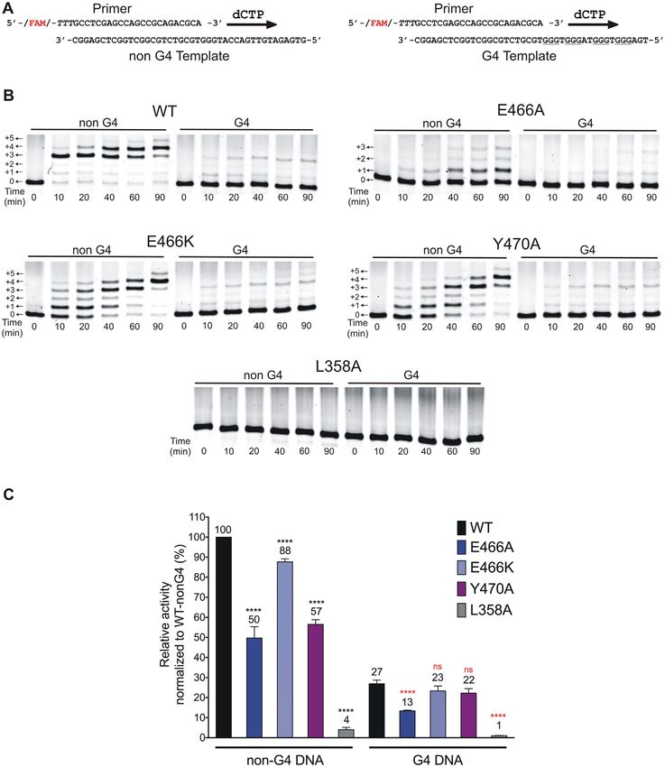
Nucleic Acids Research, 2021, Vol. 49, No. 4 2067
Table 1. Sequences of G4- and nonG4-DNA substrates used in this studya
Name Sequence Type of G4 fold
Myc 14/23 5 -FAM-AGGGTGGGTAGGGTGGGTTATGAGATGAT -3 Parallel
Myc 2/11 5 -FAM-AGGGAGGGTGGGAAGGGTTATGAGATGAT -3 Parallel
Non-G4 Myc 5 -FAM-AGCGTGCGTAGCGTGCGTTATGAGATGAT -3 -
control
KRAS 22RT 5 -FAM-AGGGCGGTGTGGGAAGAGGGAATATGAGATGAT -3 Parallel
Non-G4 KRAS 5 -FAM-AGCGCGCTGTGCGAAGAGCGAATATGAGATGAT -3 -
control
Bcl2 1245 5 -FAM-AGGGGCGGGCGCTTTAGGAAAAGGGCGGGTTATGAGATGAT-3 Parallel
Non-G4 Bcl2 5 -FAM-AGCGGCGCGCGCTTTAGGAAAAGCGCGCGTTATGAGATGAT-3 -
control
Rev1-prom 5 -FAM-AGGGCGGGGCCGGGGAGGGTATGAGATGAT -3 Parallel
Non-G4 5 -FAM-AGCGCGCGGCCGGCGAGCGTATGAGATGAT -3 -
Downloaded from https://academic.oup.com/nar/article/49/4/2065/6130841 by guest on 20 November 2021
Rev1-prom control
TBA 5 -FAM-TGGTTGGTGTGGTTGGTATGAGATGAT -3 Antiparallel
Non-G4 TBA 5 -FAM-TGCTTGCTGTGCTTGCTATGAGATGAT -3 -
control
hTelo-4 5 -FAM-TTGGGTTAGGGTTAGGGTTAGGGATATGAGATGAT -3 Hybrid
Non-G4 Telo-4 5 -FAM-TTGCGTTAGCGTTAGCGTTAGCGATATGAGATGAT -3 -
control
11-mer primer 5 - ATCATCTCATA -3 -
42-mer 5 -TGAGGGTGGGTAGGGTGGGTGCGTCTGCGGCTGGCTCGAGGC-3 Parallel
G4-templateb
42-mer non 5 -GTGAGATGTTGACCATGGGTGCGTCTGCGGCTGGCTCGAGGC-3 -
G4-templateb
23-mer 5 5 -FAM-TTTGCCTCGAGCCAGCCGCAGACGCA-3 -
FAM-primerb
a The guanine bases involved in the formation of G4-tetrads are shown in bold. The corresponding non-G4 oligonucleotides had C in place of a G (un-
derlined in each sequence). A single non-G4 Myc control sequence was used for both the Myc-14/23, and Myc-2/11 G4 sequences. Shown in italics is the
region of each oligonucleotide that is complementary to the common 11-mer primer. The 5 -FAM-labeled versions of all substrates shown above were used
in the fluorescence polarization experiments.
b The 42-mer template and 23-mer primer sequences were used exclusively in assays to determine polymerase activity. Shown in italics in the region of each
oligonucleotide that is complementary to the 23-mer primer.
Generation of mutant hRev1 proteins and purification mented with 10% (v/v) fetal bovine serum and 1% (v/v)
330–833 antibiotics for a final concentration of 100 units/mL peni-
Expression and purification of hRev1 (the polymerase
cillin, 100 g/ml streptomycin, and 0.25 g/ml ampho-
core) from Escherichia coli was performed as previously de-
tericin B (Sigma-Aldrich, St. Louis, MO, USA). Cells were
scribed (17). Mutations in the hRev1330–833 construct were
cultured in an incubator at 37◦ C in an atmosphere contain-
generated by site-directed mutagenesis, using the follow-
ing 5% (v/v) CO2 . Cultures were harvested at >80% con-
ing primers: (a) E466A: forward primer – 5 -CCCAGCT
fluency (∼15–20 × 106 cells per flask), and the cell pellet
GGCGTGGCAGTATTAC-3 ; reverse primer – 5 -GTA
was lysed in a 40 mM HEPES buffer (pH 7.5) containing
ATACTGCCACGCCAGCTGGG-3 . (b) E466K: forward
120 mM NaCl, 1 mM EDTA, 1% (v/v) Triton-X 100, and
primer – 5 -CCCCAGCTGAAATGGCAGTATTAC-3 ;
protease and phosphatase inhibitors. The clear lysate ob-
reverse primer – 5 -GTAATACTGCCATTTCAGCTGGG
tained after centrifugation was incubated with streptavidin–
G-3 . (c) W467A: forward primer – 5 -CCCAGCTGG
Sepharose beads (GE Healthcare) at 4◦ C for 16 h to enable
AGGCGCAGTATTACC-3 ; reverse primer – 5 -GGTAA
the SFB-hRev1 protein to bind. After washing the beads
TACTGCGCCTCCAGCTGGG-3 . (d) Y470A: forward
thoroughly to remove non-specifically bound proteins, the
primer 5 -GGCAGTATGCCCAGAATAAAATC-3 ; re-
SFB-hRev1 was eluted in a buffer containing 2 mg/ml bi-
verse primer – 5 -GATTTTATTCTGGGCATACTGCC
otin. The eluate was dialysed to remove biotin, followed by
-3 . (e) L358A: forward primer – 5 -CTATTCTCATTCA
concentration of the eluted protein using spin concentra-
AGAGCGCATCACATATC-3 ; reverse primer – 5 -GAT
tors (Amicon). Purity of eluted hRev11–1251 protein was con-
ATGTGATGCGCTCTTGAATGAGAATAG-3 . Mutant
firmed by SDS-PAGE followed by Ponceau staining, as well
enzymes were expressed and purified using the same pro-
as immunoblotting using an anti-FLAG-tag antibody (Sup-
tocol as wild-type enzyme (Supplementary Figure S2).
plementary Figure S3; Novus Biologicals, Centennial, CO,
Full-length hRev11–1251 was cloned into the p-BABE vec-
USA).
tor using the Gateway™ cloning system (Thermo-Fisher;
Waltham MA, USA) to express hRev1 as a N-terminal
S-protein-FLAG-Biotin (SFB)-tandem affinity tagged pro- DNA binding by fluorescence polarization
tein (Supplementary Figure S3). For expression and purifi-
cation, HEK293T cells stably expressing the SFB-tagged The affinity of the purified hRev1330–833 wild-type and mu-
hRev11–1251 protein were generated. These cells were then tant proteins, as well as the purified hRev11–1251 protein to-
cultured on a large scale in five to ten 175 cm2 tissue cul- wards all the 5 -FAM labeled non-G4 and G4 DNA sub-
ture flasks in Dulbecco’s modified Eagle medium supple- strates described in this study, was determined using fluores-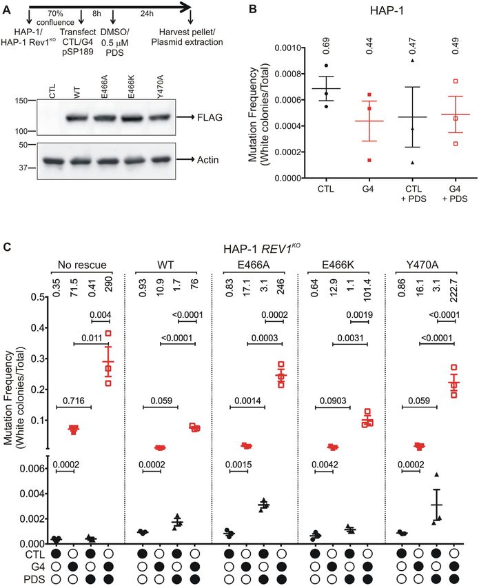
2068 Nucleic Acids Research, 2021, Vol. 49, No. 4
cence polarization on a plate-reader (Biotek SynergyH4), the reactions were quenched by the addition of 200 mM
as described previously (17). Briefly, titrations of various L-arginine. Laemmli buffer (1×) was added to each sam-
concentrations of each protein with a fixed concentration ple, and it was heated to 95◦ C for 5 min before loading on
(1 nM) of all the DNA substrates were performed in buffer a 4–20% SDS-PAGE gradient gel for electrophoresis. The
containing either 100 mM KCl or 100 mM LiCl. The change gel was stained by Coomassie Blue stain, and the band for
in fluorescence polarization at every titration was measured, hRev11–1251 protein in each lane was excised and submit-
and plotted as a function of protein concentration, and fit- ted to the UAMS Proteomics Core for tryptic digestion and
ted to a quadratic equation as described previously (17), to mass spectrometric analysis. Experiments were performed
determine the values of equilibrium dissociation constants. in duplicate. Results from the mass spectrometric analy-
sis were inspected using the Scaffold Viewer software (Pro-
teome Software Inc.). The unmodified and HPG-modified
Measurement of enzyme activity
peptides were identified for each sample. The peak inten-
In order to determine the effect of mutations on the catalytic sity for both the unmodified and HPG-modified version
Downloaded from https://academic.oup.com/nar/article/49/4/2065/6130841 by guest on 20 November 2021
efficiency of hRev1330–833 , the enzymatic activity of all the of a peptide was determined manually by calculating the
mutant hRev1 proteins was determined with non-G4 and area under the curve from the total ion chromatogram for
G4 DNA template–primer substrates and compared to the the parent ion using the XCalibur software (Thermo Fisher
activity of the wild-type protein. All assays were performed Scientific). The extent of HPG-modification at every argi-
in 40 mM HEPES buffer (pH 7.5) containing 100 mM KCl, nine identified by mass spectrometry was calculated as a
0.1 mg/mL BSA, and 5 mM dithiothreitol at 37◦ C using 50 ratio of the HPG-modified intensity to the total intensity
nM protein and 200 nM DNA. Reactions were initiated by value for that arginine (unmodified + modified). Results
adding a mixture of 5 mM MgCl2 and 1 mM dCTP. At in- were normalized across samples, by considering the HPG-
tervals of 0, 10, 20, 40, 60 and 90 min, 15 l aliquots were modification in the no-DNA sample as 100%.
withdrawn from each reaction mixture and added to tubes
containing 85 l of quench solution (95% v/v formamide,
supF forward mutagenesis assay
20 mM EDTA, 0.1% w/v bromophenol blue), and heated at
95◦ C for 5 min. Aliquots of these quenched reactions were We engineered an 18 nucleotide G4 forming sequence
loaded on to 7 M urea/14% (w/v) acrylamide gels and elec- derived from the c-Myc promoter immediately upstream
trophoresed at a constant power of 35 W for 2–3 h to sep- of the supF tRNA coding region in the pSP189 plas-
arate the products. Gels were scanned on a Typhoon Im- mid (Supplementary Figure S4). The original pSP189
ager (GE Life Sciences, Marlborough, MA, USA), and the plasmid served as a non-G4 control (20). Cloning of
bands corresponding to substrate and products in each lane the G4 fragment into psP189 was performed using three
were quantified using the ImageJ software (18). Total prod- sequential PCR reactions to insert the 18 nt sequence in
uct formation for each protein with both the DNA sub- three parts, using the following primer pairs: PCR reaction
strates was plotted as a function of time. This was converted 1, forward primer 5 -GAGCCCTGCTCGAGCTGTG
to percent product formed per unit time, by dividing the GAGGGTGGGGTTCCCGAGCGG-3 , reverse primer
sum of intensities of all product bands in a lane by the to- 5 -CCGCTCGGGAACCCCACCCTCCACAGCTCG
tal sum of intensities of all bands in that lane. The initial AGCAGGGCTC-3 . PCR reaction 2, forward primer
portion of the velocity curve was fit to a linear equation to 5 -GAGCCCTGCTCGAGCTGTGAGGGTGGGGAGG
estimate the rate of product formation. Activities were nor- GTGGGGTTCC-3 , reverse primer 5 -GGAACCCCACC
malized to the activity of wild-type enzyme (taken as 100% CTCCCCACCCTCACAGCTCGAGCAGGGCTC-3 .
with non-G4 DNA) for both DNA substrates. Relative ac- PCR reaction 3, forward primer 5 -GAGCCCTGCTC
tivity of each protein was also calculated for the non-G4 and GAGCTGTGGGGAGGGTGGGGAGGGTGGG-3 ,
G4 substrates by dividing their respective percent product reverse primer 5 - CCCACCCTCCCCACCCTCCCCACA
values. GCTCGAGCAGGGCTC-3 . After each PCR step, the
resulting products were sequenced to confirm the insertion
of a portion of the 18 nt G4 sequence. PCR product with
Chemical footprinting of hRev1 by HPG-modification of
the correct sequence served as the template for the next
arginines
PCR reaction.
Chemical labelling of hRev11–1251 was performed using the Both the plasmids were transfected into HAP-1 cells
arginine-reactive chemical HPG, as described previously (Horizon Discovery, Cambridge, UK), which either ex-
(19). Briefly, 10 M of purified hRev11–1251 protein was pressed wild-type hRev1 (HAP-1) or had the hRev1 gene
allowed to react with 0.5 mM HPG in 50 mM HEPES knocked out by CRISPR-Cas9 (REV1KO ). The HAP-1
(pH 7.5) buffer containing 100 mM KCl. Prior to addi- cells were cultured in Iscove’s modified Dulbecco’s medium
tion of HPG, the protein was pre-incubated with either (Millipore-Sigma, St. Louis, MO, USA) supplemented with
buffer (no DNA/control), 30 M of single-stranded (ss)- 10% (v/v) fetal bovine serium and 1% (v/v) antibiotics con-
Myc G4, ss-TBA G4 or ss-non G4 DNA for 15 min prior taining 100 units/mL penicillin, 100 g/ml streptomycin,
to HPG modification. All the ssDNA substrates were pre- and 0.25 g/ml amphotericin B (Sigma-Aldrich, St. Louis,
pared by heating them to 95◦ C for 5 min in 25 mM HEPES MO, USA). Cells were cultured in an incubator at 37◦ C
buffer (pH 7.5) containing 100 mM KCl, and then cool- in an atmosphere containing 5% (v/v) CO2 . Transfection
ing them overnight to room temperature. One hour after was performed using Lipofectamine-3000 (Thermo Fisher,
incubating with HPG (in the dark) at room temperature, USA) according to the manufacturer’s instructions. Ectopic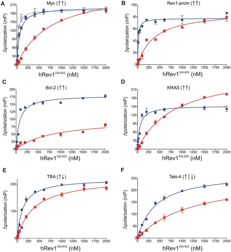
Nucleic Acids Research, 2021, Vol. 49, No. 4 2069
expression of hRev1 was confirmed by immunoblotting relatively uniform parallel-stranded quadruplex structure
(Supplementary Figure S5). The transfected cells were cul- (23,24). We wanted to investigate whether preferential bind-
tured either in the presence or absence of the G4-stabilizing ing to G4 DNA by hRev1 extended to other G4 motifs and
chemical pyridostatin (PDS; 0.5 M) for 24 h and then folds. We also wanted to test whether changing the stabil-
harvested. The pSP189 plasmids (control or G4) were ex- ity, loop-length, or inserting a base between tandem gua-
tracted from cell pellets using the plasmid miniprep kit nines would change the preference of hRev1 for binding to
(Qiagen). Following treatment with the Dpn I endonu- a parallel-stranded G4 substrate. To achieve this objective,
clease to digest parental (methylated) DNA, the plasmids we designed a panel of G4 substrates (Table 1). A non-G4
were then used to transform the MBM7070 indicator strain control sequence of the same length and with similar GC
of E. coli, and transformants were plated on LB-agar content was designed for every G4 substrate.
plates containing 100 g/ml ampicillin, 2 mg/ml 5-bromo- CD spectroscopy was used to validate the formation of
4-chloro-3-indolyl--D-galactopyranoside (X-Gal) and 1 G4 structure in 100 mM KCl buffer for all substrates (Sup-
mM isopropyl--D-1-thiogalactopyranoside (IPTG). After plementary materials and methods). A characteristic pos-
Downloaded from https://academic.oup.com/nar/article/49/4/2065/6130841 by guest on 20 November 2021
incubation at 37◦ C for 15 h, blue and white colonies were itive peak at ∼264 nm and a negative peak at ∼240 nm
observed on the plates. If necessary, the plates were incu- was observed for the parallel-stranded G4 substrates (Sup-
bated overnight at 4◦ C to allow the blue colour to inten- plementary Figure S1). The melting temperature (Tm ) for
sify. The number of blue (unmutated) and white (mutated) each G4 structure was also calculated for all substrates
colonies were counted for each plate using the Fiji version by measuring change in CD signal as a function of tem-
of ImageJ software. Mutation frequency was determined for perature (Supplementary Table S1). For DNA binding as-
each experimental sample by plotting the ratio of number of says, five parallel-stranded G4 substrates were prepared.
white colonies for a sample, to the total number of colonies Two c-MYC-derived sequences were included, Myc 14/23
(blue and white) for that sample. Each experimental condi- and Myc 2/11. These two sequences both form parallel-
tion was performed in triplicate. For every replicate, at least stranded G4 structures but they differ in loop arrangement,
10 000 colonies were counted. Further, to investigate the with the more stable Myc 14/23 possessing a 1:2:1 loop ar-
mutagenic profile of the supF gene vicinity region, plasmids rangement between runs of guanine and Myc 2/11 possess-
from representative white colonies were extracted and sub- ing a 1:1:2 loop arrangement between guanines (Table 1).
mitted for DNA sequencing. For every experimental condi- The KRAS 22RT derived substrate folds into a parallel-
tion, 10 white colonies were sequenced (Supplementary Fig- stranded quadruplex as well, but the sequence includes a
ures S6 and S7). The mutant sequences were then aligned 1:1:4 loop arrangement with single thymidine interrupting
using the T-Coffee alignment server (21). The region in the the second run of three guanines. The nomenclature for
vicinity of the supF gene was divided into four zones. The the KRAS G4 structure stems from the original motif be-
nature and frequency of occurrence of mutations was deter- ing named 32R (25). Truncation of the 32 nt sequence to
mined by examination of every zone across all the samples. a core 22 nucleotides along with mutation of a guanine
Mutations identified by sequencing were either base sub- to thymidine (22RT) stabilized the parallel-stranded struc-
stitutions (transversions and transitions) or deletions. We ture and prevented alternative G4 folds (26). The Bcl2 G4
divided the sequence surrounding the supF gene into four motif in the P1 promoter of the BCL-2 gene controls ex-
zones: zone 1––containing nts on the 5 side of the G4 mo- pression of this anti-apoptotic protein (27). The wild type
tif, zone 2––including the G4 motif, zone 3––including the Bcl2 G4 motif is 39 nucleotides long and includes six runs
supF gene and zone 4––including nts on the 3 -side of the of guanine (28). While 15 different folds are possible, two
supF gene. The mutation profile was calculated by counting major G4 species are thought to arise from the wild type
the number of base substitutions and deletions for a given 39mer sequence: Bcl2 1245 and Bcl2 2345, where the num-
number of nucleotides in each zone. Statistical analysis was bers represent the involvement of the different runs of gua-
performed using Graphpad Prism (San Diego, CA, USA). nine. For the Bcl2 1245 sequence, the third run of guanines
was mutated to three thymidines to improve the uniformity
RESULTS of the G4 structure in vitro (28). The Bcl2 1245 G4 mo-
tif folds into a parallel stranded G4 but this sequence pos-
Substrate design and characterization
sesses a 13 nt loop between the second and third guanine
We have shown that hRev1330–833 binds G4 DNA with runs. Finally, we used the QGRS algorithm to identify mul-
greater affinity than non-G4 substrates (17). Our earlier tiple putative G4 motifs in the REV1 promoter (Supple-
study used a G4-forming sequence that was based on a mo- mentary Figure S8). The highest scoring motif was found
tif found in the c-MYC promoter (Table 1, Myc 14/23). >1760 bp upstream of the transcription start site. This site
The nomenclature for the Myc G4 substrates is related to was identified previously as one of many bone fide G4 form-
changes to the wild type purine-rich 27mer G4 motif where ing sequences located in the promoters of DNA repair and
multiple runs of three (or more) guanines exist in tandem. damage tolerance genes (29,30). We designed a substrate
Mutating the 14th and 23rd guanines to thymidine prevents based on the top-scoring motif (Rev1-prom) and evaluated
formation of multiple G4 structures and improves the uni- it for G4-related properties. The Rev1-prom G4 motif is
formity of the G4 folds formed in vitro (22). Similarly, the comprised of 18 nts, including four runs of three or more
Myc 2/11 substrate was designed by mutating residues 2 guanines. It has a 1:4:1 loop arrangement between gua-
and 11 from the wild type G4 motif. The Myc 14/23 se- nines and appeared to form a parallel-stranded G4 struc-
quence was originally selected because it is an extensively ture based on CD analysis (Supplementary Figure S8). All
characterized G4 motif that is known to form a stable and the parallel-stranded G4 substrates were thermodynami-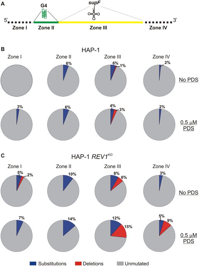
2070 Nucleic Acids Research, 2021, Vol. 49, No. 4

cally stable, based on their calculated melting temperatures    full-length hRev11–1251 (Table 2). The 20-fold preference for
(Tm between 56–90◦ C: Supplementary Table S1). As ex-           binding to G4 DNA for the full-length enzyme is compa-
pected, when 100 mM LiCl was used instead of KCl, the           rable to the 25-fold difference observed for the polymerase
stability of all G4 substrates was significantly reduced (as    core domain (a.a. 330–833). These results are in agreement
seen by much lower melting temperatures; Supplementary          with the notion that hRev1 binds to stable, parallel-stranded
Table S1).                                                      G4 structures with greater affinity than non-structured
   In addition to the parallel-stranded G4 substrates, we       DNA.
prepared a four-repeat human telomeric sequence (hTelo-            The KD,DNA values measured for the other parallel-
4) to study hRev1 interactions with a hybrid G4 structure,      stranded G4 substrates demonstrated trends in binding
while the thrombin binding aptamer (TBA) represented a          affinity that were similar to the Myc G4 motif. Interrupt-
relatively well-defined and stable anti-parallel G4 fold (Ta-   ing the tandem guanines with a single thymidine did not
ble 1). We confirmed using CD that the hTelo-4 oligonu-         alter hRev1330–833 binding to a parallel-stranded G4, as the
cleotide formed a mixed/hybrid G4 structure in 100 mM           KD,DNA value for binding to the KRAS 22RT G4 substrate

                                                                                                                                 Downloaded from https://academic.oup.com/nar/article/49/4/2065/6130841 by guest on 20 November 2021
KCl, as has been previously reported (31). Further, we also     was 16-fold lower than that measured for the non-G4 con-
confirmed that the TBA oligonucleotide sequence formed          trol sequence in KCl (Figure 1 and Table 2). Inserting a 13
an anti-parallel stranded G4 structure in KCl buffer, with      nt loop between tandem guanines also appeared to have a
a positive peak at 290 nm and a negative peak at 260 nm         minimal impact on G4 selectivity, given that hRev1330–833
(Supplementary Figure S1), as has been reported previously      bound to the Bcl-2 1245 substrate with 11-fold greater affin-
(32). The corresponding non-G4 control substrates of all se-    ity than the non-G4 control (Figure 1 and Table 2). A large
quences (non-G4 DNA) were tested similarly using CD to          difference in binding affinity between G4 and non-G4 DNA
confirm that none of them formed quadruplex structures          substrates was observed with the parallel-stranded G4 se-
(data not shown).                                               quence from the human Rev1 promoter (Rev1-prom). In
                                                                buffer containing potassium, the KD,DNA for hRev1330–833
                                                                binding to the Rev1-prom sequence was 13 ± 3 nM com-
hRev1 exhibited preferential binding to parallel-stranded G4
                                                                pared with a KD,DNA of 370 ± 70 nM for the non-G4 con-
DNA compared to anti-parallel and hybrid-type of G4 DNA
                                                                trol (Figure 1 and Table 2). When these G4 oligonucleotides
substrates
                                                                were suspended in LiCl, the difference in binding affinity for
Fluorescence polarization was used to monitor hRev1 bind-       G4 and non-G4 substrates was reduced considerably (Sup-
ing to 5 -FAM-labeled DNA for each of the different G4         plementary Table S2). Thus, it would seem that the prefer-
and non-G4 ssDNA substrates. By varying the concentra-          ential binding to parallel-stranded G4 DNA over non-G4
tion of hRev1 in the reaction mixture, we measured the          sequences is a property that is inherent to hRev1 and not
equilibrium dissociation constant (KD,DNA ) for binary com-     restricted to c-MYC-derived G4 sequences.
plex formation. The resulting titration curves were fit to a       The trends observed for hRev1 binding to the hTelo-4
quadratic equation (Figure 1). The results of the DNA bind-     substrate were quite different from those of the parallel-
ing assays are further summarized in Table 2 (ssDNA) and        stranded G4 substrates. In the presence of 100 mM KCl
Supplementary Table S3 (dsDNA).                                 buffer, conditions under which the hTelo-4 sequence was
   As observed in our previous study, hRev1 exhibited           confirmed to form a mixed/hybrid G4 structure, the mea-
greater affinity for the Myc 14/23 G4 sequence than the         sured binding constant to hRev1330–833 was 406 ± 38 nM.
non-G4 control (Figure 1 and Table 2). In this case,            This was ∼7-fold lower affinity than that measured for the
hRev1330–833 exhibited an equilibrium binding constant of       Myc-14/23 G4 substrate. A KD,DNA value of 1246 ± 196 nM
37 ± 7 nM for Myc-14/23 ss-G4 DNA in 100 mM KCl.                was measured for hRev1330–833 binding to the non-G4 se-
The value for binding constant for the corresponding non-       quence of equivalent length to the hTelo-4 substrate, which
G4 substrate was measured to be 910 ± 120 nM in 100 mM          is only ∼3-fold greater than the hTelo-4 G4 substrate. The
KCl. Thus, hRev1330–833 bound with ∼25-fold higher affin-       affinity was reduced by 1.5-fold when KCl was replaced with
ity to G4 Myc-14/23 as compared to the non-G4 control.          LiCl (634 ± 105 nM), an indication that hRev1330–833 does
This is in agreement with our observations reported earlier     not exhibit a strong preference for binding to the hybrid
(17). Similar to the Myc 14/23 G4 substrate, hRev1 bind-        hTelo-4 G4 structure.
ing to the Myc 2/11 G4 substrate was much tighter than             Similar to the hTelo-4 results, hRev1330–833 bound to the
the non-G4 control (Figure 1A and Table 2). The prefer-         anti-parallel G4 DNA substrate TBA with a KD,DNA value
ence for the 14/23 and 2/11 G4 folds was also evident from      of 127 ± 14 nM in 100 mM KCl, which is ∼2-fold higher
the fact that binding affinity was ∼5–7-fold tighter in pres-   than that measured for the Myc-14/23 G4 DNA. The ob-
ence of KCl compared to buffer containing LiCl (Supple-         served value of dissociation constant for the non-G4 con-
mentary Table S2). Substituting potassium with lithium re-      trol was 380 ± 40 nM. The ∼3-fold preference for TBA G4
duced the difference in the hRev1330–833 binding constants      DNA over the non-G4 control DNA is less than that ob-
for the Myc 14/23 G4 substrate and the non-G4 control           served for any of the parallel-stranded G4 substrates. In the
from 25-fold for potassium to around 3-fold for the same        presence of LiCl, the binding affinity did not change sub-
substrates in LiCl (Table 2 and Supplementary Table S2).        stantially for either substrate (TBA = 139 ± 17 nM, non-
For the Myc 2/11 substrate, the preference for G4 DNA was       G4 control = 310 ± 40 nM). In this regard, hRev1330–833
reduced from ∼40-fold in KCl to ∼8-fold in lithium (Table       did not bind to the anti-parallel G4 structure formed by the
2 and Supplementary Table S2). The preferential binding         TBA motif in a manner that was distinct from the non-G4
to the Myc 14/23 G4 DNA substrate was also observed for         control.
Nucleic Acids Research, 2021, Vol. 49, No. 4 2071
Downloaded from https://academic.oup.com/nar/article/49/4/2065/6130841 by guest on 20 November 2021
Figure 1. hRev1330–833 preferentially binds to G4-forming DNA sequences, with a greater affinity for parallel-stranded G4 DNA than other G4 folds.
The hRev1330–833 protein was titrated into a solution containing either single-stranded (ss)-G4-DNA (blue) or ss-non-G4-DNA (red) substrates at 1 nM.
The range of concentrations for the protein is indicated on the X-axis. The change in fluorescence polarization at each concentration was measured and
plotted as a function of the protein concentration. (A-F) Binding curves for hRev1330–833 core protein with the indicated G4 DNA substrate. In panel A,
the binding curve for Myc-14/23 is shown as a solid blue line (full circles), while that for Myc-2/11 is shown as a dotted blue line (open circles). The G4
fold is indicated by the direction of arrows in parentheses for each panel (↑↑ = parallel G4, ↑↓ = anti-parallel G4, ↑|↓ = hybrid G4). Resulting data were
fit to a quadratic equation to yield the binding dissociation constants given in Table 2. Reported values represent the mean ± SD (n = 3).
Similar binding studies for hRev1330–833 were also per- Chemical footprinting mapped the G4 DNA interacting
formed using the primer-template ds-DNA G4 and non-G4 residues in the G-loop and the insert 2 region of the human
substrates, and the observed values for equilibrium disso- Rev1 protein
ciation constants are summarized in Supplementary Table We next sought to investigate the molecular features respon-
S3, and the corresponding binding curves are shown in Sup- sible for hRev1 interactions with G4 DNA by using chem-
plementary Figure S9. In general, the primer-template sub- ical footprinting. We subjected hRev1 to chemical modifi-
strates were observed to follow the same trend as that seen cation by p-hydroxyphenyl glycol (HPG) either in the pres-
with the ss-DNA substrates. In summary, the binding trends ence of G4 or non-G4 DNA substrates. Similar treatment
observed for hRev1 and the parallel-stranded G4 substrates was also performed on protein samples without DNA to
did not seem to carry over to other G4 folds, at least in vitro. serve as a control. After the HPG reactions were quenched,2072 Nucleic Acids Research, 2021, Vol. 49, No. 4
Table 2. Equilibrium dissociation constants for hRev1 binding to ss-G4 and non-G4 DNA substratesa
KD.DNA Fold preference for G4 DNA
Non-G4 (nM) G4 (nM) (KD,NonG4 DNA /KD,G4 DNA )
hRev1 (a.a. 1–1251)
Myc 14/23 110 ± 20 5 ± 1 20
hRev1 (a.a. 330–833)
Myc 14/23 910 ± 120 37 ± 7 25
Myc 2/11 - 22 ± 3 41
Rev1-prom 370 ± 70 13 ± 3 29
Bcl-2 1245 830 ± 310 76 ± 16 11
KRAS 22RT 840 ± 90 52 ± 10 16
TBA 380 ± 40 130 ± 10 3
hTelo-4 1200 ± 200 410 ± 40 3
Downloaded from https://academic.oup.com/nar/article/49/4/2065/6130841 by guest on 20 November 2021
a Fluorescence polarization experiments were performed by titrating hRev1 (either full-length, a.a. 1–1251, or pol core, a.a. 330–833) into a solution
containing the indicated ss-DNA substrate in a buffer containing 100 mM KCl. The resulting equilibrium dissociation constant values were calculated by
fitting the resulting polarization values to a quadratic equation. Similar measurements were also performed in buffer containing 100 mM LiCl (the values
obtained are shown in Supplementary Table S2). Data represent the mean ± SD (n = 3).
the samples were subjected to tryptic digestion followed by The palm domain residue R618 was protected in both G4
LC–MS analysis of the resulting peptides (Supplementary DNA bound samples but was more protected in the Myc
Table S4 and Figure 2A). After identifying peptides con- (∼55%) compared to TBA (∼33%). We note that the chem-
taining HPG-modified arginines across all samples, the ex- ical footprinting reaction reported here was performed with
tent of HPG-modification at every arginine was calculated ssDNA for all substrates. In this respect, the diminished
and expressed as a fraction of total occurrence of peptides reactivity of R618 may be related to how the quadruplex
that contain that arginine residue (Figure 2B). A decrease structure is accommodated by hRev1 when the enzyme
in the relative abundance of the HPG-modified peptide sig- is not localized to the primer-terminus. Finally, the R696
naled protection from chemical reactivity attributed to the residue in the thumb domain was more protected by TBA
presence of the DNA substrate. By comparing results for G4 DNA than the Myc G4 DNA structure (Figure 2C,
non-G4, Myc G4 and the TBA G4 DNA, we hoped to de- compare ∼50% protection by TBA with ∼23% protection
tect regions of the enzyme that specifically interact with the by the Myc substrate), which could demarcate either a dif-
structured G4 substrates. ferent binding mode or altered conformational dynamics of
We identified 15 arginine-containing peptides from the the two G4 substrates near this site.
core polymerase domains of hRev1 that were modified by
HPG. Residues R436, R458, R618, R742 and R760 were
Mutating residues that form the hydrophobic pocket selec-
all more protected from HPG reactivity in the presence of
tively reduced the affinity of hRev1 for G4 DNA
G4 DNA than the non-G4 counterpart (Figure 2B). R436
was completely protected from modification exclusively in We were intrigued by the clustering of residues near the
the Myc-G4 DNA bound sample but was HPG-modified in hydrophobic pocket that were protected from HPG reac-
all other samples. Based on the crystal structure of hRev1 tivity in the presence of G4 substrates. This part of Rev1
in ternary complex with primer-template DNA (11), R436 is structurally distinct from other TLS pols and is directly
resides on the C-terminal end of the ␣D helix in a loop that involved in keeping the nascent template base extrahelical
sits on the backside of the finger domain (Figure 2C). Simi- (Figure 3A). We also recognized that insert-2 is unique to
lar to R436, R458 was protected only in the G4 DNA sam- multi-cellular animals (Supplementary Figure S10), which
ples (50–60% in both Myc and TBA) but not in the non- have displayed greater dependence on Rev1 function than
G4 DNA samples (∼15%). The R458 residue is located on yeast during G4 replication. We hypothesized that mutating
the N-terminal side of the ␣E helix in the insert-2 motif amino acids from insert-2, as well those in and around the
(Figure 2C). Insert-2 is comprised of 54 amino acids that template binding pocket, would selectively disrupt hRev1
were shown to serve as a ‘flap’ over a hydrophobic pocket interaction with G4 DNA substrates. To test this idea, we
that is formed by residues from the N-digit and finger do- performed site-directed mutagenesis on hRev1 – concen-
mains, as well as a short motif in the little finger domain trating our efforts on mutating residues that are in close
called the ‘G-loop’ (11). Importantly, this pocket houses proximity to the ejected template base (Figure 3B).
the ejected template base, helping endow Rev1 with its un- The following residues of hRev1330–833 were targeted
usual protein-template mechanism of nucleotide selection. to generate mutant proteins: L358A, Y470A, E466A and
Two other residues selectively protected from HPG by G4 E466K. These mutant proteins, along with the wild-type
DNA were R742 and R760, each of which reside in the little hRev1330–833 , were purified to homogeneity (Supplementary
finger (or palm-associated domain). R760 helps to form the Figure S2). The mutations did not seem to cause any dele-
G-loop, whereas R742 is part of the linker region connect- terious effects on the structural integrity of the protein, as
ing the thumb and little finger domains. witnessed by almost identical circular dichroism spectra of
Another residue that was less reactive with HPG in the the purified proteins (Supplementary Figure S2). To exam-
presence of G4 substrates was R618, which resides in the ine the effect of mutating residues in the hydrophobic pocket
palm domain and interacts with the primer backbone (11). of hRev1 on G4 selectivity, we measured the KD,DNA val-Nucleic Acids Research, 2021, Vol. 49, No. 4 2073
Downloaded from https://academic.oup.com/nar/article/49/4/2065/6130841 by guest on 20 November 2021
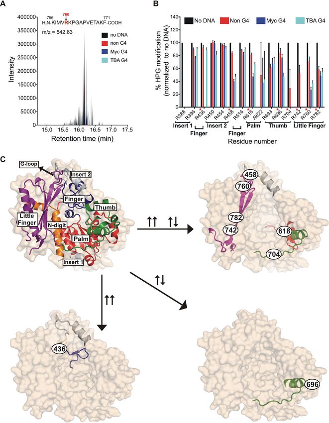
Figure 2. G4 DNA protects certain arginine residues on hRev1 from reactivity with HPG. (A) A representative total ion chromatogram is shown for a
peptide (sequence shown above the plot) containing HPG-modified Arg760 following incubation with HPG alone (black), HPG + non-G4 DNA (red),
HPG + Myc-G4 (blue) or HPG + TBA G4 (cyan). (B) For each of the 15 HPG-modified arginines detected, the fraction of modified arginine to total
arginine was calculated and values across all samples were normalized to the ‘no DNA’ sample. The location of each arginine in the hRev1 protein catalytic
domain (palm, finger, etc.), as well as the residue number, is indicated below the X-axis. The arginines highlighted in panel C are marked in red asterisks.
Values shown represent the mean ± range (n = 2). (C) The structure of hRev1330–833 (PDB 3GQC) is shown as a molecular surface (tan; semi-transparent,
in the background), as well as a cartoon showing the secondary structural elements. The domains are labeled and colored as: N-digit (orange), finger
(dark blue), thumb (green), palm (red), little finger (magenta). The insert-1 and insert-2 regions are colored light grey. Position of the G-loop in the little
finger domain is marked. The HPG-modified arginine-containing peptides protected specifically after incubation with Myc DNA (↑↑), TBA DNA (↑↓),
or common to both (↑↑ ↑↓) are shown (↑↑ = parallel G4, ↑↓ = anti-parallel G4). Position and identity of each protected arginine is shown as the residue
number inside a black oval.2074 Nucleic Acids Research, 2021, Vol. 49, No. 4
Downloaded from https://academic.oup.com/nar/article/49/4/2065/6130841 by guest on 20 November 2021
Figure 3. Mutations in insert-2 alter hRev1 binding to G4 DNA. (A) The hRev1330–833 ternary complex is shown (PDB 3GQC). The finger (blue), palm
(red), thumb (green), and little finger (magenta) domains found in all Y-family polymerases are noted. (B) A hydrophobic pocket comprised of residues from
insert-2 and the little finger domain is the site for positioning the ‘flipped’ guanine of the template during catalysis. L358 from the N-digit facilitates the
eviction of the template guanine, which is stabilized through a water-mediated bridge with residue E466 in insert-2. Additionally, residues W467 and Y470
(also from insert-2) provide the hydrophobic stacking interactions for the evicted base. Binding affinity for G4 (blue) and non-G4 (red) DNA was measured
for the (C) L358A, (D) Y470A, (E) E466A and (F) E466K hRev1 mutant proteins. The measured binding constants are listed in Table 3. Reported values
represent the mean ± SD (n = 3).
ues for mutant enzyme binding the Myc 14/23 G4 DNA overall effect of the L358A mutation was to slightly dimin-
substrate and a non-G4 DNA control. We began by mutat- ish the relative preference for G4 DNA relative to wild-type
ing the leucine residue responsible for ejecting the template enzyme (Table 3).
base into the hydrophobic pocket (L358). The L358A mu- Next, we examined the role of insert-2 in governing hRev1
tant could still bind to both G4 and non-G4 substrates (Fig- interactions with G4 DNA. We wanted to evaluate the role
ure 3C). The KD,DNA value for the G4 substrate increased 2- of aromatic amino acid side-chains located in insert-2 since
fold, from ∼40 nM for wild-type hRev1 to ∼80 nM for the these types of residues could interact with the planar face of
L358A mutant enzyme. The KD,DNA value for the non-G4 G-quartets and influence binding selectivity. Previous struc-
substrate decreased very slightly, from ∼900 nM for wild- tural reports have shown that aromatic side-chains from
type hRev1 to ∼700 nM for the L358A mutant enzyme. The residues forming the hydrophobic pocket interact with ex-Nucleic Acids Research, 2021, Vol. 49, No. 4 2075
Table 3. Equilibrium dissociation constants for mutant hRev1330–833 en- type hRev1330–833 is able to extend the nascent primer strand
zyme binding to ss-Myc-14/23 and non-G4 DNA substratesa by incorporating multiple dCMPs on both non-G4 and G4
KD,DNA Fold preference for G4 DNA DNA substrates (Figure 4B). Wild-type hRev1 readily ex-
tended the primer across the run of three guanines on the
Non-G4 (nM) G4 (nM) (KD,non-G4 DNA /KD,G4 DNA )
non-G4 template, even adding a fourth and fifth nucleotide
L358A 660 ± 140 77 ± 8 9 to the primer at later time points (Figure 4B). Apprecia-
E466A 1100 ± 130 280 ± 20 4 ble activity on the G4 DNA substrate was observed with
E466K 1300 ± 300 43 ± 16 30
Y470A 740 ± 180 180 ± 30 5
wild-type hRev1. However, the efficiency of the reaction was
reduced (∼3-fold) as compared to non-G4 DNA (Figure
a The single-stranded Myc 14/23 G4 and non-G4 DNA substrates were 4B, quantified in 4C). The enzyme added dCMP opposite
used to measure the binding affinities reported here. Data represent the the first run of three guanines of the quadruplex, but then
mean ± SD (n = 3). seemed to stall. Moreover, there is a large fraction of unre-
acted substrate left over even after 90 min, a trend that was
Downloaded from https://academic.oup.com/nar/article/49/4/2065/6130841 by guest on 20 November 2021
not observed with the non-G4 substrate.
trahelical template guanine and DNA adducts (11,33). We
Among the mutant enzymes, the L358A mutant protein
selected two residues in the insert-2 helix, W467 and Y470,
was found to have2076 Nucleic Acids Research, 2021, Vol. 49, No. 4
Downloaded from https://academic.oup.com/nar/article/49/4/2065/6130841 by guest on 20 November 2021
Figure 4. Alterations in the catalytic activity of hRev1 mutants were largely unrelated to whether G4 was present in the template strand. (A) Schematic
illustration of the primer-template DNA substrates. Arrows indicate the direction of primer extension by multiple insertions of dCMP across the template
during the time course. (B) Representative gel images for the time course monitoring dCMP insertion by the wild-type and mutant hRev1330–833 proteins
(50 nM) on non-G4 or G4 DNA substrates (200 nM) in a buffer containing 100 mM KCl. The positions of the primer and additions of dCMP (+1, +2,
etc.) are marked beside the gel images. (C) Relative enzyme activity for each protein is shown for the non-G4 and G4 DNA substrates. Relative activity is
reported as a percentage of the activity of wild-type hRev1330–833 on the non-G4 DNA substrate. The mean value for relative activity for each protein is
shown at the top of its corresponding bar. Reported values represent the mean ± SD (n = 3). Statistical significance was assessed by performing a one-way
ANOVA with multiple comparisons, and the P-values were calculated for each comparison. **** indicates P < 0.0001 for comparisons between mean
values of each mutant enzyme with that of the wild-type on non-G4 substrate, **** indicates P < 0.0001 for comparisons between mean values of each
mutant enzyme with that of the wild-type on the G4 substrate. ns; non-significant (P > 0.05).Nucleic Acids Research, 2021, Vol. 49, No. 4 2077
(35). To investigate the role of hRev1 in replication of G4 quency almost 3-fold from that observed for REV1KO cells
motifs, we inserted the Myc-derived G4-forming sequence (Figure 5C). In this regard, restoring the hydrogen-bonding
upstream and within the supF gene of the pSP189 plasmid capacity at position 466 facilitated more accurate bypass of
(Supplementary Figure S4). The supF gene, which codes a PDS-stabilized G4 structure. Expression of the Y470A
for the amber-suppressor tRNA in the lacZ gene, served as mutant resulted in trends that were very similar to the
a readout for any mutations in and around the G4-insert E466A mutant. The mutation frequency for the G4 plas-
with the unmodified pSP189 plasmid serving as the control. mid was reduced ∼4-fold in cells expressing the Y470A mu-
Both wild-type and REV1KO HAP-1 cells were grown to tant compared to REV1KO cells, but hRev1 Y470A was
∼70% confluence, followed by transfection with the con- not able to suppress the increase in mutation frequency
trol or G4-containing pSP189 plasmid either in the pres- observed for the G4 plasmid when cells were treated with
ence or absence of 0.5 M PDS or DMSO (vehicle con- PDS. In summary, complementation with wild-type and the
trol). The cells were cultured for 24 h post-transfection, har- mutant hRev1 enzymes partially suppressed the mutagenic
vested, and the replicated plasmids were retrieved (Figure G4 replication observed in REV1KO cells. Re-expressing ei-
Downloaded from https://academic.oup.com/nar/article/49/4/2065/6130841 by guest on 20 November 2021
5A). The retrieved plasmids were then used to transform ther wild-type or E466K hRev1 was able to largely elimi-
electro-competent E. coli MBM7070 cells, followed by plat- nate the increase in mutation frequency observed for cells
ing the transformants on LB-Agar plates containing X-Gal treated with PDS. G4-defective hRev1 mutants (E466A and
and IPTG. After colonies appeared, the plates were incu- Y470A) were unable to suppress PDS-induced mutations
bated at 4◦ C for 15–24 h to allow the blue color to in- on a G4-containing plasmid. The importance of residues
tensify. Finally, the blue and white colonies on each plate involved in selective G4 binding was most evident when
were counted using the Fiji version of the ImageJ software quadruplexes were stabilized by PDS.
(Figure 5A). Experiments were performed in triplicate, and
the mutation frequency was calculated by plotting ratio of
Loss of hRev1 activity resulted in an increased number of dele-
the number of white colonies to total number of colonies
tions occurring upstream of G4 DNA
counted for each experimental condition.
The control and G4 plasmids were replicated with the We extracted plasmids from mutant (white) colonies and se-
same accuracy in wild-type HAP-1 cells (Figure 5B). The quenced the products in order to determine the nature and
corresponding values for background mutation frequency position of mutations. This was done for the white colonies
were calculated to be 4.4 × 10⁻⁵ and 9.6 × 10⁻⁵ for the from both the HAP-1 cells (Supplementary Figure S6) as
control and G4 plasmids, respectively. The addition of PDS well as the REV1KO cells (Supplementary Figure S7). To
did not change the mutation frequency for either plasmid in help us understand hRev1-dependent changes in the muta-
wild-type cells (Figure 5B). Replication of the control plas- tion profiles, we divided the plasmid sequence surrounding
mid in REV1KO cells was comparable to wild-type HAP- the supF gene into four ‘zones’ (Figure 6A). We defined zone
1 cells and was unaffected by the addition of PDS (Figure I as the sequence occurring on the 5 -side of the G4 motif.
5C). There was a dramatic 200-fold increase in mutation fre- Zone II included the 18-mer Myc G4 sequence (shown in
quency when the G4-containing plasmid was replicated in green on Figure 6A), while zone III was defined as the entire
REV1KO cells (Figure 5C). This effect was further amplified supF coding region (shown in yellow on Figure 6A). Zone
by the addition of PDS, which elicited a further 4-fold in- IV was defined as the sequence on the 3 -side supF coding
crease in mutation frequency relative to what was observed region. In this way, we could evaluate the effect of hRev1 ac-
for the G4 plasmid without PDS. The mutation frequency tivity as the replisome approached the quadruplex, as well
for the PDS-stabilized G4 plasmid was >700-fold higher as mutations within the actual G4 motif or those occur-
than that observed for the control plasmid in the presence ring after the replisome had traversed the sequence. Results
of PDS (Figure 5C). These results clearly demonstrated the from DNA sequencing of 10 white colonies from each ex-
central role of hRev1 in preventing mutations at G4 DNA perimental condition were compiled, and the numbers and
sites. types of each mutation was expressed as a fraction of total
Complementation experiments were then performed to number of bases in the four zones together. Plasmids were
determine if re-expressing hRev1 would rescue the mu- harvested from different biological replicates to help ensure
tagenic replication phenotype observed in REV1KO cells. reproducibility of the sequencing results.
Equal expression of wild-type and mutant hRev1 proteins In HAP-1 cells, the largest number of mutations were ob-
was confirmed by immunoblotting (Figure 5A). Transient served in zones II and III containing the G4 motif and the
re-expression of wild-type hRev1 decreased the mutation supF gene, respectively (Figure 6B). The addition of PDS
frequency for the G4-containing plasmid ∼7-fold (Figure did not greatly alter the identity of the mutations in zones II
5C). Replication of the G4 plasmid in the presence of PDS or IV for the parental cell line, but there was a shift towards
decreased 4-fold when wild-type hRev1 was re-introduced more deletions in the supF gene (zone III). There were also
to the REV1KO cells (Figure 5C). The E466A mutant pro- base substitutions in zone I that were dependent on PDS
tein reduced the G4 mutation frequency ∼4-fold, which is treatment. Compared to wild-type HAP-1 cells, REV1KO
not that different from the wild-type enzyme (Figure 5C). cells had a higher percentage of mutated bases in all zones
However, there was a marked inability of the E466A mutant with the largest fraction of mutations in REV1KO cells being
to rescue the PDS-induced increase in mutation frequency observed in the G4 (zone II) and supF (zone III) contain-
observed for REV1KO cells (Figure 5C). PDS-induced mu- ing zones (Figure 6C). Both base substitutions and dele-
tations in the G4 plasmid were diminished when the E466K tions were observed in zones I and III, whereas zones II
hRev1 mutant was expressed – reducing the mutation fre- and IV only had base substitutions. The addition of PDSYou can also read