FEZ1 Forms Complexes with CRMP1 and DCC to Regulate Axon and Dendrite Development
←
→
Page content transcription
If your browser does not render page correctly, please read the page content below
Research Article: New Research

Development

FEZ1 Forms Complexes with CRMP1 and DCC to
Regulate Axon and Dendrite Development
Jie Yin Chua,1 Shi Jun Ng,1 Oleksandr Yagensky,2                           Erich E. Wanker,3 and              John Jia En Chua1,4,5,6

https://doi.org/10.1523/ENEURO.0193-20.2021
1
  Department of Physiology, Yong Loo Lin School of Medicine, National University of Singapore, Singapore 117593,
Singapore, 2Department of Neurobiology, Max Planck Institute for Biophysical Chemistry, Göttingen 37077, Germany,
3
  Max Delbrück Center for Molecular Medicine, Berlin-Buch 13092, Germany, 4LSI Neurobiology Programme, National
University of Singapore, Singapore 117456, Singapore, 5Healthy Longevity Translational Research Program, Yong Loo
Lin School of Medicine, National University of Singapore, Singapore 117456, Singapore, and 6Institute of Molecular
and Cell Biology, Agency for Science, Technology and Research (ApSTAR), Singapore 138673, Singapore

Abstract
Elaboration of neuronal processes is an early step in neuronal development. Guidance cues must work closely with
intracellular trafficking pathways to direct expanding axons and dendrites to their target neurons during the formation
of neuronal networks. However, how such coordination is achieved remains incompletely understood. Here, we char-
acterize an interaction between fasciculation and elongation protein zeta 1 (FEZ1), an adapter involved in synaptic
protein transport, and collapsin response mediator protein (CRMP)1, a protein that functions in growth cone guid-
ance, at neuronal growth cones. We show that similar to CRMP1 loss-of-function mutants, FEZ1 deficiency in rat
hippocampal neurons causes growth cone collapse and impairs axonal development. Strikingly, FEZ1-deficient neu-
rons also exhibited a reduction in dendritic complexity stronger than that observed in CRMP1-deficient neurons, sug-
gesting that the former could partake in additional developmental signaling pathways. Supporting this, FEZ1
colocalizes with VAMP2 in developing hippocampal neurons and forms a separate complex with deleted in colorectal
cancer (DCC) and Syntaxin-1 (Stx1), components of the Netrin-1 signaling pathway that are also involved in regulat-
ing axon and dendrite development. Significantly, developing axons and dendrites of FEZ1-deficient neurons fail to
respond to Netrin-1 or Netrin-1 and Sema3A treatment, respectively. Taken together, these findings highlight the im-
portance of FEZ1 as a common effector to integrate guidance signaling pathways with intracellular trafficking to me-
diate axo-dendrite development during neuronal network formation.
Key words: axon; CRMP1; DCC; dendrite; development; FEZ1

    Significance Statement
    Guidance cue-dependent elaboration of axons and dendrites toward their target neurons is a critical step in the
    formation of neuronal circuits during brain development. The elongating neurites require a constant supply of bio-
    molecules, but it remains unclear how guidance cues cooperate with intracellular transport. Here, we show that
    the kinesin-1 adapter fasciculation and elongation protein zeta 1 (FEZ1) forms complexes with collapsin response
    mediator protein (CRMP)1 or deleted in colorectal cancer (DCC), which are downstream effectors of the Sema3A
    and Netrin-1 signaling pathway, respectively. FEZ1-deficient neurons not only exhibit abnormal axons and den-
    drites, they were also unresponsive to Sema3A-dependent or Netrin-1-dependent regulation of axo-dendritic de-
    velopment. Our results highlight FEZ1 as a key convergence point where guidance cues and intracellular transport
    integrate to coordinate neuronal process development during neuronal network formation.

Received May 13, 2020; accepted February 14, 2021; First published March      Author contributions: J.J.E.C. designed research; J.Y.C., S.J.N., and O.Y.
  26, 2021.                                                                 performed research; E.E.W. contributed unpublished reagents/analytic tools;
  The authors declare no competing financial interests.                     J.Y.C., S.J.N., and O.Y. analyzed data; J.Y.C. and J.J.E.C. wrote the paper.

March/April 2021, 8(2) ENEURO.0193-20.2021 1–21
Research Article: New Research      2 of 21

Introduction                                                                     factor attached protein receptor (SNARE) protein family
   Guidance cue-dependent navigation of neuronal proc-                           play critical roles (Barrecheguren et al., 2017; Ulloa et al.,
esses to their cellular targets contributes to the formation                     2018). A complex of VAMP7/Syntaxin-1 (Stx1)/DCC func-
of neuronal networks during brain development (Kim                               tions in exocytosis in response to Netrin-1 chemoattrac-
and Chiba, 2004; Kolodkin and Tessier-Lavigne, 2011).                            tive signaling (Cotrufo et al., 2011). Netrin-1-induced
Extension and branching of these processes as they mi-                           aggregation of DCC receptors on the growth cone mem-
grate toward their final destinations are critically depend-                     brane also triggers VAMP2-mediated exocytosis of
ent on the delivery and insertion of new biomolecules at                         vesicles (Bouchard et al., 2008; Gopal et al., 2017; Urbina
the elongation sites (Pfenninger, 2009). While this requires                     et al., 2018). Conversely, association of VAMP2 to Nrp1
coordination between guidance cue signaling and intra-                           and PlxnA1 enables endocytosis in response to Sema3A-
cellular transport pathways, how this synchronization is                         induced repulsive signaling (Zylbersztejn et al., 2012).
achieved remains poorly understood.                                                 A sustained supply of biomolecules required for the ex-
   Guidance cues are broadly classified into attractive and                      tension of navigating neurites is largely supported by the
repulsive cues. Chemoattractive signaling effected, for in-                      Kinesin superfamily of microtubule-dependent motor pro-
stance, via Netrin-1 through its receptors deleted in colo-                      teins and their corresponding motor adapters (Hirokawa
rectal cancer (DCC) and Down’s syndrome cell adhesion                            and Takemura, 2005). In the case of Kinesin-1, adapters
molecule (DSCAM), is responsible for the attraction of                           including fasciculation and elongation protein zeta 1
commissural axons toward the midline in the spinal cord                          (FEZ1), syntabulin, and JIP family of proteins function to
(Kennedy et al., 1994; Serafini et al., 1994; Keino-Masu et                      mediate cargo binding (Gindhart et al., 2003; Su et al.,
al., 1996; Ly et al., 2008). Members of the semaphorin                           2004; Sun et al., 2011; Watt et al., 2015). Of these, FEZ1
family of proteins also mediate attractive signaling. For in-                    and syntabulin transport synaptic proteins, including Stx1
stance, Sema3C, acting in concert with the neuropilin 1                          and Synaptotagmin-1 (Cai et al., 2007; Toda et al., 2008;
(Nrp1) receptor, guides axon crossings through the cor-                          Butkevich et al., 2016). Interestingly, while enrichment of
pus callosum (Niquille et al., 2009). Other members of                           syntabulin at neurite tips has not been reported, FEZ1 is
the semaphorin family participate in repulsive signaling.                        highly enriched in the growth cones of developing neu-
Exposure of neurons to Sema3A causes neuronal growth                             rons, suggesting that the Kinesin-1/FEZ1 complex is re-
cones to shrink and collapse (Luo et al., 1993; Messersmith                      sponsible for delivering biomolecules for incorporation
et al., 1995; Fournier et al., 2000; Dent et al., 2004). Binding of              into elongating neurites (Chua et al., 2012). Indeed, using
Sema3A to its receptor Nrp1 recruits the collapsin response                      PC12 cells as model, initiation of neuritogenesis was
mediator protein (CRMP) family proteins, including CRMP1,                        found to strictly depend on FEZ1 expression (Kuroda et
to the Plexin (Plxn) co-receptor and trigger their activation via                al., 1999). Moreover, FEZ1 transport vesicles are highly
phosphorylation (Rohm et al., 2000; Deo et al., 2004).                           enriched for proteins with essential roles in neuritogene-
   In addition to their roles in navigation, guidance cues                       sis, axon guidance and axon growth (Butkevich et al.,
also determine neurite branching (Bilimoria and Bonni,                           2016). Collectively, these observations indicate that
2013; Lanoue and Cooper, 2019). Cortical neurons in                              FEZ1-mediated transport is required to support neurite
sema3A / and crmp1 / mice exhibited decreased den-                               elongation. Furthermore, the presence of guidance cue
dritic branching; conversely, hippocampal neurons ex-                            signaling pathway components (such as CRMP1, VAMP2,
posed to Sema3A exhibited increased dendritic growth                             and VAMP7) in FEZ1 transport vesicles is intriguing and
(Shelly et al., 2011; Makihara et al., 2016). Cortical neu-                      suggests that these pathways could modulate FEZ1
rons treated with Netrin-1 or Sema3A also show in-                               transport as neurites navigate toward their targets.
creased or decreased axonal branching, respectively                              However, this possibility has not been explored.
(Dent et al., 2004), and interfering Netrin-1 signaling by                          Here, we identify a new interaction between FEZ1 and
shRNA knock down of either DCC or DSCAM or by the addi-                          CRMP1 and demonstrate that both proteins colocalize in
tion of an anti-DCC antibody blocked Netrin-1-dependent                          axons and neuronal growth cones. Strikingly, FEZ1-defi-
axonal branching (Huang et al., 2015; Matsumoto and                              cient neurons develop diminished grown cones and axon
Nagashima, 2017).                                                                arborization similar to those seen when CRMP1 function
   Guidance cue signaling is also thought to coordinate                          was disrupted. Moreover, developing neurons also exhib-
the spatiotemporal insertion and removal of membranes                            ited defects in dendrite development in the absence
and proteins required for growth cone motility in attractive                     of either protein. Remarkably, FEZ1-deficient neurons
and repulsive responses (Wojnacki and Galli, 2016). Here,                        showed greater impairment, suggesting the involvement
members of the soluble N-ethylmaleimide–sensitive                                of additional signaling cues in regulating its function.
                                                                                 Indeed, FEZ1 puncta colocalizes with VAMP2, the com-
  This work was supported by funding from the National University of
Singapore.
                                                                                 mon effector SNARE protein involved in both Sema3A
  Correspondence should be addressed to John Jia En Chua at phsjcje@             and Netrin signaling. Moreover, FEZ1 is able to form a
nus.edu.sg.                                                                      separate complex with DCC and Stx1 that is required for
  https://doi.org/10.1523/ENEURO.0193-20.2021                                    Netrin-1 signaling. Confirming these observations, we
Copyright © 2021 Chua et al.
                                                                                 show that FEZ1-deficient neurons become unresponsive
This is an open-access article distributed under the terms of the Creative
Commons Attribution 4.0 International license, which permits unrestricted use,
                                                                                 to Netrin-1-dependent or Sema3A-dependent regulation
distribution and reproduction in any medium provided that the original work is   of axo-dendritic development. Collectively, these obser-
properly attributed.                                                             vations highlight a critical role of FEZ1 in both axon

March/April 2021, 8(2) ENEURO.0193-20.2021                                                                                             eNeuro.org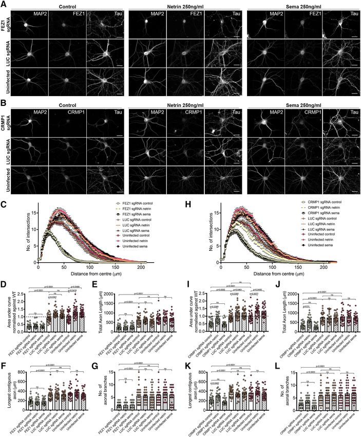
Research Article: New Research      3 of 21

and dendrite development and maturation and as a con-      were then clarified by centrifugation at 10,000  g for 10
vergence point to effect the downstream signaling of two   min at 4°C. The pull-down was performed using GFP-
established guidance cues.                                 Trap agarose beads (Chromotek) or Pierce Protein A/G
                                                           agarose beads (Thermofisher Scientific). Clarified lysates
Materials and Methods                                      were incubated with 10 ml of washed GFP-Trap bead
                                                           slurry for 1–3 h at 4°C with rotation, or with 2 ml of a-FLAG
cDNA and constructs                                        for 3–4 h, followed by 30 ml of washed Protein A/G aga-
  Plasmids expressing full-length, truncated and various   rose bead slurry for 1–2 h at 4°C with rotation. After three
phosphomutant FEZ1, and Myc-Stx1 were obtained as          washes with HEPES lysis buffer, proteins were eluted
described previously (Chua et al., 2012). Plasmids ex-     using 2 NuPAGE LDS sample buffer (Thermofisher
pressing full-length CRMP1 were obtained by shuttling      Scientific) supplemented with DTT, followed by heating at
the entry vector containing human CRMP1 cDNA (ob-          70°C for 10 min.
tained from Prof Erich Wanker; Max Delbrück Center for
Molecular Medicine) into the destination plasmids
pFLAG-CMV2 (Max Delbrück Center for Molecular              Immunoblotting
Medicine) and pcDNA6.2/N-EmGFP-DEST (Invitrogen).             Protein samples were resolved by SDS-PAGE. Semi-
Truncated cDNAs for CRMP1 were generated by PCR            dry transfer using Trans-blot Turbo (Bio-Rad) was used to
and inserted into pENTR/D-TOPO entry vectors. Primer       transfer proteins onto a nitrocellulose membrane.
sequences used are as follows (59 to 39): amino acids      Membranes were blocked for 30 min using 5% skimmed
1–462 forward, CACCATGTCGTACCAGGGCA; amino                 milk in TBST (150 mM Tris-HCl, 1.5 M NaCl, and 0.5%
acids 1–462 reverse, TCAGTTGACGTTGATGTTTCCGTC;             Tween 20, pH 7.4) at room temperature, followed by pri-
amino acids 445–572 forward, CACCATGGTCATCAG               mary antibody incubation overnight at 4°C. After three
CCAGGGCAAG, amino acids 445–572 reverse, TCAACC            washes, membranes were incubated with their respective
GAGGCTGGTGATGT; amino acids 465–572 forward,               secondary antibody at a 1:4000 dilution for 1 h at room
CACCATGGGCCGCTTCATTCCG; amino acids 465–572                temperature. Antibody dilutions and their sources are
reverse, TCAACCGAGGCT GGTGATGT. The entry vector           summarized in Table 1. After another three washes, mem-
containing human DCC cDNA was generated by PCR             branes were developed using SuperSignal West Dura
using pCMV-DCC as a template (a gift from Bert             Extended Duration Substrate (Thermofisher Scientific)
Vogelstein, Addgene plasmid #16459; http://n2t.net/        and imaged using the Azure Biosystem C300 (Azure
addgene:16459; RRID: Addgene_16459). The entry vector      Biosystems). Quantification of protein bands was per-
containing the Plxn A1 Extracellular Domain was gener-     formed using ImageJ (FIJI). Signal intensities of FEZ1 or
ated by PCR using PlxnA1-Fc-His (a gift from Woj           CRMP1 were normalized against those of the loading
Wojtowicz, Addgene plasmid #72122; http://n2t.net/         control actin. The normalized band intensities of each pro-
addgene:72122; RRID: Addgene_72122). Primer sequen-        tein for each experimental group were then divided by the
ces used are as follows (59 to 39): forward, CAC CAT GCC   corresponding intensities from the uninfected (control)
ACT GCC ACC TCT G, reverse, TCA CAG TGT CAG CAG            group and expressed as a percentage compared with the
GCT GTC CGA AT. The corresponding mammalian ex-            uninfected group. For co-immunoprecipitation, signal in-
pression plasmids were then obtained by shuttling the      tensities of the coimmunoprecipitated protein were nor-
entry plasmids with the destination plasmids. mCherry-     malized to the signal intensity of the immunoprecipitated
Nrp1 and VAMP2-GFP were gifts from Guido Serini            binding partner to obtain a semi-quantitative measure of
(Addgene plasmid #21934; http://n2t.net/addgene:21934;     binding strength.
RRID:Addgene_21934) and Reinhard Jahn, respectively.
                                                           Primary neuronal culture
Cell culture and transfection                                 Adult female Wistar rats with their corresponding litters
  Human embryonic kidney 293 (HEK293) cells were cul-      were purchased from the Centre for Animal Resources
tured in DMEM supplemented with 10% fetal bovine           and housed in temperature controlled (23 6 1°C), individ-
serum (FBS) and 0.5% penicillin/streptomycin under nor-    ually ventilated cages on a 12/12 h light/dark cycle (7 A.M.
mal growth conditions (37°C, 5% CO2) and are routinely     to 7 P.M.) with access to food and water. Rats were ac-
tested for mycoplasma contamination. Transfection was      climatized for 4 d before the start of experiments. All pro-
conducted 1 d after plating, when HEK293 cells were at     cedures were in accordance with the Principles of
70–90% confluency. Polyethylenimine (PEI; Sigma-           Laboratory Animal Care and approved by the Institutional
Aldrich) was used as the transfection reagent in a 1:3     Animal Care and Use Committee.
DNA:PEI ratio.                                                Primary hippocampal neurons were prepared from
                                                           freshly isolated hippocampal tissue from newborn rat
Co-immunoprecipitation                                     brains and immersed in dissection solution (1.5 mM CaCl2,
  Twenty-four hours after transfection, cells were lysed   4.9 mM KCl, 0.2 mM NaH2PO4, 11 mM MgCl2, 0.3 mM
with cold HEPES lysis buffer (50 mM HEPES, 150 mM          MgSO4, 130 mM NaCl, 2.7 mM NaHCO3, 0.8 mM
NaCl, 1 mM EDTA, and 1% Triton X-100, pH 7.2) supple-      Na2HPO4, 22 mM HEPES, and 5 mM glucose, pH 7.32).
mented with the cOmplete EDTA-free Protease Inhibitor      Dissociation of neurons was performed by incubation
Cocktail (Sigma-Aldrich) at 4°C for 15 min. Cell lysates   with 1 ml trypsin for 30 min at 37°C, followed by

March/April 2021, 8(2) ENEURO.0193-20.2021                                                                       eNeuro.org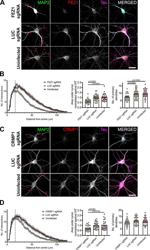
Research Article: New Research      4 of 21

Table 1: Antibodies used for immunoblotting and immunofluorescence in this study
                                                        Dilution
                                               WB                  IF            Source                             RRID
Primary antibody
  Mouse a-FLAG M2                              1:1000              -             Sigma-Aldrich                      AB_262044
  Mouse a-CRMP1                                1:1000              1:200         Santa Cruz Biotechnology           AB_10846086
  Mouse a-VAMP2                                -                   1:200         Synaptic Systems                   AB_887811
  Rabbit a- b actin                            1:1000              -             Synaptic Systems                   AB_11042458
  Rabbit a-FEZ1                                1:1000              1:400         In house
  Rabbit a-GFP                                 1:20,000            -             Synaptic Systems                   AB_887725
  Mouse a-MAP2                                 -                   1:1000        Merck                              AB_477256
  Rabbit a-MAP2                                -                   1:1000        Synaptic Systems                   AB_2147096
  Mouse a-Myc                                  1:1000              -             In house
  Guinea Pig a-Tau                             -                   1:400         Synaptic Systems                   AB_1547385
Secondary antibody
  Goat a-mouse IgG (H 1 L)-HRP conjugate       1:4000              -             Bio-Rad                            AB_11125547
  Goat a-rabbit IgG (H 1 L)-HRP conjugate      1:4000              -                                                AB_11125142
  Donkey a-mouse IgG (Cy2)                     -                   1:200         Jackson ImmunoResearch             AB_2340827
  Donkey a-mouse IgG (Cy3)                     -                   1:200                                            AB_2315777
  Donkey a-rabbit IgG (Cy2)                    -                   1:400                                            AB_2340612
  Donkey a-rabbit IgG (Cy3)                    -                   1:200                                            AB_2307443
  Donkey a-guinea pig IgG (Cy5)                -                   1:400                                            AB_2340462

neutralization with 2 ml 5% serum media (MEM Eagle                 (Invitrogen) diluted 1:10,000 in water for 10 min, coverslips
modified, 21 mM D-glucose, 200 mM L-glutamine, MEM-                were then mounted onto a glass slide using Fluoro-Gel
vitamin, Mito 1 serum extender, and 5% FBS). To tritu-             (Electron Microscopy Sciences) and stored. Images of
rate, digested tissue was passed through a fire-polished           stained neurons were acquired using the 63 oil-immersion
Pasteur pipette until large tissue fragments were dis-             objective on the Axio Observer (Zeiss).
persed. Neurons were pelleted by centrifugation at 500               Line scan analyses, Pearson correlation coefficient
g for 5 min. After resuspending in serum media, cells were         (PCC) analyses, Sholl analyses, neurite tracing and image
plated on PDL-coated coverslips in wells containing                segmentation were performed using ImageJ (FIJI), and
DMEM/F12 supplemented with B27.                                    growth cone area was calculated using Zen. To obtain
                                                                   PCC values, the EzColocalization plugin was used
                                                                   (Stauffer et al., 2018). Merged images containing only the
Immunofluorescence
                                                                   desired growth cones or axons were split into individual
  Primary hippocampal neurons were fixed using 4%                  channels and input into the plugin. Growth cones were
paraformaldehyde in PBS (Santa Cruz Biotechnology).                only selected if they could be traced via a continuous
Neurons were first permeabilized by incubation with 0.3%           axon back to the cell body. Negative control values were
Triton X-100 for 10 min, followed by blocking for 30 min           obtained by flipping the image in the green channel hori-
using 10% normal goat serum (NGS) in PBS. Primary and              zontally. Line scan analyses were performed essentially
secondary antibody solutions were prepared using 10%               as described previously (Barszczewski et al., 2008).
NGS as a diluent as summarized in Table 1, which were              Briefly, regions of interest (three lines of 10 pixels width
then incubated with the coverslips in a dark humidified            and 115 pixels length) drawn randomly across regions
chamber for 1 h at room temperature in succession. After           that correspond to the proximal, middle and distal re-
each incubation, coverslips were washed before proceed-            gions from the axon, and growth cones to obtain signal
ing to the next step. After incubation with Hoescht                intensities. For growth cones, the lines were spread

Table 2: CRISPOR scores for FEZ1 and CRMP1 gRNA sequences
#guideId                                                FEZ1                                                    CRMP1
targetSeq                                               AATCAGCTTCAAG                                           CGACTTCGACGC
                                                        TCCATGGAGG                                              CTACAGCGTGG
mitSpecScore                                            73                                                      96
cfdSpecScore                                            86                                                      98
offtargetCount                                          154                                                     13
targetGenomeGeneLocus                                   exon:Fez1                                               Exon 1
Doench ’16-Score                                        NotEnoughFlankSeq                                       65
Moreno-Mateos-Score                                     NotEnoughFlankSeq                                       24
Out-of-Frame-Score                                      NotEnoughFlankSeq                                       61
Lindel-Score                                            NotEnoughFlankSeq                                       -
GrafEtAlStatus                                          GrafOK                                                  -
grafType                                                                                                        -

March/April 2021, 8(2) ENEURO.0193-20.2021                                                                                 eNeuro.org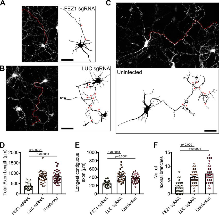
Research Article: New Research      5 of 21

Figure 1. FEZ1 interacts with CRMP1. A, Co-immunoprecipitation of FLAG-FEZ1 FL and EmGFP-CRMP1 FL. Cell lysates of
HEK293 transiently expressing FLAG-FEZ1 and EmGFP-CRMP1 were collected and immunoprecipitated using a-FLAG. EmGFP-
CRMP1 efficiently co-immunoprecipitated with FLAG-FEZ. B, Mapping of interaction domains on FEZ1. Lysates from HEK293 cells
co-expressing the EmGFP-FEZ1 constructs and FLAG-CRMP1 were immunoprecipitated using GFP-TRAP agarose beads. Only
FEZ1 constructs containing the coiled-coil domain (amino acids 1–392 and amino acids 1–308) coimmunoprecipitated CRMP1.

March/April 2021, 8(2) ENEURO.0193-20.2021                                                                           eNeuro.org
Research Article: New Research      6 of 21

continued
Truncated FEZ1 lacking its C terminus interacted approximately three times more strongly with FLAG-CRMP1. C, Co-immunopreci-
pitation of wild-type (WT) or various phosphomutants (QUAD, S58A, or S316A) of FEZ1 tagged to EmGFP, co-expressed with
FLAG-CRMP1. Transfected HEK293 cell lysates were collected and immunoprecipitated using GFP-TRAP agarose beads. All FEZ1
phosphomutants co-immunoprecipitated CRMP1. Compared with WT FEZ1, the QUAD and S58A FEZ1 mutants interacted slightly
more strongly with CRMP1, but this increase did not reach statistical significance. The S316A FEZ1 mutant displayed a similar inter-
action level to WT FEZ1. D, Mapping of interaction domains on CRMP1. Lysates from HEK293 cells co-expressing FLAG-FEZ1 and
various EmGFP-CRMP1 constructs were immunoprecipitated using GFP-TRAP agarose beads. All versions of CRMP1 were able to
interact with FEZ1, suggesting that the amidohydrolase family domain is not critical for this interaction. CRMP1 (amino acids 1–462)
bound to FEZ1 more strongly than full-length CRMP1. Red asterisks indicate non-specific bands. Statistical significance was deter-
mined using one-way ANOVA. Data were obtained from three independent experiments. All error bars represent SEM. Reciprocal
and control coimmunoprecipitations are shown in Extended Data Figure 1-1.

out as evenly as possible to cover the entire growth               unconcentrated viruses. For viral concentration, 15 ml of
cone. For axons, the first line was drawn at the end of            filtered supernatant was concentrated at 3220  g for
the axon before it becomes a growth cone, the third                30 min at 4°C in an Amicon Ultra 15-ml centrifugal filter
line was drawn at the start where it projects from soma            (Merck). Primary neurons were infected with lentiviruses
and the second line was drawn between the first and                1 d after plating.
third lines (equidistant where possible). Colocalization
is deemed to occur when both channels display
closely correlating similar peaks and troughs. For axon            shRNA knock down of FEZ1
analyses, neurons were doubly stained for MAP2 and                    shRNA targeting FEZ1 (59-GAGGACCTCGTGAATG
Tau. Only continuous processes that were concur-                   AATTT-39) or Luc (59-CGTACGCGGAATACTTCGA-39)
rently Tau1 and MAP2 – were taken as axons and used                were inserted into the FHUG1W vector (a kind gift from Dr.
for subsequent analyses. Axon branches were defined                Oliver Schlueter). Primary rat day in vitro (DIV)1 neurons were
as protrusions measuring at least 20 mm (Winkle et al.,            infected with lentiviruses expressing either shRNA and sub-
2014). For analyses of FEZ1 or CRMP1 sgRNA-treated                 sequently fixed at DIV3, DIV7, and DIV14 and stained for Tau
neurons, only neurons negative for staining of either              and MAP2. Neurons were imaged on a Zeiss Axio Observer
protein (confirmed by immunofluorescence staining)                 S1 microscope. The tiling function was used to obtain images
were included in the analyses.                                     for a complete coverslip. Axon and dendrite lengths were de-
                                                                   termined using NeuronJ (FIJI).
CRISPR-Cas9 system to knock down CRMP1 and
FEZ1 expression                                                    Netrin-1 and Sema3A treatment
   Guide RNAs (gRNAs) targeting human FEZ1 gene (59-                  Six days after infection of primary neurons with lentivi-
AATCAGCTTCAAGTCCATGG-39) were designed using                       ruses to knock down CRMP1 or FEZ1, neurons were
the online CRISPR design tool (http://crispr.mit.edu/).            treated with 250 ng/ml of Netrin-1 or 250 ng/ml of
gRNAs for CRMP1 (59-CGACTTCGACGCCTACAGCG-39)                       Sema3A (RnD Systems) and incubated for 24 h in normal
were designed using CRISPOR (Haeussler et al., 2016;               growth conditions before fixing and immunostaining was
Concordet and Haeussler, 2018). CRISPOR scores                     performed. For the untreated control, PBS (vehicle con-
for both gRNA sequences are summarized in Table 2.                 trol) was added in place of Netrin-1 and Sema3A.
The Luc gRNA sequence was used as control (59-
CCGGGCTTTAACGAATATGA-39). All gRNAs were in-
serted into LentiCRISPRv2 plasmid (Addgene; plasmid                Statistical analyses
#52961; RRID:Addgene_52961) at the BsmBI restriction                  Prism GraphPad (version 8) was used to perform statis-
enzyme site using the GeCKO protocol (Sanjana et al.,              tical analyses. To calculate statistical significance, one-
2014; Shalem et al., 2014). Viruses were produced as de-           way ANOVA with Bonferroni correction was applied when
scribed previously (Yagensky et al., 2019). Briefly,               comparing three conditions. Kruskal–Wallis analysis with
13  106 HEK293 cells were seeded and grown under nor-             Dunn’s test was performed when comparing three condi-
mal conditions. A day later, 10 mg of transfer plasmid, 5 mg       tions for populations with non-Gaussian distributions. A
of pMDLg/pRRE (Addgene; plasmid #12251; RRID:                      two-tailed Mann–Whitney test was used when comparing
Addgene_12251), 2.5 mg of pRSV-rev (Addgene; plasmid               two conditions directly.
#12253; RRID:Addgene_12253), and 2.5 mg of pMD2.G
plasmids (Addgene; plasmid #12259; RRID:Addgene_                   Results
12259) were transfected into HEK293 cells in 3% FBS
DMEM using the PEI transfection reagent in a 1:3 DNA:              The coiled-coil region of FEZ1 interacts with CRMP1,
PEI ratio. After 4–6 h of incubation, media were changed           a protein participating in axonal outgrowth and
into 20 ml of DMEM/F12 (Thermofisher Scientific) and in-           growth cone guidance
cubated for an additional 24 h. To harvest the lentiviruses,         A previous effort to systematically identify new proteins
media from each plate of cells were collected and spun             involved in presynaptic function using a yeast 2 hybrid
down at 1000 rpm for 5 min at 4°C. The supernatant was             screen uncovered an interaction between FEZ1 and
passed through a 0.45 mm filter and saved as                       CRMP1 (Chua et al., 2012). CRMP1 has been identified to

March/April 2021, 8(2) ENEURO.0193-20.2021                                                                                 eNeuro.org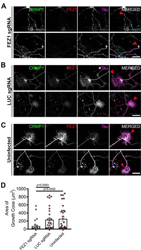
Research Article: New Research      7 of 21

Figure 2. FEZ1 and CRMP1 colocalize in hippocampal neurons. A, Distribution of FEZ1 and CRMP1 in growth cones and axons of
hippocampal neurons at 1, 4, and 7 DIV. Puncta for FEZ1 and CRMP1 (red arrowheads) can be observed in the growth cones.

March/April 2021, 8(2) ENEURO.0193-20.2021                                                                          eNeuro.org
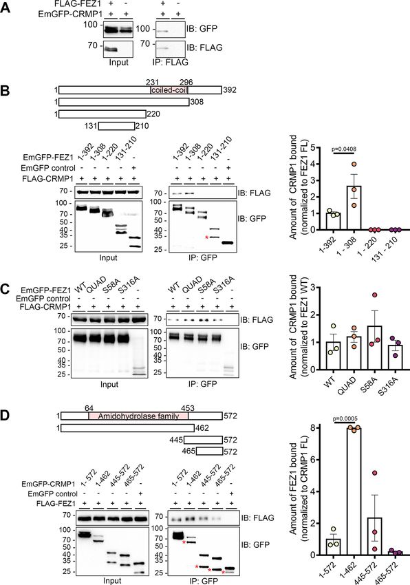
Research Article: New Research      8 of 21

continued
Scale bars: 20 mm. B, PCC analysis of colocalization extent of both proteins in growth cones and axons. Control values were ob-
tained by obtaining the PCC with the rotated image of one channel; n = 15 (DIV1), n = 9 (DIV4), n = 8 (DIV7) collected across three in-
dependent experiments. Statistical significance was determined using Kruskal–Wallis analysis. All error bars represent SEM. C, D,
Line scan analysis for colocalization between FEZ1 and CRMP1 in growth cones and axons respectively. Merged images are
shown. x- and y-axes, distance (mm) and gray value respectively; axon 1, distal; axon 2, intermediate; axon 3, proximal (relative to
cell body). Vertical columns in red represent regions of colocalization. Scale bars: 10 mm.

be present on FEZ1 transport vesicles isolated from the rat          at these sites by mutating them to alanine residues can affect
brain (Butkevich et al., 2016). To additionally validate that the    its binding to some of its interaction partners (Chua et al.,
two proteins interact, we performed co-immunoprecipitation           2012). To determine whether phosphorylation of these serine
assays using FLAG-FEZ1 and EmGFP-CRMP1 co-ex-                        residues might also influence the interaction between FEZ1
pressed in HEK293 cells. Immunoprecipitation of FEZ1 re-             and CRMP1, we separately co-expressed three EmGFP-FEZ1
covered CRMP1 from transfected cell lysates containing               phosphomutants [S58A, S316A, and QUAD (S58A, S134A,
both proteins but not in the absence of FEZ1, thereby sup-           S301A, S316A)] with FLAG-CRMP1 and used GFP-TRAP
porting that FEZ1 forms a complex with CRMP1 (Fig. 1A).              beads to pulldown FEZ1 (Fig. 1C). All phosphomutants re-
   To identify the region of FEZ1 responsible for binding            tained their ability to bind CRMP1 (Fig. 1C). Thus, the binding
CRMP1, we separately co-expressed full-length FEZ1 or                between the two proteins is unaffected by FEZ1 phosphoryla-
one of the three truncated versions of FEZ1 (amino acids             tion, which is similar to what has been reported for its interac-
1–308, amino acids 1–220, and amino acids 131–210)                   tion with Stx1 (Chua et al., 2012).
fused to EmGFP with FLAG-CRMP1 in HEK293 cells. Co-
expression of EmGFP alone with FLAG-CRMP1 served as
                                                                     The CRMP1 amidohydrolase domain is involved in
a negative control. FLAG-CRMP1 was resolved as a sin-
                                                                     FEZ1 binding
gle band of ;70 kDa on immunoblots. Full-length and
                                                                        CRMP1 contains a central amidohydrolase family domain
truncated versions of FEZ1 were resolved at ;97, 90, 75,
                                                                     comprising of a triosephosphate isomerase-like barrel. The
and 45 kDa, respectively, as previously reported (Chua et
                                                                     TIM barrel is a common structure involved in many enzyme-li-
al., 2012). The protein is phosphorylated at multiple sites,
                                                                     gand interactions (Kurochkina, 2010), suggesting the involve-
accounting for the appearance of additional minor bands
                                                                     ment of this domain in protein interactions. To identify
observed (Chua et al., 2012; Butkevich et al., 2016). An
unexpected smaller band was observed for the shortest                whether this region in CRMP1 is involved in FEZ1 binding, we
FEZ1 peptide (indicated by an asterisk) that could have              co-expressed FLAG-FEZ1 separately with three truncated
arisen as a result of non-specific cleavage. Using GFP-              versions of EmGFP-CRMP1 corresponding to its N-terminal
TRAP beads to pulldown the FEZ1 peptides, only FEZ1                  and central amidohydrolase family domain (amino acids 1–
fragments containing the coiled-coil domain (amino acids             462) and the C-terminal regions (amino acids 445–572 and
                                                                     amino acids 465–572). CRMP1 was then immunoprecipitated
1–392 and amino acids 1–308) were observed to interact
                                                                     using GFP-TRAP beads. Supporting the previous observa-
efficiently with FLAG-CRMP1 (Fig. 1B). Truncated FEZ1
                                                                     tions, reciprocal coimmunoprecipitation with EmGFP-CRMP1
peptides lacking the coiled coil domain (amino acids 1–
                                                                     full-length also coimmunoprecipitated FLAG-FEZ1 (Fig. 1D).
220 and amino acids 131–210) did not pulldown CRMP1,
                                                                     Sequential deletion of CRMP1 progressively diminished its in-
indicating that this protein domain is critical for the inter-
                                                                     teraction with FEZ1, with the exception of CRMP1 (amino
action (Fig. 1B). Remarkably, FEZ1 (amino acids 1–308)
                                                                     acids 1–462) where binding to FEZ1 was significantly in-
displayed a greater affinity (;3-fold) to interact with
                                                                     creased. Interestingly, the C-terminal CRMP1 alone (amino
CRMP1 as compared with full-length FEZ1 (Fig. 1B), sug-
                                                                     acids 465–572) also retained residual binding to FEZ1, which
gesting that the C-terminal region of FEZ1 might play an
                                                                     was enhanced when a small portion of the conserved ami-
inhibitory role in this interaction.
                                                                     dohydrolase family domain was included (amino acids
                                                                     445–572). No FEZ1 was detectable in the negative
Phosphorylation of FEZ1 does not regulate its                        control where CRMP1 was omitted. In addition to the
interaction with CRMP1                                               larger bands corresponding to the expected band
  Four phosphorylation sites (S58, S134, S301, and S316)             sizes for each truncated CRMP1 peptide, a smaller ac-
were previously identified in FEZ1. Abolishing phosphorylation       companying band could also be observed (Fig. 1D,

Table 3: Summary of PCC values (colocalization of FEZ1 and CRMP1)
                                                                                                     PCC
                                 Days in vitro                           FEZ1 1 CRMP1                                Control
GC                               1                                       0.8311 6 0.0277                               0.108 6 0.0472
                                 4                                       0.8214 6 0.0275                              0.1104 6 0.0570
                                 7                                       0.8024 6 0.0252                               0.214 6 0.0404
Axons                            1                                       0.9026 6 0.009                              0.02804 6 0.0134
                                 4                                        0.794 6 0.0222                             0.02467 6 0.0121
                                 7                                       0.8094 6 0.0265                              0.0216 6 0.0107

March/April 2021, 8(2) ENEURO.0193-20.2021                                                                                   eNeuro.org
Research Article: New Research 9 of 21 Figure 3. FEZ1-deficient neurons display collapsed growth cones. A–C, Immunostaining against CRMP1, FEZ1, and Tau in hippo- campal neurons treated with FEZ1 sgRNA, LUC sgRNA, or uninfected controls, respectively, 6 DPI. Majority of the FEZ1-deficient neurons displayed smaller growth cones compared with the controls (red arrowheads). Scale bars: 20 mm. D, Quantification of growth cone sizes in FEZ1-deficient or control LUC sgRNA and uninfected neurons. On average, growth cones in control neurons were ;4- to 5-fold larger than those in FEZ1-deficient neurons. Statistical significance was determined using Kruskal–Wallis analy- sis. FEZ1 sgRNA n = 30, LUC sgRNA n = 31, uninfected n = 33, collected over three independent experiments. All error bars repre- sent SEM. Western blottings for CRIPSR/Cas9-mediated FEZ1 knock-down in neurons are shown in Extended Data Figure 3-1. asterisks). Reciprocal coimmunoprecipitations with showing that detection of this interaction could not be FLAG-FEZ1 coprecipitate both long and short pep- attributable to non-specific binding. Taken together, tides, suggesting that the smaller peptides did not cor- these results indicate that the two halves of CRMP1 respond to non-specific interactions (Extended Data may possess independent interaction regions for FEZ1 Fig. 1-1A). Furthermore, as expected, immunoprecipi- with the amidohydrolase family domain-containing re- tation of the CRMP1 truncated peptides did not co-im- gion binding more strongly to FEZ1 (Fig. 1D). It is re- munoprecipitate Munc18 (Extended Data Fig. 1-1B) mains unclear why, on their own, each half of CRMP1 March/April 2021, 8(2) ENEURO.0193-20.2021 eNeuro.org
Research Article: New Research 10 of 21
Figure 4. FEZ1-deficient neurons display defects in axonal development. A–C, Tiled images of FEZ1 sgRNA, LUC sgRNA, and unin-
fected neurons immunostained for Tau. Respective axon traces are shown juxtaposed. Axons of FEZ1-deficient neurons are signifi-
cantly less branched as compared with control neurons (branch points indicated by red arrowheads). The length of the longest
contiguous axon (red dotted lines) in FEZ1-deficient neurons was also shorter than in control neurons. D–F, Quantification of total
axon length, longest contiguous axon length, and number of axon branches in FEZ1 sgRNA, LUC sgRNA, and uninfected neurons,
respectively. All quantified variables were significantly decreased in FEZ1-deficient neurons as compared with control groups.
Statistical significance was determined using Kruskal–Wallis analysis. FEZ1 sgRNA n = 31, LUC sgRNA n = 32, uninfected n = 34, ob-
tained from three independent experiments. Error bars represent SEM. Scale bars: 50 mm. Developmental abnormalities in shRNA-
mediated FEZ1 knock-down hippocampal neurons are shown in Extended Data Figure 4-1.
appears to exhibit stronger binding as compared with (Chua et al., 2012). These observations suggest that the
the full-length protein. FEZ1-CRMP1 complex could function to coordinate de-
livery of cargoes to neurite tips.
To determine this, we first examined the localization of
FEZ1 and CRMP1 colocalize in growth cones of both proteins in developing hippocampal neurons. As
developing neurons previously reported, FEZ1 puncta are distributed in cell
The CRMP family of proteins participate in Sema3A sig- bodies, axons and growth cones (Fig. 2A; Ikuta et al.,
naling and are involved in regulating several aspects of 2007; Chua et al., 2012). In comparison, CRMP1 shows a
neuronal development (Yamashita and Goshima, 2012). more uniform distribution in neurons, especially through-
In particular, CRMP1 participates in neurite outgrowth out the length of neurites. As previously reported, CRMP1
and growth cone dynamics in developing neurons is also present in growth cones, with some regions show-
(Higurashi et al., 2012). Likewise, FEZ1 is readily detecta- ing punctate distribution (Fig. 2A, arrowheads; Higurashi
ble in growth cones of developing hippocampal neurons et al., 2012). Colocalization analysis via PCC revealed a
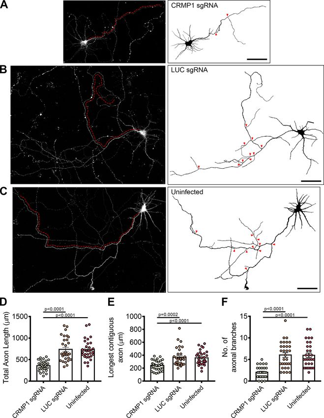
March/April 2021, 8(2) ENEURO.0193-20.2021 eNeuro.orgResearch Article: New Research 11 of 21 Figure 5. CRMP1-deficient neurons display defects in axonal development. A–C, Tiled images of CRMP1 sgRNA, LUC sgRNA, and uninfected neurons immunostained for Tau. Respective axon traces are shown juxtaposed. Axons of CRMP1-deficient neurons are significantly shorter and less branched as compared with control neurons (branch points indicated by red arrowheads; longest con- tiguous axon for each neuron is indicated by red dotted line). D–F, Quantification of total axon length, longest contiguous axon length and number of axon branches in CRMP1 sgRNA, LUC sgRNA, and uninfected neurons, respectively. All three variables were significantly decreased in CRMP1-deficient neurons as compared with control groups. Statistical significance was determined using Kruskal–Wallis analysis. CRMP1 sgRNA n = 32, LUC sgRNA n = 31, uninfected n = 31, obtained from three independent experiments. Error bars represent SEM. Scale bars: 50 mm. Western blottings for CRIPSR/Cas9-mediated CRMP1 knock-down in neurons are shown in Extended Data Figure 5-1. March/April 2021, 8(2) ENEURO.0193-20.2021 eNeuro.org
Research Article: New Research     12 of 21

Figure 6. Loss of FEZ1 impairs dendritic development. A, Images of FEZ1 sgRNA, LUC sgRNA, and uninfected neurons stained for
MAP2, Tau, and FEZ1 6 DPI. Branch points are denoted by red arrowheads. B, Loss of FEZ1 reduces dendritic branching and num-
ber of primary dendrites. Dotted line indicates the peak of intersections for FEZ1-deficient neurons. Statistical significance was de-
termined using Kruskal–Wallis analysis. FEZ1 sgRNA n = 32, LUC sgRNA n = 37, uninfected n = 34, collected over three independent
experiments. C, Images CRMP1 sgRNA, LUC sgRNA, and uninfected neurons stained for MAP2, Tau, and CRMP1 at 6 DPI. Branch
points are denoted by red arrowheads. D, Loss of CRMP1 reduces overall dendritic branching but does not affect maximum number
of crossings and number of primary dendrites. Dotted line indicates the peak of intersections for CRMP1-deficient neurons.
Statistical significance was determined using Kruskal–Wallis analysis. CRMP1 sgRNA n = 32, LUC sgRNA n = 31, uninfected n = 31,
collected over three independent experiments. All error bars represent SEM. Scale bars: 20 mm.

significant overlap of both proteins in growth cones and             examined colocalization of both proteins using line scan
axons. In comparison, the coefficient significantly de-              analysis. Supporting the previous analysis, correlating signal
creased when rotated images from one channel were                    peaks indicative of colocalization could be observed in
used for the analyses (Table 3; Fig. 2B). We further                 growth cones and axons (Fig. 2C,D, vertical red columns).

March/April 2021, 8(2) ENEURO.0193-20.2021                                                                                 eNeuro.org
Research Article: New Research 13 of 21
Table 4: Summary of Sholl analyses in FEZ1 and CRMP1 knock-down neurons
sgRNA Maximum no. of crossings Distance from centre of cell body (mm) Total area under curve
CRMP1 knock-down CRMP1 11.8 6 0.47 16 0.72 6 0.044
LUC 13.16 6 0.85 20 1 6 0.0576
Uninfected 12.9 6 0.66 18 1 6 0.075
FEZ1 knock-down FEZ1 10.15 6 0.568 16 0.635 6 0.038
LUC 12.16 6 0.8 16 0.98 6 0.047
Uninfected 13.05 6 0.69 20 1 6 0.055
Together, these results indicate that FEZ1 and CRMP1 can expression, we also observed impairments in axon devel-
form a complex in axons and, in particular, in growth cones. opment where mean axon length was significantly shorter
in FEZ1 knocked down neurons (Extended Data Fig. 4-1).
Loss of FEZ1 causes growth cone collapse and These results implicate FEZ1 in axon elongation and arbo-
decreased axonal branching rization during neurite outgrowth.
To further examine whether the FEZ1/CRMP1 complex A previous study has shown that CRMP1 is involved in
might be involved in supporting neurite extension, we em- neurite outgrowth of dorsal root ganglion neurons (Higurashi
ployed a lentiviral-based CRISPR-Cas9 system to ablate et al., 2012). To investigate whether neurite extension was
FEZ1 expression using FEZ1 sgRNAs in developing hip- also affected in CRMP1-deficient hippocampal neurons, we
pocampal neurons. As a control, we used a sgRNA target- generated a lentiviral vector expressing sgRNA targeting
ing a scrambled luciferase sequence (LUC sgRNA; Yagensky CRMP1 (CRMP1 sgRNA). Following infection, endogenous
et al., 2019). Neurons at 1 DIV were infected with the corre- CRMP1 protein levels were reduced by ;60% as determined
sponding viruses and lysates immunoblotted to probe for by immunoblotting (Extended Data Fig. 5-1). We repeated
FEZ1 expression. Approximately 80% of endogenous FEZ1 the axonal measurements on CRMP1-deficient neurons. As
protein was eliminated 6 d post virus infection (DPI; Extended before, CRMP1 knock-down was first confirmed using immu-
Data Fig. 3-1A). Abrogation of FEZ1 expression in individual nofluorescence, and only these neurons were used for analy-
neurons was first confirmed using immunofluorescence stain- sis. Indeed, CRMP1-deficient neurons exhibited significantly
ing (Fig. 3A). Only neurons successfully knocked down for shorter total axon lengths, shorter contiguous axons and
FEZ1 were selected for analyses. We could not study the ef- fewer axon branches as compared with control groups (Fig.
fect of FEZ1 loss in neurons younger than 6 DIV as endoge- 5A–C, quantifications in D–F). Collectively, the phenotypes
nous FEZ1 could still be detected from 1 to 4 DPI (Extended present in FEZ1-deficient neurons agree well with those re-
Data Fig. 3-1B). ported in neurons when CRMP1 function was perturbed,
FEZ1 has previously been found to deliver biomolecules thereby implicating that FEZ1 and CRMP1 work together in a
to the growth cones (Chua et al., 2012); and if FEZ1 was common pathway.
involved in CRMP1 transport, a general mislocalization of
CRMP1 would have been expected. However, CRMP1 FEZ1-deficient neurons display more severe dendritic
distribution in neurons was largely unchanged in the ab- defects than CRMP1-deficient neurons
sence of FEZ1 (Fig. 3A–C), indicating that FEZ1 is not crit- In addition to its functions in axon, FEZ1 is also found
ical for its transport. However, using Tau as a label for in dendrites and has been implicated in dendritic develop-
both axons and growth cones (Mandell and Banker, 1996; ment. FEZ1 expression is maintained in adulthood (Sakae
Biswas and Kalil, 2018), we observed that growth cones et al., 2008) and previous studies in adult hippocampal
were significantly smaller in FEZ1-deficient neurons but neurogenesis have shown that shRNA knock down of
were still present at axon ends in control neurons (Fig. FEZ1 increased dendritic arborization in dentate granule
3A–C, red arrowheads). Indeed, the average growth cone cells (Kang et al., 2011; Watanabe et al., 2014). To exam-
area of these neurons was at least 4-fold smaller as com- ine whether these effects could be recapitulated during
pared with the control neurons (Fig. 3D). postnatal neuron development, we immunostained wild-
To directly examine whether loss of FEZ1 function di- type and FEZ1-deficient neurons for MAP2 and per-
rectly affected neurite extension, we measured and com- formed Sholl analyses as a measure of dendritic
pared total axon (Tau1 and MAP2–) length as well as the development.
length of the longest contiguous axon (Fig. 4A–C, red dot- In stark comparison to what was observed during adult
ted lines) between control and FEZ1-deficient neurons. neurogenesis, FEZ1-deficient postnatal developing neu-
Both parameters were significantly decreased in the latter rons exhibited fewer MAP21 primary dendrites as com-
group (Fig. 4A–C, quantifications in D,E). We also ob- pared with wild-type neurons (Kang et al., 2011). Dendritic
served decreased axonal branching in FEZ1-deficient branching was also reduced (Fig. 6A, red arrowheads in-
neurons as compared with both sets of control neurons dicate branch points). Sholl analyses indicated that FEZ1-
(Fig. 4A–C, red arrowheads show branch points, quantifi- deficient neurons displayed fewer intersections with con-
cations in F). Indeed, several neurons lacking FEZ1 dis- centric circles radiating outwards from the cell body as
played no branch points, a phenotype not observed in compared with the controls, confirming that dendritic de-
either set of control neurons surveyed. Using lentiviral velopment in these neurons is significantly impaired (Fig.
shRNA as an alternative strategy to knock down FEZ1 6B). Likewise, impaired dendritic development was also
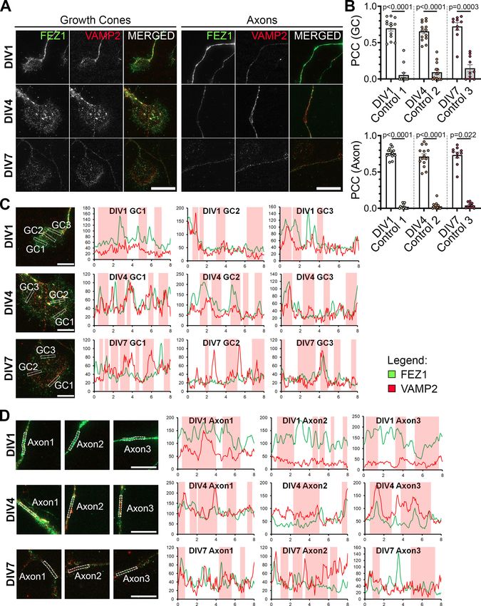
March/April 2021, 8(2) ENEURO.0193-20.2021 eNeuro.orgResearch Article: New Research 14 of 21 Figure 7. FEZ1 and VAMP2 co-localize in growth cones and axons. A, Immunostaining of FEZ1 and VAMP2 in growth cones and axons of hippocampal neurons at 1, 4, and 7 DIV. Punctate staining of FEZ1 and VAMP2 are observed. Scale bars: 20 mm. B, PCC analysis of fluorescence intensities of both proteins in growth cones. Control values were obtained by measuring the PCC with the March/April 2021, 8(2) ENEURO.0193-20.2021 eNeuro.org
Research Article: New Research     15 of 21

continued
rotated image of one channel. DIV1 n = 13, DIV4 n = 14, DIV7 n = 10, collected over three independent experiments. Statistical signifi-
cance (experimental vs control) was determined using Kruskal–Wallis analysis. All error bars represent SEM. C, D, Line scan analy-
sis for co-localization between FEZ1 and VAMP2 in growth cones and axons. Merged images are shown. x- and y-axes, distance
(mm) and gray values, respectively; axon 1, distal; axon 2, intermediate; axon 3, proximal (relative to cell body). Vertical columns in
red represent regions of colocalization. Scale bars: 10 mm. Additional line scans are shown in Extended Data Figure 7-1.

observed in shRNA-treated FEZ1 knocked down neurons                  Butkevich et al., 2016; Gopal et al., 2017; Urbina et al.,
(Extended Data Fig. 3-1). These results indicate that, un-           2018). Like Sema3A/CRMP1, Netrin-1/DCC signaling is
like adult neurogenesis, FEZ1 is required to promote den-            involved in regulating axonal and dendritic development
dritic development and branching in postnatal developing             (Kim and Chiba, 2004; Stoeckli, 2018). Thus, we hypothe-
hippocampal neurons.                                                 sized that the more severe impairment on dendritic devel-
   Dendritic development defects were also present in                opment observed in FEZ1-deficient neurons could be
neurons of Sema3a and CRMP1 knock-out mice (Su et                    potentially mediated via Netrin-1 signaling pathways.
al., 2007; Makihara et al., 2016). In agreement with these             To determine this, we first investigated whether VAMP2
reports, we also observed reduced dendritic branching in             could also colocalize with FEZ1 in growth cones of devel-
CRMP1-deficient neurons (Fig. 6C, red arrowheads).                   oping neurons. In agreement with previous findings,
Sholl analyses confirmed that neurons lacking CRMP1 in-              punctate distribution of VAMP2 was detectable along
deed show less severe defects in dendritic development               axons and in growth cones (Fig. 7A; Baumert et al., 1989;
as compared with FEZ1-deficient neurons (Fig. 6B,D;                  Coco et al., 1999; Cotrufo et al., 2011). The number of
Table 4). The mean peak number of intersections for                  VAMP2 puncta increased from DIV1, peaking at DIV4 be-
CRMP1-deficient neurons was 11.8 6 0.47 at distance of               fore decreasing at DIV7. PCC analysis showed partial co-
16 mm from the center of the cell body; intersections                localization of FEZ1 and VAMP2 in growth cones and
began to decline thereafter (Fig. 6D, dotted line). This             axons at all stages studied. Correlation significantly de-
peak was comparable in control neurons but the number                creased when rotated images from one channel were
of intersections in CRMP1-deficient neurons declined                 used as controls (Table 5; Fig. 7B). Line scan analyses
faster than in controls (Fig. 6D). Contrasting strongly with         also supported the PCC analyses (Fig. 7C,D, red vertical
this, FEZ1-deficient neurons showed a significantly small-           columns; additional line scans in Extended Data Fig. 7-1).
er peak (Mann–Whitney two-tailed test, p = 0.0369) of                These results suggest that FEZ1, in concert with VAMP2,
10.2 6 0.57 at 16 mm (Table 4; Fig. 6B, dotted line).                could also participate in Netrin-1 signaling. We were un-
Supporting this, the number of primary dendrites was sig-            able to examine localization between FEZ1 and VAMP7
nificantly reduced in FEZ1-deficient neurons compared                because of the lack of a suitable compatible antibody for
with control neurons, but not in CRMP1-deficicient neu-              staining the latter.
rons (Fig. 6B,D). Moreover, the overall reduction in dendri-
tic network complexity, as represented by the normalized             FEZ1 complexes with components of Sema3A and
area under curve, was slightly greater in FEZ1-deficient             Netrin-1 signaling pathways
neurons while those in control neurons remained compa-                  We wondered whether colocalization between FEZ1
rable (Table 4; Fig. 6B,D). Neurons doubly infected with             and VAMP2 could indicate that the 2 proteins interact.
lentiviruses targeting FEZ1 and CRMP1 were not viable,               Immunoprecipitation of HEK293 lysates co-expressing
which hindered our attempts to further examine whether               VAMP2-GFP and FLAG-FEZ1 using GFP-TRAP show that
there could be an additive effect on dendrite development            both proteins can interact (Fig. 8A). FLAG-FEZ1 was not
when both proteins were simultaneously eliminated (data              detected in control coIPs where VAMP2-GFP was re-
not shown). Nevertheless, the greater reduction in dendri-           placed by GFP. We further examined whether FEZ1 could
tic development arising from loss of FEZ1 as compared                also interact with the Sema3A receptor complex Nrp1/
with CRMP1 suggested that FEZ1 is likely to participate in           PlxnA1 (Deo et al., 2004; Zylbersztejn et al., 2012). As we
other dendritic development signaling pathways in addi-              were unable to obtain any plasmids expressing full-length
tion to those mediated by Sema3A/CRMP1.                              PlxnA1, the co-immunoprecipitation experiments were
                                                                     performed using the extracellular domain (ED) of PlxnA1,
                                                                     which is still able to form a complex with Nrp1 (Takahashi
FEZ1 colocalizes with VAMP2 in growth cones, a                       et al., 1999). Immunoprecipitation of FLAG-FEZ1 with
SNARE protein shared by Sema3A and Netrin                            a-FLAG antibodies co-immunoprecipitated mCherry-
signaling                                                            Nrp1 and EmGFP-PlxnA1 ED but not when FLAG-FEZ1
  In addition to its interaction with CRMP1, FEZ1 inter-             was omitted (Fig. 8B). This indicated that FEZ1 can also
acts with Stx1 in neuronal growth cones, which works                 interact with the Sema3A receptor complex.
downstream of the Netrin-1 receptor DCC to mediate                      Binding of Netrin-1 to DCC allows the latter to bind Stx1
axonal outgrowth in response to Netrin-1 signaling                   to effect subsequent exocytosis for neurite growth
(Cotrufo et al., 2011; Chua et al., 2012). Moreover,                 (Cotrufo et al., 2011). As FEZ1 colocalizes with Stx1 in
VAMP2, a SNARE protein used by both Netrin-1 and                     growth cones, we wondered whether FEZ1 might also
Sema3A signaling pathways is present in FEZ1 transport               form part of this DCC complex to mediate Netrin-1 signal-
vesicles isolated from rat brains (Zylbersztejn et al., 2012;        ing (Chua et al., 2012). To examine this, co-

March/April 2021, 8(2) ENEURO.0193-20.2021                                                                                  eNeuro.org
Research Article: New Research 16 of 21
Table 5. Summary of PCC values (colocalization of FEZ1 and VAMP2)
PCC
Days in vitro FEZ1 1 VAMP2 Control
GC 1 0.6972 6 0.0378 0.05331 6 0.0324
4 0.655 6 0.0328 0.09171 6 0.0313
7 0.7232 6 0.0498 0.1467 6 0.0466
Axons 1 0.760 6 0.0192 0.02056 6 0.00819
4 0.712 6 0.0358 0.0272 6 0.0121
7 0.7334 6 0.0386 0.03947 6 0.0112
immunoprecipitation assays were performed using increased dendritic arborization when treated with
EmGFP-DCC, FLAG-FEZ1 and Myc-Stx1 in HEK293 Sema3A is in agreement with previous findings. However,
cells. Indeed, Stx1 and FEZ1 co-immunoprecipitated with when treated with Netrin-1, CRMP1-deficient neurons
DCC pulled down using GFP-TRAP, but not when DCC showed a significant increase in dendritic complexity
was absent, indicating that a complex between FEZ1, against untreated controls (Fig. 9B,H,I), indicating that
Stx1, and DCC can be formed. Together, these data high- CRMP1-deficient neurons can still respond to Netrin-1
light a possible role for FEZ1 in both Sema3A-induced signaling. Strikingly, FEZ1-deficient neurons responded
and Netrin-1-induced guidance (Fig. 8C). neither to Sema3A nor Netrin-1 (Fig. 9A,C,D). Indeed, the
level of dendritic complexity in these neurons was compa-
rable to untreated FEZ1-deficient neurons. Thus, the lack
FEZ1 is required for both Netrin-1 and Sema3A of responsiveness to either stimulus in these neurons indi-
regulated neurite development cated that FEZ1, unlike CRMP1, is a common effector of
Treatment with Netrin-1 has been reported to increase both signaling pathways.
dendritic arborization during neuronal development We further investigated whether similar effects could be
(Goldman et al., 2013; Winkle et al., 2016). Similarly, hip- seen in developing axons. Netrin-1 treatment was previ-
pocampal neurons exposed to Sema3A also exhibited in- ously observed to increase axon length and branching,
creased dendritic development (Shelly et al., 2011). To whereas Sema3A reportedly inhibited axonal develop-
determine whether FEZ1 indeed functions as a common ment (Shelly et al., 2011; Winkle et al., 2016). We did not
effector downstream of both signaling pathways, we observe any changes in total axon length, longest contig-
tested how dendritic development of FEZ1-deficient or uous axon length and axonal branching when control neu-
CRMP1-deficient neurons were affected by Netrin-1 and rons were treated with Netrin-1 or Sema3A likely since the
Sema3A treatments. treatments were only applied after axons have developed
Neurons infected with FEZ1, CRMP1, and LUC sgRNA (Fig. 9E–G,J–L). While exposure of CRMP1-deficient neu-
were incubated with Netrin-1 or Sema3A for 24 h at 6 DPI. rons to Sema3A did not cause any change, treatment of
As with previous reports, treatment with either Netrin-1 or these neurons with Netrin-1 induced a significant increase
Sema3A increased dendritic arborization of neurons in in total axon length, the longest contiguous axon length
the control groups (Fig. 9A,B, FEZ1 in C,D, CRMP1 in H,I). and a slight increase in axonal branching (Fig. 9J–L).
Since CRMP1 has been shown to mediate Sema3A sig- These increases were completely abolished in Netrin-1-
naling (Deo et al., 2004; Yamashita et al., 2007), our ob- treated FEZ1-deficient neurons that were also unrespon-
servation that CRMP1-deficient neurons did not exhibit sive to Sema3A treatment (Fig. 9E–G). Taken together,
Figure 8. FEZ1 forms complexes with components of Netrin-1 and Sema3A signaling pathways. A, FEZ1 interacts with VAMP2.
Lysates from HEK293 cells expressing FLAG-FEZ1 and VAMP2-GFP were immunoprecipitated using GFP-TRAP. FEZ1 co-immuno-
precipitates with VAMP2. B, FEZ1 interacts with Nrp1 and PlxnA1. FLAG-FEZ1 was immunoprecipitated using a-FLAG from
HEK293 cell lysates expressing FLAG-FEZ1, mCherry-Nrp1, and EmGFP-PlxnA1 ED. Nrp1 and PlxnA1 ED were both co-immuno-
precipitated, indicating an interaction between FEZ1 and the Sema3A receptor complex. C, FEZ1 forms a complex with DCC and
Stx1. HEK293 cell lysates transiently expressing the three proteins were immunoprecipitated using GFP-TRAP. Both FLAG-FEZ1
and Myc-Stx1 co-immunoprecipitated with EmGFP-DCC, indicating that the three proteins can form a complex. Data were obtained
from three independent experiments.
March/April 2021, 8(2) ENEURO.0193-20.2021 eNeuro.orgResearch Article: New Research 17 of 21 Figure 9. FEZ1 is involved in Netrin-1 and Sema3A-induced dendritic development. A, B, Images of FEZ1 sgRNA or CRMP1 sgRNA, LUC sgRNA, and uninfected neurons treated with Netrin-1 or Sema3A. Neurons were fixed and stained for MAP2, Tau, and FEZ1, or CRMP1 24 h post-treatment. Scale bars: 20 mm. C, D, Sholl analyses and AUC of FEZ1-deficient and control neurons treated with Netrin-1 or Sema3A. Dendritic complexity of control neurons but not FEZ1-deficient neurons were observed to increase after Netrin-1 or Sema3A treatment. FEZ1 sgRNA control n = 32, netrin n = 35, sema n = 33; LUC sgRNA control n = 31, netrin n = 32, March/April 2021, 8(2) ENEURO.0193-20.2021 eNeuro.org
You can also read