Progress in the Use of Antisense Oligonucleotides for Vaccine Improvement - MDPI
←
→
Page content transcription
If your browser does not render page correctly, please read the page content below
Review
Progress in the Use of Antisense Oligonucleotides for
Vaccine Improvement
Alexander Batista-Duharte 1,2,*, Luis Sendra 2, Maria José Herrero 2,*, Damiana Téllez-Martínez 1,
Iracilda Zeppone Carlos 1 and Salvador Francisco Aliño 2
 1 School of Pharmaceutical Sciences, Department of Clinical Analysis, São Paulo State University (UNESP),
   Rod. Araraquara-Jaú - Km 1, 14800-903 Araraquara, SP, Brazil; batistaduhartea@gmail.com (A.B.-D.);
   damianatellezm@gmail.com (D.T.-M.); carlosiz@fcfar.unesp.br (I.Z.C.)
 2 Pharmacology Department, Faculty of Medicine, Universidad Valencia, Av. Blasco Ibáñez 15, 46010

   Valencia, Spain; luis.sendra@uv.es (L.S.); salvador.f.alino@uv.es (S.F.A.)
 * Correspondence: batistaduhartea@gmail.com (A.B.-D.); Maria.Jose.Herrero@uv.es (M.J.H.)

 Received: 31 December 2019; Accepted: 11 February 2020; Published: 17 February 2020

 Abstract: Antisense oligonucleotides (ASOs) are synthetically prepared short single-stranded
 deoxynucleotide sequences that have been validated as therapeutic agents and as a valuable tool in
 molecular driving biology. ASOs can block the expression of specific target genes via
 complementary hybridization to mRNA. Due to their high specificity and well-known mechanism
 of action, there has been a growing interest in using them for improving vaccine efficacy. Several
 studies have shown that ASOs can improve the efficacy of vaccines either by inducing antigen
 modification such as enhanced expression of immunogenic molecules or by targeting certain
 components of the host immune system to achieve the desired immune response. However, despite
 their extended use, some problems such as insufficient stability and low cellular delivery have not
 been sufficiently resolved to achieve effective and safe ASO-based vaccines. In this review, we
 analyze the molecular bases and the research that has been conducted to demonstrate the potential
 use of ASOs in vaccines.

 Keywords: antisense oligonucleotide; vaccines; adjuvants; infectious disease; cancer

1. Introduction
     For more than two centuries, vaccines have contributed to the eradication or control of important
diseases, and they have participated greatly in the increased life expectancy and the improvement of
sanitary conditions throughout the world [1]. However, despite the great success of vaccination in
public health, there are still many challenges. The lack of effective vaccines against several diseases
such as HIV/AIDS, malaria, and leishmaniasis; the re-emergence of other diseases such as
tuberculosis; and the appearance of new pathogenic organisms or known pathogens with increased
virulence stimulate the search for more effective vaccines than those available today [2,3].
     The last decades have been marked by important advances in vaccine research and
development. The technology of recombinant DNA and the synthesis of peptides, the development
of modern bioinformatic tools, and the use of improved adjuvants and delivery systems have allowed
the development of more effective and safer vaccines based on rational designs [2,4]. Moreover, the
successful completion of the human genome project in the early 2000s ushered the genomics
revolution, which is beginning to have a great impact on vaccine research [4,5].
     For many years, nucleic acids and short nucleotide molecules have been used as vaccine
components. Their use has ranged from DNA [6,7] or RNA vaccines [8,9], to oligonucleotide
sequences containing unmethylated cytidine phosphate guanosine (CpG) motifs with significant
immunostimulatory (adjuvant) properties [10,11]. More recently, strategies to manipulate the
expression of genes controlling the immune response or the expression of antigens of interest are
being used to improve immunogenicity and vaccine efficacy.

Biomolecules 2020, 10, 316; doi:10.3390/biom10020316                      www.mdpi.com/journal/biomolecules
Biomolecules 2020, 10, 316                                                                        2 of 23

      Antisense oligonucleotides (ASOs) are synthetically prepared short single strands (usually 18–
21 deoxynucleotides in length), complementary to a preRNA or an mRNA sequence of the target
gene. ASOs modify the expression of specific target genes, by either splicing modifications or by
recruiting RNase H leading to RNA degradation of RNA–DNA hetero-duplex, thus blocking the
expression of the target gene [12,13]. The availability of human genome sequence information, freely
and publicly, offers the possibility to obtain inexpensive specific synthetic oligonucleotides designed
against a specific target gene. The strengths of their pharmacological effects evidenced in in vitro and
in vivo models have favored the development of several drugs based on oligonucleotides that have
been approved by the FDA [14]. Currently, several groups are making efforts to improve vaccine
efficacy using ASOs with encouraging advances achieved over the last years, but some problems are
still hampering further progress on these fronts. These are mainly related to ASOs bioavailability and
the occurrence of potential off-target effects. In this review, we focus on the recent design of ASOs
that have yielded promising results in terms of vaccine immunogenicity improvement. Current
challenges and opportunities are also analyzed.

1.1. Earlier Uses of Oligonucleotides in Vaccines
      In 1893, William Coley reported that a mixture of bacterial cell lysate, named Coley’s toxin, could
reduce the progression of some carcinomas [15]. Since the first description of the possible anti-tumor
effect of Coley’s toxin, there was much debate about its mechanism of action. More than 60 years after
Coley’s report, Taliaferro and Jaroslow [16] reported that preparations of nucleases-degraded DNA
and RNA could partially restore hemolysin production after a single intravenous injection of sheep
red blood cells (RBC) in rabbits that received 400 r total body X radiation. However, the development
of synthetic oligonucleotides for medical use was only possible after the discovery of two chemical
modifications, namely 2’fluoro (2´-F) substitutions [17,18], and Phosphorothioate (PTO) chemistry
[19]. Another important 2´ modification was developed in 1969, 2´-O-Methyl (2´-O-Me) [20], a major
alternative in many synthetic oligonucleotides. These chemical modifications improved the cellular
uptake of oligonucleotides and conferred protection against enzymatic degradation.
      Several studies showed that oligonucleotides can stimulate the production of specific antibodies
in mature animals after concurrent administration of an antigen with either DNA or RNA digest
[21,22]. At the same time, the immunogenic capacity of nucleic acids and their influence in
autoimmune diseases was demonstrated [23]. Moreover, Field et al. identified that complexes of
polyinosinic and polycytidylic acids (poly (I:C)) were highly active as inducers of interferon [24]. The
biological basis for this observation was understood more than three decades later when Toll-like
receptor 3 (TLR3) was reported to be the receptor for double-stranded RNA [25]. Related to these
findings, Tokunaga et al. identified bacterial DNA as the underlying component of a fraction
extracted from Mycobacterium bovis strain BCG that elicited an antitumor response in different in vitro
and in vivo models [26]. After that, these researchers cloned mycobacterial genes, synthesized diverse
oligodeoxynucleotides (ODNs), and observed that certain palindromes in these ODNs were
responsible for activating the immune response [27,28].
      In 1995, Krieg et al. reported that unmethylated CpG dinucleotides (CpG ODN) within bacterial
DNA activate host defense mechanisms leading to innate and adaptive immune responses [29]. CpG
ODN is a ligand of Toll-like receptor 9 (TLR-9) in antigen-presenting cells (APCs). CpG ODN/TLR-9
interaction induces an innate immune response that promotes the subsequent development of
adaptive immunity [10]. CpG ODN can be divided into classes A, B, C, P, and S [30]. Their utility as
vaccine adjuvants has been evaluated in different clinical trials and the achieved results indicate that
CpG ODN augments the induction of vaccine-specific cellular and humoral responses [11]. In 2017,
the FDA approved HEPLISAV-B, the first vaccine with a CpG ODN as an adjuvant for hepatitis B
vaccines [31]. On the other hand, it has been reported that CpG ODN can induce high levels of pro-
inflammatory cytokines, with potential risk for developing or worsening autoimmune diseases and
systemic inflammatory response syndrome (SIRS) [32–35].

1.2. Birth of ASOs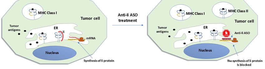
Biomolecules 2020, 10, 316                                                                       3 of 23

     In 1978, Zamecnik and Stephenson used a synthetic ASO, which was complementary to 13
nucleotides of Rous sarcoma virus (RSV) RNA, to inhibit the translation of the viral RNA and
subsequently block the virus replication in a chick embryo fibroblasts culture [36,37]. One year later,
Donis-Keller reported that RNase H catalyzes the cleavage of the RNA strand in RNA/DNA
heteroduplexes [38] in a site-specific manner. That report demonstrated for the first time that ASOs
can work through an enzyme-mediated process in addition to steric blocking. The decade of the 80s
was marked by other advances. In 1983, Simons and Kleckner showed evidence of the existence of
naturally occurring antisense RNAs and suggested a role in the regulation of gene expression [39].
After that report, other authors successfully inhibited mRNA translation by anti-sense RNA [40–43].
Moreover, in that decade, different methods for the automatic synthesis of oligonucleotides were
developed [44,45] and the first antisense patent was presented in 1987, although this was publicly
available from 1995 [46].
     Despite the advances achieved, the experimental and clinical use of unmodified ASOs was
limited as they were easily degraded by intracellular endonucleases and exonucleases, usually via 3′-
5′ activity [47]. Thus, diverse chemical modifications have been developed to protect them against
nuclease degradation, increase their affinity and potency, extend their tissue half-life, and reduce the
undesired off-target effects [Table 1].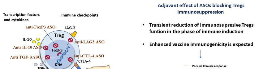
Biomolecules 2020, 10, 316 4 of 23
Table 1. Summary of three generations of the most studied ASOs chemical modifications.
Chemical Modifications Characteristics Mechanisms Clinical Use Limitations
First Generation
High affinity for various cellular
proteins and components of the innate
immune system, such as Toll-like
First generation ASOs promote degradation PTO is the most widely used modification
receptors (TLRs), with
Either a sulfur atom (PTO), or a of target mRNA by RNase H enzyme. They of ASOs. Fomivirsen, is a PTO-modified
Phosphorothioate (PTO), proinflammatory effects.
methyl group (MPO) substitutes also confer higher solubility, resistance to ASO, used as local treatment of
Methylphosphonate Commonly reported side effects
the non-bridging oxygen atoms in nuclease degradation, antisense activity and cytomegalovirus (CMV) retinitis in
(MPO) following systemic administration of
the phosphodiester bond. longer plasma half-life as compared with patients with acquired immunodeficiency
PTO ASOs include fever, activated
phosphodiester oligonucleotides. syndrome (AIDS) [48].
partial thromboplastin time
prolongation, thrombocytopenia,
and leukopenia.
Second Generation
Mipomersen is used as an adjunct therapy
2’-O-Methyl (2’-OMe) and 2’-O-
The PTO DNA induces RNase H cleavage for homozygous familial
Methoxyethyl (2’-MOE) are the A subset of 2´-MOE-modified ASOs
while 2-OME or 2-MOE on both sides (5- hypercholesterolemia [49].
most widely studied. induced pro-inflammatory cytokines
and 3-directions) confers nuclease- Nusinersen was approved for spinal
Chimeric ‘gapmer’ ASOs consist in and type I interferons (IFN-α/β) and
ASOs with 2’-O-alkyl resistance, and they can exert activity by a muscular atrophy treatment [50].
a central ‘gap’ region containing interaction with innate immune
modifications of the ribose. steric interference of translation process. Apatorsen is a HSP27 targeting ASO that
10 DNA or PTO DNA monomers, receptors such as TLR9, melanoma-
Chimeric ‘gapmer’ ASOs They are safer than PTO-modified ASOs and is being studied in phase II clinical trials in
flanked on both 5’ and differentiation associated-5 (MDA-5)
exhibit enhanced affinity towards the patients with metastatic castration
3’extremities by alkyl modified and IFN-β promoter stimulator-1 (IPS-
complementary RNA with better tissue resistant prostate cancer [51] and
nucleotides such as 2′-OM or 2’- 1).
uptake and longer in vivo half-life. Untreated Stage IV Non-Squamous-Non-
MOE.
Small-Cell Lung Cancer [52].
Third Generation
The potential of PNA as drugs in gene
therapy has been hampered by the poor
intrinsic uptake of PNA by living cells.
PNA block the protein expression, by steric
PNA is a synthetic DNA in which Current strategies for improving PNA PNA do not activate the RNase H to
hindrance, forming sequence-specific duplex
the deoxyribose phosphate delivery into the cytosolic space and cleave the target hybridized RNA.
Peptide nucleic acid (PNA) with the targeted mRNA. They are
backbone is replaced by nucleus include microinjection, PNA have low solubility and cellular
biologically stable and have good
polyamide linkages. electroporation, co-transfection with DNA, uptake.
hybridization properties.
or conjugation to lipophilic moieties,
nanoparticles, cell-penetrating peptides
(CPPs), oligo-aspartic acid, or nuclear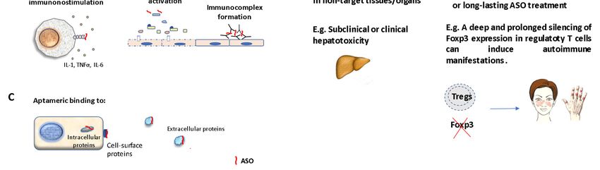
Biomolecules 2020, 10, 316                                                                                                                                                                  5 of 23

                                                                                                                  localization signal (NLS) peptides to
                                                                                                                      enhance cellular internalization
                              PMOs are neutral ASOs. The                                                         Eteplirsen was approved for Duchenne
                                                                 The mechanism of PMO is the translational                                                      PMOs exhibit reduced cellular uptake.
                             pentose sugar is substituted by a                                                   muscular dystrophy (DMD) treatment
                                                                  arrest mediated by steric interference of                                                      Conjugation with peptides such as
Phosphoramidate morpholino    morpholino ring and the inter-                                                    [53]. Other potential applications include
                                                                   ribosomal assembly. PMO show fewer                                                             arginine-rich peptide (ARP) can
      oligomer (PMO)             nucleotide linkages are                                                       the treatment of viral infections, antibiotic-
                                                                  nonspecific properties and lesser toxicity                                                      enhance its cellular uptake and
                             phosphoramidate bonds in place                                                     resistant bacterial infections, and cancers
                                                                                 than PTO.                                                                               antisense efficacy.
                                of phosphodiester bonds.                                                                            [54].
                                                                                                                                                                 LNA does not activate RNase. LNA
                              LNAs are chemically modified       LNA modifications improve the affinity of
                                                                                                                                                                nucleotides can be incorporated at the
                                 nucleotides with a ribose       ASO hybridization towards mRNA target,
                                                                                                                 Diverse LNAs are currently in clinical          ends of RNA and DNA sequences to
 Locked nucleic acid (LNA)    containing a methylene bridge            by increase of the DNA/RNA
                                                                                                                 trials by several biotechnology firms.            form chimeric oligonucleotides
                             between the 2′-oxygen and the 4′-    heteroduplexes thermal stability. LNAs
                                                                                                                                                                 resulting in restoration of RNase H-
                                   carbon of the ribose.               avoid nuclease degradation.
                                                                                                                                                                    mediated cleavage of mRNA.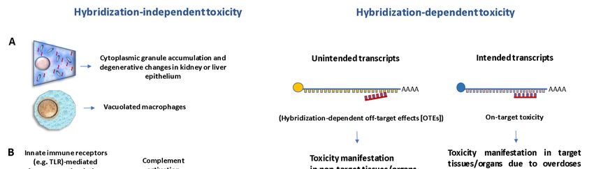
Biomolecules 2020, 10, 316                                                                                6 of 23

     After these first reports, notable progress has been made in ASOs pharmacology and now
different therapeutic ASOs against different diseases, including neurodegenerative, cardiovascular,
metabolic, inflammatory, infectious, and neoplastic diseases are being tested in clinical trials, and
several ASOs have been approved by the US Food and Drug Administration (FDA) to be used in
humans. Fomivirsen (Vitravene) was the first antisense drug approved in 1998 for cytomegalovirus
(CMV) retinitis [48]. In January 2013, the FDA approved mipomersen (Kynamro, Genzyme) for
homozygous familial hypercholesterolemia (HoFH) [49]. Both fomiversen and mipomersen were
developed by Isis Pharmaceuticals. More recently, nusinersen was approved for spinal muscular
atrophy [50] and eteplirsen for Duchenne Muscular Dystrophy [53]. There are many other ASOs in
different phases of clinical trials [14,54–56].

2. Molecular Basis of ASOs
     ASOs and other forms of genetic therapies have emerged as an attractive therapeutic alternative
to monoclonal antibodies. Factors such as structure, stability, cellular, bioavailability, lack of
immunogenicity, specificity, relative low toxicity, and low cost indicate a great future perspective for
these molecules (Table 2).

                       Table 2. Key advantages of ASOs compared to monoclonal antibodies.

                                             ASOs                                     mAb
    Molecular Weight                      ~6 to10 kDa                               ~150 kDa
                             Relatively simple structure. Usually
         Structure                 13–20 mer with chemical           Glycoproteins with complex structure
                                         modifications
                              Highly stable. Lyophilization and       Low stability. Cold chain through the
    Thermal Stability            freezing does not modify its        storage, handling, and transportation is
                                      biological activity                           necessary
         Cellular            ASOs can penetrate the cells and act
                                                                      They are unable to penetrate the cells
      Bioavailability               on intracellular targets
                                                                      Highly immunogenic by xenogeneic
     Immunogenicity          ASOs are not properly immunogenic         differences, e.g., between mice and
                                                                                     humans
                                 Highly specific but off target     Highly specific but cross-reactivity can be
        Specificity
                                 interaction can be observed                         observed
                                                                     Different grades of toxicity have been
          Toxicity                  Relatively low toxicity
                                                                                    described
                               ASOs are obtained synthetically.
                                                                      The production for pharmacological
    Development and               The use of vehicles can add
                                                                        proposes requires high level of
     Manufacturing              complexity to the manufacture
                                                                           technological complexity
                                            process

2.1. Pharmacokinetics
     Different factors such as the type of chemical modifications, the electric charge, and the use of
delivery systems are pivotal factors in the pharmacokinetic properties of ASOs, independently of
nucleotide sequence [57–59]. Following intravenous or subcutaneous administration, ASOs quickly
pass from the injection site to the circulation, and the highest plasma concentrations for first and
second-generation of ASOs are reached within 3–4 h with a rapid distribution phase to tissues in
minutes, followed by a slow elimination from tissues lasting a few hours [58]. Double-stranded RNA
ASOs and single-stranded neutral-backbone ASOs such as morpholinos exhibit low serum protein
binding, with limited tissue distribution, and they are rapidly eliminated by renal filtration [58,60].
In contrast, first-generation PTO can interact with albumin and other serum proteins, extending their
circulation from minutes to a few hours and their tissue distribution [61]. Studies suggest that PTO-
modified ASOs can also interact with a large number of proteins on the cell surface and in the
extracellular matrix and can be endocytosed into intracellular vesicles [62–64] After that, they bind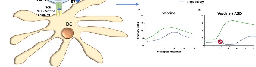
Biomolecules 2020, 10, 316                                                                            7 of 23

to proteins that transport them into the nucleus and perhaps promote the hybridization to RNAs [64–
66]. The clearance and elimination of PTO are facilitated by enzymatic degradation mediated by
endo- and exonucleases that results in small-molecular-weight fragments that are easily eliminated
in urine [58]. Liver, kidneys, bone marrow, adipocytes, and lymph nodes are the major systemic
tissues of distribution [58,62] Second-generation ASOs with 2’-O-alkyl modifications of the ribose are
metabolized more slowly than the first and third generation of ASOs, and the clearance half-lives
occur in 2–4 weeks [58].

2.2. Mechanism of Action of ASOs
     ASOs are designed to specifically block the transfer of the genetic information for the protein
synthesis by binding to a target RNA through Watson–Crick base pairing: A-T, C-G interaction [47].
They can modulate the processing, stability, or activity of specific RNAs by different mechanisms
that have been extensively analyzed in previous reviews [56,67,68]. Briefly, ASOs can directly stick
to pre-RNA or mRNA molecules and prevent the formation of the 5'-mRNA cap, modulate
alternative splicing, dictate the location of the polyadenylation site, and recruit RNAse H to cleave
and degrade the RNA target in the ASO-RNA complex. ASOs can also sterically block the ribosomal
subunits from attaching or running along with the mRNA transcript, hampering the translation
process (Figure 1).

      Figure 1. Main mechanisms of action of antisense oligonucleotides. (A) Normal gene and protein
      expression in the absence of ASO. (B) In cytoplasm, ASOs can bind to a complementary mRNA
      region. ASO-mRNA heteroduplex can induce the activation of RNase H, leading to mRNA
      degradation. Alternatively, ASOs can block the translation process without promoting RNA
      degradation by steric interference of ribosomal assembly. (C) ASO can enter the nucleus and hinder
      mRNA maturation by inhibition of 5′ cap formation, RNase H-mediated pre-RNA cleavage, and
      inhibition of mRNA splicing.

2.3. Toxicology of ASOs
     In addition to the Watson–Crick interaction, ASOs can form other interactions, such as
electrostatic links with polycations and positively charged proteins, noncanonical base-pairing with
themselves and other nucleic acids, and sequence-specific interaction with proteins [69]. These non-
antisense interactions can cause different toxicity manifestations [70–75].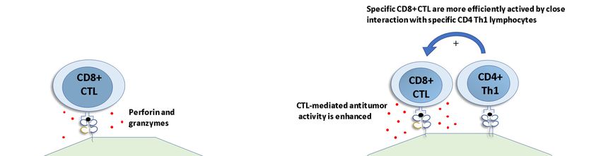
Biomolecules 2020, 10, 316                                                                             8 of 23

     The toxicity of ASOs is classified into two categories: hybridization-independent (non-
pharmacologic) or hybridization-dependent (pharmacologically based) mechanisms [72] (Figure 2).
Hybridization-independent toxicity is related to the specific chemical modification of the ASO and it
does not involve base-pairing interactions. There are three general subcategories of hybridization-
independent toxicities: (a) ASO effects by excessive accumulation, which is manifested by basophilic
cytoplasmic granule accumulation, predominantly in kidney or liver epithelium that can produce
degenerative changes. Another common histologic finding is the presence of increased granular
macrophages or increased vacuolated macrophages related to cellular activation and
proinflammatory cytokine production [72]; (b) immunomodulation (proinflammatory mechanisms,
caused by toll-like receptors (TLR) and other innate immune receptors mediated mechanisms,
complement proteins activation, and immune complexes causing immune cell activation and
inflammatory reactions [72–74]. The major contributor to the proinflammatory effect is the backbone
chemistry, although base-pair sequence and base modifications can also contribute. Proinflammatory
effects are often observed as lymphoid hyperplasia in lymph nodes and spleen, associated to PTO
ASOs, in rodents and monkeys, while reversible glomerulonephritis and vasculitis after
administration of some types of ASOs have also been reported in monkeys [72]; (c) ASO interactions
with extracellular, cell-surface, and/or intracellular proteins with high affinity and specificity (named
as aptameric binding) [72,74]. According to Frasier [72], accumulation-related effects are the most
encountered changes in preclinical toxicity studies.

      Figure 2. Types of ASOs-mediated toxicity: (1) Hybridization-independent toxicity represent those
      effects that are not due to Watson–Crick base pairing between an ASO and RNA. This type of toxicity
      occurs by three possible mechanisms: (A) ASOs accumulation effect is manifested as cytoplasmic
      granule accumulation, degenerative changes in kidney or liver epithelium, and presence of
      vacuolated macrophages. (B) Proinflammatory mechanisms due to ASOs interaction with innate
      immune receptors, inducing macrophages activation, complement activation, and immunocomplex
      formation. (C) Aptameric binding to intracellular cell surface or extracellular proteins. (2)
      Hybridization-dependent toxicity can be caused by partial or complete ASO interaction with
      unintended transcripts (hybridization-dependent off-target effects [OTEs]); or with intended
      transcripts (on-target toxicity).

     The hybridization-dependent toxicity can be caused by either (a) hybridization-dependent off-
target effects (OTEs) due to complete or partial complementary recognition of unintended transcripts
or (b) hybridization-dependent toxicity (on-target toxicity). OTEs have been detected in preclinical
Biomolecules 2020, 10, 316 9 of 23
studies after systemic administration of gapmers using biochemical markers of hepatotoxicity in the
blood, while it has not been observed in clinical trials involving therapeutic ASOs to date [70]. Some
authors also consider the non-pharmacologic effects as a form of off-target toxicity. On the other
hand, on-target toxicity can occur after over-silencing intended transcripts or as a result of long-
lasting treatment, and the adverse effects are specific to individual ASOs. For example, long lasting
knockdown of genes involved in the immunosuppressive activity of regulatory T cells (Tregs) can
potentially cause autoimmune manifestations.
Although the chemical modifications of ASOs have been important for their stability and
delivery, the introduction of some structural modifications can increase the risk of toxicity. Swayze
et al. [76] evaluated the toxicity of several ASOs containing either 2'-O-methoxyethylribose (MOE) or
locked nucleic acid (LNA). They showed that incorporation of LNAs in some sequences induced
hepatotoxic effects as early as 4 days after a single administration, whereas MOE-modified
nucleotides in the same sequences did not cause toxicity. In another work, the authors observed that
many unintended transcripts were downregulated in mice treated with hepatotoxic LNA ASOs,
whereas in mice treated with non-hepatotoxic LNA ASOs, the transcript knockdown was highly
selective [77].
Two forms of thrombocytopenia have been reported following several ASOs treatments. The
most common form is mild, reversible, and dose-dependent thrombocytopenia, typically observed at
high doses in monkeys and rodents after treatment with first-generation ASOs. In humans, transient
thrombocytopenia without hemorrhagic manifestation has been reported after the use of first-
generation ASOs such as oblimersen, aprinocarsen, ISIS 2503, and ISIS 5132 and less frequently with
second-generation ASOs, including mipomersen, ISIS 104838, LY2275796 for cancer. A second form
of thrombocytopenia with marked platelet depletion and hemorrhages is a rare but serious adverse
event related to repeated exposure of ASOs [72,74,78].
2.4. Strategies to Improve ASOs Cell Targeting and Overcome Toxicity
Diverse reports have shown that naked ASOs are poorly internalized by cells, and they tend to
be localized in endosomes, where they are unavailable for antisense purposes [47]. Thus, difficulties
in biodistribution and cellular internalization are the main obstacles for clinical applications of ASOs.
Diverse methods have been developed to improve cellular uptake of ASOs and their pharmacological
activity, to increase their stability, to reduce the therapeutic dose, and to limit the off-target effects
[79–82]. Conjugation of ASOs to molecules that can bind to certain ligands in the cell can improve
their cell uptake and internalization. Small hydrophobic molecules, such as cholesterol [83], lipids
[84], or fluorinated chains [85,86] may increase ASOs stability and their membrane permeation.
Multivalent N-acetylgalactosamine (GalNAc) conjugation is another important way for ASOs
delivery to hepatocytes [87]. Basic peptides such as Drosophila melanogaster homeotic transcription
factor, Antennapedia peptide [88], and Tat protein of HIV-1 [89] have also been used to increase ASOs
passage through the plasma membrane by a receptor- and transporter-independent mechanism
delivering them directly into the cytoplasm and, hence, ultimately the nucleus.
In addition to direct conjugation of ASOs with defined molecules, the use of nanoparticles as
vehicles for ASOs has been widely evaluated. The first generation of ASOs vehicles were liposomes,
which are sphere-shaped vesicles consisting of one or more bilayers of phospholipids and cholesterol
[90]. The ASO can be encapsulated into the aqueous compartment of the liposome or can be bound
to the liposome surface by electrostatic interactions. Under physiological conditions, positively
charged liposomes have high affinity for the negatively charged cell membranes and can easily bind
to cells. Because these liposomes use the endosomal pathway to deliver ASOs into cells, they can be
formulated with certain molecules inducing endosomal membrane destabilization, such as
chloroquine and 1,2-dioleoyl-sn-glycero-3-phosphatidylethanolamine, to allow the scape of ASOs
from the endosomes and be actively transported in high concentration to the nucleus [91–95].
Lipid nanoparticles (LNP) are other important formulations that have been used to enhance the
delivery of ASOs to target tissues. Delivery using LNPs increases the stability and circulation time of
ASOs [96,97]. LNPs contain ionizable amino lipids that self-assemble into nanoparticles when mixedBiomolecules 2020, 10, 316 10 of 23
with polyanionic oligonucleotides. The electrostatic interaction of LNPs with polyanionic nucleic
acids promotes their encapsulation, allowing the escape to cell cytoplasm from the endosomal
compartment [98]. Several ligands for overexpressed receptors on the target cell surface can be linked
to LNPs, to facilitate the cellular uptake. These ligands include cell transferrin [99], penetrating
peptides [100], folate [101], polysaccharides [102] and antibodies [103].
Besides liposomes and LNPs, cationic polymers, including poly-L-lysine [104,105],
polyalkylcyanoacrylate nanoparticles [106–108]; polyethyleneimine (PEI) [109,110], and poly(amido
amide) (PAMAM) dendrimers [111] have been also developed for ASOs delivery. These polyamines
cause endosomal rupture via a “molecular sponge” mechanism and are less used than the cationic
liposomes due to their toxicity [47]. Moreover, niosomes are an interesting alternative to liposomes
for ASOs delivery. They are vesicles composed of non-ionic surfactants, amphipathic compounds
with an overall neutral charge [80,112,113].
3. ASOs in Vaccines
The use of ASOs for vaccine improvement has been mainly based on the following strategies:
(1) antigen modification; (2) targeting the host immune system by overexpression/inhibition of
molecules involved in the immune response. In the following sections, we will analyze the main
advances in these areas and the challenges still to be solved.
3.1. Antigen Modification
The first attempts using ASOs for antigen manipulation started in 1990. Goudsmit´s group used
a phosphate-methylated ASO complementary to the tat responsive region (TAR) of the HIV-1 isolate
CBL-4 (RUT) to reduce the viral infectivity [114,115]. However, some technical errors and
interpretation of results that were subsequently corrected by the same authors caused the retraction
of the article published in Science [116], as well as the conclusion issued that the observed inhibitory
effect of viral infectivity should be ascribed to the phosphate methylation of natural DNA.
Tumor cells escape from immune surveillance by means of mechanisms to prevent tumor
antigens recognition by the immune system. Several methods have been developed to increase the
immunogenicity of the tumor cells. The most efficient methods can force tumor cells to present their
own tumor antigens to the immune system [117]. In the early 1990s, the group led by Dr. Ostrand–
Rosenberg demonstrated that tumor cells transfected with MHC class II molecules can generate a
potent tumor cell vaccine, which protects against challenge with the parental tumor [118]. Moreover,
supra-transfecting MHC class II+ tumor cells with li gene, coding for li protein (CD74), the invariant
chain that normally blocks the binding of self-peptide fragments to MHC class II molecules,
abrogated the immunogenicity of the modified cells [119].
Based on this principle, Qiu et al. treated cancer cells expressing, naturally or by induction, MHC
class II molecules and Ii protein, with anti-Ii ASO to induce the MHC-II–mediated presentation of
diverse antigenic peptides to helper T cells (Figure 3). In each line of transfected tumor cells, the ASO
profoundly suppressed Ii protein in 35% ± 55% cells, without affecting the expression of MHC class
II molecules. The absence of the Ii protein increased the range of cancer-related epitopes presented to
CD4+ helper T cells and generated effective tumor cell vaccines [120]. They also created several
antisense Ii-reverse gene constructs (Ii-RGC) that inhibited Ii expression in A20 B lymphoma cells in
vitro and Renca renal adenocarcinoma tumors in vivo. Subcutaneous Renca tumors in BALB/c mice
were treated by intratumoral injection with a plasmid containing the gene for MHC class II
transactivator (CIITA) and Ii-RGC. A subtherapeutic dose of IL-2 was also used to upregulate the
activation of T cells. Significant tumor reduction and a decrease in the progression rates of the
established tumors in the groups injected with Ii-RGC were observed, compared to the groups treated
with IL-2 plus empty plasmid controls (p < 0.002) [121]. In another study, a single recombinant
adenovirus with both interferon-gamma (IFN-γ) and Ii-RGC (rAV/IFN-γ/Ii-RGC) genes efficiently
induced the MHC Class II+/Ii- phenotype in MC-38 colon adenocarcinoma cells and Renca tumors.
Injection of tumor nodules with rAV/Ii-RGC and rAV/CIITA/IFN-γ, associated with a suboptimal
dose of rAV/IL-2 induced a potent antitumor immune response. Control mice developed growingBiomolecules 2020, 10, 316 11 of 23
tumors by day 8 after injection. On the other hand, mice treated with rAV/CIITA/IFN-γ + rAV/IL-2 +
rAV(wild type) showed delayed tumor growth in three of five mice, with tumor re-growing in two
of these mice, resulting in one of five mice being tumor-free on day 60. Mice treated with
rAV/CIITA/IFN-γ + rAV/IL-2 + rAV/Ii-RGC showed tumor regression in three of four animals.
Finally, tumor-free mice were challenged on day 63 with Renca cells. Naive mice injected with the
same number of Renca cells developed tumors while those tumor-free mice did not develop tumors
in a follow-up of 34 days post-challenge. Similar results were observed in repeated experiments
under the same conditions [122].
Figure 3. Tumor cells are forced to present their own tumor antigens to the immune system by anti-li
ASO treatment. Left, MHC class I presents endogenous tumor antigens to CD8+ cytotoxic T cells
(CTL). Ii protein blocks the binding of endogenous antigens to MHC class II in the endoplasmic
reticulum (ER). Right, anti li-ASO blocks Ii protein expression, and endogenous tumor antigens are
also presented by MHC class II molecules and recognized by specific Th1 lymphocytes. The
simultaneous presentation of tumor antigens by both MHC class I to CTL and MHC II to Th1
lymphocytes induces a stronger antitumor response. (Adapted from [117]).
Rubenstein et al. evaluated the effect of bispecific ASOs targeting BCL-2 and epidermal growth
factor receptor (EGFR) in the in vitro growth and prostatic antigen expression on androgen-sensitive
human prostate adenocarcinoma (LNCaP) cells. Cultured cells were treated with 6.25 µM of either
mono or bispecific ASOs and significant inhibition of the cellular growth was observed after
treatment with bispecific ASOs. Interestingly, the bispecific ASO treatment also enhanced the
expression of non-targeted proteins: prostate-specific cell surface antigens (PSMA), and IFN-γ.
However, monospecific ASOs directed solely against BCL-2 did not stimulate the production of these
proteins. The authors concluded that enhanced expression of cell surface differentiation antigens
(such as PSMA) could increase their recognition and targeting by antitumor immunologic
mechanisms and increase the effectiveness of tumor vaccines [123]. In other studies, the authors
showed that LNCaP cells treated with ASOs directed against BCL2 administered in a nanoparticle
suspension of lipofectin as vehicle exhibited non-target effects by suppressing the expression of
apoptosis promoter caspase-3 [124]. In addition, they observed compensatory enhanced expression
of other molecules such as (a) apoptosis inhibitor serine/threonine protein kinase (AKT1) [125], (b)
androgen receptor (AR) and their co-activators p300 [126,127], (c) interleukin-6 (IL6) [128], (d)
programmed death 1 (PD1) and its ligand PDL1, and (e) FAS-ligand, which activate apoptosis [129].
These and other reports of this group suggest that the use of ASOs to suppress BCL2 to restoreBiomolecules 2020, 10, 316 12 of 23
apoptosis can lead to altered expression of non-targeted genes with different effects, including the
stimulation of tumor proliferation [130].
3.2. Targeting Host Immune Mechanisms
It has been demonstrated that different isoforms of transforming growth factor-beta (TGF-β)
with immunosuppressive activity are overexpressed in different malignant tumors such as
melanoma, gliomas, prostate, gastric, colorectal, ovarian, gastric, and non-small cell lung cancers
(NSCLC). Enhanced TGF-β2 expression in malignant cells is suggested to be a pivotal factor for tumor
progression by inducing immunosuppression, metastasis, angiogenesis, and proliferation [131–137].
Tumor-infiltrating tolerogenic DCs and suppressor T cells are related to tumor-associated
immunosuppression and tumor escape. These processes are mediated by TGF-β and IL-10 expression
[133]. Elevated levels of TGF-β are inversely correlated with prognosis in patients with NSCLC
[135,137].
The immunosuppressive effect of a TGF-β-producing autologous tumor vaccine was abrogated
and rendered immunogenic when suppressing its TGF-β secretion with antisense strategy [138]. In
that study, Tzai et al. used an MBT-2 tumor cell line [MBT-2/TGF-beta(-)#3] treated with ASOs against
TGF-β and demonstrated that the amounts of this protein were significantly decreased in both
irradiated and non-irradiated MBT-2/TGF-beta(-)#3 after 48 h of in vitro culture. This was associated
to an increased expression of MHC class I molecule and Fas on the surface of MBT-2 tumor cells. This
tumoral transformation enhanced vaccine immunogenicity and promoted a better survival rate in
vaccinated mice when they were challenged with a two-fold higher amount of wild-type MBT-2
tumor cells.
Using a “double-punch” approach to overcome the escape of glioblastoma cells to the immune
surveillance, [139] blocked the TGF-β production by TGF-β ASO. They used polybutyl cyanoacrylate
nanoparticles (NPs) as vehicle for delivery of TGF-β ASO (NP-anti-TGF-β), to increase the immune
response induced by active specific immunization with tumor cells infected with Newcastle-Disease-
Virus (NDV). Glioblastoma cells were implanted into the brain of Fischer rats and then received
intracutaneous vaccination with 1 × 10⁵ F98 cells infected with NDV. In addition, the rats were
intraperitoneally injected with 9.34 nmol of TGF-β2 ASOs attached to 2.5 mg NPs coated with
Polysorbate 80, suspended in sodium chloride solution. This treatment was repeated on days 1, 2, 10,
11, and 12 after tumor implantation. Three control groups were also used: one group was not treated
at all, another group was treated by immunization only at days 0 and 10 and the third group only
received ASOs attached to NPs without immunization. The treatment with NP-anti-TGF-β after
immunization led to a rat mean survival rate of 25 days, which was significantly longer than the
control animals’ survival. Moreover, the enhanced rat survival rate induced by the combined
treatment was associated with reduced levels of TGF-β and increased rates of activated CD25+ T cells
with significant differences to the control groups.
Belagenpumatucel-L (LucanixR), an allogeneic tumor cell vaccine gene-modified with TGF-β
antisense, has been evaluated in locally advanced and metastatic NSCLC patients with an
unfavorable response to chemotherapy. Results from a phase 2 trial showed a clear dose-dependent
increase in overall survival (OS) with no significant adverse events [140]. A phase III trial that
enrolled 270 patients treated with belagenpumatucel-L confirmed that the treatment was well
tolerated. In contrast, there was no difference in survival between patients receiving
belagenpumatucel-L compared with the placebo group, and there were no differences in progression-
free survival [141].
Trabedersen [AP12009; OT-101] is a synthetic ASO that hybridizes with RNA sequences to block
TGF-β translation, which is being used against advanced tumors overproducing TGF-β2 [142,143]. It
has been reported that Trabedersen reduces the levels of this cytokine in human pancreatic cancer
cell lines [136,142]. During a phase I/II clinical trial, Trabedersen improved OS in a subset of patients
with advanced pancreatic cancer who received ASO treatment followed by subsequent
chemotherapy. Levels of IL-8 and IL-15 were positively associated with OS across 12 of these patients
and have been suggested as potential predictive biomarkers for this associated therapy in pancreaticBiomolecules 2020, 10, 316 13 of 23
cancer [144]. Trabedersen was also tested on patients with glioblastoma and anaplastic astrocytoma
[145]. The ASO treatment exhibited an improved profile of efficacy and safety compared to that of
conventional chemotherapy. More recently, it was reported that targeting TGF-β expression with two
new ASOs named ISTH1047 and ISTH0047 results in strong anti-glioma activity in vitro and in vivo
[146].
Another study demonstrated that immunization with C4HD, a hormone-dependent ductal
breast tumor cell line, pretreated with PTO ASO against type I insulin-like growth factor receptor
and irradiated, provided protection against C4HD wild-type tumor challenge. The ASO treatment
induced expression of CD86 and heat shock protein 70 in the tumor cells. These molecules are
involved in the induction of the immunogenic phenotype. Immunized mice exhibited a tumor growth
inhibition of 53.4%, 61.6%, and 60.2% when compared with PBS-treated mice, wild-type C4HD cell-
injected mice, and PTO ASO-treated C4HD cell-injected mice, respectively. The specificity of the
antitumor effect was proved since no cross-protection was observed against other syngeneic
mammary tumor cell lines. In addition, immunization induced splenocytes to produce Ag-dependent
IFN-γ, indicating the presence of an antitumor Th1 response. Moreover, a cellular CD8+-dependent
immune response, acting through the Fas/Fas ligand death pathway, was observed [147].
Our group evaluated the effect of silencing Foxp3 on antitumor efficacy of a genetically modified
tumor cell vaccine against B16 mouse melanoma cells. Miguel et al. transplanted B16 mouse tumor
cells to mice prior to treating them with irradiated GM-CSF (granulocyte and macrophage colony-
stimulating factor) tumor-producing cells combined with anti-Foxp3 2'-O-methyl phosphorotioate-
modified oligonucleotides (2'-OMe-PS-ASOs). Antitumor response and mice survival rate improved
in animals treated with therapeutic vaccine combined with Foxp3 antisense when compared to
vehicle-treated control. In that study, an ASO against CTLA4 was also evaluated, but this resulted
less efficacious than anti-Foxp3. These data supported the hypothesis that silencing Foxp3 can be a
potential adjuvant strategy to improve antitumor vaccines based on the reduction of Treg-mediated
immunosuppressive effects in the tumor microenvironment [148].
ASOs as Vaccine Adjuvants in Subunit Vaccines
In the last years, there has been a growing interest in the rational design of vaccines using
defined molecules with well-characterized cellular and molecular mechanisms of action. One of the
current directions of this approach is the development of subunit vaccines that contain only the
minimal microbial component necessary to stimulate long-lasting protective or therapeutic immune
responses [149]. In the meantime, another direction is targeting immune regulatory networks with
molecular adjuvants for improving vaccine immunogenicity with the lowest possible toxicity [150].
Several ASOs have been evaluated as adjuvants to enhance the immune response in experimental
vaccines. These ASOs were designed against suppressor components such as cytokines [151,152],
checkpoints [153,154], or transcription factors [148] (Figure 4).Biomolecules 2020, 10, 316 14 of 23
Figure 4. Some examples of the use of ASOs as vaccine adjuvant by modulating the regulatory T cells
(Tregs) response. Left, molecules involved in Tregs function that are currently being studied as target
for vaccine improvement with ASOs. CTLA-4, T-lymphocyte antigen4; DC, dendritic cells; IL-,
interleukin; LAG-3, lymphocyte activation gene-3; TGFβ, transforming growth factor beta; TCR, T
cells receptor. Right, (A) Normal post-vaccination immune response without Tregs modulation. (B)
ASO-mediated transitory Tregs depletion/inhibition elicit a stronger vaccine immune response.
Using a neonatal mouse model of respiratory syncytial virus (RSV) infection, Ripple et al.
evaluated if local inhibition of IL-4Rα expression using an ASO specific for IL-4Rα during primary
RSV infection would prevent Th2-biased responses to secondary RSV infection and improve long-
term pulmonary function. Mice were initially infected with RSV at one week after birth and re-
infected at six weeks of age. Intranasal administration of IL-4Rα ASO during primary RSV infection
does not hinder viral clearance; however, the ASO treatment abolished the pulmonary dysfunction
normally observed following reinfection in the adult with a significant response (p < 0.05) compared
with non-treated mice. The parameters evaluated were lung resistance in response to increasing
doses of methacholine (MeCh) and histology after secondary RSV infection, including a measure of
% inflammation and mucus index. This protection was achieved by decreasing the Th2 immune
modulation responses associated with an increased Th1 immune activation (i.e., elevated Th1 cell
numbers and type I antibodies and cytokines). The authors suggested that vaccine strategies based
on IL-4Rα ASO might offer significant benefits to preventing RSV-mediated pulmonary disease in
infants [151].
In a recent report, Zhang et al. [152] evaluated the effect of an interleukin 10 (IL-10)- PTO
targeted ASO as an immune adjuvant in intradermal vaccination using ovalbumin (OVA), a standard
T-dependent antigen. Their results showed that the specific antibody titer of OVA increased 100-fold
upon the addition of IL-10 ASO as adjuvant compared to that of OVA alone (p < 0.01). According to
the authors, IL-10 ASO potentiated the immune response in a similar way to that of Freund’s
incomplete adjuvant, used as the positive control, without detectable cell or tissue toxicity. They also
confirmed that IL-10 ASO enhanced the T-mediated specific immune responses by temporal
inhibition of the IL-10 produced by local DCs.
The synergistic effect of two ASOs against cytotoxic T lymphocyte antigen 4 (CTLA-4), a widely
studied checkpoint inhibitor of T-cell proliferation and activation, was evaluated in experimental
vaccines. These vaccines were prepared with either recombinant PCV2b capsid protein or inactivated
foot-and-mouth disease virus (FMDV) in ICR and BALB/c mice. The sequences of these anti-CTLA-4
ASOs, named CMD-1 and CMD-2, were complementary to conserved regions that are identicalBiomolecules 2020, 10, 316 15 of 23
between human and mouse CTLA-4 mRNA present in 3' untranslated region (3' UTR) [153]. The
authors found that CMD-1 inhibited the antigen-induced CTLA-4 up-regulation on the CD4+ T cells
and enhanced the antibody response against both recombinant PCV2b capsid protein and inactivated
FMDV in both ICR and BALB/c mice compared with the control group without ASOs (p < 0.05).
Moreover, CMD-1 promoted high expression levels of CD80 and CD86 on the CD11c+ populations
and the recalled proliferation of CD4+ T cells and CD19+ B cells.
In another recent study, the same group designed an interfering ASO (LIO-1) against
lymphocyte activation gene-3 (LAG3) to enhance the immune response induced by both ISA35-
formulated recombinant protein vaccines and ISA35-formulated inactivated influenza virus vaccines.
LAG3 is a transmembrane protein expressed on activated T cells that triggers inhibitory signals for
the activation of B cells to produce antibodies. The authors demonstrated that LIO-1 induced the
degradation of LAG3 mRNA and decreased the LAG3 expression on CD4+ T cells, promoting the
activation and increasing the production of IFN-γ, IL-2, and IL-6 CD4+ T cells re-stimulated with
specific antigens. Moreover, they found that LIO-1 enhanced the antibody responses induced by both
vaccine formulations in mice [154].
4. Challenges and Opportunities for ASOs Application in Vaccinology
Since the discovery, more than two decades ago, that ASOs could be used in clinical
pharmacology to modulate protein expression, several antisense drugs have been approved for
clinical use in the last years. Nowadays, there is a great interest in ASOs-based drugs, due to the
development of more specific and nuclease resistant structures, as well as more efficient vehicles that
enhance the ASOs delivery to target tissues. The application of ASOs to improve vaccines is more
recent but undoubtedly with promissory perspectives. They can be used for antigen modification of
whole cell immunogens or as vaccine adjuvant by enhancing the host immune response.
Although most of the uses of ASOs in vaccines have been directed to the transformation of tumor
cells to increase their immunogenicity [117,155], various factors including the tumor
microenvironment complex [156], constitute real challenges to achieve homogeneous results.
Only recently it has been reported that it is possible to successfully use ASOs as part of vaccine
formulation for single antigens [151–154]. This strategy can avoid the administration of systemic
higher doses of ASOs, potentially reducing undesired events such as off-target effects and adjuvant-
mediated immunotoxicity [157,158]. However, despite their specificity and broadness of use, some
problems remain unsolved in ASOs for vaccine use. The experiences in the use of ASOs as adjuvants
to improve the immune response are still scarce, and the possibility of toxicity by immune
overstimulation needs to be deeply studied. ASO toxicities including off-target effects can be both
sequences- and chemistry-dependent, and thus, each ASO molecule must be considered
independently during toxicological studies [159]. In this way, bioinformatic tools are being developed
to identify suitable target regions and to analyze potential off-target effects of therapeutic ASOs [157].
A recent guideline offers a set of recommendations and standards for designing and evaluating
experiments using ASOs and double-stranded RNAs that help to achieve a better interpretation of
data in the pharmacological evaluation of these molecules [69]. This list summarizes several of the
current trends in ASOs research for vaccine development:
Discovery of new suitable genes to improve vaccine protective immunogenicity against specific
infectious or tumoral disease using ASOs.
Development of bioinformatic tools and in vitro systems for ASOs screening to vaccine
application.
Discovery of delivery systems that can promote effective ASOs cellular uptake in the immune
system.
Studies of stability and antigen-ASOs compatibility in vaccine formulations.
Immunotoxicity studies to discover potential consequences of immune overstimulation.
Studies of efficacy/safety in different genetic contexts.
5. Concluding RemarksBiomolecules 2020, 10, 316 16 of 23
Roughly 40 years have passed since the birth of synthetic ASOs, meanwhile the medical
application of ASOs has advanced rapidly in understanding and clinical/regulatory acceptance.
Herein, we have reviewed recent progress in ASOs research focusing on prophylactic and therapeutic
vaccine applications. The widespread availability of various types of ASOs with well-characterized
structures and mechanisms of action suggests that this is an emerging field of potential application
for the next generation of vaccines. Experimental and clinical evidence shows that ASOs can be used
to control the expression of certain genes, favoring the induction of stronger antigen immune
responses. Recent reports suggest that ASOs can be use as vaccine adjuvant, but further studies are
necessary to provide a better understanding of the ASOs-mediated immunostimulation and potential
risk of toxicity. The next few years promise relevant achievements in this emergent area.
Funding: This work was supported by Fundacão de Amparo à Pesquisa do Estado de São Paulo (FAPESP, grant
No. 2018/15187-2).
Conflicts of Interest: The authors declare no conflict of interest.
References
1. Rappuoli, R.; Mandl, C.W.; Black, S.; De Gregorio, E. Vaccines for the twenty-first society. Nature Rev.
Immunol. 2011, 11, 865–872.
2. Finco, O.; Rappuoli, R. Designing vaccines for the twenty-first century society. Front. Immunol. 2014, 5, 12.
3. Afrough, B.; Dowall, S.; Hewson, R. Emerging viruses and current strategies for vaccine intervention. Clin.
Exp. Immunol. 2019, 196, 157–166.
4. Rauch, S.; Jasny, E.; Schmidt, K.E.; Petsch, B. New Vaccine Technologies to Combat Outbreak Situations.
Front. Immunol. 2018, 9, 1963.
5. Dormitzer, P.R.; Grandi, G.; Rappuoli, R. Structural vaccinology starts to deliver. Nat. Rev. Microbiol. 2012,
10, 807–813.
6. Myhr, A.I. DNA Vaccines: Regulatory Considerations and Safety Aspects. Curr. Issues Mol. Biol. 2017, 22,
79–88.
7. Ghaffarifar, F. Plasmid DNA vaccines: Where are we now? Drugs Today (Barc) 2018, 54, 315–333.
8. Geall, A.J.; Mandl, C.W.; Ulmer, J.B. RNA: The new revolution in nucleic acid vaccines. Semin. Immunol.
2013, 25, 152–159.
9. Kramps, T.; Elbers, K. Introduction to RNA Vaccines. Methods Mol. Biol. 2017, 1499, 1–11.
10. Bode, C.; Zhao, G.; Steinhagen, F.; Kinjo, T.; Klinman, D.M. CpG DNA as a vaccine adjuvant. Expert Rev.
Vaccines 2011, 10, 499–511.
11. Scheiermann, J.; Klinman, D.M. Clinical evaluation of CpG oligonucleotides as adjuvants for vaccines
targeting infectious diseases and cancer. Vaccine 2014, 32, 6377–6389.
12. Campbell, J.M.; Bacon, T.A.; Wickstrom, E. Oligodeoxynucleoside phosphorothioate stability in subcellular
extracts, culture media, sera and cerebrospinal fluid. J. Biochem. Biophys. Methods 1990, 20, 259–267.
13. Hyjek, M.; Figiel, M.; Nowotny, M. RNases H: Structure and mechanism. DNA Repair (Amst). 2019, 84,
102672.
14. Stein, C.A.; Castanotto, D. FDA-Approved Oligonucleotide Therapies in 2017. Mol. Ther. 2017, 25, 1069–
1075.
15. Coley, W.B. The treatment of malignant tumors by repeated inoculations of Erysipelas, with a report of ten
original cases. Am. J. Med. Sci. 1893, 105, 487–511.
16. Taliaferro, W.H.; Jaroslow, B.N. The restoration of hemolysin formation in x-rayed rabbits by nucleic acid
derivatives and antagonists of nucleic acid synthesis. J. Infect. Dis. 1960, 107, 341–350.
17. Reist, E.J.; Benitez, A.; Goodman, L. The synthesis of some 5′-thiopentofuranosylpyrimidines. J. Org. Chem.
1964, 29, 554–558.
18. Codington, J.F.; Doerr, I.L.; Fox, J.J. Nucleosides. XVIII. Synthesis of 2’-fluorothymidine, 2’-
fluorodeoxyuridine, and other 2’-halogeno-2¢-deoxy nucleosides. J. Org. Chem. 1964, 29, 558–564.
19. Eckstein, F. Nucleoside phosphorothioates. J. Am. Chem. Soc. 1966, 88, 4292–4294.
20. Bobst, A.M.; Rottman, F.; Cerutti, P.A. Effect of the methylation of the 2’-hydroxyl groups in polyadenylic
acid on its structure in weakly acidic and neutral solutions and on its capability to form ordered complexes
with polyuridylic acid. J. Mol. Biol. 1969, 46, 221–234.Biomolecules 2020, 10, 316 17 of 23
21. Braun, W.; Nakano, M. Influence of oligodeoxyribonucleotides on early events in antibody formation. Proc.
Soc. Exp. Biol. Med. 1965, 119, 701–707.
22. Braun, W.; Nakano, M. Antibody formation: Stimulation by polyadenylic and polycytidylic acids. Science
1967, 157, 819–821.
23. Steinberg, A.D.; Baron, S.; Talal, N. The pathogenesis of autoimmunity in New Zealand mice, I. Induction
of antinucleic acid antibodies by polyinosinic-polycytidylic acid. Proc. Natl. Acad. Sci. U. S. A. 1969, 63,
1102–1107.
24. Field, A.K.; Tytell, A.A.; Lampson, G.P.; Hilleman, M.R. Inducers of interferon and host resistance. II.
Multistranded synthetic polynucleotide complexes. Proc. Natl. Acad. Sci. U. S. A. 1967, 58, 1004–1010.
25. Alexopoulou, L.; Holt, A.C.; Medzhitov, R.; Flavell R.A. Recognition of double-stranded RNA and
activation of NF-kappaB by Toll-like receptor 3. Nature 2001, 413, 732–738.
26. Tokunaga, T.; Yamamoto, H.; Shimada, S.; Abe, H.; Fukuda, T.; Fujisawa, Y.; Furutani, Y.; Yano, O.;
Kataoka, T.; Sudo, T.; et al. Antitumor activity of deoxyribonucleic acid fraction from Mycobacterium bovis
BCG. I. Isolation, physicochemical characterization, and antitumor activity. J. Natl. Cancer Inst. 1984, 72,
955–962.
27. Yamamoto, S.; Yamamoto, T.; Shimada, S.; Kuramoto, E.; Yano, O.; Kataoka, T.; Tokunaga, T. DNA from
bacteria, but not from vertebrates, induces interferons, activates natural killer cells and inhibits tumor
growth. Microbiol. Immunol. 1992, 36, 983–997.
28. Kuramoto, E.; Yano, O.; Kimura, Y.; Baba, M.; Makino, T.; Yamamoto, S.; Yamamoto, T.; Kataoka, T.;
Tokunaga, T. Oligonucleotide sequences required for natural killer cell activation. Jpn. J. Cancer Res. 1992,
83, 1128–1131.
29. Krieg, A.M.; Yi, A.K.; Matson, S.; Waldschmidt, T.J.; Bishop, G.A.; Teasdale, R.; Koretzky, G.A.; Klinman,
D.M. CpG motifs in bacterial DNA trigger direct B-cell activation. Nature 1995, 374, 546–549.
30. Vollmer, J.; Krieg, A.M. Immunotherapeutic applications of CpG oligodeoxynucleotide TLR9 agonists. Adv.
Drug Deliv. Rev. 2009, 61, 195–204.
31. Campbell, J.D. Development of the CpG adjuvant 1018: A case study. Methods Mol. Biol. 2017, 1494, 15–27.
32. Krieg, A.M. CpG motifs in bacterial DNA and their immune effects. Annu. Rev. Immunol. 2002, 20, 709–760.
33. Sacher, T.; Knolle, P.; Nichterlein, T.; Arnold, B.; Hämmerling, G.J.; Limmer, A. CpG-ODN-induced
inflammation is sufficient to cause T-cell-mediated autoaggression against hepatocytes. Eur. J. Immunol.
2002, 32, 3628–3637.
34. Tadema, H.; Abdulahad, W.H.; Lepse, N.; Stegeman, C.A.; Kallenberg, C.G.; Heeringa, P. Bacterial DNA
motifs trigger ANCA production in ANCA-associated vasculitis in remission. Rheumatology (Oxford) 2011,
50, 689–696.
35. Guerrier, T.; Youinou, P.; Pers, J.O.; Jamin, C. TLR9 drives the development of transitional B cells towards
the marginal zone pathway and promotes autoimmunity. J. Autoimmun. 2012, 39, 173–179.
36. Zamecnik, P.C.; Stephenson, M.L. Inhibition of Rous sarcoma virus replication and cell transformation by
a specific oligodeoxynucleotide. Proc. Natl. Acad. Sci. U. S. A. 1978, 75, 280–284.
37. Stephenson, M.L.; Zamecnik, P.C. Inhibition of Rous sarcoma viral RNA translation by a specific
oligodeoxyribonucleotide. Proc. Natl. Acad. Sci. U. S. A. 1978, 75, 285–288.
38. Donis-Keller, H. Site specific enzymatic cleavage of RNA. Nucleic Acids Res. 1979, 7, 179–192.
39. Simons, R.W.; Kleckner, N. Translational control of IS10 transposition. Cell 1983, 34, 683–691.
40. Izant, J.G.; Weintraub, H. Inhibition of thymidine kinase gene expression by anti-sense RNA: A molecular
approach to genetic analysis. Cell 1984, 36, 1007–1015.
41. Harland, R.; Weintraub, H. Translation of mRNA injected into Xenopus oocytes is specifically inhibited by
antisense RNA. J. Cell Biol. 1985, 101, 1094–1099.
42. Melton, D.A. Injected anti-sense RNAs specifically block messenger RNA translation in vivo. Proc. Natl.
Acad. Sci. U. S. A. 1985, 82, 144–148.
43. Matsukura, M.; Zon, G.; Shinozuka, K.; Robert-Guroff, M.; Shimada, T.; Stein, C.A.; Mitsuya, H.; Wong-
Staal, F.; Cohen, J.S.; Broder, S. Regulation of viral expression of human immunodeficiency virus in vitro
by an antisense phosphorothioate oligodeoxynucleotide against rev (art/trs) in chronically infected cells.
Proc. Natl. Acad. Sci. U. S. A. 1989, 86, 4244–4248.
44. Sinha, N.D.; Biernat, J.; McManus, J.; Köster, H. Polymer support oligonucleotide synthesis XVIII: Use of
beta-cyanoethyl-N,N-dialkylamino-/N-morpholino phosphoramidite of deoxynucleosides for theYou can also read