Dyf-7 is responsible for the low-levels of ivermectin resistance in Caenorhabditis elegans strains IVR6 and IVR10
←
→
Page content transcription
If your browser does not render page correctly, please read the page content below
dyf-7 is responsible for the low-levels of
ivermectin resistance in Caenorhabditis
     elegans strains IVR6 and IVR10

                                   Patrick Janukavicius

                                 Department of Biology

                               McGill University, Montreal

 A thesis submitted to McGill University in partial fulfillment of the requirements of the
                               Master of Science degree

                              © Patrick Janukavicius, 2012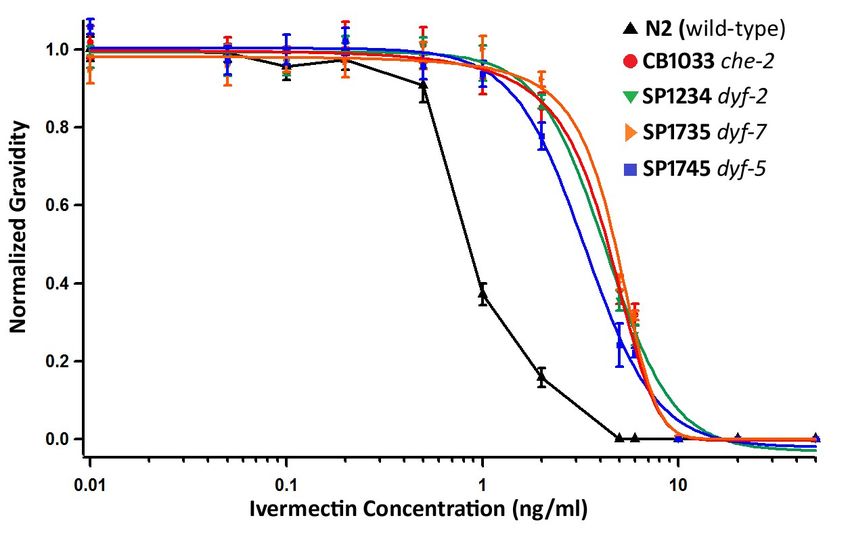
Abstract

       Ivermectin has been a very useful drug for the control of diseases caused by helminths.

However, the occurrence of drug resistant parasites is a major concern. In an attempt to mimic

the conditions under which ivermectin resistance is selected for in the field, James and Davey

(2009) generated the IVR6 and IVR10 Caenorhabditis elegans strains from a wild-type strain by

growing them in the presence of sub-lethal doses of ivermectin for several generations. To

better understand the mechanisms by which ivermectin resistance might arise, I have

investigated the IVR6 and IVR10 strains. I found that the IVR6 and IVR10 strains are dye-filling

defective (Dyf), a phenotype previously associated with ivermectin resistance. Our results

indicate that IVR6 and IVR10 have the same level of ivermectin resistance. We discovered a

frame shift mutation in the dyf-7 gene of both strains. The location of dyf-7 on the X-

chromosome is consistent with the results of our resistance mapping experiment. IVR6 and

IVR10’s dye-filling phenotype is roughly 80% penetrant and we show that ivermectin can select

for the phenotype. Only dye-filling defective worms can grow at 10 ng/ml ivermectin. We have

tested four other dye-filling defective strains, including a strain carrying a mutant dyf-7 allele

and all were ivermectin resistant. Preliminary results indicate that dyf-7 confers levamisole

resistance, suggesting an alternate mechanism for multidrug resistance. Taken together, my

results show that an allele of the dyf-7 gene is the cause of ivermectin resistance in the IVR6

and IVR10 strains and that the dendrite morphology phenotype of Dyf genes is essential for

their ability to confer resistance.

                                                  2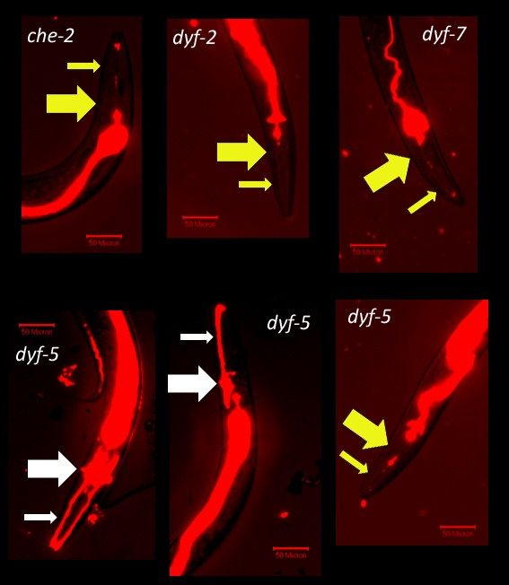
Résumé

       L’Ivermectine est un médicament qui a été très utile pour contrôler des maladies

causées par les nématodes parasitiques. Cependant, le développement chez les parasites d’une

résistance à ce médicament reste inquiétant. Ainsi, en reproduisant les conditions dans

lesquelles les parasites développent une résistance à l’ivermectine, James et Davey (2009) ont

généré deux souches de Caenorhabditis elegans, l’IVR6 et l’IVR10, en les cultivant sur une dose

non-létale pour quelques générations. Afin de mieux comprendre les mécanismes impliqués

dans le développement de la résistance à l’ivermectine, j’ai étudié les souches d’IVR6 et

d’IVR10. J’ai découvert que celles-ci sont déficientes en absorption de teinture, soit un

phénotype associé à la résistance à l’ivermectine. Les résultats obtenus présentaient le même

niveau de résistance pour les deux souches. De plus, nous avons découvert une mutation dans

le gène dyf-7, aussi chez les deux souches. L’usage de la cartographie génétique qui utilise les

polymorphismes pour un nucléotide (SNPs), m’a permis de déterminer que le locus de

résistance est en accord avec le locus de dyf-7. Le phénotype de déficience d’absorption de

teinture est pénétrant à 80% chez l’IVR6 et l’IVR10. Quant à l’ivermectine, celui-ci peut

sélectionner pour ce phénotype. Seulement les vers déficients en absorption de teinture

peuvent survivre sur 10 ng/ml d’ivermectine. J’ai examiné quatre autres souches qui sont

déficientes en absorption de teinte, incluant une souche avec un allèle différent de dyf-7; elles

démontrent toutes une résistance à l’ivermectine. Des expériences préliminaires indiquent que

le dyf-7 induit une résistance à un autre médicament, lévamisole. Ce qui me suggère un

nouveau moyen de développer la multirésistance. Pris dans leur ensemble, mes résultats

montrent que l'allèle du gène dyf-7 est la cause de la résistance à l'ivermectine chez les souches

                                                3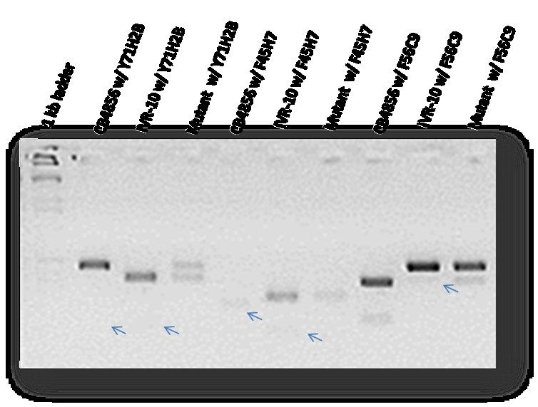
d’IVR6 et d’IVR10 et que la malformation des dendrites chez les souches Dyf est essentielle au

développement de la résistance.

                                               4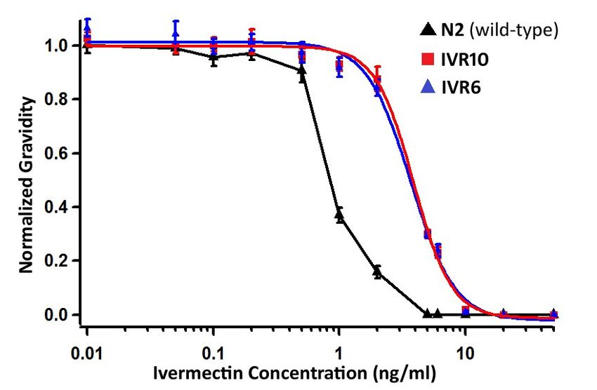
Acknowledgements

       First, I would like to thank my supervisor Joseph Dent for his guidance. He provided me

with the opportunity to work on six different and very interesting research projects in the lab,

allowing me to learn a broad range of techniques. He was also responsible for discovering that

IVR6 and IVR10 were dye-filling defective.

       Next, I would like to thank the three undergraduate students who I had the privilege to

supervise. Laura Tiseo, did a fantastic job setting up the crosses for SNP mapping and began

much of the analysis. She will be starting graduate school in September 2012. Mi Tan worked

briefly in the lab in the summer of 2011. She helped to finish off the SNP mapping experiment

and hopes to go to medical school. Finally, I had the pleasure of working with James Bae for

the last ten months starting in the summer of 2011. He has made innumerable contributions to

the present work, including, many of the ivermectin dose response curves and many of the

Dye-filling assays. James will either continue his education in medical school or graduate

studies.

       I need to thank Roger Prichard and Ludmel Urdanata from McGill’s Institute of

Parasitology. Ludmel was responsible for the sequencing of IVR6 and IVR10 and subsequent

analysis. She is also leading investigations into the role that dyf-7 plays in ivermectin resistance

in the parasite Haemonchus contortus.

       I would like to thank all the Dent lab members I had the chance to work with. In

particular Claudia Wever, who began training me as an undergraduate and taught me how to

clone novel ligand-gated ion channel subunits. She continued teaching me various techniques

throughout my four years in the Dent lab and was incredibly supportive. I would like to thank
                                                 5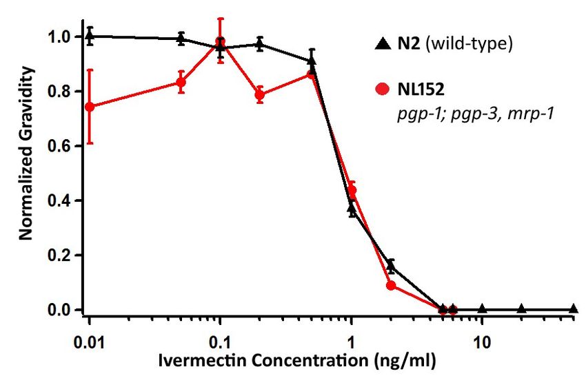
Danny Feingold, who I also started working with as an undergraduate. He supervised me

through the process of developing antibodies against ion channel subunits in Drosophila

melanogaster. He is also responsible for the title of my thesis. Chris Farnet also needs to be

extended a wonderful thank-you. He has provided critical input into the experimental design of

the current project. Importantly, he always had an open ear for my ideas and would help refine

my thought process. I would like to thank Nela Durisic, who supervised me in the production

and purification of fluorescent proteins and Stephanie Bourque who I had the pleasure of

teaching Western blots to.

       I need to extend a huge thank-you to Chiara Gamberi from Paul Lasko’s lab. She taught

me how to prepare antigen for antibody production and how to do Western Blots.

       Finally, I would like to thank my supervisory committee members Ken Hastings, Siegfried

Hekimi and Roger Prichard for their intellectual input into my projects and advice.

       This project was supported with funding by the Fonds de recherche du Québec - Nature

et technologies.

                                                6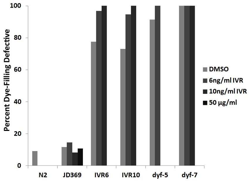
Table of Contents
Abstract                                                      2
Résumé                                                        3
Acknowledgements                                              5
Table of Contents                                             7
List of Figures                                               8
List of Tables                                                8
Literature Review                                             9
Introduction                                                  9
The scope of the parasitic nematode problem                   9
River blindness                                               10
Human Intestinal helminths                                    11
Lymphatic filariasis                                          12
Heartworm                                                     12
The ruminant parasite Haemonchus contortus                    13
Evidence that drug resistance is an issue                     13
C. elegans as a model organism                                14
Anthelmintic drugs: mode of action                            15
Mechanisms of drug resistance                                 16
Ivermectin resistance                                         17
Dye-filling defective genes                                   18
DYF-5                                                         19
DYF-2                                                         19
DYF-7                                                         19
CHE-2                                                         20
Cross-resistance of dye-filling defective genes to paraquat   20
The effects of ivermectin on gene expression                  20
The IVR6 and IVR10 strains of C. elegans                      21
Specific aims of this project                                 22
Materials and Methods                                         24
Worm Strains                                                  24
Sequencing                                                    24
Mapping                                                       24
Ivermectin Plates                                             25
Egg Preparation                                               25
Ivermectin Dose Response Curves                               26
Ivermectin & Dye-filling Defective Selection Experiment       26
Dye-Filling Experiment                                        26

                                                7
Imaging 27
Levamisole Dose Response Curves 27
Results 28
IVR6 and IVR10 have a partially penetrant dye-filling defective phenotype 28
Sequencing results 29
Mapping ivermectin resistance 31
IVR6 and IVR10 have equal ivermectin resistance 34
ABC transporters and ivermectin resistance 36
Dye-filling defective strains are ivermectin resistant 38
Ivermectin selects for the dye-filling defective phenotype 41
Levamisole resistance 43
Discussion 46
The dye-filling defective phenotype confers ivermectin resistance 46
The role of the ABC transporters in ivermectin resistance 47
We find that IVR6 and IVR10 have the same levels of ivermectin resistance 48
Dyf-7 confers levamisole resistance 50
dyf-7(vu268) maintains some wild-type function 50
Conclusion 51
Bibliography 52
Appendix A 58
List of Figures
Figure 1: IVR6 and IVR10 are mostly dye-filling defective 29
Figure 2: Analysis of whole genome sequencing reveals a frameshift mutation in 31
the dyf-7 gene of IVR6 and IVR10
Figure 3: SNP Mapping data shows that the location of dyf-7 correlates with 33
ivermectin resistance
Figure 4: IVR6 and IVR10 have the same ivermectin resistance 36
Figure 5: The pgp-1, pgp-3 and mrp-1 triple mutant is not more sensitive to 37
ivermectin than wild-type
Figure 6: The dye-filling phenotypes of four strains 39
Figure 7: Dye-filling defective strains are ivermectin resistant 40
Figure 8: Ivermectin Selects for the dye-filling defective phenotype 43
Figure 9: Dose response curves for wild type worms grown on Ivermectin vs. 45
Levamisole
List of Tables
Table 1: Four homozygous, non-synonymous single nucleotide polymorphisms 31
(SNPs) are found in IVR10 but not IVR6
8Literature Review
Introduction
Parasitic nematodes are a major health concern for humans and animals and some
species are harmful to agricultural crops. In the literature review, I will discuss the health
impacts and treatment of several human and animal helminthic diseases as well as the
recurring issue of drug resistance.
Next, I will discuss the utility of C. elegans as a model organism for parasitic infections. I
will explain the mode of action of three important anthelmintic drugs and discuss mechanisms
that confer drug resistance.
The scope of the parasitic nematode problem
The economic, social and physical burden of parasites is enormous. It is estimated that
30% of humans carry nematode infections, with 800 million people being burdened by multiple
parasite species1. The infections are especially prevalent amongst the poorest human
populations. Globally, 807 million people have ascariasis, 604 million have trichuriasis, 576
million have hookworm infections, 207 million have schistosomiasis, 120 million have lymphatic
filariasis, and 37 million have onchocerciasis2. The list of symptoms ranges from indigestion,
nausea and diarrhea to anemia, blindness and death3,4.
9For the agricultural industry, nematodes cause a significant economic burden. Parasitic
nematodes cause greater than 10% loss of crop production for farmers or $80 billion dollars
globally5. The best estimate of the costs of animal parasites comes from the sale of
anthelmintic drugs6. The belief is that the economic burden of parasites is equal or greater to
the amount consumers are willing to spend on anthelminthic drugs. In 1999, 3.5 billion US$
were spent on anthelmintic drugs globally. The most was spent for dogs and cats which
accounted for 1.5 billion US$ in drug use, cattle accounted for 1.1 billion, sheep 379 million,
swine 303 million and the remainder was administered to other animals6.
River blindness
Onchocerciasis, a disease caused by the helminth Onchocerca volvulus, is commonly
known as river blindness because prolonged infection can cause blindness and because the
disease transmitting vector, the black fly, is abundant along rivers. The parasite is found in
West Africa and a few isolated locations in South America7. It is estimated that 37 million
people are infected by Onchocerca volvulus8. Ivermectin has been used effectively in the
prevention and treatment of onchocerciasis since 1987, when the drug company Merck offered
to donate the drug “for as long as necessary to eliminate onchocerciasis as a public health
problem”9. A single annual dose of the drug significantly reduces the burden caused by the
microfilariae (larva) but is not effective against the macrofilariae (adults)10. Treating the
microfilariae significantly reduces itching and prevents the occurrence of blindness.
10Human Intestinal helminths
Humans living in low-income areas are especially susceptible to intestinal helminth
infections. For example, prevalence of ascariasis, trichuriasis and hookworm infections are as
high as 50-80% in some areas7.
Infections with the parasite Ascaris lumbricoides can lead to serious health
complications, some requiring surgery or causing morbidity3. The related ascarid, A. summ can
infect both pigs and humans4. It can reach larval and tissue-migratory stages in humans.
However, it rarely reaches adulthood in human hosts. Ascariasis is caused by ingesting eggs of
the parasite from soil or contaminated food. Young children are especially at risk of being
infected because of their natural tendency to put things in their mouths 3.
Researchers estimate that from 604 million2 to 1.049 billion people carry Trichuris
trichiura infections including, 347 million children11. T. trichiura is a parasitic nematode that
migrates to the human cecum and large bowel11. The adult female will reach 3 to 5 cm, lay
3000 to 20, 000 eggs per day and can live up to 8 years. The eggs develop into infective larva in
moist soil after approximately 3 weeks and are able to live up to one year in soil before
infecting a host. T. trichiura infections are often asymptomatic. However, Symptoms tend to
be worse in children who can experience diarrhea, rectal bleeding, anemia and finger clubbing,
a deformity of the hands where the fingertips thicken.
Another intestinal helminth is commonly known as hookworm. Hookworm infection is
caused by the helminths Necator americanus or Ancylostoma duodenale and is usually
transmitted through contaminated soil. Larva can burrow through human skin and migrate to
11the lungs within 10 days12. The adults live in the human intestine. Usually, the most severe
health impact is iron-deficiency anemia, although other symptoms are possible such as cough,
nausea, vomiting, and pneumonitis. Children co-infected with hookworm and T. trichiura were
associated with higher levels of anemia than expected, suggesting a synergistic effect of co-
infection13.
Lymphatic filariasis
There are two parasites mainly responsible for the estimated 120 million cases of
lymphatic filariasis. The disease is also known as elephantiasis because of the swelling induced
by the infection. Wuchereria bancrofti is responsible for 90% of the cases and Brugia malayi is
responsible for most of the remainder14. Strikingly, hosts can be infected with millions of
worms and still be asymptomatic. Still, 40 million people have clinical symptoms of the disease.
Inflammatory responses due to dead worms in the body are the primary cause for
elephantiasis.
Heartworm
In 2001 alone, there were 244, 291 cases of canine heartworm in the USA. Dirofilaria
immitis, the parasite which causes heartworm, can also infect a range of other mammals
including horses, cats and humans15. The infection is particularly dangerous in cats where fatal
cardiac reactions can occur due to a single worm16. Mosquitoes, which transmit the infection,
become infected with eggs when taking a blood meal from an infected host. The parasite takes
11-15 days developing within the mosquito to reach its infective stage. When the mosquito
feeds on another victim it can transmit the infection through the puncture wound.
12The ruminant parasite Haemonchus contortus
The ruminant parasite Haemonchus contortus causes significant economic loss to
farmers. The parasite is extremely successful and can infect sheep and goats17. It can survive in
a broad range of climates and trade in livestock leads to the spread of the parasite. A single
ruminant can host thousands of H. contortus and each female lays up to 10 000 eggs a day. This
means that H. contortus populations are huge and are often larger on the pasture than in the
host.
Evidence that drug resistance is an issue
Resistance to the first widely administered anthelmintic, phenothiazine, was reported in
1957, 17 years after the drug came to market18. Ever since, resistance has been reported for
new anthelmintic drugs shortly after they released onto the market. For example, ivermectin
was released onto the market in 1981 and the first report of resistance occurred in 1987. Here,
I will explore evidence for drug resistance in animal parasites and the human parasite
Onchocerca volvulus.
Animal parasites are developing resistance to all the major classes of anthelmintic drugs.
For example, the genetic diversity created by the large population size and high reproduction
rates of H. contortus raises serious concerns for the development of resistance to anthelmintic
drugs17. James Wyk et al. found a strain of H. contortus that was resistant to all five major
classes of anthelmintics currently in use19. Further, Wyk et al. report that 90% of sheep farms
in South Africa are resistant to at least one class of anthelmintic drugs. In New Zealand,
researchers investigated the drug resistance of intestinal helminthes parasitic to cattle and
13found ivermectin resistance on 92% of farms, albendazole resistance on 76% of farms and
levamisole resistance on 6% of farms20.
In the past, ivermectin has proven to be a useful drug for treating patients infected with
Onchocerca volvulus and preventing blindness. However, the continual use of ivermectin as a
means of controlling river blindness leads to concerns about the selection for drug resistance in
the parasite. In fact, a study of the efficacy of ivermectin in Ghana found that some adult
Onchocerca volvulus populations are resistant to ivermectin8.
However, some hope remains for the ivermectin’s efficacy in the treatment of river
blindness. A study published in 2012, found that the prevalence of Onchocerca volvulus in
certain communities in Kenya dropped to 0% after 15 to 17 years of treament 21, a reasonable
timeframe for a microfilaricide. Available baseline data indicates that prevalence of the
infection was a median of 52% before treatment. This study brings hope that ivermectin alone
might be able to eliminate river blindness from endemic areas.
C. elegans as a model organism
C. elegans is an excellent model organism for studying parasitic nematodes. C. elegans
is a harmless free-living species but it is related to parasitic nematodes that can harm plants 22
and infect animals.
C. elegans is a small organism that is found in garden soil and feeds on bacteria23. It can
easily be grown in a laboratory on an agar medium with bacteria growing on it. They will feed
on the bacteria and go through four larval stages. As adults they are a mere 959 somatic cells
14and about 1 mM in length. They undergo their life-cycle in 3 days. Most C. elegans are self-
fertilizing hermaphrodites, laying about 300 eggs24. However, there are also males and this
allows for genetic crosses.
There are features of C. elegans that make it a valuable research tool. First, researchers
have determined the entire ontogeny of the organism, mapping out the entire cell lineage from
egg to adult25. Moreover, It’s known that the nervous system is made up of only 302 neurons23.
In 1986, researchers completed the mapping of the nervous system having determined all
branches and connections. Additionally, C. elegans was the first animal to have its genome
entirely sequenced. This has made C. elegans an excellent research tool for understanding the
mode of action of anthelmintic drugs and mechanisms of resistance especially since many
anthelmintic drugs act on the nervous system26.
Anthelmintic drugs: mode of action
To treat nematode infections there are a number of drugs available27. Three important
classes of anthelmintics are the benzimidazoles which include albendazole, the nicotinic
agonists which include levamisole, and the macrocyclic lactones which include ivermectin.
Many anthelmintic drugs, such as ivermectin, levamisole and Fipronil target ion
channels28,29. Drugs that targeting ion channels usually function in one of two ways. They can
either prevent channels from opening, such as Fipronil or activate ion channels in the case of
ivermectin and levamisole. Either method works to deregulate an organisms control over their
nervous system.
15Ivermectin binds to the glutamate-gated chloride channels (GluCls) of C. elegans26 and
H. contortus30,31. The activation of the GluCls in an irreversible fashion leads to
hyperpolarisation of neurons and muscles preventing the contracting of the pharynx of
nematodes and causing starvation32. Recently, x-ray crystallography revealed that ivermectin
binds to GluCls in the transmembrane domain proximal to the extracellular domain33.
Levamisole binds to cationic nicotinic acetylcholine receptors in nematodes causing the
depolarization of muscle cells34, instead of the hyperpolarisation caused by ivermectin. The
drug results in spastic paralysis which is believed to contribute to the expulsion of helminths
from the host.
Other drugs, such as albendazole have other cellular targets35. Albendazole works
primarily by preventing the polymerization of microtubules by binding to tubulin.
Mechanisms of drug resistance
There are several ways to develop drug resistance. One way, is caused by drug induced
selection for changes in the drug target. The drug target undergoes changes such that it can no
longer interact with the drug. Another way is to reduce drugs’ accessibility to their target.
There are at least a couple ways to accomplish reduced access, including, decreased drug
permeability, increased efflux of the drug or increased metabolism of the drug. In the section
that follows I will examine mechanisms that have been proposed for ivermectin resistance.
16Ivermectin resistance
It is well established that the target of ivermectin is the glutamate-gated chloride
channels33. Mutations in the drug target confer ivermectin resistance. In C. elegans, the
glutamate channel AVR-15 is the most sensitive target to ivermectin, followed by AVR-14 and
GLC-126. The glutamate channel GLC-3 is sensitive to ivermectin when expressed in Xenopus
oocytes36, and it is the fourth most important ivermectin target in C. elegans (Dent, personal
communication). C. elegans quadruple mutants, lacking all four ivermectin sensitive subunits
are more than 50, 000-fold resistant to ivermectin. Moreover, it has been found that selection
at the glutamate-gated chloride channels can confer ivermectin resistance in parasites37,38.
Another proposed mechanism of drug resistance is the efflux of drugs by ABC
transporters. The ABC transporter genes use the energy generated from the hydrolysis of ATP
to pump substrates in or out of cells. Investigation of the substrates for ABC transporters in C.
elegans suggests that MRP- 1 and PGP-1 are able to pump anions conjugated to glutathione39.
Further, it is believed that PGP-3 pumps colchicine and chloroquine since a strain with a PGP-3
deletion mutant allele is sensitive to these drugs. There is also evidence that MRP-3, MRP-4
and MRP-8 are able to mediate sensitivity to ivermectin40. It has been suggested that an
increased expression of the ABC transporter proteins would lead to an increase in the efflux of
ivermectin and increased resistance.
An important mechanism of ivermectin resistance is decreased drug permeability.
When wild-type C. elegans are incubated with a dye, DiI, their sensory neurons will adsorb the
dye41. This phenotype is known as dye-filling. It has been shown that C. elegans with a dye-
17filling defective phenotype have morphological defects in their sensory amphid neurons41 and
are ivermectin resistant26. Through analogy to the permeability of the dye, it is believed that
dye-filling defective C. elegans also have decreased permeability to ivermectin leading to low-
levels of ivermectin resistance.
Moreover, phenotypes similar to the dye-filling defective phenotype in C. elegans have
been shown to contribute to ivermectin resistance in parasites. Li et al. have investigated the
structure of sensory neurons in Haemonchus contortus42. Similar to dye-filling defective C.
elegans strains, they found that ivermectin resistant H. contortus strains had shorter sensory
cilia than ivermectin susceptible strains43. The molecular biology and effects on neural
morphology of the dye-filling defective genes will be covered in more detail in the next section.
Dye-filling defective genes
The study of neuronal development in C. elegans has benefitted from the easy to
visualize dye-filling defective (Dyf) phenotype. Wild-type worms will normally have their
sensory amphid and phasmid neurons fill with the dye, DiI, after incubating the worms in the
dye41. The sensory neurons of Dyf mutants do not fill with dye and have been related to
various defects in neural development and morphology, from dendrite extension defects, to
shortened sensory cilia.
There are 31 characterized Dyf genes and likely more to be uncovered44,45. Various dye-
filling defective mutants have been characterized as defective in osmosensation (Osm), dauer
formation (Daf), chemotaxis (Che), and dye-filling (Dyf).
18My research has characterized the ivermectin resistance conferred by mutant alleles of
four known dye-filling defective genes. I found that strains with mutant alleles of che-2, dyf-2,
dyf-5 or dyf-7 have low-levels of ivermectin resistance and that dyf-7 confers low-levels of
levamisole resistance. The following paragraphs describe what is known about the molecular
function of these genes and their effects on neural morphology.
DYF-5
Previous researchers have characterized the effect of dyf-5 mutants on neural
morphology46. The gene is expressed in ciliated neurons. dyf-5 mutants have longer cilia that
do not enter the sensory pore. dyf-5 homologues in Chlamydomas and Leishmania also affect
cilia morphology47,48. DYF-5 is a MAP kinase that affects the movement speed of the kinesin
motor protein OSM-3 through phosphorylation. osm-3 null mutants are also dye-filling
defective44.
DYF-2
DYF-2 forms part of intraflagellar transport particles and is involved in anterograde
transport49, or movement away from the cell body. dyf-2 is expressed in 7 of the amphid
neurons, the oxygen sensors AQR and PQR and in the phasmid neurons.
DYF-7
DYF-7 is an extracellular matrix protein required to anchor the dendrites to the sensory
pore during development50. The protein has a zona pellucida domain, similar to that found on
the egg of most animals that allows the sperm to bind. DYF-7 interacts with the protein DEX-1,
19which has a zonadhesin domain similar to that found on sperm. dex-1 or dyf-7 null mutants
cause dendrite extension defects in the sensory neurons and the dye-filling defective
phenotype.
CHE-2
CHE-2 is involved in sensory cilium formation51. che-2 mutants have very short cilia,
however expression of the gene under a heat shock promoter that allows production of the
gene at different stages of development shows that proper extension and anchoring is possible
in the adult. After analysing the homology of che-2 to other genes, Fujiwara et al. believe that
the protein mediates the protein to protein interactions of intraflagellar transport proteins.
Cross-resistance of dye-filling defective genes to paraquat
The genes that cause the dye-filling defective phenotype have been associated to
paraquat resistance. Starting in 2004, publications have demonstrated the importance of the
Dyf genes in resistance to paraquat44,45, a drug used to catalyze the formation of reactive
oxygen species52. In one publication, 37 Dyf mutants were tested and 27 of them were
resistant to paraquat, including the genes che-2, dyf-2, dyf-5 and dyf-7 used in our study.
The effects of ivermectin on gene expression
There are several different studies that investigated the effects of ivermectin on gene
expression in nematodes as a way of understanding the physiological response to ivermectin,
including possible de-toxification mechanisms. James et al. (2009) and Ardelli et al. (2008)
found that ivermectin causes an increase in the expression of the ABC transporters40,53. These
20reports suggest the ABC transporters in nematodes mediate ivermectin resistance. Further, in
the mite Sarcoptes scabiei, Mounsey et al. found that there is an increase in transcription of
glutathione s-transferase and a p-glycoprotein in the presence of ivermectin, suggesting
increased metabolism and efflux of ivermectin as a mechanism of resistance54.
However, Laing et al. found that the genes that were most upregulated in the presence
of ivermectin were not transporters but genes related to the metabolism of lipids55. Ivermectin
causes a decrease in pharyngeal pumping. Therefore, the worm gets less food and needs to
metabolize its energy stores, explaining why ivermectin might cause an increase in fat
metabolism. The Laing et al. approach to expression analysis appears to be the most reliable
because it is the first study to look at the expression of all genes. Other studies looking at the
effects of ivermectin on gene expression looked only at genes that were believed to be involved
in ivermectin efflux.
The IVR6 and IVR10 strains of C. elegans
It is thought that resistance in parasites develops when exposed to sub-lethal
concentrations of ivermectin over several generations. In contrast, previous studies in C.
elegans have relied on mutagens to generate resistance alleles in a single generation26,32.
Catherine James and Mary Davey set out to make ivermectin resistant C. elegans strains that
developed resistance by a mechanisms that is more similar to what is thought to occur in the
field 53. To do so they exposed C. elegans to sub-lethal doses of ivermectin over several
generations.
21James and Davey started out with wild-type C. elegans which can grow on a maximum
of 2 ng/ml ivermectin and after 44 weeks they had selected for two ivermectin resistant strains
IVR6, and IVR10 that grew on a maximum of 6ng/ml and 10 ng/ml ivermectin, respectively.
James and Davey produced several lines of evidence that supported their theory that
the ABC transporters could mediate ivermectin resistance. First, they showed that four
different groups of chemicals believed to block the pumping of the ABC transporters, such as
the drug verapamil, could reverse the ivermectin resistance of the IVR6 and IVR10 strains.
Next, they showed that the strains IVR6 and IVR10 were multidrug resistant. They
demonstrated resistance to the anthelmintic drugs moxidectin, levamisole, albendazole, and
pyrantel. This was evidence that the ABC transporters were involved in resistance since the
transporters are believed to pump multiple substrates. James and Davey also showed the
increased expression of some of the ABC transporters, in particular pgp-1 and mrp-1, using
quantitative PCR. Based on these data, Catherine James and Mary Davey concluded that the
ABC transporters were conferring ivermectin resistance in the IVR6 and IVR10 strains of C.
elegans.
Specific aims of this project
The primary goal of this project is to investigate the mechanisms of resistance in the
IVR6 and IVR10 strains of C. elegans. We discovered that the IVR6 and IVR10 strains are dye-
filling defective, a phenotype associated with ivermectin resistance26. Both strains have a
frameshift mutation in their dyf-7 gene. We show that the location of dyf-7 is consistent with
the source of ivermectin resistance in mapping experiments. Although the penetrance of the
22dye-filling defective phenotype in IVR6 and IVR10 is incomplete, only dye-filling defective
worms can survive on 10 ng/ml ivermectin.
Our findings that IVR6 and IVR10 are dye-filling defective also led us to explore the
resistance properties of four other Dyf strains, CB1033 che-2(e1033); SP1234 dyf-2(m160);
SP1735 dyf-7(m537); and SP1745 dyf-5(mn400). We found that all four strains are ivermectin
resistant and that dyf-7(m537) confers levamisole resistance.
Our data indicates that the neural morphological defects of the dye-filling defective
strains are essential for their ability to confer ivermectin resistance.
23Materials and Methods
Worm Strains
C. elegans strains were maintained using standard practices as described by Brenner56, except
with HB101 bacteria instead of OP50. The IVR6 and IVR10 strains were a kind gift from
Catherine James and Mary Davey (University of Technology Sydney, Sydney, Australia)53. The
strains were thawed every month to prevent the selection of additional ivermectin resistance
mechanisms. Other strains used include, wild-type N2 Bristol, CB1033 che-2(e1033); CB4856;
JD369 avr-14(vu47) I; glc-3(ok321), avr-15(ad1051), glc-1(pk54) V; SP1234 dyf-2(m160); SP1735
dyf-7(m537); SP1745 dyf-5(mn400); NL152 pgp-1(pk17) IV; pgp-3(pk18), mrp-1(pk89)I.
Sequencing
Genomic DNA from IVR6 and IVR10 were sequenced by Genome Quebec using an
Illumina Genome Analyzer (Illumina). The sequences were aligned to the 2008 version of the
Bristol N2 sequence available at UCSC Genome Browser using the Burrows-Wheeler Alignment
tool (BWA)57.
Mapping
The protocol used for mapping is from Davis et. al 200558. Prior to mapping, IVR10 was
propagated on 10 ng/ml ivermectin plates. First, we crossed the mapping strain CB4856 with
IVR10. Next, we selected cross progeny by selecting for worms that have dye-filled sensory
neurons41,59,60. We intended to select only cross-progeny. However, we selected some IVR10
24due to the incomplete penetrance of the Dyf phenotype [see the results section for details
about the penetrance of dyf-7(vu268)]. We allowed the F1 generation to self-fertilize. Next, we
performed an egg-preparation and placed eggs of the F2 generation on 10ng/ml ivermectin
plates to select resistance progeny. Following the Davis protocol, we performed SNP (Single
Nucleotide Polymorphism) mapping by PCR amplification and digesting with the restriction
endonuclease DraI58. The recombination frequency was calculated as the number of SNPs that
matched the mapping strain CB4856 divided by the total number of SNPs (IVR10 + CB4856).
Ivermectin Plates
To make ivermectin plates we followed the procedure in Dent et. al (1997)32. In brief,
ivermectin plates were prepared using the standard recipe for NGMSR61. Ivermectin was
dissolved in DMSO and added to the medium immediately before pouring to a final
concentration of 1% DMSO. Control plates used for normalizing the ivermectin dose response
curves had 1% DMSO and no ivermectin.
Egg Preparation
To collect C. elegans eggs we used a method called an egg preparation, also called an egg-prep.
Worms are rinsed off a starved plate with 1 mL M9 Salt Solution and pipetted into a 15 mL
conical tube. The tube is filled with M9 Salt Solution to 7 mL and then 2 mL of 2 M NaOH and 1
mL of bleach is added. The solution is placed on a vortex for 3 minutes and then spun down in
a clinical centrifuge at full speed for 1 minute. The supernatant is removed until there remains
only ~0.5 mL and then is resuspended in 5 mL M9 Salt Solution. The spin and resuspension can
25be repeated, if desired. The eggs remaining in solution can then be pipetted onto a plate.
Ivermectin Dose Response Curves
Ivermectin dose response curves were performed similarly to Dent et. al (2000)26.
Worms underwent an egg-preparation and placed on a range of concentrations of ivermectin
plates and a 1% DMSO control. The eggs resuspended in a M9 salts solution was titrated so
that an aliquot of 35µL per plate resulted in approximately 50 eggs per plate. Gravidity was
scored after 4 days (~96 hours) by the presence of eggs in the uterus. Males, while rare, were
not counted. Survival on ivermectin was normalized based on the number of gravid adults on
DMSO control plates. Three plates of each concentration were used in two trials for a total of
six plates per concentration.
Ivermectin & Dye-filling Defective Selection Experiment
Worms were egg-prepped and the eggs were placed on either DMSO (control), 6 or 10
ng/ml ivermectin plates for most strains or up to 50 µg/ml for the JD369 strain. After 5 or 6
days dye-filling experiments were performed on the surviving animals.
Dye-Filling Experiment
Dye-filling experiments were performed as in Starich et al. (1990)41. Briefly, adult
worms were collected in 1 mL of M9 buffer. 5 μL of 0.4% the dye, DiI (1,1'-dioctadecyl-3,3,3'3'-
tetramethylindocarbocyanine perchlorate), dissolved in dimethylformamide, was added to the
tube and it was allowed to rock on a shaker for one hour at 20°C. The worms were washed once
with M9 buffer and cultured on NGM plates.
26Imaging
To make slides for imaging, 50 uL of 2% agarose in M9 was melted and placed onto a
microscope slide. A second slide was used to flatten the drop into an agarose pad. Worms
were placed on the slide in drops of M9 with 5 mM levamisole to paralyse the worms. Images
were taken using the Olympus IX81, a motorized inverted microscope (Olympus). The resultant
image was black and white. Color was added to the image using a red filter with the software
ImageJ (Image / Lookup Tables / Red).
Levamisole Dose Response Curves
To make levamisole plates we followed the standard NGMSR procedure 61 and added
levamisole that was dissolved in H2O. Control plates were standard NGMSR plates and were
used to normalize the curve.
27Results
IVR6 and IVR10 have a partially penetrant dye-filling defective phenotype
Previous research associated low-levels of ivermectin resistance with the dye-filling
defective phenotype26. The level of ivermectin resistance in the IVR6 and IVR10 strains is
relatively low. They can grow at a maximum of 10 ng/ml ivermectin18. I wanted to determine
whether the IVR6 and IVR10 strains were dye-filling defective because this phenotype would
influence the resistance-levels of these strains.
We found that the majority of worms from the IVR6 and IVR10 strains are dye-filling
defective (Figure 1). However, the penetrance of the phenotype is incomplete. A subset of
IVR6 and IVR10 worms dye-fill as in wild-type. Another subset of worms has only one set of the
sensory amphid neurons dye-fill. C. elegans have 8 pairs of bilaterally symmetrical amphid
neurons in the head that anchor to the sensory pore41. Previous research has shown that the
two sets of amphid sensory neurons anchor independently50, which provides an explanation for
the observed phenotype.
Our discovery that the IVR6 and IVR10 strains have a partially penetrant dye-filling
defective phenotype was significant and suggested that the phenotype would play a role in the
ivermectin resistance of these strains, but some questions remained. First, what gene was
responsible for the dye-filling defective phenotype? Second, could IVR6 and IVR10 worms with
wild-type dye-filling survive on ivermectin? Are there other resistance mechanisms involved or
only the dye-filling defective phenotype?
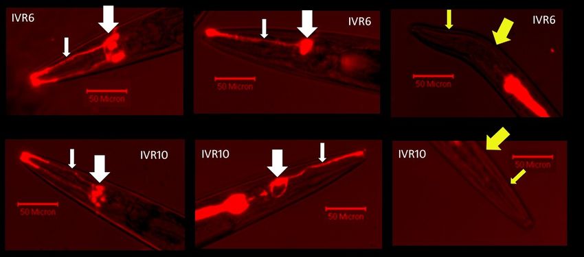
28Figure 1: IVR6 and IVR10 are mostly dye-filling defective. The penetrance of the dye-flling
defective phenotype is incomplete. TOP (from left to right): IVR6 with both bilateral sets of
amphid neurons dye-filled. IVR6 with a single set of amphid neurons dye-filled. IVR6 dye-filling
defective. Bottom (from left to right): IVR10 with both bilateral sets of amphid neurons dye-
filled. IVR10 with a single set of amphid neurons dye-filled. IVR10 dye-filling defective. The
small arrow points to the dendrites and the large arrow points to the cell bodies (white = dye-
filling, yellow = no dye-filling).
Sequencing results
Having found that the IVR6 and IVR10 strains were dye-filling defective, we wanted to
determine which gene was responsible for this phenotype. Additionally, we wanted to search
for genetic differences between the IVR6 and IVR10 strains, which James and Davey concluded
have different levels of ivermectin resistance18. We decided to perform whole genome
sequencing in search of candidate dye-filling defective genes and ivermectin resistance genes.
We performed whole genome sequencing on the IVR6 and IVR10 strains and found a
mutation in the open reading frame of the gene dyf-7 (Figure 2). A two nucleotide CT deletion
29causes a frameshift in dyf-7 in the second to last exon. We named this allele dyf-7(vu268). dyf-
7(vu268) was a candidate gene for ivermectin resistance in IVR6 and IVR10.
There were, however, many other SNPs, insertions and deletions. There were 107 SNPs
in coding regions that differed between IVR6 and IVR10. Of these SNPs, only 4 were both
homozygous and led to non-synonymous substitutions (See Table 1).
The pgp-6 and dyf-7 mutations were not found in our initial anaysis. To limit the
number of false positive mutations we found from regions with poor quality reads, we required
a minimum coverage of 19 reads. The pgp-6 mutation was not counted because the coverage
was too low (only 13 reads for IVR10 and 4 reads for IVR6). The dyf-7 mutation on the other
hand was not discovered in our initial analysis because it was the same in IVR6 and IVR10 and in
our initial analysis we were searching for differences between IVR6 and IVR10. To find the
mutations in pgp-6 and dyf-7 we looked through all the suspected candidate genes, including
the glutamate-gated chloride channels, the ABC transporters and the dye-filling defective genes
and searched for differences between IVR10 and wild-type. All other candidate genes were
wild-type.
Finding the frameshift mutation in dyf-7 suggested a role for the dyf-7(vu268) allele in
the ivermectin resistance of IVR6 and IVR10, as well as the dye-filling defective phenotype.
However, given the total number of SNPs uncovered, additional experiments were required to
determine whether dyf-7(vu268) or some of the other SNPs were contributing to the ivermectin
resistance of these strains.
30Figure 2: Analysis of whole genome sequencing reveals a frameshift mutation in the dyf-7
gene of IVR6 and IVR10. A. Sequences from dyf-7 wild-type and IVR6/IVR10 strains’ dyf-
7(vu268) which has a two nucleotide deletion. B. A gene model of the dyf-7 gene with an
arrow indicating the location of the mutation.
Chromosome Location Ref. Read Cov. Genes Nucleot. AA
change change
I 10520034 T A 20 F59C6.5 GAT-GAA Asp-Glu
V 10794768 G A 20 D1054.11 GCT-ACT Ala-Thr
X 5832712 G T 19 F13D11.4 CCC-ACC Pro-Thr
X 10872824 C C 13 pgp-6 GCT-GGT Ala-Gly
Table 1: Four homozygous, non-synonymous single nucleotide polymorphisms (SNPs) are
found in IVR10 but not IVR6. The genes F59C6.5, D1054.11 and F13D11.4 contain amino acids
in IVR10 that are different from IVR6. ‘Ref.’ refers to the nucleotide in the reference strain.
‘Read’ refers to the sequence of IVR10. ‘Cov.’ stands for coverage and indicates the number of
reads obtained from sequencing for that nucleotide. ‘Nucleot. Change’ and ‘AA change’
indicate the nucleotide change (underlined) and the resulting amino acid change.
Mapping ivermectin resistance
Having found many SNPs in our sequencing analysis we wanted to determine which of
these, if any, were linked to ivermectin resistance. To determine whether the dyf-7(vu268)
allele was conferring ivermectin resistance we performed a mapping experiment on IVR10. Of
principle concern to us, was the possibility that ivermectin resistance was multigenic. In fact, at
31the time of sequencing we were assuming at least two mechanisms of ivermectin resistance,
one mechanism providing up to 6 ng/ml ivermectin resistance and a second mechanism
allowing for survival on 10 ng/ml, corresponding to the reported resistance levels of IVR6 and
IVR10, respectively53. It was difficult to rule out the possibility that non-candidate genes or
mutations in non-coding regions could confer ivermectin resistance. James and Davey had
shown the increased expression of ABC transporters in IVR6 and IVR1053, and there could be a
mutation in the regulatory regions of one of these transporters.
All chromosomes were tested for linkage to ivermectin resistance using the
chromosome mapping method58. The X-chromosome was clearly linked to ivermectin
resistance, but it was hard to rule out linkage on most of the other chromosomes, except the
fifth which was clearly not linked (Supplementary Figures 1-4). The chromosome mapping
method pools 30 F2 cross-progeny. If there are restriction fragment length polymorphisms
(RFLP) of each genotype, which would be expected for non-linked regions, it is very difficult to
judge the relative intensities of the two types. Interval mapping is much more accurate. We
can genotype individual F2 worms across all available RFLPs by pooling the F3 progeny, which
provides sufficient amounts of DNA to work with while reflecting the genotype of the parent F2.
Next, all chromosomes, except the fifth, were interval mapped; a method that is more
accurate than chromosome mapping and is used to determine which region of the
chromosome is linked to the desired phenotype. The X chromosome was the only chromosome
clearly linked to ivermectin resistance (Figure 3 and Supplementary Figure 5). The
recombination frequency on the X-chromosome reached zero at +2 map units from the defined
32center of the chromosome. Our mapping results correlate well with the location of dyf-7 found
at 1.46 map units. The fact that dyf-7 had a frameshift mutation and that its location correlates
with resistance mapping supports its involvement in ivermectin resistance in IVR10.
However, mapping has not ruled out the possibility that the mrp-4 gene may contribute
to ivermectin resistance. The mrp-4 gene is located at 1.73 map units. The expression levels of
mrp-4 were not reported for IVR6 and IVR10 by James and Davey53, but Ardelli and Prichard
found that mrp-4 can mediate ivermectin sensitivity40. However, in our analysis we did not
detect any obvious mutations in the mrp-4 gene.
Figure 3: SNP Mapping data shows that the location of dyf-7 correlates with ivermectin
resistance. The image shows the X-chromosome with the location of dyf-7. The names of the
restriction length polymorphisms are indicated under the X-chromosome and the chromosome
position on top (map units). The recombination frequency indicates the percentages of RFLPs
scored that were mapping strain RFLPs compared to the total number of RFLPs scored. We
analysed the same 36 worms or 72 chromosomes for each RFLP on the X-chromosome.
If a region is not linked to resistance the expectation is a recombination frequency close
to 50%. The recombination frequencies we found are lower than expected at random. We
believe that the lower than expected frequency suggests imperfect selection for cross progeny
33between IVR10 and CB4856. In fact, we analysed the same 10 worms across 5 chromosomes
and 30% (3/10) did not have any CB4856 SNPs across 19 RFLPs examined suggesting that
roughly 30% were uncrossed worms from the IVR10. When setting up crosses, we selected for
cross progeny between IVR10 hermaphrodites and CB4856 males by selecting for dye-fillers.
However, roughly 20% of IVR10 worms dye-fill as in wild-type. This means that when selecting
for dye-fillers in the F1 population, we selected for not only cross-progeny, but also some self-
progeny. We realised this shortcoming at the time, but could not think of a better way to select
cross-progeny.
IVR6 and IVR10 have equal ivermectin resistance
Results from our mapping experiment suggested that the dyf-7(vu268) allele was
conferring ivermectin resistance. However, we found that this allele was present in both IVR6
and IVR10. We wanted to determine the ivermectin resistance levels of the IVR6 and IVR10
strains, to see whether they were different as reported by James and Davey53.
We performed ivermectin dose response curves for the IVR6 and IVR10 strains and we
determined that both strains have the same ivermectin sensitivity. Using a Mann-Whitney
statistical test at individual concentrations we found no statistical difference between the
strains (Figure 4). Both strains showed about 4-fold higher ivermectin resistance than N2. The
results we found for IVR6 (4.1-fold increase in ivermectin resistance) were comparable to those
proposed by James and Davey for IVR6 (4.4-fold increase)53. However, for IVR10 our results
differed from James and Davey. We found that IVR10 has a 4.3 fold-increase in ivermectin
resistance but James and Davey proposed a 19-fold increase. It’s important to note that the
34assay we used to score ivermectin resistance was different than the method used by James and
Davey. We measured ivermectin resistance using a growth assay scoring gravidity or the
worms’ ability to reach adulthood. James and Davey used an MTT dye assay, which is
colorimetric and uses metabolic activity as a readout for viability62.
Gravidity in our dose response curves was scored after 4 days, but we found that some
worms of both the IVR6 and IVR10 strains were gravid in the presence of 10ng/ml ivermectin
after 6 days. 10 ng/ml ivermectin was the maximum concentration for the viability of both
strains that we observed. The next concentration we tested was 20 ng/ml ivermectin and no
worms could reach adulthood. James and Davey reported that IVR10 grows at 10 ng/ml
ivermectin but not at 15 ng/ml, similar to what we found. However, they claim that IVR6 grows
at 6 ng/ml ivermectin but not at 10 ng/ml ivermectin53.
35Figure 4: IVR6 and IVR10 have the same ivermectin resistance. Dose response curves for
N2( ), IVR10 ( ) and IVR6 ( ) show the survival of worms to adulthood, scored by gravidity,
normalized relative to DMSO control plates. The error bars represent standard error (n=6).
ABC transporters and ivermectin resistance
James and Davey reported that increased expression of the ABC transporters were
associated with ivermectin resistance in the IVR6 and IVR10 strains. We were interested in how
the ABC transporters affect ivermectin sensitivity. Since James and Davey reported the highest
expression levels for pgp-1 and mrp-1, we investigated the ivermectin sensitivity of the strain
NL152 pgp-1(pk17) IV; pgp-3(pk18), mrp-1(pk89)I . If those transporters were responsible for
pumping ivermectin we would expect the mutant to be more ivermectin sensitive than wild-
type because the worms would lose some ability to efflux the drug. Previous studies indicate
that single mutants are sufficient to notice a shift in the ivermectin sensitivity by mutant alleles
36of the mrp-3, mrp-4 and mrp-8 genes. However, we found that the ivermectin sensitivity of
NL152 was not significantly different from wild-type using the Mann-Whitney U test at
concentrations ranging from 0.5 to 2ng/ml (Figure 5). Our results suggest that pgp-1, pgp-3 and
mrp-1 are not involved in the efflux of ivermectin.
Figure 5: The pgp-1, pgp-3 and mrp-1 triple mutant is not more sensitive to ivermectin than
wild-type. The y-axis measures gravidity of the N2 ( ) and NL152 pgp-1(pk17) IV; pgp-3(pk18),
mrp-1(pk89)I ( ) strains, by scoring the presence of eggs in the uterus, normalized to the
number of gravid adults on control DMSO plates. The error bars represent standard error
(n=3).
37Dye-filling defective strains are ivermectin resistant
Previous studies have suggested that ivermectin resistance is a phenotype common to
all dye-filling defective C. elegans strains26. In this study we wanted to support this claim by
investigating strains with known neural morphology defects in their amphid sensory neurons
that have not previously been characterized for ivermectin resistance. We investigated four
different strains with mutations in che-2, dyf-2, dyf-5, and dyf-7. All alleles tested are thought
to be null mutations46,49–51.
We investigated whether the strains were dye-filling defective. The strains with null
alleles of che-2, dyf-2 and dyf-7 were completely dye-filling defective (Figure 6). However, the
strain carrying a null allele of dyf-5 showed incomplete penetrance for the dye-filling defective
phenotype. Similar to IVR6 and IVR10, dyf-5 was most frequently fully dye-filling defective but
on occasion either the left or right set of bilaterally-symmetric amphid neurons or both sets of
amphid neurons were dye-filled.
38Figure 6: The dye-filling phenotypes of four strains. (Top Row – from left to right): Strains with
che-2(e1033), dyf-2(m160) and dyf-7(m537) mutant alleles, respectively, were completely dye-
filling defective. (Bottom Row – From left to right): A strain carrying the allele dyf-5(mn400),
sometimes dye-fills like wild-type, has a single set of amphid neurons dye-fill or is dye-filling
defective. The small arrow points to the dendrites and the large arrow points to the cell bodies
(white = dye-filling, yellow = no dye-filling). There is some dye in the gut because the worms
eat the dye during incubation but it passes through the gut without appearing to cross into the
intestinal cells.
39Next, we investigated the ivermectin sensitivity of these dye-filling defective strains and
found that they were all ivermectin resistant (Figure 7). They all had low-levels of ivermectin
resistance consistent with levels we found for IVR6 and IVR10 (che-2: 4.8-fold, dyf-2: 4.7-fold,
dyf-7: 5.2-fold, dyf-5: 3.7-fold increase in ivermectin resistance relative to wild-type). These
dose response curves were scored over 4 days but che-2, dyf-2 and dyf-7 strains were gravid on
10 ng/ml ivermectin after 6 days. This suggests that the dye-filling defective phenotype alone is
able to confer resistance up to 10 ng/ml ivermectin. Only the dyf-5 mutant strain was not able
to propagate on 10 ng/ml ivermectin.
Figure 7: Dye-filling defective strains are ivermectin resistant. The strains tested are wild-type
N2 Bristol ( ), CB1033 che-2(e1033) ( ); SP1234 dyf-2(m160) ( ); SP1735 dyf-7(m537) ( )
and SP1745 dyf-5(mn400) ( ). The number of gravid adults was normalized to the number of
gravid adults on DMSO control plates. The error bars represent standard error (n=6).
40Only worms with the dye-filling defective phenotype can grow at 10ng/ml ivermectin
I showed that the penetrance of the dye-filling defective phenotype in the IVR6 and
IVR10 strains is incomplete. My hypothesis was that only the dye-filling defective worms are
ivermectin resistant. To test this hypothesis we did the dye-filling experiments on worms that
grew on different concentrations of ivermectin.
I found that only dye-filling defective worms are able to survive on 10 ng/ml ivermectin
(Figure 8). Grown from egg to adulthood on DMSO control plates IVR6 and IVR10 are ~80%
dye-filling defective. Grown at 6 ng/ml ivermectin there is an increase in the percentage of
IVR6 and IVR10 adults that are dye-filling defective. By 10 ng/ml ivermectin all worms reaching
adulthood are dye-filing defective. This indicates that the dye-filling defective phenotype is
conferring ivermectin resistance since only dye-filling defective worms can survive on higher
doses of ivermectin.
Selection for the dye-filling defective phenotype by ivermectin is also observed for the
strain with a dyf-5 null mutation. This strain showed that at 6 ng/ml ivermectin all the adults
were dye-filling defective compared to 91% dye-filling defective on DMSO control plates (Figure
8). This strain did not reach adulthood on 10 ng/ml ivermectin plates. This might be caused by
a worsening of the dye-filling defective phenotype with age in the dyf-5 stain. We observed that
there were more dye-filling defective adults than larva (results not shown).
We hypothesize that ivermectin is selecting for the dye-filling defective phenotype and
not causing it. Since wild-type cannot grow beyond 2ng/ml of ivermectin we needed another
strain to use as a control. To test this claim the JD369 strain was used. The JD369 strain is an
41You can also read