High Risk Clone: A Proposal of Criteria Adapted to the One Health Context with Application to Enterotoxigenic Escherichia coli in the Pig ...
←
→
Page content transcription
If your browser does not render page correctly, please read the page content below
Article

High Risk Clone: A Proposal of Criteria Adapted to the One
Health Context with Application to Enterotoxigenic
Escherichia coli in the Pig Population
Maud de Lagarde 1, Ghyslaine Vanier 1, Julie Arsenault 2,3 and John Morris Fairbrother 1,2,*

                                          1 OIE Reference Laboratory for Escherichia coli, Faculty of Veterinary Medicine, Université de Montréal,
                                            Saint-Hyacinthe, QC J2S2M2, Canada; maud.de.lagarde@umontreal.ca (M.d.L.);
                                            ghyslaine.vanier@umontreal.ca (G.V.)
                                          2 Swine and Poultry Infectious Research Center (CRIPA-FQRNT), Faculty of Veterinary Medicine,

                                            Université de Montréal, Saint-Hyacinthe, QC J2S2M2, Canada; julie.arsenault@umontreal.ca
                                          3 Groupe de Recherche en Epidémiologie des Zoonoses et Santé Publique (GREZOSP),

                                            Faculty of Veterinary Medicine, Université de Montréal, Saint-Hyacinthe, QC J2S2M2, Canada
                                          * Correspondence: john.morris.fairbrother@umontreal.ca

                                          Abstract: The definition of a high risk clone for antibiotic resistance dissemination was initially es-
                                          tablished for human medicine. We propose a revised definition of a high risk clone adapted to the
                                          One Health context. Then, we applied our criteria to a cluster of enrofloxacin non susceptible
                                          ETEC:F4 isolates which emerged in 2013 in diseased pigs in Quebec. The whole genomes of 183
                                          ETEC:F4 strains isolated in Quebec from 1990 to 2018 were sequenced. The presence of virulence
Citation: de Lagarde, M.; Vanier, G.;
                                          and resistance genes and replicons was examined in 173 isolates. Maximum likelihood phylogenetic
Arsenault, J.; Fairbrother, J.M. High     trees were constructed based on SNP data and clones were identified using a set of predefined cri-
Risk Clone: A Proposal of Criteria        teria. The strains belonging to the clonal lineage ST100/O149:H10 isolated in Quebec in 2013 or later
Adapted to the One Health Context         were compared to ETEC:F4 whole genome sequences available in GenBank. Prior to 2000, ETEC:F4
with Application to Enterotoxigenic       isolates from pigs in Quebec were mostly ST90 and belonged to several serotypes. After 2000, the
Escherichia coli in the Pig Population.   isolates were mostly ST100/O149:H10. In this article, we demonstrated the presence of a ETEC:F4
Antibiotics 2021, 10, 244. https://       high risk clone. This clone (1) emerged in 2013, (2) is multidrug resistant, (3) has a widespread dis-
doi.org/10.3390/antibiotics10030244       tribution over North America and was able to persist several months on farms, and (4) possesses
                                          specific virulence genes. It is crucial to detect and characterize high risk clones in animal popula-
Academic Editor: Laura J. Rojas
                                          tions to increase our understanding of their emergence and their dissemination.

Received: 29 January 2021
                                          Keywords: Escherichia coli; ETEC:F4; fluoroquinolones non-susceptibility; antimicrobial resistance;
Accepted: 25 February 2021
                                          pigs; genomics; multidrug resistance; North America
Published: 28 February 2021

Publisher’s Note: MDPI stays neu-
tral with regard to jurisdictional
claims in published maps and insti-       1. Introduction
tutional affiliations.                         It is now well recognized that antimicrobial resistance threatens environmental, ani-
                                          mal, and public health [1]. The dissemination of antimicrobial resistance genes through
                                          human populations, animal populations, and the environment is no longer a subject for
                                          debate [2]. There are several known mechanisms of such dissemination that can be
Copyright: © 2021 by the authors. Li-
                                          broadly classified as horizontal gene transfer (HGT), mainly through mobile genetic ele-
censee MDPI, Basel, Switzerland.
                                          ments, or clonal dissemination. The term “high risk clone” emerged around 2011 [3] and
This article is an open access article
                                          has been used to describe bacterial clones that enhance the dissemination of antibiotic
distributed under the terms and con-
                                          resistance [4,5]. These clones represent a major concern not only because they pose a sub-
ditions of the Creative Commons At-
tribution (CC BY) license (http://crea-
                                          stantial challenge for the treatment of patients but also because they are very efficient
tivecommons.org/licenses/by/4.0/).
                                          vehicles for mobile genetic elements carrying antimicrobial genes, and therefore promote
                                          the spread of these genes. The dissemination of ST131-H30-Rx Escherichia coli causing

Antibiotics 2021, 10, 244. https://doi.org/10.3390/antibiotics10030244                                               www.mdpi.com/journal/antibiotics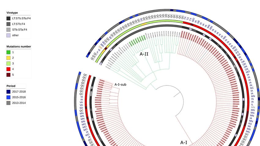
Antibiotics 2021, 10, 244                                                                                          2 of 19

                            mainly urinary tract infection and septicemia in humans, which is resistant to fluoroquin-
                            olones and third generation cephalosporins, is one of the best examples of this problem
                            [6]. In its current definition, the term “high risk clone” is mainly adapted for human path-
                            ogens, whereas antimicrobial resistance should be addressed using a more global ap-
                            proach [7]. Therefore, it may be timely to revisit the present criteria [8] and to adapt them
                            to the One Health concept [9].
                                  The factors driving the emergence and the dissemination capacity of high risk clones
                            are not well understood. Several fitness characteristics, particularly the speed of replica-
                            tion, have been associated with high resistance to fluoroquinolones [10] and have been
                            demonstrated to confer growth advantage to several clones, particularly in extended-
                            spectrum β-lactamase (ESBL) producing E. coli [11]. Therefore, fluoroquinolone use lead-
                            ing to fluoroquinolone resistance could contribute to the selection and the spread of high
                            risk clones [12,13].
                                  Enterotoxigenic E. coli (ETEC) causes diarrhea in piglets during the neonatal and
                            post-weaning periods, resulting in important economic losses for the swine industry [14].
                            ETEC strains produce one or several enterotoxins, which induce the secretion by epithelial
                            cells of water and electrolytes into the intestinal lumen. These ETEC strains colonize the
                            host via fimbriae which adhere to the intestinal mucosa. ETEC associated with diarrhea
                            in young pigs most commonly possess F4 (K88) or F18 fimbriae, although geographic var-
                            iations have been reported [15]. ETEC isolates are grouped into pathovirotypes based on
                            the presence of the different fimbriae and one or more of the enterotoxins heat labile toxin
                            (LT) and heat stable toxins (STa and STb). Pigs with diarrhea associated with ETEC have
                            been commonly treated with ampicillin (alone or in combination with clavulanic acid),
                            trimethoprim, sulfonamides, aminoglycosides (i.e., neomycin), ceftiofur, spectinomycin,
                            or enrofloxacin, depending on the country [15]. In a previous article, we reported the pres-
                            ence of a cluster of ETEC:F4 isolates sharing at least 55% of similarity based on their pulse
                            field gel electrophoresis (PFGE) profile in diseased pigs in Quebec, which emerged in 2013
                            [16]. Most of these isolates were LT:STb:STa:F4 and non-susceptible (intermediate or re-
                            sistant) to enrofloxacin. This antimicrobial has been used off-label in Quebec since the
                            2000s. In addition, fluoroquinolone non-susceptibility in E. coli isolated from diseased pigs
                            has been observed in other countries [17,18] and has been associated with enrofloxacin
                            treatment previously in herds [19]. Moreover, clonality has been demonstrated among
                            ETEC isolates causing disease in pigs in the USA [20]. Our hypothesis is that a high risk
                            fluoroquinolone non-susceptible pathogenic ETEC clone has emerged in pigs in various
                            countries, thus representing a threat for swine and public health. The objectives of this
                            study were to (1) propose a new definition including revised criteria for a high risk clone
                            adapted to the One Health context, (2) further characterize ETEC:F4 isolates sampled in
                            Quebec, using a genomic approach, and (3) compare these isolates with other ETEC:F4
                            worldwide both phylogenetically and with respect to the presence of virulence and anti-
                            microbial resistance genes and mobile genetic elements, in order to assess the presence of
                            a high risk clone based on well-defined criteria.
                                  The detection and surveillance of these putative new clonal isolates is crucial to iden-
                            tify and develop control strategies in the field.

                            2. Results
                            2.1. Proposal of Criteria for a High Risk Clone in the One Health Context
                                  Until now, clones have qualified as being “high risk” when they have met 6 criteria:
                            (i) worldwide spread, (ii) carriage of multiple antimicrobial characteristics, (iii) efficient
                            colonisation and persistence in host, (iv) effective transmission, (v) increased pathogenic-
                            ity, and (vi) increased virulence causing recurrent infection [6,21]. Other important as-
                            pects, such as the emergence of the pathogen, environmental persistence, and zoonotic
                            transmission, were not included in these characteristics. Therefore, we propose a list of
                            new criteria, presented in Table 1, to adapt the “high risk” clone definition to a One-Health
Antibiotics 2021, 10, 244                                                                                                          3 of 19

                                  context. According to our revised criteria, a clone qualifies to be high risk for antibiotic
                                  resistance dissemination if (1) it is emergent, the definition of emergence of a pathogen is
                                  as described in Wang et al. [22] and is based on OIE recommendations; (2) it carries mul-
                                  tiple resistance genes associated with phenotypic resistance; (3) it has a high capacity of
                                  dissemination, the definition of persistence was adapted from Falkow et al. [23] and the
                                  concept of environmentally maintained source is described in Blackburn et al. [24]; and
                                  (4) it is highly pathogenic. The definitions of these criteria are detailed in Table 1.

      Table 1. Definition of criteria for determination of a high risk bacterial clone for antibiotic resistance dissemination in the
      context of the One Health concept.

                    Criterion                                                                Application to ETEC:F4 clone A-I de-
                                                               Definition
                                                                                                   tected in pigs in Quebec
                                       The clone is newly recognized, newly
                                      evolved or has occurred previously but
                                                                              The clone A-I emerged in North Amer-
             1/Emergence               shows an increase in incidence or ex-
                                                                                               ica in 2013.
                                      pansion in geographical, host or vector
                                                      range.
                                                                               The clone A-I carries at least the genes
                                                                               tet(A) and the blaTEM-1 which are associ-
                                       The clone carries multiple resistance
                                                                               ated with phenotypic resistance to tet-
                                      genes associated with phenotypic mul-
                                                                              racyclines and penicillins, respectively.
 2/Carriage of multiple antimicrobial tidrug resistance. The resistance genes
                                                                                 Several replicons such as the IncFII
            characteristics            can be carried by mobile genetic ele-
                                                                                have been identified in the clone A-I.
                                      ments or by the chromosome (and then
                                                                              Moreover, it also carries parC and gyrA
                                         results from mutations) or both.
                                                                               mutations responsible for non-suscep-
                                                                                     tibility to fluoroquinolones.
   3/High capacity of dissemination
                                                                                The clone A-I has been detected in at
 through one or a combination of the The clone is likely to disseminate due
                                                                              least one province of Canada as well as
      following characteristics:                         to:
                                                                                      in many states of the USA.

                                              -       High probability of transmission
                                                                                       The clone A-I has been observed in dif-
             3a/ High infectivity             following exposure to an infected host
                                                                                       ferent batches of pigs on the same farm
                                                       or environmental source
                                                                                                     for 6 months.
                                                                and/or
                                              -       Long-term persistence and shed-
        3b/ Long-term persistence                   ding in colonized individuals
                                                                and/or
                                              -       Presence of the pathogen in mul-
                                                tiple animal species and/or in human
        3c/ Multiplicity of sources
                                              population and/or in environmentally
                                                          maintained source.
                                                                                          The clone A-I is associated with a
                                                The clone can cause severe disease in
           4/High Pathogenicity                                                         higher risk of mortality that observed
                                                      animals or/and in humans.
                                                                                               for other clonal lineages.

                                  2.2. Identification of Clonal Lineages and Clones
                                       We sequenced a total of 183 isolates originating from diseased pigs in Quebec, Can-
                                  ada, between 1990 and 2018, randomly selected in each cluster of a preliminary PFGE
                                  study or from the APZEC database. After removing 10 isolates due to unsatisfactory raw
                                  data or assembling quality, 36 isolates from 1990 to 2012 as well as 21, 21, 22, 26, 24, and
                                  23 isolates for each year from 2013 to 2018, respectively, were retained. The sequencing
                                  quality information about the 173 isolates is available in supplemental data Table S1.
Antibiotics 2021, 10, 244 4 of 19
A total of 3,366,150 positions were found in all analyzed genomes. The percentage of
reference genome covered by all isolates was 61.95% with a reference genome size of
5,433,365 pb (ECL19664). After the removal of the recombination signal with the Gubbins
software, the total number of SNPs used to build the tree was 19,229. The resulting SNP
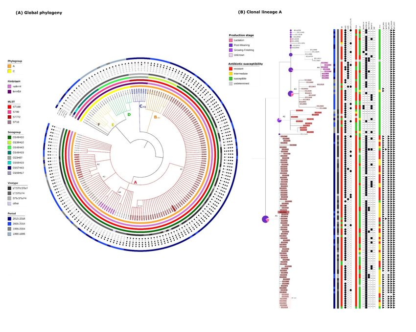
tree of the 173 isolates is shown in Figure 1A. Based on the definitions described in the
Materials and Method, 6 clonal lineages (A, B, C, D, E, and F) and 6 clones (A1, A2, A3,
A4, B1 and C1) were identified. One sub-clone within the clone A1, designated A1-sub
was also detected. Two singletons were also identified (ECL01947 and ECL20395). The
SNPmax of each group is presented in detail in Figure S1.
In the 1990s and the early 2000s, clonal lineages D, E, and F were predominant (Figure
1A). Clonal lineage D was the oldest, 10/12 isolates being obtained between 1990 and 1995.
All isolates (D, E and F) were from the phylogroup C, MSLT ST90 and presented the gene
fimH54. However, isolates of the three clonal lineages belonged to different serotypes, re-
spectively O149:H43, O149:H19 and O159:H19 and no clone was detected within these
lineages according to the set of predefined criteria.
The predominant clonal lineage A, comprising 129/173 isolates, was first observed in
1995. The phylogeny of these 129 isolates has been enlarged and is presented in more de-
tail in Figure 1B. These 129 isolates are O149:H10, ST100, phylogroup A and possess no
known fimH gene. Within this clonal lineage, 4 clones (A1, A2, A3 and A4) were identified.
Isolates from the clones A2 and A3 were identified around the same period (1995 to 2014),
prior to isolates from the clones A1 and A4. Isolates from the clone A1 were first observed
in 2013 and have continued to be observed over the 5-year period until the end of the
study. The subclone (A1-sub) was composed of isolates sampled between 2015 and 2018.
Isolates belonging to the clone A4 were isolated during the same period as the clone A1
(between 2013 and 2018) but represent a much smaller proportion of isolates from the
clonal lineage A.
The clonal lineage B/clone B1, including 11 isolates, was first observed in 2009, and
continued to be found until the end of the study period (Figure 1A). These isolates are
O138:H10, ST100, phylogroup A and present no known fimH gene.
More recently, in the last 2 years of the study period, the clonal lineage C/clone C1,
belonging to a new sequence type, has been observed. These isolates are O23:H37, ST772,
phylogroup A and possess the gene fimH54.
In summary, ETEC:F4 isolates in Quebec in the early 1990s were predominantly ST90,
belonging to 3 different serotypes and carrying the fimH54 gene. In the mid-1990s, isolates
belonging to ST100 appeared and became predominant. They belong to 2 main O sero-
types and carry no fimH gene. The clonal lineage A (ST100/O149:H10) is the most wide-
spread and is composed of several clones. In the late 2010s, isolates belonging to another
clonal lineage have appeared. They belong to the ST772 and the serotype O23:H37 and
constitute a clone.Antibiotics 2021, 10, 244 5 of 19
Figure 1. (A)Global phylogeny: Circular Tree based on SNP phylogeny. The length of the branches is not proportionate to
the phylogenetic distance. Gubbins was used to eliminate the recombination signal. The total SNP number was 19,299.
Each branch with a bootstrap value under 1 was collapsed. The capital letters designate each clonal lineage. Filled triangles
indicate the presence of the corresponding mutation, empty triangles indicate the absence of the corresponding mutation.
(B)Clonal lineage A (same phylogeny is used): The number associated with the letter A designates each clone. The identi-
fication name for each isolate belonging to a clone is highlighted in a specific color. Circles represent the proportion of the
age of the pigs within each corresponding branch. Columns entitled Tet, Amp, Cef, Enro, represent respectively the sus-
ceptibility to tetracycline, ampicillin, ceftiofur and enrofloxacin (code for the corresponding color is available on the fig-
ure).
2.3. Virulence Gene Profiles of the Different Clonal Lineages.
The virulence gene profiles of the isolates were established based on the detection of
these genes in the VirulenceFinder and VFDB databases. ETEC:F4 isolates of the same
clonal lineage mostly belonged to the same virotype (Figures 1A and 1B). All isolates from
clonal lineages D, E, F and B1 (apart from one in B1) were LT:STb:F4 and isolates from C1
were STa:STb:F4. Isolates from the clonal lineage A belonged to the virotype LT:STb:F4
except for isolates from clones A1 and A2 which possessed various combinations of en-
terotoxins, most isolates being LT:STb:STa:F4 (49/80 and 9/11 respectively).
Clonal lineage A differed from the other lineages by the absence of the group of genes
coding for the siderophore Yersiniabactin (irp, fyuA and ybt family genes), by the presenceAntibiotics 2021, 10, 244 6 of 19
of genes involved in the type VI secretion system (aec family genes) and the presence of
genes coding for elements of the E. coli common pilus (ECP) and of the hemorrhagic E. coli
pilus (HEP). Clonal lineage A was also distinguished by the presence of ompA (interacts
with specific receptors for initiating the pathogenic process), rpoS (involved in the regula-
tion of the stress response) and tar/cheM (involved in the chemotactic response) (Table 2
and Table S2). The ehxA gene encoding enterohemolysin (associated with haemolysin se-
cretion) was detected in all isolates belonging to the clonal lineage C but in none of the
isolates belonging to the other clonal lineages. The iha gene (encoding for a homologue
adhesin (IrgA)), was detected in all isolates belonging to the clonal lineage B but in none
of the isolates belonging to the other clonal lineages. The lfpA gene (encoding for a fimbrial
major protein) was present in all isolates belonging to clonal lineages D, E and F but none
of the isolates belonging to other clonal lineages. The porcine attaching-effacing associated
gene (paa) was detected only in isolates belonging to the clonal lineage A. However, its
presence was not systematic (83/129) and was not specific to a clone. There was no viru-
lence gene identified exclusively in isolates belonging to a particular clone and in all iso-
lates from this clone. All these data are available in Tables S3 and S4.
Table 2. Virulence genes specific of the different clonal lineage (the details are available in Tables S2 and S3) *See Table S1
for the complete list of genes.
tar/cheM
methyl-ac-
ompA
Fimbrial Siderophore E. coli rpoS cepting
Enterohe- Type VI Hemor- outer
Genes major Adhesin Yersiniabac- Common sigma S chemotaxis
molysin secretion rhagic E. coli mem-
Clonal lineage protein (Iha) tin * Pilus (sigma protein II
(ehxA) system * pilus (HEP)* brane
(lfpA) (ECP) * 38) factor [peritri-
protein A
chous fla-
gella]
A
Absent Absent Present Absent Present Present Present Present Present Present
(ST100/O149)
B
Present Absent Absent Present Absent Absent Absent Absent Absent Absent
ST772/O23
C
Absent Absent Present Present Absent Absent Absent Absent Absent Absent
ST100/O138
D
Absent Present Absent Present Absent Absent Absent Absent Absent Absent
ST90/O149H19
E
Absent Present Absent Present Absent Absent Absent Absent Absent Absent
ST90/O157
F
Absent Present Absent Present Absent Absent Absent Absent Absent Absent
ST90/O149H43
2.4. Resistance Gene, Multidrug Resistance (MDR) and Replicon Profiles of the Different Clonal
Lineages.
The resistance gene and replicon profiles of the isolates were established based on
the detection of these genes in the ResFinder, PlasmidFinder and ARG-Annot databases.
Isolates from clonal lineage E (n=10) were mostly non-susceptible to tetracycline (9/10)
and ampicillin (7/10) and showed no or little non-susceptibility to enrofloxacin (0/10),
cephalosporins (1/10), florfenicol (3/10), TMS (4/10), and aminoglycosides (5/10) (Figure
S2). Genes detected and likely responsible for these non-susceptibilities were tet(B) for
tetracycline, blaTEM-1B for ampicillin, cmlA1 or catA1 for florfenicol, sul1 and drf1 for TMS,
and aad or aph for aminoglycosides.
Half of the isolates from clonal lineage D (n = 12) were non-susceptible to tetracycline
(6/12), otherwise they were infrequently non-susceptible to ampicillin (2/12), enrofloxacin
(0/12), cephalosporins (1/12), florfenicol (2/12) and TMS (2/12), and aminoglycosides (6/12)
(Figure S2). Genes detected and likely responsible for these non-susceptibilities wereAntibiotics 2021, 10, 244 7 of 19
tet(A) or tet(B) for tetracycline, blaTEM-1B for ampicillin, cmlA1 or catA1 for florfenicol, sul1
and drf1 for TMS, and aad for aminoglycosides (Tables S5 and S6). IncFIB and IncFII repli-
cons were present in isolates from both clonal lineages D and E. However, the IncFIC
replicon was only present in the clonal lineage E (Table S7). One isolate in D carried a
blaIMP (responsible for carbapenemase activity) (Table S8).
Isolates from clonal lineage B1 (n = 11) were mostly non-susceptible to tetracycline
(11/11), to TMS (11/11), to aminoglycosides (9/11), and to ampicillin (6/11) and infre-
quently non-susceptible to enrofloxacin (0/11), cephalosporins (5/11), and florfenicol
(2/11) (Figure S2). Genes detected and likely responsible for these non-susceptibilities
were tet(A) and tet(M) for tetracycline, blaTEM-1B for ampicillin, cmlA1 or catA1 for
florfenicol, sul1 or sul2 and drf1 for TMS, and aad or aph for aminoglycosides (Tables S5
and S6). The plasmid profile was also similar among B1 isolates with the presence of IncI1
and IncFIB (Table S7).
Isolates from clonal lineage A (n = 129) were mostly non-susceptible to tetracycline
(115/129) and to ampicillin (113/129). They were infrequently non-susceptible to TMS
(55/129), to aminoglycosides (61/129), to cephalosporins (31/129), and to florfenicol
(40/129) (Figure S2). Concerning the enrofloxacin non-susceptibility there was a clear sep-
aration between isolates belonging to the clone A1, which were mostly non-susceptible
(74/80) and those of the clonal lineage A not belonging to the clone A1 which were very
rarely non-susceptible (1/49). Therefore, isolates belonging to A1 were mostly MDR
(71/80).
Isolates from the clonal lineage A carried two mutations, parC (E62K) and gyrA
(N652H), classified as «unknown» (meaning that the phenotypic significance is not estab-
lished) in ResFinder. All isolates from the clone A1 carried these two mutations as well as
two other mutations in the parC (S80I) and gyrA (S83L) genes, classified as «known» in
ResFinder (meaning that these mutations were associated with a non susceptible pheno-
type). Isolates from the clonal lineage A also carried the tet(A) (109/129) or the tet(B)
(11/129) which confer tetracycline resistance (Figure 1B). The tet(B) gene was mainly pre-
sent in isolates from the clone A4. Except for isolates from the clone A3, they also carried
the blaTEM-1 (104/129) gene which confers ampicillin resistance. All A1 isolates carried the
replicon FIB(K) which was found only in two other isolates within the clonal lineage A
(one in the clone A2 and one in an isolate belonging to no clone (Table S7).
Isolates from the most recently occurring clonal lineage C1 (n = 7) were all non-sus-
ceptible to tetracycline (7/7), to aminoglycosides (7/7), and to TMS (7/7), therefore, they
were all MDR. Moreover, they were mostly non-susceptible to florfenicol (6/7) and infre-
quently non-susceptible to enrofloxacin (1/7), cephalosporins (3/7), and ampicillin (3/7)
(Figure S2). Genes detected and likely responsible for these non-susceptibilities were aah
gene for aminoglycosides, tet(A) for tetracycline resistance and sul3 and dfrA12 for TMS.
It is noticeable that one of the isolates was non-susceptible to quinolones, although this
isolate carried a qnr gene. Also, despite the presence of cmlA1 (supposedly conferring re-
sistance to phenicols) two of these isolates were susceptible to florfenicol. Two isolates
carried the blaCTX-M-1 gene and were resistant to ceftiofur. The plasmid profile was also sim-
ilar among C1 isolates, IncFIB(K) and (PB71) being always present.
Overall, the AmpC gene blaCMY-2 was detected in 23/173 isolates, belonging to clonal
lineages A (22/23) and E (1/23). In our isolates, this gene was first detected in 2005. It is
interesting to note that when the blaCMY-2 was present, the replicon IncA/C was also present,
suggesting a link between them (data not shown). In our isolates, the ESBL gene blaCTX-M-1
was first detected in 2013 and was found in 15/173 isolates belonging to recent clonal lin-
eages (A(9/15), B(4/15) and C(2/15)). However, neither blaCMY-2 nor blaCTX-M-1 was specific to
a clone within the clonal lineage A.
2.5. Mortality Risk, Production Phase of Affected Pigs, and Persistence on Farm of the Different
Clonal Lineages.Antibiotics 2021, 10, 244 8 of 19
Necropsy reports were available for 106 cases. Overall, mortality was observed in
47% of these cases. The risk of mortality was significantly (p = 0.003) higher in cases in-
fected with isolates from clonal lineage A (54%) compared to clonal lineage B (0%) but
was not associated with other clonal lineages. Within clonal lineage A, no significant dif-
ference in the mortality risk was observed between cases infected with isolates from dif-
ferent clones (p = 0.09) (see Tables S9 and S10).
Information on the phase of production of the pigs was available for 158 cases. Over-
all, most cases of ETEC:F4 infection (73%) were detected in pigs in the weaning phase in
nurseries. No statistically significant association was found between clonal lineage and
phase of production (p = 0.5), nor between clones and phase of production within the
clonal lineage A (p = 0.7) (Tables S11 and S12).
In 2 farms located in Montérégie, Québec, 2 isolates belonging to the clone A1 were
identified 6 and 8 months apart, respectively. In both cases, the farms were housing two
stages of production: a farrowing barn and a nursery. For both cases, the first isolate was
identified in the farrowing barn and the second isolate was identified in the nursery,
strongly suggesting that the clone persisted on farm. The type of cleaning and rotation
were not known for either farm. No other pair of isolates was sampled at different times
on the same site, according to the available information.
2.6. Presence of Isolates Belonging to the Clonal Lineage A in North America.
Eighty-seven whole genome sequences in Enterobase were described as ETEC and
ST100 isolated from diseased pigs between 2013 and 2018 and were available in GenBank.
Among these, 7 were duplicates and were removed from our analysis. The 112 isolates
belonging to the clonal lineage A and sampled in Quebec after 2013 and the 80 isolates
identified in Enterobase were used for the second phylogenetic analysis. All the isolates
described in Enterobase were confirmed to belong to the ST100, O149:H10 serotype, phy-
logroup A, and to carry no fimH gene in silico. These isolates originated from various
states of the USA except for one from a sample in Switzerland. The available metadata for
these isolates are presented in Table S13.
A total of 3,675,678 positions were found in all analyzed genomes. The percentage of
reference genome covered by all isolates was 67.65% with a size of reference genome of
5,433,365 pb (the same isolate was used as reference). After removal of the recombination
signal the total number of SNPs used to build the tree was 3,119. As expected, because
isolates belonged to the same clonal lineage, the number of differing SNPs was low (min
= 0, max = 1,007, median =38), indicating that isolates were very similar. The resulting SNP
tree of the 192 isolates is illustrated in Figure 2 and distance matrix is presented in Table
S14.
Two clones were identified and designated as A-I and A-II and one subclone desig-
nated A-I-sub. The clone A-I (Figure 2 and Figure S3) comprised 150 isolates, originating
from both Canada and USA. All isolates from the clone A1 and only isolates from the
clone A1 in the previous analysis belonged to the clone A-I. Therefore, we will use the
designation clone A-I to refer to the isolates from both analyses. The clone A-II in this
analysis comprised the same isolates as those of the clone A4 in the previous phylogenetic
analysis.
Clone A-I isolates possessed various combinations of enterotoxin virulence genes but
were mainly LT:STb:STa:F4 (98/150). These virulence profiles and those identified in Table
2 did not differ between isolates of clone A-I and those of the clonal lineage A.
All isolates from clone A-I carried a mutation in each of the parC and gyrA genes (S80I
and S83L respectively), classified as «known» in ResFinder, and a mutation in each of the
parC and gyrA genes (E62K and N652H, respectively), classified as «unknown» in
ResFinder. In 16 isolates, an additional mutation was detected in the gyrA gene (Figure 2
and Figure S4). Fourteen of these 16 isolates were sampled in the USA and 2/16 in Quebec.
Most of the clone A-I isolates also carried blaTEM-1 (142/150) and tet(A) (134/150); however,
susceptibility phenotype results were not present in the metadata available in Enterobase.Antibiotics 2021, 10, 244 9 of 19
In the isolates sampled in the USA, the ampC gene blaCMY-2 was detected in 16/80 iso-
lates and the ESBL gene blaCTX-M-1 was detected in only 1/80 isolates. The A-I isolates also
carried replicons of the incompatibility group FIB (Table S7).
Figure 2. Circular Tree based on SNP phylogeny. The length of the branches is not proportionate to the phylogenetic
distance. All isolates belong to the clonal lineage A (ST100-O149H10-phylogroupA-no fimH gene) and originated from
2013 or after. Each branch with a bootstrap value under 1 was collapsed. QC = Quebec, SW = Switzerland, MN (in grey) =
Manitoba, ON = Ontario, MI = Michigan, MN (in blue) = Minnesota. MO = Missouri, NC = North Carolina, TX = Texas, IA
= Iowa, KS = Kansas, WI = Wisconsin, IN = Indiana, IL = Illinois, OK = Oklahoma, NE = Nebraska, SD = South Dakota, VA
= Virginia. A-I and A-II designate each clone and A-I-sub designates the subclone within the clone A-I. The 3 isolates
colored in black are the three outsiders (more than 70 SNPs of differences in the pairwise distance comparison). The iden-
tification name for each isolate belonging to a clone is highlighted in a specific color.
2.7. Application of the Criteria for “High Risk” Clone to the Clones A-I, A2, A3, A4, C1 and B1.
As illustrated in Table 3, the clone A-I emerged in 2013, which is multidrug resistant
and carries several genes associated with multidrug resistance, has disseminated effi-
ciently throughout North America and is associated with severe diarrhea and/or sudden
death. Therefore, the clone A-I qualifies as a high risk clone. Other clones in the clonal
lineage A (A2, A3, A4) are not consistently MDR and did not disseminate as efficiently as
the clone A-I. The clone B1 is not consistently MDR and does not predominate currently.
The clone C1 emerged around the year 2014, is MDR and pathogenic. Until now, there is
no proof that it has disseminated throughout North America.Antibiotics 2021, 10, 244 10 of 19
Table 3. Revised criteria for high risk clone applied to the clones identifies in our study.
Criteria/Clone A-I A2 A3 A4 B1 C1
Emergence yes no no no no yes
Multidrug resistance yes no no no no yes
Potent dissemination yes no no no no no
Pathogenicity yes yes yes yes yes yes
3. Discussion
The main objective of this study was to redefine the term “high risk clone”. Indeed,
bacteria (and other microorganisms) lodge in multiple ecological niches, such as humans,
animals, or the environment. A comprehensive and integrated approach considering the
complex interrelationships between these niches is needed in the development of global
strategies tackling antimicrobial resistance [25]. In this regard, the One Health approach,
defined as “the collaborative effort of multiple health science professions, together with
their related disciplines and institutions -working locally, nationally, and globally -to at-
tain optimal health for people, domestic animals, wildlife, plants, and environment” [26]
is essential to investigate all the multifaceted mechanisms for antimicrobial resistance dis-
semination. Yet, the current definition of a high risk clone is mainly adapted to human
medicine alone [6,21]. The second objective was to determine if enrofloxacin-non-suscep-
tible ETEC:F4 isolates from diseased pigs isolated in Quebec belonged to a high risk clone.
Our analytical approach permitted us to examine the phylogenetic variation of
ETEC:F4 isolates from diseased pigs with time in Quebec. Prior to 2000, ETEC:F4 isolates
were mostly ST90 and belonged to several serotypes. On the other hand, after 2000, iso-
lates were mostly ST100, belonging to only one serotype, O149:H10, and carrying no fimH
gene. We designated these isolates as the clonal lineage A. Within this clonal lineage we
identified several clones and subclones. In addition, in a second phylogenetic analysis, we
compared isolates from the clonal lineage A to other ST100/O149:H19 isolates sampled in
other geographical locations. All enrofloxacin-non-susceptible isolates belonged to a clone
designed as A-I. As the clone A-I of the second analysis included all isolates from the clone
A1 of the first analysis, we will use the designation clone A-I to refer to the isolates from
both analyses. In the next paragraphs we will discuss each revised criterion for a high risk
clone and its application to the clone A-I.
We demonstrated that the clone A-I appeared in 2013 and became predominant in
2015 (data from this study and [16]), consequently meeting the emergence criteria (cf Table
1). The clonality of ETEC isolates from various states of the USA and belonging to clonal
lineage A had already been demonstrated [20]. In addition, we demonstrated the interna-
tional distribution of the clone A-I, as it is also present in Canada, at least in Quebec. Thus,
we demonstrated that isolates of the clone A-I spread in Canada and in the USA, possibly
via the frequent displacement of pigs between the two countries. As an illustration, in
2015, Canada exported almost 6 million live hogs to the USA for feeding or harvest and
imported 528 million pounds of USA pork meat (https://www.pork.org/facts/stats/cana-
dian-statistics/). A wider distribution of this clone was difficult to assess, as whole genome
sequences of E. coli from other continents were not available in Enterobase within the
study period. Indeed, the common use of whole genome sequencing occurred later in vet-
erinary medicine than in human medicine, due mainly to its high cost. Consequently, vet-
erinary isolates are less frequently available in databases. In particular, it would have been
very interesting to include sequences from isolates in Japan where PFGE-based cluster
lineages demonstrating fluoroquinolone non-susceptibility have been reported [18]. This
wide distribution is the result of a wide capacity of dissemination (cf Table1). Yet, this
capacity of dissemination can be driven by several means. In our definition of a high risk
clone, we wished to include all factors of importance that could increase the likelihood of
clonal dissemination to human and/or animal populations, to match as many field situa-
tions as possible. Therefore, we established three inherent sub-criteria. These criteria wereAntibiotics 2021, 10, 244 11 of 19
grouped as the observed persistence of a clone in a population may be the result of a high
transmissibility among hosts, of a high persistence in colonized individuals (especially if
asymptomatic) and/or from recurrent exposure to a contaminated environment and/or
other host species, and thus it may be difficult to disentangle their individual impacts.
Our data suggests that the clone A-I might persist on certain farms for 6 months or longer.
It is a good illustration of a situation where both environmental persistence and transmis-
sion between pigs on the farm may have occurred. Whichever scenario(s) is/are at stake,
the clone is still a risk for the swine health and therefore is considered a high risk clone in
a One Health context. Nevertheless, these characteristics should be investigated further to
determine which mechanism(s) is/are involved to develop fighting strategies. As an ex-
ample, if persistence in the environment is the main mechanism involved in transmission
between batches of pigs, protocols including efficient disinfection should be advised to
farms that harbour the clone. On the other hand, if the transmissibility between animals
is high, vaccination may be advised. This distinction could not be made with the design
of our study. To summarize, the clone A-I has a great capacity of dissemination through
either high infectivity or environmental persistence or both and, as a result, has been iden-
tified throughout a large territory and therefore fulfils our third criterion to be a high risk
clone.
It is noteworthy that isolates from the clones A2, A3 and A4 did not spread as widely
as the clone A-I. We believe that the sequence of events, first the acquisition of a higher
pathogenicity (isolates from the clone A2 and A3 were identified prior to isolates from
A-I) then the acquisition of the resistance to fluoroquinolone, was required to lead to the
clone A-I being qualified as high risk. It is not known if the acquisition of fluoroquinolone
resistance alone would have allowed the resulting clone to spread as efficiently as the
clone A-I through increased fitness mechanisms [10].
To support the critical role of the One Health approach, the inclusion of samples from
other origins than pigs, such as environmental samples near the farms and/or human sam-
ples such as caretakers, would have been advisable. These samples would have allowed
us to assess the potential zoonotic transmission or the persistence in the environment.
These two criteria are acknowledged in the multiplicity of sources sub-criterion of our
new definition of high risk clone. Indeed, an animal population could be a reservoir for a
clone that does not endanger the host population but could threaten public health, such
as the Shiga-toxin producing E. coli O157:H7. In our case, due to the retrospective nature
of our study, environmental and human samples could not be analyzed as they were not
available. Therefore, environmental persistence or zoonotic transmission were not evalu-
ated directly. The zoonotic transmission is unlikely, though, as ETEC: F4 are usually not
pathogenic for humans.
Most isolates belonging to the clone A-I were MDR, therefore fulfilling the second
criterion (Table 1). Indeed, most isolates presented non susceptibility to enrofloxacin, tet-
racycline and ampicillin. Enrofloxacin non susceptibility was driven by at least 2 muta-
tions in parC (S801and E62K) and 2 mutations in gyrA (S83L and N652H). In 16 isolates,
mostly from the USA, an additional mutation in the gyrA gene was detected. The mutation
parC E62K was detected in all our isolates and has been described in the literature as a
“real” mutation [27] although not unanimously [28]. As it was present in all our isolates
(including those from the USA), and as isolates only possessing this mutation were sus-
ceptible to fluoroquinolones, we considered this mutation as clinically not relevant. The
gyrA N652H mutation, classified as «unknown» in ResFinder, was present in all isolates
from the clonal lineage A (from USA and from Quebec). It is not described in the literature
to the authors’ knowledge. Both mutations should be further investigated for MIC levels
to evaluate their possible role in sequential acquisition of clinically significant resistance
to fluoroquinolones. Ampicillin and tetracycline resistance were driven by blaTEM-1
(142/150) and tet(A) (134/150) respectively. In addition, isolates belonging to the clone A-I
were associated with several plasmids such as the IncFIB(K) replicon. This plasmid familyAntibiotics 2021, 10, 244 12 of 19
has been described as “an epidemic plasmid” [29] and is largely associated with specific
resistance genes in Enterobacteriaceae [30].
BlaCMY-2 is the predominant ESBL/AmpC gene in the ETEC:F4 of swine origin (39/192
isolates) compared to the blaCTX-M-1 (16/192). However, blaCTX-M-1 was mostly identified in
isolates from Quebec (15/16). The monitoring of this tendency must be pursued to deter-
mine if the blaCTX-M family is overtaking the rest of the ESBL/AmpC genes in animal popu-
lations in North America, as happened in Europe [31]. Nevertheless, the presence of
ESBL/AmpC genes in 31 isolates belonging to the clone A-I is concerning as the presence
of these genes results in a great capacity to disseminate, thus conferring on the clone an-
other “fitness” advantage. Hence, a subclone carrying systematically such a gene could
emerge in the near future, as has occurred for ST131-H30-Rx [32].
Our evaluation of clinical severity was based on retrospective evaluation of non-
standardized necropsy reports, which is imperfect. However, it was our only means of
evaluating objectively the clinical aspects of increased pathogenicity. We limited our eval-
uation to one criterion: death of affected pigs on the farm. Although the clone A-I itself
was not associated with a higher proportion of death in pigs on the farm compared to
other isolates within the clonal lineage A, greater mortality was associated with isolates
in the clonal lineage A than that observed with other clonal lineages. Thus, this clone also
fulfills the pathogenicity criteria (Table 1). The presence of genes coding for different pili
(ECP and HEP) or for T6SS and the presence of known virulence factors such as ompA are
likely to contribute to an increase in pathogenicity of this clonal lineage. Indeed, each of
these elements are known key virulence factors. ECP plays a dual role in early-stage bio-
film development and host cell recognition [33]. HCP mediates invasion of epithelial cells
or interbacterial connections leading to biofilm formation [34]. T6SS is a multi-protein
complex dedicated to the delivery of toxins [35]. Finally, despite its conservation through-
out evolution among pathogenic and non-pathogenic bacteria, ompA interacts with spe-
cific receptors for initiating the pathogenic process in some Gram-negative bacterial infec-
tions [36]. However, the presence of these genes alone is not enough to prove that they are
responsible for the increased pathogenicity. Further functional studies are needed to en-
sure the role of each of these elements.
The definition of a clone remains a challenge for all microbiologists. The definition of
a bacterial clone, based on whole genome sequencing (WGS) data, should not be based
only on a fixed number of SNPs. In the current context, where WGS is being increasingly
used as a result of decreased costs, the analytical approaches used in different studies are
still subject to a great lack of homogeneity and standardisation because of the many pos-
sibilities that they offer. The definition of a clone that we propose, based on the bootstrap
values, the number of isolates in the branch and number of SNP differences in relation to
the mutation risk adjusted for the period between sampling of isolates, allow reproduci-
bility and minimize over-interpretation of the data. Nevertheless, these criteria are debat-
able. Firstly, the mutation rate is not constant between bacterial species or even across
time within the same species. It is known, particularly in E. coli, to depend on the level of
stress due to evolution pressure [37]. However, we needed to determine a fixed mutation
rate to establish a SNP cut-off which was based on E. coli variability during an outbreak
[38]. Secondly, the bootstrap value of 1 is very stringent. However, considering the large
number of isolates taken into consideration in the study, we chose to be strict to enforce
the robustness of our analysis. In another context, for example if fewer isolates are avail-
able for the phylogenetic analysis, a bootstrap value of 0.99 or 0.95 might also have been
acceptable. Thirdly, we fixed the minimal number of isolates per branch at three to reduce
the likelihood of a grouping only due to chance. Finally, although the lag of time between
the detection of isolates was taken into account in the definition of a cluster, it could not
be considered in the maximum-likelihood approach used to create the phylogeny tree,
which represents a limitation.
As observed with all laboratory techniques, there is an inherent error rate associated
with whole genome sequencing. However, this is difficult to evaluate considering all theAntibiotics 2021, 10, 244 13 of 19
steps of analysis involved in establishing a phylogenetic tree, including the sequencing
itself, assembling, multiple alignment, recombination signal removal, or tree building. We
evaluated the error rate of our pipeline based on a total of four isolates that were dupli-
cated (for 2 isolates there were 2 duplicates, for 1 isolate there were 3 duplicates, and for
1 isolate there were 4 duplicates). We observed that the error rate was very variable de-
pending on the quality of the raw data. However, for the mean quality of our data, we
estimate the error rate to be from 30 to 60 SNP between two duplicates (data not shown).
This number strengthens our finding that isolates belonging to the designated clone A-I
are very similar, because the SNPs we did detect could be related to this measured error
rate.
Although our data demonstrate the capacity of WGS to refine the analysis for detect-
ing clones, they also show that the combination of MLST and serotype data can accurately
discriminate clonal lineages. On the other hand, when we compared these results to those
obtained with PFGE in our previous study, we observed that only two thirds of the iso-
lates analyzed with both methods were classified correctly by the PFGE when the cut-off
of 55% of similarity was used to define a cluster [16]. Price et al. found a similar misclas-
sification for the E. coli ST131 [32]. Although PFGE has proven useful in the past, we feel
that the use of this method should be limited to specific situations such as outbreak inves-
tigations and its results interpreted with great caution, and that PFGE should be replaced
by WGS as much as possible.
An additional clone C1, comprising isolates belonging to ST772 and serogroup O23
and having a specific virotype STa:STb:F4 infrequently described in Quebec previously
[16], was first observed in 2016. As all these isolates are MDR (non susceptible to tetracy-
cline, TMS and one antimicrobial in the aminoglycoside category), they should be moni-
tored during the coming years to evaluate if they could develop into a high risk clone and
represent a threat for porcine health in Quebec and in North America. The absence of
quinolone non susceptibility is of note and might represent a fitness disadvantage for this
clone.
4. Materials and Methods
4.1. Proposal of Criteria for a High Risk Clone in a One Health Context and Application
A list of previously used criteria to define a high risk clone for antibiotic resistance
dissemination in human medicine was established based on a literature review. This list
was used as a starting point for discussion among the study members. These criteria were
adapted to fulfill the gaps that the current definition could present for bacterial pathogens
as regards to the One Health concept, which could include the addition, removal or up-
dating of criteria. The proposed criteria were then applied to putative ETEC:F4 high-risk
clones detected in the study.
4.2. Isolate Selection
In a preliminary study, ETEC:F4 isolates, coming from diseased (mostly diarrhea or
sudden death) pigs were examined by PFGE as described previously [39]. These isolates
were randomly selected from the Animal Pathogenic and Zoonotic E. coli (APZEC) data-
base (http://www.apzec.ca/) [16] as follows: 90 isolates from between 1990 and 2012, and
46 or 47 isolates per year from 2013 to 2016 inclusively. A phylogenetic tree based on the
PFGE profiles of these 274 isolates was generated using the UPGMA method. The tree
was composed of 10 clusters using a 55% similarity cut-off. Between 45 and 55% of isolates
were randomly selected from each cluster for whole genome sequencing, for a total of 183
isolates. To follow more recent trends, we also sequenced a random selection of ETEC:F4
isolates in the APZEC database from 2017 (n = 24) and 2018 (n = 23), that had not been
included in the PFGE study.
4.3. Antimicrobial SusceptibilityAntibiotics 2021, 10, 244 14 of 19
Antimicrobial susceptibility results were extracted from the APZEC database when
available. The isolates had been tested for susceptibility to 10 antimicrobial agents using
the disk-diffusion (Kirby-Bauer) assay, at either the Animal Health Laboratory of the Min-
istère de l’Agriculture, des Pêcheries et de l’Alimentation du Québec (MAPAQ) or the
Diagnostic Service of the Faculty of Veterinary Medicine, as previously described [16,40].
The antibiotics to be tested were chosen based on clinical relevance. Isolates were consid-
ered to be multidrug resistant (MDR) if they were non-susceptible (resistant or interme-
diate) to at least one antimicrobial in three or more classes of antimicrobial tested [41].
4.4. DNA Extraction, Library Preparation and Whole Genome Sequencing
DNA was extracted from an overnight culture of each isolate in LB broth using the
QIAamp DNA Mini Kit. Briefly, 1mL of LB broth was centrifuged for each isolate. Then,
the pellet was washed with several buffer solutions and ethanol. The supernatant was
filtered through the QIAamp Mini spin column (Qiagen, Toronto, ON, Canada) and re-
suspended with distilled water. All these steps were performed according to the manu-
facturer’s instructions. The libraries were prepared using the Nextera XT DNA library
prep Kit (Illumina, Vancouver, BC, Canada), also according to the manufacturer’s instruc-
tions. The isolates were sequenced using Illumina Miseq technology, generating 300 bp
paired end reads from libraries. The mean total amount of quality filtered raw sequence
was 212.7 MB (minimum 55.23 and maximum 608.49) per isolate and the mean coverage
was 61x (minimum 16x and maximum 196x (Table S1)).
4.5. Quality Assessment and Assembly
The Galaxy (https://usegalaxy.org/) platform [42] was used for in-silico analysis.
FastQC and MultiQC were used to evaluate the quality of raw data [43,44]. Short-read
sequences were assembled using SPAdes (Galaxy Version 3.12.0+galaxy1, Center for Al-
gorithmic Biotechnology, St-Petersburg, Russia)) [45], and assembly quality was evalu-
ated using Quast (Galaxy Version 5.0.2+galaxy1, Center for Algorithmic Biotechnology,
St-Petersburg, Russia) [46]. An assembly was rejected if the number of contigs was > 400,
if the N50 was < 40000, or if the number of contigs was between 300 and 400 and the N50
< 50000.
4.6. MLST, Serotype, Phylogroup and FimH
MLST [47], O and H serotype [48], and the fimH subtype [49] were determined by the
analysis of generated FASTA files using the Center of Genomic Epidemiology (CGE) plat-
form (http://www.genomicepidemiology.org/). The fimH gene is part of the fim operon,
which encodes type 1 fimbriae found in most E. coli strains. The default parameters were
used for each application. Phylogroups were determined with in-silico PCR using the
Clermont Typing platform (http://clermontyping.iame-research.center/) [50].
These parameters (MLST, serotype, phylogroup and fimH subtype) will be referred
to as phylogenetic characteristics.
4.7. Virulence and Resistance Gene and Replicon Determination
To determine the presence of virulence and resistance genes and replicons, the blast
option in Geneious Prime 2020.1.2 was used with a custom-made comprehensive data-
base. To create this database, FASTA files from ARG-Annot [51], PlasmidFinder [52], Vir-
ulenceFinder [53], ResFinder [54,55] and VirulenceFactorDataBase (VFDB) [56] were
downloaded. For each individual database, we used the most recent version as of 1 June
2020. The comprehensive database (concatenate, makeblastdb) was then created using the
Galaxy platform. Duplicate genes were removed with the tool sRNAtoolbox [57]. The
SAS.9.4 software (SAS, SAS Institute Inc., Cary, NC, USA was used to filter the hits. Hits
were eliminated if coverage was less than 60% and homology less than 90%. Then, for
each isolate, the best hit for each gene or replicon was selected based on the followingAntibiotics 2021, 10, 244 15 of 19
criterion, applied sequentially: maximum homology, maximum bit score, minimum e-
value, maximum coverage and maximum length. These hits were considered to be present
in the isolate.
The presence of chromosomal point mutations was determined using PointFinder on
the CGE platform [54].
4.8. Phylogenetic Analysis
Initial multiple alignment with the default parameters was performed using CSIphy-
logeny on the CGE platform [58]. The oldest isolate non-susceptible to enrofloxacin was
used as reference. The recombination signal [59] in the SNP output was eliminated using
Gubbins [60] on the Galaxy platform (Galaxy Version 0.1.0). The maximum-likelihood
phylogenetic tree was generated using FastTree v2.1.10 [61] on the Galaxy platform. The
SNP phylogenies were annotated with the relevant metadata using iTOL
(http://itol.embl.de) [62].
A clonal lineage was defined as a gathering of isolates that belong to the same ST
(MLST), the same serogroup, the same phylogroup and same fimH gene.
The clones were defined based on three criteria. Firstly, only branches from nodes
with a bootstrap value of 1 were retained in the tree. Secondly, a candidate for a clone was
only considered if the grouping included three or more isolates. Thirdly, the maximum
number of SNPs between pairs of isolates within a group, defined as the SNPmax was < M
× T × P where M is the mutation rate of E. coli, which has been described as 3×10⁻⁶ per year
per site [38], T is the number of years between two isolates and P is the number of positions
analyzed in all genomes for each phylogenetic analysis. A sub-clone was defined as a
clone (defined with the same criteria) within a clone.
Singletons were defined as unique isolates in terms of phylogenetic characteristics
(MLST, serotype, fimH gene and phylogroup).
4.9. Mortality Risk and Stage of Production.
The mortality associated with isolates was evaluated for all cases with a necropsy
report available from the the Animal Health Laboratory, MAPAQ or the Diagnostic Ser-
vice of the Faculté de médecine vétérinaire (FMV) of the Université de Montréal. A “case”
was defined as a gathering of samples coming from the same farm on the same day, but
not necessarily coming from the same animal [16]. Isolates belonging to several pathovi-
rotypes may be found in one case; however, only one isolate belonging to a specific patho-
type was randomly selected from a particular case. Each necropsy report was reviewed
blinded to the clonal lineage identified. The case was considered as being associated with
mortality if the presence of one or more deaths on the farm was reported in the anamnesis
or if the pig was submitted dead (either by natural causes or by euthanasia on the farm)
for necropsy. Chi square tests were performed to compare the mortality risk between
clonal lineages and between clones within the clonal lineage A (see results). When the
global comparison with the chi square test was significant (p < 0.05), pairwise comparisons
between clonal lineages were performed and the Bonferroni correction was applied. As
there were few isolates in the clonal lineages D, E and F, these were merged into one cat-
egory for the analysis.
The phase of production was extracted from the APZEC database for each case and
categorized as lactation, weaning, and growing-finishing phase. Exact chi square tests
were performed to determine if clonal lineages or clones within the clonal lineage A (see
results) were significantly associated with one phase of production. When the global com-
parisons with the chi square test were significant (p < 0.05), pairwise comparisons between
clonal lineages were performed and the Bonferroni correction was applied. We also
merged isolates in the clonal lineages D, E, and F into one category for this analysis.
The name and location (6-digit postal code) of the farm were also retrieved from the
database to assess the persistence of the clone on a farm.Antibiotics 2021, 10, 244 16 of 19
4.10. Selection of Other Whole Genome Sequences from GenBank.
To evaluate if isolates from other parts of the world cluster with those of the clone
detected in Quebec, the Enterobase database was searched (https://enterobase.war-
wick.ac.uk/) [63]. As the clone A1 (see Figure 1 and results) seemed to have emerged in
Quebec in 2013, the period of analysis was restricted to 2013–2018. The search criteria for
isolates were as follows: sampled in 2013 or later, in swine, and determined to be from the
clonal lineage ETEC ST100, O149H10 and phylogroup A. All isolates corresponding to
these criteria were selected and sequences were downloaded from GenBank when avail-
able. Phylogenetic analysis was performed as described above, including all isolates
(those from Quebec in the present study and those found in GenBank) belonging to the
clonal lineage A (see results) sampled from 2013 to 2018.
5. Conclusions
In this article, we proposed an adaptation of the high risk clone definition to the One
Health concept. Based on this definition, we demonstrated the presence of a high-risk en-
rofloxacin-non-susceptible ETEC:F4 clone circulating in the pig population in North
America. This clone was determined using three well-defined criteria applied to a WGS
phylogenetic tree to ensure replicability. The surveillance of this clone and other potential
emerging clones is essential. Hence, we need to identify this type of clone to increase our
understanding of the conditions that promote their emergence and their dissemination, to
implement fighting strategies on the field.
Supplementary Materials: The following are available online at www.mdpi.com/2079-
6382/10/3/244/s1: Figure S1: Number of maximum SNPs in relation to the number of years between
two isolates within each branch tree with a bootstrap value of one and composed of 3 isolates or
more. The bolded branches in the table are considered as clones. In this phylogenetic analysis the
number of SNPs per year should not exceed 10 (M×P = 3×10−6 × 3,366,150); FigureS2: Phenotypical
resistance of 173 isolates sampled in Quebec between 1990 and 2018, classified by clonal lineages.
An isolate was considered as resistant to aminoglycosides if it was resistant to neomycin or gen-
tamicin or spectinomycin.; Figure S3: Number of maximum SNPs in relation to the number of years
between two isolates within each branch tree with a bootstrap value of one and composed of 3 iso-
lates or more. The bolded branches in the table are considered as clones. In this phylogenetic anal-
ysis, the number of SNPs per year should not exceed 11 (M×P = 3×10-6 × 3,675,678); Figure S4: Circular
Tree based on SNP phylogeny. The length of the branches is proportionate to the phylogenetic dis-
tance. All isolates belong to the clonal lineage A (ST100-O149H10-phylogroupA-no fimH gene) and
originate from 2013 or after. The 3 isolates colored in black are the three outsiders (more than 70
SNPs of differences in the pairwise distance comparison). QC = Quebec, SW = Switzerland, MN (in
grey) = Manitoba, ON = Ontario, MI = Michigan, MN (in blue) = Minnesota. MO = Missouri, NC =
North Carolina, TX = Texas, IA = Iowa, KS = Kansas, WI = Wisconsin, IN = Indiana, IL = Illinois, OK
= Oklahoma, NE = Nebraska, SD = South Dakota, VA = Virginia. Each isolates’ label belonging to a
clone is highlighted in a specific color. Circle represents the proportion of the age of the pigs within
each corresponding branch; Table S1: Assembling quality data for the 183 isolates sequenced; Table
S2: Details of the genes absent (highlighted in gray) of present (not highlighted) in the isolates be-
longing to the clonal lineage A, classified by function. The nomenclature is the same as used in the
VFDB database; Table S3: Results from VirulenceFinder database; Table S4: Results from VFDB da-
tabase; Table S5: Results from ResistanceFinder database; Table S6: from ARG-Annot database; Ta-
ble S7: Results from PlasmidFinder database; Table S8: Results for the bla genes found both in Re-
sistanceFinder and ARG-Annot databases; Table S9: Number of isolates associated with a disease
severity score per clonal lineage. The clonal lineages D, E and F were grouped because the number
of isolates for which necropsy reports were available was low; Table S10: Number of isolates asso-
ciated with a disease severity score per clone within the clonal lineage A; Table S11: Number of
isolates associated with each stage of production. The clonal lineage D, E and F were grouped be-
cause the number of isolates for each clonal lineage was low; Table S12: Number of isolates associ-
ated with each stage of production score per clone within the clonal lineage A; Table S13: Metadata
for the 173 isolates used in the first phylogeny analysis; Table S14: Distance Matrix for the second
phylogenetic analysis, composed of 192 isolates sampled both in Quebec and in the USA; Raw data
sequences are available on GenBank under the bioproject PRJNA679562.You can also read