Mitochondrial maturation drives germline stem cell differentiation in Caenorhabditis elegans - Nature
←
→
Page content transcription
If your browser does not render page correctly, please read the page content below
Cell Death & Differentiation
                                    https://doi.org/10.1038/s41418-019-0375-9

                                        ARTICLE

                                    Mitochondrial maturation drives germline stem cell differentiation
                                    in Caenorhabditis elegans
                                    Nikolaos Charmpilas1,2 Nektarios Tavernarakis
                                                                 ●
                                                                                                   1,3

                                    Received: 23 November 2018 / Revised: 27 May 2019 / Accepted: 3 June 2019
                                    © The Author(s) 2019. This article is published with open access

                                    Abstract
                                    The C. elegans germline recapitulates mammalian stem cell niches and provides an effective platform for investigating key
                                    aspects of stem cell biology. However, the molecular and physiological requirements for germline stem cell homeostasis
                                    remain largely elusive. Here, we report that mitochondrial biogenesis and function are crucial for germline stem cell identity.
                                    We show that general transcription activity in germline mitochondria is highly compartmentalized, and determines
                                    mitochondrial maturation. RPOM-1, the mitochondrial RNA polymerase, is differentially expressed as germ nuclei progress
                                    from the distal to the proximal gonad arm to form oocytes. Mitochondria undergo changes from globular to tubular
1234567890();,:
                  1234567890();,:

                                    morphology and become polarized, as they approach the proximal gonad arm. Notably, this mitochondrial maturation
                                    trajectory is evolutionarily conserved. We find that a similar transition and temporal mitochondrial RNA polymerase
                                    expression profile characterizes differentiation of mammalian stem cells. In C. elegans, ATP, and ROS production increases
                                    sharply during maturation. Impaired mitochondrial bioenergetics causes gonad syncytium tumor formation by disrupting the
                                    balance between mitosis and differentiation to oocytes, which results in a marked reduction of fecundity. Consequently,
                                    compensatory apoptosis is induced in the germline. Sperm-derived signals promote mitochondrial maturation and proper
                                    germ cell differentiation via the MEK/ERK kinase pathway. Germ cell fate decisions are determined by a crosstalk between
                                    Insulin/IGF-1 and TGF-β signaling, mitochondria and protein synthesis. Our findings demonstrate that mitochondrial
                                    transcription activity determines a shift in mitochondrial bioenergetics, which in turn regulates germline stem cell survival
                                    and differentiation. Perturbation of mitochondrial transcription hinders proper germ cell differentiation and causes germline
                                    tumor development.

                                    Introduction                                                             hermaphrodite nematodes possess a germ cell population
                                                                                                             undergoing rapid proliferation. Germ cells are topologically
                                    In adult Caenorhabditis elegans animals, somatic cells                   isolated from surrounding somatic tissues and are enclosed
                                    are postmitotic and terminally differentiated. Yet, adult                in two U-shaped gonads. At the distal tip each gonad, the
                                                                                                             distal tip cell (DTC), a somatic cell of mesenchymal origin,
                                                                                                             preserves the mitotic identity of nearby germ cell nuclei
                                                                                                             through GLP-1 (Notch)/LAG-2 (Delta) signaling. The DTC
                                    Edited by N. Chandel                                                     forms a plexus that surrounds adjacent nuclei [1]. The
                                                                                                             majority of germline nuclei divide once within the pro-
                                    Supplementary information The online version of this article (https://
                                    doi.org/10.1038/s41418-019-0375-9) contains supplementary                liferative region [2]. Upon escaping from DTC’s vicinity,
                                    material, which is available to authorized users.                        they invariably progress toward meiosis I (pachytene,
                                                                                                             diplotene, and diakinesis), become enclosed by a cell
                                    * Nektarios Tavernarakis
                                      tavernarakis@imbb.forth.gr
                                                                                                             membrane after the gonad turn and ultimately form oocytes,
                                                                                                             which are fertilized upon reaching the spermatheca in the
                                    1
                                         Institute of Molecular Biology and Biotechnology, Foundation for    proximal arm [3, 4]. The C. elegans germline shares several
                                         Research and Technology-Hellas, Heraklion, Greece                   features analogous to mammalian stem cell niches; thus,
                                    2
                                         Department of Biology, University of Crete, Heraklion, Greece       providing an effective platform toward the delineation
                                    3
                                         Department of Basic Sciences, School of Medicine, University of     of cellular and molecular mechanisms underlying stem
                                         Crete, 70013 Heraklion, Crete, Greece                               cell fate [5].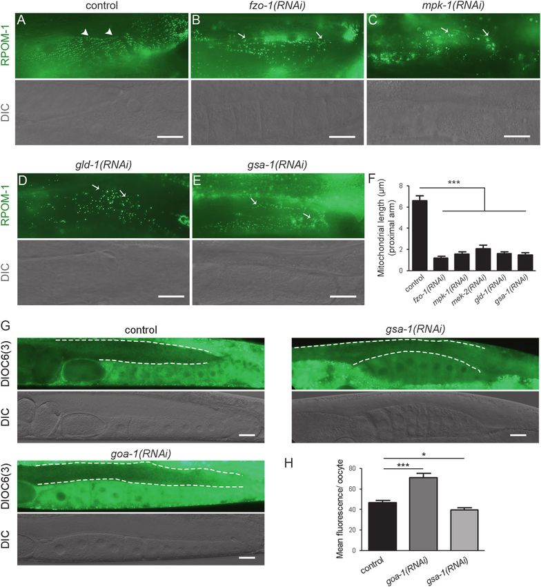
N. Charmpilas, N. Tavernarakis

   A distinguishing characteristic of stem cells relates to   Materials and methods
energy metabolism and mitochondrial function (reviewed
in [6]). Mitochondria originate from endosymbiotic            Strains and genetics
events of ancestral eukaryotic cells with once free-living
proteobacteria [7]. Albeit they principally rely on nuclear   We followed standard procedures for maintaining C. elegans
gene products for their function, they are considered         strains. Rearing temperature was set at 20 °C for most of our
semiautonomous, since they retain their own genome            experiments unless noted otherwise, but was raised at 25 °C
(mtDNA). mtDNA mutations have been associated with            when germline expression was assessed to avoid transgene
diverse human diseases [8]. The human mtDNA encodes           silencing that occurs at lower temperatures. The following
13 electron transport chain (ETC) components, as well as      nematode strains, which are available from CGC, were uti-
two rRNAs (12S and 16S rRNAs) and the whole reper-            lized in this study: N2: wild-type Bristol isolate, CU5991: fzo-
toire of tRNA molecules [9]. These untranslated RNA           1(tm1133) II, CU6372: drp-1(1108) IV, and DA631: eat-3
species, together with exclusively nuclear-encoded            (ad426) II; and him-8(e1489) IV, MT7686: ced-9(n2812)/
mitochondrial ribosomal proteins, form the 55S mito-          qC1 [dpy-19(e1259) glp-1(q339)] III, MT1522: ced-3(n717)
chondrial ribosome, which synthesizes mtDNA-derived           IV, CB1370: daf-2(e1370) III, DR40: daf-1(m40) IV,
ETC components in the organelle [10]. Transcription of        RB1206: rsks-1(ok1255) III, KX80: ife-5(ok1934) II, and
the mitochondrial genome is mediated by a tripartite          CB4037: glp-1(e2141ts) III. We used the JJ1850: unc-119
complex, comprising a dedicated RNA polymerase                (ed3) III; Is[his-72(1kb 5′UTR)::his-72::SRPVAT::GFP::his-
and two auxiliary transcription factors, TFAM and             72 (1KB 3'UTR)] and the RW10226: Is[ppie-1mCherry::H2B::
TFB2M [11–14].                                                pie-1 3′UTR]; Is[phis-72HIS-24::mCherry::let-858 3′UTR]
   Although mitochondria have been implicated in the          strains for monitoring germline nuclei in vivo, the AD189:
maintenance of stemness and the progression toward            unc-119(ed3) III; Is[ppie-1GFP::EGG-1+ unc-119(+)] strain
differentiation, the contribution of the mitochondrial        for monitoring oocyte membranes in vivo and the OD95: unc-
genome remains elusive. To gain relevant insight, we          119(ed3) III; Is[ppie-1mCherry::HIS-58; ppie-1GFP::PH
examined the involvement of mitochondrial genome              (PLC1delta1) +unc-119(+)] strain for simultaneously mon-
expression in C. elegans germ cell homeostasis. We find        itoring oocyte membranes and germline nuclei in vivo.
that perturbations in general mitochondrial transcription     JK4472: WT; Is[plag-2MYR::tdTomato + pttx-3GFP] and
and mitochondrial bioenergetics reduces fecundity and         JK4475: WT; Is[plag-2MYR::GFP + pttx-3DsRed] were used
causes sterility. Depletion of RPOM-1, the homolog of         for monitoring the DTC’s membrane plexus, while DG1575:
the mammalian mitochondrial RNA polymerase, gen-              WT; Is[plim-7GFP + rol-6(su1006)] was used for monitoring
erates a tumor phenotype in the pachytene gonad region        gonad sheath morphology. All the experiments were per-
due to impaired germ cell differentiation, and leads to       formed using day 1 (D1) gravid adult animals that were
gonad collapse at elevated temperatures. Induction of         precisely synchronized at the L4 larval stage one day earlier.
apoptosis acts in a protective, compensatory manner to        For glp-1(e2141) mutants specifically, we placed eggs on
restrict tumor size. Gonadal mitochondrial biogenesis is      plates at the restrictive temperature of 25 °C for 48 h. Then we
coupled with extrinsic Insulin/IGF-1 and TGF-β signal-        transferred L4 larvae on fresh control or rpom-1(RNAi) plates
ing pathways that converge on the germline. Attenuation       and we placed them for one more day at 20 °C prior to
of general protein synthesis in the germline alleviates the   nuclear staining and microscopic observation. Culture of glp-
sequelae of RPOM-1 deficiency. Expression of mito-             1(e2141) mutants at 25 °C for 48 h during this developmental
chondrial RNA polymerase gradually increases in the           window renders glp-1(e2141) mutants completely sterile [15].
course of germ cell differentiation, concurrent with          The following strains were generated in the current study:
morphological and functional adaptations, associated          IR1677: WT; Ex[pCEOP1740RPOM-1::GFP::unc-54 3′UTR],
with enhanced metabolic activity of germline mitochon-        IR1968: unc-119(ed3) III; Ex[pCEOP1740RPOM-1::GFP::
dria. These adaptations are accompanied by increased          rpom-1 3′UTR +unc-119(+)], IR1966: unc-119(ed3) III; and
ATP and ROS production in the proximal gonad                  Is[ppie-1Perceval::tbb-2 3′UTR +unc-119(+)].
arm. Notably, upregulation of mitochondrial RNA
polymerase expression, the consequent establishment of        Molecular cloning
tubular mitochondrial morphology and associated bioe-
nergetic profile are evolutionarily conserved during           Gene inactivation was achieved by bacterial feeding of C.
mouse stem cell differentiation. Thus, coordinated            elegans with RNAi clones expressing double-stranded RNA
expression of the mitochondrial genome is a key com-          targeting the gene of interest. All RNAi treatments were
ponent of a regulatory program that determines germ cell      performed continuously from hatching (L1 larval stage) till
differentiation.                                              the day of observation (D1). The following primers were
Mitochondrial maturation drives germline stem cell differentiation in Caenorhabditis elegans

used for generation of RNAi constructs used in the present                  simultaneously ligated the two inserts with the linearized
study. For rpom-1(RNAi): 5′-ATGAGAAGACTGGAACG                               vector in a single reaction. An extended pie-1 promoter was
AATTGTC-3′ and 5′-TAGTGTTCAATCCCTCACCAAT                                    amplified using the 5′-GTCGACTCGTATTTCTCAGTCA
C-3′, for hmg-5(RNAi): 5′-GGATCCAATGTTGGGAACA                               TTTTTGTG-3′ and 5′-CCATGGATCGTTTTGTATTCT
ATTTC-3′ and 5′-ACCGGTGGTTGATCTGCATTTT                                      GTGTGCTGG-3′ primer pair and was inserted upstream of
C-3′, for tfbm-1(RNAi): 5′-ATGGCTTCTGCTTCACGTCT                             the Perceval::tbb-2 cassette using SalI-NcoI restriction sites.
CC-3′ and 5′-CATCTTAGGCTCTGCCACGTATTG-3′,
for gld-1(RNAi): 5′-CACTCCAACTTACGGTGTTTCG-3′                               Nematode strain generation
and 5′-TCTACCGACGAAGTTATACTGAAATG-3′, for
mpk-1(RNAi): FW: 5′-GTTCATGGGCAACTTTTTGA                                    We used both microinjection into C. elegans gonads and
G-3′ and 5′-GATTACTGAGCATTTCTGCGAG-3′, for                                  bombardment with gold nanoparticles to generate trans-
mek-2(RNAi): 5′-CGTAATCCGTTGGGACTCAG-3′ and                                 genic strains. To avoid undesired transgene silencing in the
5′-CGAGAATCCTGCGAGAACTG-3′, for goa-1(RNAi):                                germline, in microinjections we used low micromolar con-
5′-CCACATACAGTGAGTGAGTAGAG-3′ and 5′-CTCT                                   centrations (5 ng/μL) of both the reporter plasmid and the
CTGTCAGCCGAACC-3′, and for gsa-1(RNAi): 5′-AGCA                             cotransformation marker and an excess of PvuII-digested E.
AAAAAGAACGAGCAAC-3′ and 5′-TGTCCTCCCAGA                                     coli genomic fragments (50 ng/μL), as previously described
GTACAAGA-3′.                                                                [16].
   For cloning the minimal 2.1 kb operon promoter
upstream of CEOP1740 operon, which contains the rpom-1                      Mitochondrial DNA quantification
gene, we used the 5′-CCATGAAATTGAGGATTCTGA
AAC-3′ and 5′- ATTTCCGTTTAGTAGCGATTTTTAAC                                   mtDNA was quantified by quantitative real-time PCR-based
AG-3′ primer pair. The corresponding product was inserted                   method as previously described [17]. The 5′-GTTTATG
in TOPO and then in pPD95.77 linearized with PstI-BamHI                     CTGCTGTAGCGTG-3′ and 5′-CTGTTAAAGCAAGTGG
digestion. For cloning the 3.5 kb rpom-1 full length cDNA,                  ACGAG-3′ primer pair was used for measuring mtDNA
we utilized C. elegans total RNA as a template to synthesize                levels. These primers hybridize in the atp-6 gene which is
the rpom-1 single-strand cDNA with the PrimeScript™                         located in the mitochondrial genome (mtDNA). The results
Reverse Transcriptase kit (Taqara) and the 5′-                              were normalized to genomic DNA amplified with the 5′-
ACCGGTGGACTAAAAAAATAAACAGAATCCTTGA                                          TGGAACTCTGGAGTCACACC-3′ and 5′-CATCCTCCT
C-3′ gene-specific reverse primer. Then, the previous pri-                   TCATTGAACGG-3′ primer pair, which hybridizes to the
mer was used in the same reaction with the 5′-GGATCCA                       genomic region of ama-1 gene. Quantitative PCR was
TGAGAAGACTGGAACGAATTGTC-3′ forward primer                                   performed using a Bio-Rad CFX96 real-time PCR system,
to synthesize the full length rpom-1 cDNA. Expand high-                     and was repeated three times.
fidelity DNA polymerase (Roche) was used in the PCR
reaction to minimize errors. The amplified fragment was                      Measurements of mitochondrial activity
inserted upstream of GFP in the pPD95.77 expression
vector linearized with BamHI-AgeI restriction digestion.                    Staining with mitochondrial dyes (TMRE, Mitotracker
For amplifying the 0.1 kb rpom-1 3′UTR fragment, we used                    ROS) to assess functional mitochondria and ATP mea-
the 5′-GAATTCAGTTAGAAGTGTTTTTTTTGTTG-3′                                     surements in whole animals were performed as previously
and 5′-GGGCCCCATTTTCTGATTCCAGCG-3′ pair of                                  described [18].
primers. Upon ligation into TOPO vector, we isolated the
corresponding fragment using EcoRI-ApaI restriction                         mRNA quantification
digestion. This was then inserted in pPD95.77 linearized
with EcoRI-ApaI digestion downstream of the rpom-1                          Total RNA from synchronized D4 animals was extracted
cDNA::GFP cassette to replace the unc-54 3′UTR fragment.                    using the TRIzol reagent (Invitrogen). The following sets of
For creating the pie-1 promoter Perceval::tbb-2 3′UTR                       real-time primers were used in the current study: for mea-
plasmid, we extracted the Perceval from the pRsetB-his7-                    suring hmg-5 mRNA levels: FW: 5′-CGTCCAAGTGT
Perceval plasmid (catalog number 20336, Addgene) using                      TCCTCCAAGTG-3′ and 5′-CTTCGCTTCGTCTGTGTA
XbaI-XhoI digestion. In parallel, we amplified the tbb-2 3′                  CTTCTTT-3′, for measuring tfbm-1 mRNA levels: 5′-CAC
UTR using the 5′-CTCGAGCAATAAATGCAAGATC                                     AAGAAAGATAGCAAAACACGC-3′ and 5′-CGAGATG
CTTTCAAGC-3′ and 5′-GAATTCTTTCCTCTTTTTGTT                                   CTGTAACGGCGG-3′, and for measuring rpom-1 mRNA
GGGTCACTC-3′ pair of primers. We digested pPD95.77                          levels: 5′-GGTGTCGGCTGGTATCCTCAAC-3′ and 5′-T
with XbaI-EcoRI digestion to remove GFP and we                              GGCACAATCTCCTGAGTAGCC-3′.
N. Charmpilas, N. Tavernarakis

Egg laying assays                                             Immunofluorescence of C. elegans gonads

For assessing egg laying, we placed single, precisely syn-    A large number of synchronized D1 adult worms grown on
chronized gravid adult nematodes (at D1–D2 of adulthood)      NGM plates were washed two repetitive times with M9
in individual 33 mm NGM plates seeded with a standard         buffer and 20 μL of 20 mM levamisole was added on the
OP50 bacterial lawn. The number of eggs that each adult       worm pellet to anaesthetize the animals. About 7–8 μL of
worm produced in the next 12 h were measured. At least 20     the mixture was placed on top of polylysine-treated slides
individual measurements were acquired per experimental        and the animals were dissected using a 8 mm insulin syr-
condition. The egg laying assays were performed at 20 °C,     inge, until the gonads were released from the animals due to
apart from the one corresponding to the mitochondrial         mechanical pressure. The extracted tissue was fixed with
dynamics mutants (fzo-1, drp-1, and eat-3) that were per-     4% paraformaldehyde in PBS for 20 min at room tem-
formed at 25 °C.                                              perature. The fixative was removed by washing the speci-
                                                              men twice with PBS. Then, the gonads were permeabilized
Antimycin A treatment                                         with 0.2% Triton X-100 in PBS 1× for 5 min and rinsed
                                                              twice with PBS. Next, the tissue was incubated in blocking
Antimycin A (catalogue number A8674, Sigma-Aldrich)           solution (1% BSA in PBS-Tween 0.02%) for 1h at room
was supplemented on top of NGM plates seeded with OP50        temperature. The incubation with the rabbit anti-phospho-
E. coli bacterial food at a final concentration of 50 nM per   Histone H3 (Serine 10) (06-570, Sigma-Aldrich) antibody
plate. The plates were previously placed in a UV-             (diluted 1:300 in blocking solution) was performed over-
crosslinker for 15 min to kill off bacteria before Anti-      night in the dark at 4 °C and the slides were sealed to avoid
mycin A addition. L4-staged nematodes were placed on          evaporation. The sample was washed thrice with PBS-
Antimycin A-containing plates for 2 days prior to micro-      Tween 0.02% to remove the primary antibody excess.
scopic observation. The entire experiment was performed at    Finally, the gonads were incubated for 1 h with the anti-
25 °C to avoid undesired silencing of the Perceval-           rabbit IgG AlexaFluor® 488 (catalogue number ab150077,
expressing transgene.                                         Abcam) secondary antibody (diluted 1:500 in PBS-Tween
                                                              0.02%) and DAPI (final concentration 2 μg/mL) to stain
Nuclear staining of nematodes                                 germline nuclei, rinsed three times with PBS and mounted
                                                              with glycerol prior to observation.
We synchronized adult worms of the genotypes of interest
at the first day of adulthood (D1). We stained with Hoechst    Staining germline mitochondria in live animals
33342 to monitor germline nuclei at this stage. First we
washed NGM plates with M9 buffer and collected the            To stain germline mitochondria, mitochondrial dyes were
animals in a 1.5 mL Eppendorf tube. We let the worms          added on top of standard NGM plates seeded with OP50 E.
pellet with gravity and we washed once with PBS-              coli bacterial food. The plates were previously placed in a
Tween20® 0.01%. We centrifuged at 3000 rpm for 1 min          UV-crosslinker for 15 min to kill off bacteria and avoid
to pellet the animals and we removed the supernatant.         undesired catabolism of compounds. TMRE (Tetra-
Fixation was performed with cold methanol 100% for            methylrhodamine, ethyl ester, perchlorate, catalog number
maximum 5 min at −20 °C. The Eppendorf tubes were             T-669; Molecular Probes, Invitrogen) and Mitotracker Red
spinned and the fixative was removed. Upon fixation, cell       CM-H2X ROS (catalog number M-7513; Molecular Probes,
membranes were permeated by washing once with PBS-            Invitrogen) were administered by food in a final con-
Tween20® 0.1%. Upon centrifugation at 3000 rpm for            centration of 1 μΜ per plate and DIOC6(3) (3,3′-Dihex-
1 min, the worm pellet was stained by adding 300 μL           yloxacarbocyanine Iodide, CAS Number 53213-82-4,
of diluted Hoechst 33342 solution (final concentration         Sigma-Aldrich) in a final concentration of 2 μΜ per plate.
1 μg/mL) for 5 min in the dark. The Eppendorf tubes were      The plates were allowed to dry, constantly protected from
centrifuged, the supernatant was removed and the pellet was   light, and then wild-type eggs harvested from hypochlorite-
washed for a final time with PBS-Tween20® 0.1% to              treated, well-fed adult animals were placed on top of the
remove excessive Hoechst stain. The supernatant was           bacterial lawn. The larvae that hatched were fed with the
aspirated; 20 μL 80% glycerol was added in each Eppendorf     dyes for three consecutive days at 25 °C (when His-72::GFP
tube and the samples were mounted in microscopic slides       transgenic animals were used) or for four consecutive days
prior to observation. The transition zone nuclei were dis-    at 20 °C (when wild-type animals were used), until they
tinguished due to the polarized localization of chromosomes   produced D1–2 gravid adult nematodes. Adult animals were
in germ nuclei [19].                                          anaesthetized with 20 mM levamisole and were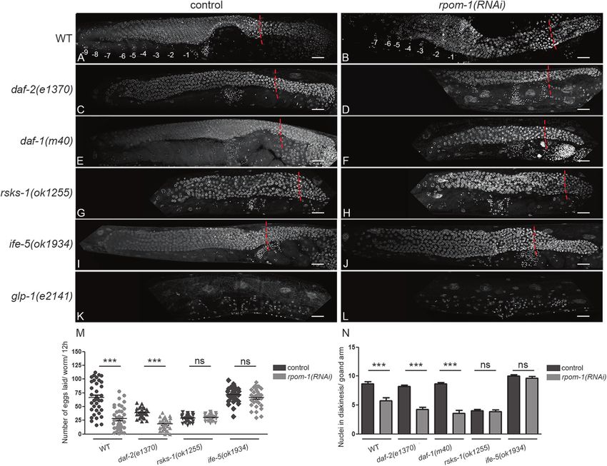
Mitochondrial maturation drives germline stem cell differentiation in Caenorhabditis elegans

microscopically examined using a ZEISS LSM 710 con-                         which shares some properties, but has distinct functions
focal microscope.                                                           from TFB2M [23]. Rpom-1 gene encodes the mammalian
                                                                            POLRMT homolog RPOM-1. Alignment of the catalytic
Mammalian cell culture and immunofluorescence                                domain of RPOM-1 and its putative homologs revealed
                                                                            extensive conservation from yeast to mammals (Fig. S1).
Standard cell culture procedures were followed. J1                          We created RNAi constructs to specifically target hmg-5,
embryonic stem cells were initially cultured for four pas-                  rpom-1 and tfbm-1 transcripts (Fig. S2A), since mutants
sages on top of Mitomycin C-treated mouse fibroblasts and                    for the respective genes are lethal and sterile. We
then on gelatin in LIF cytokine-containing ES medium to                     found that RNAi-mediated knockdown of hmg-5, rpom-1,
preserve their pluripotent identity. Large globular ES                      and tfbm-1 compromised mitochondrial function, as evi-
colonies were acquired. LIF was removed and the ES                          denced by a decrease in TMRE (Tetramethylrhodamine,
medium was replaced with EB medium to favor unbiased                        ethyl ester, perchlorate) and MitoTracker Red CM-
differentiation toward multiple cell lineages. Cells were                   H2XROS stainings, as well as a reduction in ATP levels
grown for up to 48 h on EB medium on 12-well plates on                      (Fig. S2B-D). Importantly, RPOM-1 depletion dramati-
top of cover slips. For immunofluorescence experiments,                      cally reduced mtDNA content, at levels comparable to
cells were washed three times with PBS before they were                     HMG-5 depletion (Fig. S2E). This can be attributed to the
fixed with 4% paraformaldehyde in RT. The cover slips                        fact that POLRMTs also provide RNA primers for the
were washed again three times with PBS. The blocking was                    initiation of mtDNA replication by the mitochondrial-
performed with 0.2% Triton X, 10% FBS for 1 h. Mouse                        specific DNA polymerase. Taken together, these results
monoclonal anti-MTCO1 (catalogue number ab14705,                            highlight the importance of intact transcription machinery
Abcam), rabbit polyclonal anti-POLRMT (catalogue num-                       residing in mitochondria for preserving full organelle
ber PA5-28196, Thermo Scientific), and mouse monoclonal                      function.
anti-OCT4 (catalogue number sc-5279, Santa Cruz Bio-                           We noticed that rpom-1 knockdown dramatically
technology) were used as primary antibodies according to                    reduced the brood size of wild-type nematodes (Fig. 1a,
the manufacturer’s instructions. The appropriate primary                    b). The defect was even more pronounced in subsequent
antibody combination was added in blocking solution and                     generations, indicating the existence of a maternal-effect
was incubated overnight with the specimen at 4 °C. Next                     phenotype (Fig. 1c). Intriguingly, we detected the for-
day, the specimen was rinsed thrice with PBS and was                        mation of germline tumors in the pachytene syncytium
incubated with anti-rabbit IgG AlexaFluor® 488 (catalogue                   region of animals fed with bacteria expressing rpom-1
number ab150077, Abcam), anti-mouse IgG Alexa-                              (RNAi) (Fig. 1d). We reasoned that this phenotype could
Fluor® 594 (catalogue number ab150116, Abcam) fluor-                         be a consequence of elevated mitotic activity in the pro-
escent secondary antibodies and Hoechst 33342 diluted in                    liferative region of the gonad. To test this hypothesis, we
PBS for an hour in RT. Finally, the slides were mounted                     stained extruded gonads from control and rpom-1(RNAi)-
with prolong® gold antifade reagent (catalogue number                       treated animals with an antibody specific to phosphory-
9071, cell signaling) and stored, protected from light at 4 °C              lated serine 10 of histone H3 (anti-pH3), a widely-used
prior to microscopic observation.                                           marker for mitotic-phase nuclei [24, 25]. RPOM-1
                                                                            depletion did not cause any significant change in the
                                                                            number of mitotic nuclei (Fig. 1e, f). We then investi-
Results                                                                     gated whether germ nuclei accumulate due to their
                                                                            inability to properly differentiate and produce oocytes. By
Intact mitochondrial function is indispensable for                          utilizing a fluorescent reporter strain for EGG-1, a protein
fecundity in C. elegans                                                     that localizes to oocyte membranes, we detected three
                                                                            times less EGG-1::GFP positive oocytes in rpom-1
The mitochondrial genome of C. elegans encodes 12 ETC                       (RNAi)-treated animals compared to their control coun-
protein-coding genes lacking the ATP8 gene of its human                     terparts, as well as less nuclei in diplotene and diakinesis
counterpart [20]. The following proteins have been pro-                     (Fig. 1g–i). Furthermore, we did not observe any gross
posed to engage in mtDNA metabolism: HMG-5, a puta-                         morphological defect in somatic tissues that support the
tive nematode homolog to the mammalian TFAM, is a                           gonads, such as the gonad sheath (Fig. S3Α) and the DTC
high-mobility group protein that regulates mitochondrial                    (Fig. S3Β) or any change in the number of sperm nuclei
DNA content and has important roles in replication,                         that the animals produce (Fig. S3C, D). These observa-
transcription, and packaging of mtDNA into nucleoids                        tions confirm that impaired differentiation of germ cells to
[21, 22]. C. elegans genome encodes a TFB1M homolog,                        oocytes leads to germline tumor development in rpom-1
a protein with 16S rRNA methyltransferase activity,                         (RNAi)-treated animals.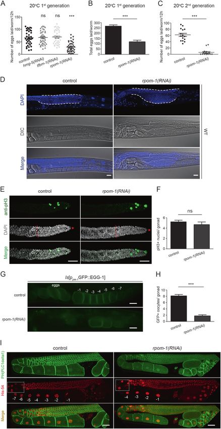
N. Charmpilas, N. Tavernarakis
Mitochondrial maturation drives germline stem cell differentiation in Caenorhabditis elegans
Fig. 1 RPOM-1 depletion causes germline tumor formation in C. ele- Germline apoptosis counterbalances tumor
gans. a, b The brood size of rpom-1(RNAi)-treated hermaphrodites is formation
significantly reduced (up to 50%) compared to their control counterparts.
Unpaired t-test was used for the estimation of statistical significance (n >
40; ***P < 0.001). c Egg laying measurement of animals treated with We surveyed for cellular responses triggered by aberrant
control or rpom-1(RNAi) for two subsequent generations. d DAPI staining mitochondrial biogenesis. RPOM-1 depletion resulted in
of day 1 control and rpom-1(RNAi)-treated WT animals. Inhibition of accumulation of apoptotic corpses in the gonad syncytium,
mitochondrial transcription results in germ nuclei arrest in the pachytene
which were clearly evident using differential interference
region of prophase I in the germline syncytium. The dashed lines sur-
round the germline syncytium. e Phosphorylated histone H3 antibody contrast (DIC; Nomarski) microscopy and a CED-1::GFP
staining of extruded gonads for the detection of mitotic nuclei in the distal reporter strain (Fig. 2a). CED-1 is a transmembrane receptor
gonad arm. Red dashed lines highlight the border between the mitotic which normally clusters around apoptotic corpses before
region and the transition zone, marked by the appearance of crescent-
they are engulfed and removed by gonad sheath cells
shape nuclei, while the red asterisk marks the relative position of the distal
gonad arm tip. f Quantification of phosphorylated histone H3 positive [26, 27]. Furthermore, both HMG-5 and TFBM-1 depletion
germ nuclei in control and rpom-1(RNAi)-treated hermaphrodites. efficiently induced apoptosis, although to a lesser extent
g Representative images of EGG-1 positive oocytes in the proximal arm than rpom-1(RNAi) (Fig. 2b), indicating that distinct signals
from control and RPOM-1-depleted gonads. h Quantification of EGG-1
emanating from dysfunctional mitochondria may trigger
positive oocytes in control and rpom-1(RNAi)-treated hermaphrodites.
i Confocal image of day 1 transgenic worms expressing a cell membrane programmed cell death. Inhibition of apoptosis in homo-
tagged GFP and a histone-54 fused mCherry. Animals treated with rpom- zygous ced-3/caspase mutants can lead to the accumulation
1(RNAi) display a lower number of nuclei in diplotene (compare dashed of germ cell corpses that cannot be removed [28]. Notably,
rectangles) and fewer mature oocytes in diakinesis. −1 denotes the most
rpom-1 knockdown in ced-3(n717) homozygous mutants
proximal oocyte. Unpaired t-test was used for the estimation of statistical
significance (n > 40; ***P < 0.001). Error bars, s.e.m. Images were aggravated the pachytene arrest phenotype observed in
acquired using a ×40 objective lens. Scale bars, 20 μm wild-type nematodes. By contrast, loss of function mutants
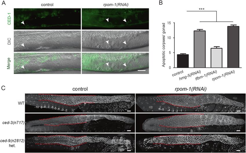
Fig. 2 Induction of apoptosis alleviates germline tumor development. for multiple comparisons). c RPOM-1 depletion in ced-3(n717)/cas-
a Apoptosis induction following rpom-1 downregulation, as monitored pase-deficient animals causes pronounced tumor formation, even more
using the CED-1::GFP reporter in combination with DIC microscopy. severe than in wild-type worms. Heterozygous ced-9(n2812)/BCL-2
Arrows highlight apoptotic corpses in the syncytium area. b A two- animals exhibit no sign of germ nuclei arrest in pachytene. The red
fold induction in the number of early apoptotic corpses can be detected dashed lines surround the pachytene region of the gonads. −1 denotes
upon RPOM-1 depletion. Hmg-5 and tfbm-1 downregulation also the most proximal oocyte. Images were acquired using a ×40 objective
trigger apoptosis (n = 40; ***P < 0.001, one-way ANOVA was used lens. Error bars, s.e.m. Scale bars, 20 μm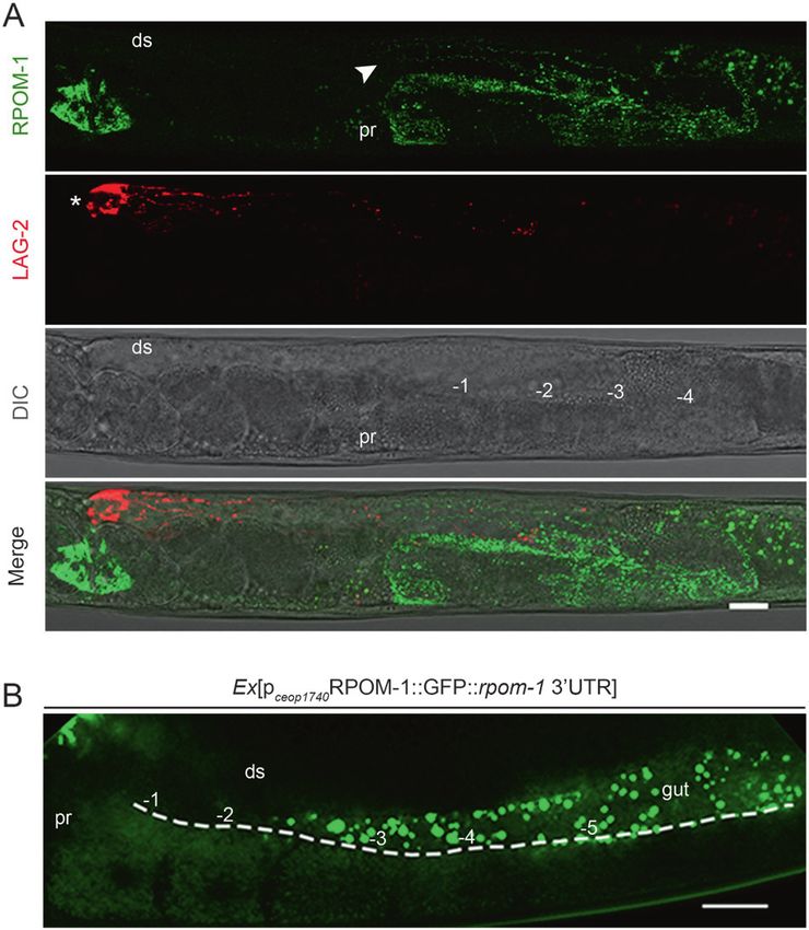
N. Charmpilas, N. Tavernarakis

for ced-9(n2814)/BCL-2, where apoptosis is induced,                      (Fig. 3a, b), it differentially affected daf-2(e1370)/IGFR
exhibit no signs of tumor formation upon rpom-1 down-                    mutants by producing dwarf gonads at permissive tempera-
regulation (Fig. 2c). Hence, induction of apoptosis com-                 tures (20 °C) and aggravating their proliferative defects
pensates for the tumor phenotype caused by the inhibition                (Fig. 3c, d). TGF-β signaling is also reported to affect the
of mitochondrial transcription.                                          balance between mitosis and differentiation in the C. elegans
                                                                         germline, in response to environmental cues, such as con-
Mitochondrial transcription acts in parallel with                        centration of dauer pheromone or population density [30].
signaling pathways converging on the germline                            RPOM-1 depletion in daf-1(m40)/TGFR mutants behaved
                                                                         similarly to daf-2(e1370)/IGFR mutants, since it produced
We next investigated the impact of RPOM-1 depletion in                   gonads with further compromised proliferative potential
mutants with reported defects in germline homeostasis.                   compared to their respective controls (Fig. 3e, f). In sharp
Insulin/IGF-1 signaling promotes proliferation of germline               contrast the gonads of rsks-1(ok1255)/S6K mutants, which
stem cells [29]. While rpom-1 downregulation caused tumor                exhibit attenuated global protein synthesis, were identical to
formation in the germline syncytium of wild-type animals                 those of control animals (Fig. 3g, h). Importantly, ife-5

Fig. 3 Mitochondrial transcription acts in parallel with signaling       counterparts upon treatment with rpom-1(RNAi). k, l Glp-1/ Notch loss
pathways converging on the germline. Hoechst 33342 staining of D1        of function produces germline-less animals at restrictive temperatures.
adult animals in control conditions and upon rpom-1 silencing. a, b      The red dashed lines indicate the border between the mitotic region
Inhibition of mitochondrial transcription causes pachytene arrest in     and the transition zone. m Egg laying measurements in wild-type, daf-
otherwise wild-type animals. In contrast, treatment of daf-2/ IGFR (c,   2(e1370), rsks-1(ok1255) and ife-5(ok1934) mutants. n Quantification
d) and daf-1/ TGFR (e, f) homozygous mutants with rpom-1(RNAi) at        of the number of germ nuclei reaching diakinesis in wild-type, daf-2
20 °C produces dwarf gonads and augments their reported defects.         (e1370), daf-1(m40), rsks-1(ok1255) and ife-5(ok1934) genetic back-
Mutants with attenuated protein synthesis rates, such as rsks-1/ S6K     grounds. (n > 40; ***P < 0.001, unpaired t-test). Error bars, s.e.m.
(g, h) and ife-5/eIF4E (i, j) are indistinguishable from their control   Images were acquired using a ×40 objective lens. Scale bars 20 μm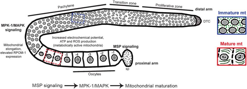
Mitochondrial maturation drives germline stem cell differentiation in Caenorhabditis elegans

(ok1934)/eIF4E mutants, similarly to rsks-1(ok1255) mutant
animals, remained unaffected by RPOM-1 depletion (Fig. 3i,
j). Furthermore, the egg-laying defect caused by RPOM-1
deficiency was completely absent in protein synthesis-
defective nematodes, as evident by counting the brood size
of individual animals and the number of diakinesis-staged
nuclei (Fig. 3m, n). This indicates that reduced protein
synthesis provides a selective advantage under conditions of
impaired mitochondrial ATP production. Finally, rpom-1
(RNAi)-treated glp-1(e2141)/Notch loss of function mutants
was indistinguishable from controls under restrictive
temperatures, producing germline-less hermaphrodites
(Fig. 3k, l). Numerous mutant strains for vital germline
components, such as P-granules, are fully fertile at standard
growth conditions (20 °C), but become sterile following a
switch to a higher temperature (25 °C) [31]. The defects
accompanying RPOM-1 depletion became more prominent
when the animals were raised at 25 °C. The gonads at that
temperature virtually collapse, barely produce a few diplotene
and diakinesis-staged germ nuclei and accumulate dead
corpses (Fig. S4). Together, these findings suggest that the
precise coordination of signaling pathways (Insulin/IGF-1,
                                                                            Fig. 4 Rpom-1 expression is compartmentalized. a RPOM-1 abun-
TGF-β, and Notch/Delta) and internal gonad processes                        dance (in green) is low in the distal arm and the mitotic region of the
(mitochondrial transcription and protein synthesis) shapes the              gonads, becomes evident at the onset of the pachytene region
balance between mitosis and meiosis in the C. elegans                       (arrowhead) and substantially increases close to the turn and in the
germline.                                                                   proximal arm, where the oocytes mature. LAG-2::myr::tdTomato (in
                                                                            red) marks the distal tip cell (white star) and its membrane projections.
                                                                            b A reporter strain overexpressing RPOM-1 under the control of its
rpom-1 expression is compartmentalized                                      endogenous promoter and 3′UTR reveals higher expression in the
                                                                            oocytes and lower in the syncytium. Ds; distal, pr; proximal, −1
To monitor rpom-1 expression in vivo, we generated a                        denotes the most proximal oocyte. Images were acquired using ×40
                                                                            and ×63 objective lenses. Scale bars, 20 μm
translational reporter by fusing GFP to the carboxyl termi-
nus of full-length rpom-1 cDNA regulated by its endo-
genous operon promoter. rpom-1 is expressed in several                      UTR. On a similar note, RPOM-1 was enriched in the
somatic tissues, including muscles, intestine, vulva, and                   oocytes of the proximal gonad arm (Fig. 4b). The expres-
neuronal cells in the nerve ring and the tail, in a pattern                 sion of the operon’s promoter per se gradually increases in a
reminiscent of proteins localizing in the mitochondrial                     distal to proximal manner (Fig. S7B). IFET-1, which loca-
matrix (Fig. S5). rpom-1 is also strongly expressed in the                  lizes to P-granules and is required for normal gonad
germline of hermaphrodite animals in a punctuate pattern.                   development, is a general translational repressor in the
In the gonads, mitochondria surround and enwrap germ cell                   germline [32]. Treatment of our reporter animals with ifet-1
nuclei in the syncytium region (Fig. S6A, B). We observed                   (RNAi) de-repressed rpom-1 expression distally (Fig. S7C).
a clear colocalization of RPOM-1 with TMRE, a potential-                    Hence, rpom-1 expression in the germline is compartmen-
dependent mitochondrial dye (Fig. S6C). We crossed                          talized, increases as germ nuclei progress to the proximal
RPOM-1::GFP transgenic animals with plag-2MYR::tdTo-                        arm and form oocytes and is directly regulated by IFET-1 at
mato reporter animals, to visualize RPOM-1 expression in                    the translational level.
regard to the position of the DTC. We noticed that rpom-1
expression is low in the mitotic region distally, but increases             Transition to tubular mitochondria is a hallmark of
profoundly as germ cell nuclei mature to form oocytes                       differentiation
(Fig. 4a, Fig. S7A). Interestingly, RPOM-1 expression
increases at the onset of pachytene, the exact same region                  We noticed that mitochondrial morphology alters in the
where germ nuclei arrest upon RPOM-1 depletion. To                          course of germ cell differentiation. The distal gonad arm
achieve a faithful reconstitution of the endogenous rpom-1                  was abundant with globular mitochondria, while elongated
expression pattern, we also generated a reporter strain that                organelles prevailed in the proximal arm (Fig. 5a, e). In the
expresses a transgene carrying the endogenous rpom-1 3′                     gonad turn both globular and tubular mitochondria coexist
N. Charmpilas, N. Tavernarakis (Fig. 5b). In that region, an actin-dependent cytoplasmic elongated mitochondria are associated with elevated ETC streaming deposits cytoplasmic material and mitochondria activity and vice versa [34, 35]. Mitochondrial shape is to the oocytes [33]. Accumulating evidence suggests that malleable and alters to fulfill physiological demands, in
Mitochondrial maturation drives germline stem cell differentiation in Caenorhabditis elegans
Fig. 5 Transition from globular to tubular mitochondria is a pre- prevents oocyte maturation [40]. Interestingly, mitochon-
requisite for germline homeostasis. a Confocal image of an adult C. dria failed to elongate in the proximal arm of gsa-1(RNAi)-
elegans gonad. In the distal arm, mitochondria have a globular shape
treated animals (Fig. 6a, e, f) and the existing organelles
(arrow), which gradually switches to a more elongated/ tubular one in
the oocytes of the proximal arm (arrowhead). b The turn of the gonad, were not efficiently polarized (Fig. 6g, h). Conversely,
shown in magnification, is the site where the shape alteration occurs. inhibition of GOA-1, a negative regulator of oocyte
There, both globular (arrow) and tubular (arrowhead) mitochondria maturation, boosted mitochondrial potential in the proximal
can be observed. c DAPI staining of wild-type and fzo-1(tm1133)/
arm (Fig. 6g, h). Altogether, these findings suggest that
Mitofusin homozygous mutants. The red dashed line marks the gonad
turn. d Day 1 adult fzo-1(tm1133)/Mitofusin mutant animals, bearing germ nuclei differentiation is intertwined with mitochon-
defects in mitochondrial fusion, produce significantly fewer germ drial maturation and the latter appears to rely on two
nuclei in diplotene as well as nuclei in diakinesis compared to their important signaling pathways, namely MPK-1/MAPK and
control counterparts. (n > 40; ***P < 0.001, unpaired t-test). e Quan-
MSP.
tification of mitochondrial length in the proximal and distal arm of the
gonads. f Animals with perturbed mitochondrial dynamics (fusion-
fission), such as fzo-1, drp-1 and eat-3 mutants become sterile when Mitochondria functionally mature en route to germ
exposed to a mild heat stress (25 °C). One-way ANOVA was used for cell differentiation
the estimation of statistical significance (n > 40; ***P < 0.001). Error
bars, s.e.m. Images were acquired using X40 and X63 objective lenses.
Ds; distal, pr; proximal, −1 denotes the proximal-most oocyte. Scale The established interplay between mitochondrial morphology
bars, 20 μm and metabolic activity [34] prompted us to test mitochondrial
maturation within the germline. We cloned Perceval, a fluor-
response to stress and other intracellular or environmental escent sensor for adenylate nucleotides [41], downstream of
signals. Specialized dynamin GTPases FZO-1/Mitofusin and the pie-1 promoter, to uniformly express it in the germline. We
EAT-3/OPA-1 mediate fusion, while DRP-1 is required for also utilized the tbb-2 3′UTR to avoid undesired silencing of
fission [36]. Interestingly, fzo-1(tm1133) mutant animals our transgene [42]. Interestingly, we detected expression in the
possess significantly less germ nuclei in diplotene as well as oocytes, but not in the gonad syncytium (Fig. 7a). This is
oocytes in diakinesis, producing fewer offspring (Fig. 5c, d). consistent with the notion that ATP is produced proximally, in
Furthermore, while fzo-1(tm1133) and drp-1(tm1108) the area that is abundant with tubular mitochondria. Treatment
mutants were viable and fertile at 20oC, they became sterile with Antimycin A, a bacterial toxin that inhibits ETC complex
when the rearing temperature was shifted to 25oC, producing III, dampened fluorescence in oocytes and fertilized eggs,
mainly dead eggs and negligible offspring (Fig. 5f). Toge- proving that Perceval can indeed detect changes in mito-
ther, these findings support the notion that tubular mito- chondrial ATP production (Fig. S8). To further verify the
chondria in the proximal arm represent the outcome of a previous finding, we stained whole animals with dyes that
maturation process, which is essential for oocytes to cope stain mitochondria in a membrane potential-dependent man-
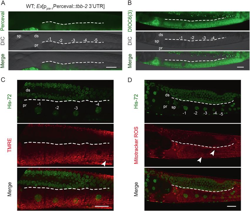
with their high-energy demands. ner, such as TMRE, DIOC6(3) (3,3′-Dihexyloxacarbocyanine
To shed light on the molecular mechanism that governs Iodide), and MitoTracker Red CM-H2XROS. In congruence
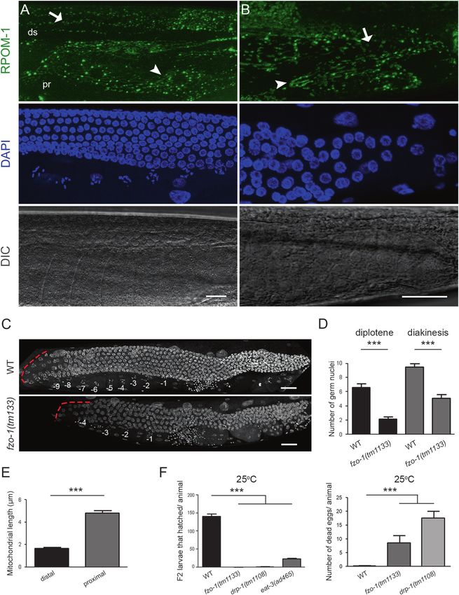
the alteration of mitochondrial morphology, we focused on with Perceval findings, mitochondrial membrane potential and
well characterized pathways associated with germ cell dif- ROS levels were elevated in developing oocytes (Fig. 7b–d).
ferentiation in C. elegans. Spatial activation of MPK-1/ Interestingly, rpom-1 knockdown inhibited mitochondrial
MAPK signaling is crucial for germ cell exit from pachy- polarization in the proximal gonad arm as indicated by DIOC6
tene. MPK-1 deficient mutants exhibit pachytene arrest, (3) staining (Fig. S9). We postulate that mitochondrial elon-
phenocopying RPOM-1 depletion [37]. Interestingly, tub- gation is a primary step in a maturation process, which results
ular organelles were absent upon knockdown of mek-2 or in enhanced mitochondrial polarization and concomitant ATP
mpk-1, the homologs of mammalian MEK and ERK kina- and ROS production in oocytes.
ses, respectively (Fig. 6a, c, f). GLD-1 is an RNA-binding
protein that binds to the 3′UTR of target mRNAs, repres- Elevated POLRMT expression and switch to tubular
sing their translation. In gld-1 loss of function mutants germ mitochondria are conserved during evolution
nuclei exit meiosis and return to mitosis, forming germline
tumors [38]. Similarly to FZO-1/Mitofusin depleted ani- We next wondered whether a similar mechanism controls
mals, gld-1(RNAi)-treated animals contain exclusively mouse stem cell differentiation. We employed J1 cells,
globular mitochondria in the proximal gonad arm (Fig. 6a, derived from the inner cell mass of mouse blastocysts and
b, f). Gonad sheath cells are known to respond to sperm- grown on spherical colonies in the presence of LIF cyto-
derived signals (major sperm proteins) and promote oocyte kine. We removed LIF from our culture medium and let the
maturation through activation of the Gαs-adenylate cyclase- cells differentiate in an unbiased manner. We stained with
protein kinase A pathway [39]. Inhibition of gsa-1, the antibodies for POLRMT and MTCO1, the cytochrome c
worm Gs alpha subunit of heterotrimeric G proteins, oxidase subunit I encoded by the mitochondrial genome.N. Charmpilas, N. Tavernarakis Fig. 6 Sperm-derived signals promote mitochondrial maturation. one-way ANOVA was used for multiple comparisons). g DIOC6(3) a Mitochondria in the proximal gonad arm are tubular under control staining reveals that mitochondria polarize in the course of germ cell conditions (arrowheads). b Knockdown of fzo-1 leads to mitochondrial differentiation. Inhibition of GOA-1, a negative regulator of sperm network fragmentation and globular mitochondria in the proximal signaling, boosts mitochondrial potential in the proximal gonad arm. gonad arm (arrows). c Inhibition of MPK-1/MAPK signaling via mpk- Treatment with gsa-1(RNAi) results in a failure of mitochondria to 1(RNAi) results in a failure of mitochondria to elongate proximally polarize proximally. h Quantification of the DIOC6(3) fluorescence (arrows). d Mitochondria are exclusively globular in the proximal arm per oocyte of the proximal gonad arm. The dashed lines surround the of gld-1(RNAi)-treated animals (arrows). e GSA-1 inhibition results in germline syncytium. (n = 40; ***P < 0.001, unpaired t-test). Error failure of oocyte maturation as well as mitochondrial elongation. bars, s.e.m. Images were acquired using a X40 objective lens. Scale f Quantification of mitochondrial length in proximal gonad arm bar, 20 μm oocytes upon the respective RNAi treatments (n = 40; ***P < 0.001,
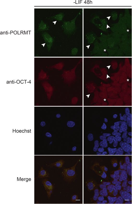
Mitochondrial maturation drives germline stem cell differentiation in Caenorhabditis elegans

Fig. 7 Mitochondria functionally mature during germ nuclei differ-          proximal gonad arm. c TMRE staining reveals increased electro-
entiation. a The ATP/ADP sensor Perceval was overexpressed in the           chemical potential in the oocytes of the proximal arm. d Mitochondrial
C. elegans germline under the control of pie-1 promoter, to achieve         ROS production increases as the germ nuclei mature and give rise to
germline-specific expression. Perceval emission increases upon ATP           oocytes. Arrowheads highlight tubular mitochondria in the proximal
binding. Fluorescence could be mainly detected in the oocytes, indi-        gonad arm. Sp; spermatheca, ds; distal, pr; proximal, −1 denotes the
cating increased ATP production in the proximal arm. b DIOC6(3)             most proximal oocyte. Images were acquired using ×40 and ×63
mitochondrial dye preferentially stains energized mitochondria in the       objective lenses. Scale bars, 20 μm

The staining was much weaker at the core of the stem cell                   in line with the C. elegans findings, mouse stem cells
colonies and progressively increased as cells differentiated                exhibit low POLRMT expression, while differentiation is
and extended membrane projections typical of differentiated                 accompanied by an increase in POLRMT expression and
cells. Furthermore, 48 h after LIF removal, we observed                     the appearance of elongated mitochondria.
POLRMT-positive tubular mitochondria (Fig. S10). We
also simultaneously stained with POLRMT and OCT-4
antibodies. OCT-4 is a key pluripotency transcription factor                Discussion
that shuttles between the nucleus and the cytoplasm. Its
nuclear retention enhances reprogramming efficiency and is                   The term stem cell niche refers to the specific micro-
associated with pluripotency [43]. Notably, OCT-4 reten-                    environment, which ensures that stem cells are protected
tion in the nucleus was associated with reduced POLRMT                      from harmful agents, divide and differentiate to constantly
expression, while cytoplasmic OCT-4 coincided with                          replenish organs [44]. Each C. elegans gonad hosts a unique
increased POLRMT expression (Fig. 8). Collectively, and                     stem cell niche in an otherwise postmitotic organism [45].
N. Charmpilas, N. Tavernarakis

                                                                     facilitate oocyte membrane organization [49]. MPK-1
                                                                     phosphorylation itself is positively regulated by MSP sig-
                                                                     naling [50]. Thus, mitochondrial bioenergetics is likely a
                                                                     nodal point modulated by these cascades, in stem cells.
                                                                         Our findings indicate that expression of mitochondrial
                                                                     RNA polymerase progressively increases during germ cell
                                                                     differentiation. This is accompanied by mitochondrial
                                                                     elongation and manifestation of several hallmarks of
                                                                     metabolic activity, such as increased electrochemical
                                                                     potential, ATP, and ROS production. Rpom-1 mRNA is one
                                                                     of the numerous targets of FBF-1, a Pumilio family, RNA-
                                                                     binding protein that negatively regulates the expression of
                                                                     mRNAs implicated in meiotic entry [51, 52]. In addition,
                                                                     localized transcription and mRNA translation likely con-
                                                                     tribute to the subcellular compartmentalization of RPOM-1.
                                                                     Taken together, our observations indicate that upon per-
                                                                     turbation of germ cell bioenergetics, germ nuclei stall in the
                                                                     pachytene stage and fail to differentiate, generating fewer
                                                                     oocytes. Consistent with this notion, mitochondrial ATP
                                                                     synthase function is required for the maturation of mito-
                                                                     chondrial cristae in Drosophila ovaries [53]. Similarly, the
                                                                     pluripotent state of mammalian stem cells has been linked to
                                                                     decreased mitochondrial respiration, in favor of anaerobic
                                                                     glycolysis [54]. Previous studies have demonstrated that
                                                                     mitochondrial mass, mtDNA copy number, and oxygen
                                                                     consumption increase during stem cell differentiation [55–
                                                                     57]. By contrast, successful induction of pluripotent stem
                                                                     cell (iPSC) lines is marked by a reduction of ETC function
                                                                     [58]. Hence, a switch to enhanced mitochondrial respiration
Fig. 8 Mammalian stem cell differentiation upon LIF removal is       is a prerequisite for stem cell differentiation across species.
accompanied by an increase in POLRMT expression. Elevated
POLRMT expression is observed in cells with increased cytoplasmic        Conserved signaling pathways implicated in lifespan reg-
OCT-4 abundance (arrowheads). In contrast, adjacent areas with       ulation and dauer formation, also influence germline home-
increased nuclear OCT-4 abundance (stars) display lower POLRMT       ostasis. For instance, Insulin/IGF-1 promotes germ cell
expression. Images were acquired using a ×40 objective lens. Scale   proliferation, while DAF-16/FOXO is beneficial for stem cell
bars, 20 μm
                                                                     pool maintenance during ageing [29, 59]. Furthermore,
                                                                     TORC1 and RSKS-1/S6K are required for efficient pro-
This is the main tissue where mitochondrial DNA replica-             liferation of germ cell progenitors [60]. In addition, ASI
tion occurs [46]. A previous study showed that mutation of           neuron-derived TGF-β signals determine the balance between
a germline-specific mitochondrial ATPase subunit impairs              mitosis and differentiation in the C. elegans germline [30].
fecundity [47]. We find that perturbation of mitochondrial            Attenuation of Insulin/IGF-1 and TGF-β signaling, combined
biogenesis, energy production, and dynamics, collectively            with perturbation of mitochondrial transcription generates an
referred to as bioenergetics, can profoundly affect germ cell        atrophic germline, and exacerbates proliferation defects,
differentiation. We describe a maturation process, whereby           indicating that mitochondria act in concert with extrinsic
globular, immature mitochondria are gradually converted to           growth stimuli to dictate mitosis versus differentiation deci-
elongated, functional organelles to support increased oocyte         sions. Similarly, energy is diverted to stress resistance and
energy demands (Fig. 9). This switch is tightly regulated by         maintenance mechanisms in mutants with reduced protein
two core signaling pathways associated with oocyte pro-              synthesis [61, 62]. In this context, as the disposable soma
duction and maturation, namely MAPK/ERK and MSP. The                 concept postulates, damage repair takes precedence over
MAPK/ERK pathway has pleiotropic functions in the C.                 protein synthesis for germline maintenance and reproduction.
elegans germline [37]. The DDX-19 helicase and GSK-3                     The precise balance between mitosis and differentiation
kinase have been shown to be direct MPK-1 targets in vivo            is of utmost importance for tissue and organismal home-
[48]. In addition, MPK-1 phosphorylates NOS-3, promoting             ostasis. Our work provides novel insights on how mito-
degradation of TRA-1 by the FEM-CUL2 complex, to                     chondrial bioenergetics dictates cell fate decisions and
Mitochondrial maturation drives germline stem cell differentiation in Caenorhabditis elegans
Fig. 9 Intact mitochondrial bioenergetics safeguards germline home- metabolic activity is manifested by enhanced ATP and ROS produc-
ostasis. Expression of rpom-1 (the nematode orthologue of POLRMT) tion, as well as increased electrochemical potential in the proximal
increases progressively as the germ nuclei mature and mitochondria gonad arm. Mitochondrial maturation is under the control of MPK-1/
acquire an elongated, tubular shape. The boost in mitochondrial MAPK and MSP signaling pathways
integrates mitochondria at the core of the developmental holder. To view a copy of this license, visit http://creativecommons.
org/licenses/by/4.0/.
modules that shape the C. elegans germline. Perturbation of
mitochondrial function obstructs germ nuclei differentiation
and causes cancer-like phenotypes. A main challenge for References
future research is to delineate the molecular underpinnings
1. Byrd DT, Knobel K, Affeldt K, Crittenden SL, Kimble JA. DTC
of the germline mitochondrial metabolic switch as well as niche plexus surrounds the germline stem cell pool in Cae-
its temporal and spatial regulation. norhabditis elegans. PLoS ONE. 2014;9:e88372.
2. Fox PM, Schedl T. Analysis of germline stem cell differentiation
Acknowledgements We thank A. Pasparaki and G. Tsikala for following loss of GLP-1 notch activity in Caenorhabditis elegans.
excellent technical support. We acknowledge E. Kyriakakis, K. Pali- Genetics. 2015;201:167–84.
karas, and M. Markaki for critically reading the manuscript. We thank 3. Crittenden SL, Leonhard KA, Byrd DT, Kimble J. Cellular ana-
G. Yellen and Addgene for providing the pRsetB-his7-Perceval plas- lyses of the mitotic region in the Caenorhabditis elegans adult
mid. Nematode strains used in this work were provided by the Cae- germ line. Mol Biol Cell. 2006;17:3051–61.
norhabditis Genetics Center, which is funded by the National Centre 4. Hirsh D, Oppenheim D, Klass M. Development of the reproduc-
for Research Resources of the National Institutes of Health, and S. tive system of Caenorhabditis elegans. Dev Biol.
Mitani (National Bioresource Project) in Japan. We thank A. Fire for 1976;49:200–19.
plasmid vectors. This work was supported by the European Research 5. Albert Hubbard EJ. Caenorhabditis elegans germ line: a model for
Council (GA695190- MANNA), and the General Secretariat for stem cell biology. Dev Dyn. 2007;236:3343–57.
Research and Technology of the Greek Ministry of Education 6. Folmes CD, Ma H, Mitalipov S, Terzic A. Mitochondria in
(THALIS MIS380228 GEnAge). pluripotent stem cells: stemness regulators and disease targets.
Curr Opin Genet Dev. 2016;38:1–7.
7. Archibald John M. Endosymbiosis and eukaryotic cell evolution.
Compliance with ethical standards Curr Biol. 2015;25:R911–R21.
8. Tuppen HAL, Blakely EL, Turnbull DM, Taylor RW. Mito-
Conflict of interest The authors declare that they have no conflict of chondrial DNA mutations and human disease. Biochim et Bio-
interest. phys Acta. 2010;1797:113–28.
9. Schon EA, DiMauro S, Hirano M. Human mitochondrial DNA:
Publisher’s note: Springer Nature remains neutral with regard to roles of inherited and somatic mutations. Nat Rev Genet.
jurisdictional claims in published maps and institutional affiliations. 2012;13:878–90.
10. Amunts A, Brown A, Toots J, Scheres SHW, Ramakrishnan V.
Open Access This article is licensed under a Creative Commons The structure of the human mitochondrial ribosome. Science.
Attribution 4.0 International License, which permits use, sharing, 2015;348:95–8.
adaptation, distribution and reproduction in any medium or format, as 11. Ringel R, Sologub M, Morozov YI, Litonin D, Cramer P,
long as you give appropriate credit to the original author(s) and the Temiakov D. Structure of human mitochondrial RNA polymerase.
source, provide a link to the Creative Commons license, and indicate if Nature. 2011;478:269–73.
changes were made. The images or other third party material in this 12. Litonin D, Sologub M, Shi Y, Savkina M, Anikin M, Falkenberg
article are included in the article’s Creative Commons license, unless M, et al. Human mitochondrial transcription revisited: only TFAM
indicated otherwise in a credit line to the material. If material is not And TFB2M are required for transcription of the mitochondrial
included in the article’s Creative Commons license and your intended genes in vitro. J Biol Chem. 2010;285:18129–33.
use is not permitted by statutory regulation or exceeds the permitted 13. Shi Y, Dierckx A, Wanrooij PH, Wanrooij S, Larsson N-G,
use, you will need to obtain permission directly from the copyright Wilhelmsson LM, et al. Mammalian transcription factor A is aN. Charmpilas, N. Tavernarakis
core component of the mitochondrial transcription machinery. 33. Wolke U, Jezuit EA, Priess JR. Actin-dependent cytoplasmic
Proc Natl Acad Sci USA. 2012;109:16510–5. streaming in C. elegans oogenesis. Development.
14. Sologub M, Litonin D, Anikin M, Mustaev A, Temiakov D. TFB2 2007;134:2227–36.
is a transient component of the catalytic site of the human mito- 34. Mishra P, Chan DC. Metabolic regulation of mitochondrial
chondrial RNA polymerase. Cell. 2009;139:934–44. dynamics. J Cell Biol. 2016;212:379–87.
15. Berman JR, Kenyon C. Germ-cell loss extends C. elegans life 35. Liesa M, Shirihai Orian S. Mitochondrial dynamics in the reg-
span through regulation of DAF-16 by kri-1 and lipophilic- ulation of nutrient utilization and energy expenditure. Cell Metab.
hormone signaling. Cell. 2006;124:1055–68. 2013;17:491–506.
16. Kelly WG, Xu S, Montgomery MK, Fire A. Distinct require- 36. Youle RJ, van der Bliek AM. Mitochondrial fission, fusion, and
ments for somatic and germline expression of a generally stress. Science. 2012;337:1062–5.
expressed Caernorhabditis elegans gene. Genetics. 1997; 37. Lee M-H, Ohmachi M, Arur S, Nayak S, Francis R, Church D,
146:227–38. et al. Multiple Functions and Dynamic Activation of MPK-1
17. Cristina D, Cary M, Lunceford A, Clarke C, Kenyon C. A regu- Extracellular Signal-Regulated Kinase Signaling in Caenorhabdi-
lated response to impaired respiration slows behavioral rates and tis elegans Germline Development. Genetics. 2007;177:2039–62.
increases lifespan in Caenorhabditis elegans. PLoS Genet. 38. Francis R, Barton MK, Kimble J, Schedl T. gld-1, a tumor sup-
2009;5:e1000450. pressor gene required for oocyte development in Caenorhabditis
18. Palikaras K, Lionaki E, Tavernarakis N. Coordination of mito- elegans. Genetics. 1995;139:579.
phagy and mitochondrial biogenesis during ageing in C. elegans. 39. Kim S, Spike C, Greenstein D. Control of oocyte growth and
Nature. 2015;521:525–8. meiotic maturation in C. elegans. Adv Exp Med Biol. 2013;757:
19. Lui DY, Colaiacovo MP. Meiotic development in Caenorhabditis https://doi.org/10.1007/978-1-4614-015-4_10.
elegans. Adv Exp Med Biol. 2013;757:133–70. 40. Govindan JA, Cheng H, Harris JE, Greenstein D. Gαo/iand gαs
20. Okimoto R, Macfarlane JL, Clary DO, Wolstenholme DR. The signaling function in parallel with the MSP/Eph receptor to con-
mitochondrial genomes of two nematodes, Caenorhabditis ele- trol meiotic diapause in C. elegans. Curr Biol. 2006;16:1257–68.
gans and Ascaris suum. Genetics. 1992;130:471–98. 41. Berg J, Hung YP, Yellen G. A genetically encoded fluorescent
21. Sumitani M, Kasashima K, Matsugi J, Endo H. Biochemical reporter of ATP:ADP ratio. Nat Methods. 2009;6:161–6.
properties of Caenorhabditis elegans HMG-5, a regulator of 42. Merritt C, Rasoloson D, Ko D, Seydoux G. 3′UTRs are the pri-
mitochondrial DNA. J Biochem. 2011;149:581–9. mary regulators of gene expression in the C. elegans germline.
22. Kukat C, Larsson N-G. mtDNA makes a U-turn for the mito- Curr Biol. 2008;18:1476–82.
chondrial nucleoid. Trends Cell Biol. 2013;23:457–63. 43. Oka M, Moriyama T, Asally M, Kawakami K, Yoneda Y. Dif-
23. Cotney J, McKay SE, Shadel GS. Elucidation of separate, but ferential role for transcription factor Oct4 nucleocytoplasmic
collaborative functions of the rRNA methyltransferase-related dynamics in somatic cell reprogramming and self-renewal of
human mitochondrial transcription factors B1 and B2 in mito- embryonic stem cells. J Biol Chem. 2013;288:15085–97.
chondrial biogenesis reveals new insight into maternally inherited 44. Morrison SJ, Spradling AC. Stem cells and niches: mechanisms
deafness. Hum Mol Genet. 2009;18:2670–82. that promote stem cell maintenance throughout life. Cell .
24. Goto H, Tomono Y, Ajiro K, Kosako H, Fujita M, Sakurai M, 2008;132:598–611.
et al. Identification of a novel phosphorylation site on histone H3 45. Joshi PM, Riddle MR, Djabrayan NJV, Rothman JH. C. elegans
coupled with mitotic chromosome condensation. J Biol Chem. as a model for stem cell biology. Dev Dyn. 2010;239:1539–54.
1999;274:25543–9. 46. Bratic I, Hench J, Henriksson J, Antebi A, Bürglin TR, Trifunovic
25. Van Hooser A, Goodrich DW, Allis CD, Brinkley BR, Mancini A. Mitochondrial DNA level, but not active replicase, is essential
MA. Histone H3 phosphorylation is required for the initiation, but for Caenorhabditis elegans development. Nucleic Acids Res.
not maintenance, of mammalian chromosome condensation. J Cell 2009;37:1817–28.
Sci. 1998;111:3497–506. 47. Kawasaki I, Hanazawa M, Gengyo-Ando K, Mitani S, Maruyama
26. Zhou Z, Hartwieg E, Horvitz HR. CED-1 is a transmembrane I, Iino Y. ASB-1, a germline-specific isoform of mitochondrial
receptor that mediates cell corpse engulfment in C. elegans. Cell. ATP synthase b subunit, is required to maintain the rate of
2001;104:43–56. germline development in Caenorhabditis elegans. Mech Dev.
27. Li X, Johnson RW, Park D, Chin-Sang I, Chamberlin HM. 2007;124:237–51.
Somatic gonad sheath cells and Eph receptor signaling 48. Arur S, Ohmachi M, Nayak S, Hayes M, Miranda A, Hay A, et al.
promote germ-cell death in C. elegans. Cell Death Differ. 2012; Multiple ERK substrates execute single biological processes in
19:1080–9. Caenorhabditis elegans germ-line development. Proc Natl Acad
28. Gumienny TL, Lambie E, Hartwieg E, Horvitz HR, Hengartner Sci USA. 2009;106:4776–81.
MO. Genetic control of programmed cell death in the Cae- 49. Arur S, Ohmachi M, Berkseth M, Nayak S, Hansen D, Zarkower
norhabditis elegans hermaphrodite germline. Development. D, et al. MPK-1 ERK controls membrane organization in C.
1999;126:1011–22. elegans oogenesis via a sex determination module. Dev Cell.
29. Michaelson D, Korta DZ, Capua Y, Hubbard EJA. Insulin sig- 2011;20:677–88.
naling promotes germline proliferation in C. elegans. Develop- 50. Miller MA, Nguyen VQ, Lee M-H, Kosinski M, Schedl T, Caprioli
ment. 2010;137:671–80. RM, et al. A sperm cytoskeletal protein that signals oocyte meiotic
30. Dalfó D, Michaelson D, Hubbard EJA. Sensory regulation of maturation and ovulation. Science. 2001;291:2144–7.
reproduction via TGFβ signaling through the stem cell niche. Curr 51. Kershner AM, Kimble J. Genome-wide analysis of mRNA targets
Biol. 2012;22:712–9. for Caenorhabditis elegans FBF, a conserved stem cell regulator.
31. Spike CA, Bader J, Reinke V, Strome S. DEPS-1 promotes P- Proc Natl Acad Sci USA. 2010;107:3936–41.
granule assembly and RNA interference in C. elegans germ cells. 52. Crittenden SL, Bernstein DS, Bachorik JL, Thompson BE, Gal-
Development. 2008;135:983–93. legos M, Petcherski AG, et al. A conserved RNA-binding protein
32. Sengupta MS, Low WY, Patterson JR, Kim H-M, Traven A, controls germline stem cells in Caenorhabditis elegans. Nature.
Beilharz TH, et al. ifet-1 is a broad-scale translational repressor 2002;417:660–3.
required for normal P granule formation in C. elegans. J Cell Sci. 53. Teixeira FK, Sanchez CG, Hurd TR, Seifert JRK, Czech B, Preall
2013;126:850–9. JB, et al. ATP synthase promotes germ cell differentiationYou can also read