Extended Amygdala Neuropeptide Circuitry of Emotional Arousal: Waking Up on the Wrong Side of the Bed Nuclei of Stria Terminalis - Frontiers
←
→
Page content transcription
If your browser does not render page correctly, please read the page content below
REVIEW
                                                                                                                                         published: 09 February 2021
                                                                                                                                    doi: 10.3389/fnbeh.2021.613025

                                           Extended Amygdala Neuropeptide
                                           Circuitry of Emotional Arousal:
                                           Waking Up on the Wrong Side of the
                                           Bed Nuclei of Stria Terminalis
                                           William J. Giardino*† and Matthew B. Pomrenze*†

                                           Department of Psychiatry and Behavioral Sciences, Stanford University School of Medicine, Stanford, CA, United States
                            Edited by:
                      Yuval Silberman,
         Pennsylvania State University,    Sleep is fundamental to life, and poor sleep quality is linked to the suboptimal function of
                        United States
                                           the neural circuits that process and respond to emotional stimuli. Wakefulness (“arousal”)
                       Reviewed by:
                       Dennis Sparta,
                                           is chiefly regulated by circadian and homeostatic forces, but affective mood states also
   University of Maryland, Baltimore,      strongly impact the balance between sleep and wake. Considering the bidirectional
                         United States
                                           relationships between sleep/wake changes and emotional dynamics, we use the term
                  Antonio Figueiredo,
   University of Maryland, Baltimore,      “emotional arousal” as a representative characteristic of the profound overlap between
   United States in collaboration with     brain pathways that: (1) modulate wakefulness; (2) interpret emotional information;
                          reviewer DS
                      Yanhua Huang,
                                           and (3) calibrate motivated behaviors. Interestingly, many emotional arousal circuits
             University of Pittsburgh,     communicate using specialized signaling molecules called neuropeptides to broadly
                         United States
                                           modify neural network activities. One major neuropeptide-enriched brain region that is
                   *Correspondence:        critical for emotional processing and has been recently implicated in sleep regulation is
                     William J. Giardino
                  willgiar@stanford.edu    the bed nuclei of stria terminalis (BNST), a core component of the extended amygdala
                 Matthew B. Pomrenze       (an anatomical term that also includes the central and medial amygdalae, nucleus
               pomrenze@stanford.edu
                                           accumbens shell, and transition zones betwixt). The BNST encompasses an astonishing
    †
        These authors have contributed     diversity of cell types that differ across many features including spatial organization,
                   equally to this work
                                           molecular signature, biological sex and hormonal milieu, synaptic input, axonal output,
                   Specialty section:
         This article was submitted to     neurophysiological communication mode, and functional role. Given this tremendous
 Emotion Regulation and Processing,        complexity, comprehensive elucidation of the BNST neuropeptide circuit mechanisms
                a section of the journal
                                           underlying emotional arousal presents an ambitious set of challenges. In this review,
 Frontiers in Behavioral Neuroscience
                                           we describe how rigorous investigation of these unresolved questions may reveal key
          Received: 01 October 2020        insights to enhancing psychiatric treatments and global psychological wellbeing.
          Accepted: 15 January 2021
         Published: 09 February 2021       Keywords: bed nuclei of the stria terminalis, extended amygdala, neuropeptide, arousal, circuit, sleep,
                                           wakefulness, bed nucleus of stria terminalis (BNST)
                             Citation:
     Giardino WJ and Pomrenze MB
          (2021) Extended Amygdala
 Neuropeptide Circuitry of Emotional
                                           INTRODUCTION
   Arousal: Waking Up on the Wrong
            Side of the Bed Nuclei of
                                           Precise control of wakefulness (‘‘arousal’’) is essential for generating the adaptive forms of
                      Stria Terminalis.    reward-seeking and stress resilience that encourage healthy survival (Tsujino and Sakurai,
 Front. Behav. Neurosci. 15:613025.        2009; Eban-Rothschild et al., 2017). Thus, neuronal wakefulness systems were shaped by
   doi: 10.3389/fnbeh.2021.613025          evolution to confer high sensitivity for detecting, interpreting, and acting upon emotional stimuli.

Frontiers in Behavioral Neuroscience | www.frontiersin.org                        1                                     February 2021 | Volume 15 | Article 613025
Giardino and Pomrenze BNST Neurocircuitry of Emotional Arousal
Healthy sleep/wake cycles are essential for optimal cognition and mechanisms underlying emotional arousal presents an
emotion, and poor sleep quality can lead to deleterious changes ambitious set of challenges. In this review, we describe how
in the physiological function of brain circuits that gate behavioral rigorous investigation of these unresolved questions may reveal
responses to emotional stimuli (Koob and Colrain, 2020). In key insights to enhancing psychiatric treatments and global
mental health conditions of addiction, anxiety, and depression, psychological wellbeing.
maladaptive responses to hedonically-valenced stimuli are linked
to distinct activity patterns in brain arousal pathways. Indeed, SPATIALLY-DEFINED BNST CELL TYPES
stress is a major factor driving insomnia (inability to sleep),
and various sleep-related disturbances are common among The BNST is a ventromedial forebrain complex surrounded
individuals enduring stress-related psychiatric conditions. On on all sides by the hypothalamus, thalamus, striatum, septum,
the other hand, experiencing pleasure and anticipating future and lateral ventricles. Given the wide-ranging descriptions of
reward can also extend wakefulness and prevent healthy sleep, ‘‘BNST’’, we primarily discuss the multiple distinct neuronal
highlighting the ability of both positive and negative hedonically- populations corresponding to those encompassed within
valenced stimuli to shift the thresholds of arousal (Eban- adult mouse (Mus musculus) brain stereotaxic coordinates
Rothschild et al., 2018). A further fascinating example of the link approximately +0.45 to −0.35 mm anterior/posterior (A/P),
between emotion and arousal is the sleep disorder narcolepsy with 0.40 to 1.20 mm medial/lateral (M/L) bilaterally off the
cataplexy, in which powerful feelings of euphoria or aversion can midline, and −4.0 to −5.0 mm dorsal/ventral (D/V). Various
interrupt wakefulness by triggering rapid intrusion of a sleep-like systems of nomenclature have been proposed for labeling
state (Adamantidis et al., 2020). These profound neuroscientific unique BNST subcompartments, but the classification of BNST
mysteries hint at the commonalities among (and/or interactions cellular populations based solely on spatial location remains
between) brain pathways that calibrate wakefulness, process unstandardized and highly subjective (Bota et al., 2012; Lebow
emotional information, and generate motivated behaviors. and Chen, 2016; Barbier et al., 2021). This persisting lack of
Intriguingly, many emotional arousal circuits use specialized consensus for definitive BNST spatial subdivisions reflects the
modulatory signaling molecules called neuropeptides to fine-tune challenges faced by early anatomists, who first divided BNST on
the coordination of broad neural network activity (Ryabinin the M/L axis, only to be challenged by developmental biologists
et al., 2012; Schank et al., 2012; Giardino and de Lecea, 2014; who inferred a predominantly A/P axis, followed by synaptic
Kash et al., 2015; Li et al., 2017). One major neuropeptide- physiologists, neurochemists, and others who emphasized a D/V
enriched emotional processing network is the extended amygdala axis (corresponding to divergent patterns of monoaminergic
(an anatomical term referring to neurons spanning the bed innervation, for example; De Olmos and Ingram, 1972; Krettek
nuclei of stria terminalis (BNST), central and medial amygdalae and Price, 1978; Weller and Smith, 1982; Bayer and Altman,
(CeA, MeA), nucleus accumbens shell (NAcSh), and the 1987; Dong et al., 2000; Egli and Winder, 2003; Bota et al.,
transition zones betwixt; Alheid, 2003). While communication 2012; McElligott et al., 2013; Radley and Johnson, 2018).
via neuropeptide signaling likely allows the BNST to perform Although functional associations with BNST divisions across
sophisticated control of emotional arousal circuitry, the primary each of the anatomical axes have sparked valuable hypotheses,
mechanisms underlying changes in synthesis, storage, and release variability in the degree to which unique BNST features differ
of peptide neuromodulators from BNST neurons remain largely across distinct spatial dimensions limits the holistic impact
undescribed. This is due in part to the complex patterns of relying solely on such descriptors to functionally parcellate
of more than 10 discrete neuropeptides that are distributed the BNST.
in varying combinations of multi-neuropeptide co-expression For example, the term ‘‘ventral BNST’’ commonly refers
amongst up to forty unique cellular subpopulations (Moffitt et al., to neurons located directly ventral to (beneath) the anterior
2018; Welch et al., 2019; Rodriguez-Romaguera et al., 2020). commissure (a prominent white matter tract that forms a
The BNST encompasses a particularly astonishing diversity wide horizontal band when viewed in the coronal plane).
of cell types that differ along spectrums of several features, However, pioneering neuroanatomists acknowledged more
including spatial organization, molecular signature, biological than 30 years ago that, while the commissure may be a
sex and hormonal milieu, synaptic input, axonal output, useful landmark for dividing general areas of the BNST,
neurophysiological messaging, and functional role (Kash et al., ‘‘it does not necessarily always define strict cytoarchitectonic
2015; Lebow and Chen, 2016; Vranjkovic et al., 2017; Ch’ng et al., boundaries, since a component of the dorsal area may well
2018; Beyeler and Dabrowska, 2020). be separated and come to lie in the ventral area’’ (Ju and
Historically, an all-encompassing framework for the BNST Swanson, 1989). In other words, the commissure forms a
cell groups and connections driving emotional behaviors wide horizontal shape only at certain points along the rodent
was limited by existing pharmacological and neurochemical BNST A/P axis, and the commissure’s departure from view
approaches. Recent advances in genetic, optical, and in the caudal BNST reveals contiguous cellular populations
computational tools for mapping, manipulating, and monitoring that may have been ‘‘divided’’ on a D/V axis in rostral
brain activity have revolutionized functional annotation sections purely incidentally. Indeed, Ju and Swanson (1989)
of behavioral neurocircuits (Saunders et al., 2015; Nectow noted that ‘‘Immunohistochemical studies with antisera to
and Nestler, 2020; Xia and Kheirbek, 2020). Nevertheless, several peptides also indicate very similar staining patterns
comprehensive elucidation of the BNST neuropeptide within these (D/V) regions. It seems clear to us, therefore,
Frontiers in Behavioral Neuroscience | www.frontiersin.org 2 February 2021 | Volume 15 | Article 613025Giardino and Pomrenze BNST Neurocircuitry of Emotional Arousal
that the anterior commissure simply passes through the BNST’’ doing so, we posit that emphasis on non-spatial aspects of BNST
(Ju and Swanson, 1989). neurons (such as molecular markers, physiological features,
Upon eschewing the commissure as a monolithic landmark, long-range projection targets, and sources of upstream neural
Swanson and colleagues identified at least five different BNST inputs) may hold the key for accelerating discovery on functional
cellular populations residing ventrally to the commissure (Ju contributions of BNST circuitry to behavior and sleep/wake
and Swanson, 1989; Ju et al., 1989; Dong et al., 2001a,b; arousal states.
Dong and Swanson, 2003, 2004a,b, 2006a,b,c). Although they
originally used the abbreviation ‘‘vBNST’’ to refer only to a MOLECULARLY-DEFINED BNST CELL
particular ventralmost subnucleus within the ventral BNST TYPES
complex (Ju and Swanson, 1989; Ju et al., 1989; Dong
et al., 2000, 2001a,b; Dong and Swanson, 2003, 2004a,b, Similar to the rest of the extended amygdala, the BNST contains
2006a,b,c), modern widespread usage of the term ‘‘vBNST’’ many cell types marked by a myriad of neurotransmitters,
generally translates to ‘‘ventral BNST writ large’’, and the neuropeptides, receptors, enzymes, and regulatory proteins (Bota
commissure remains a major dividing line for ascribing any et al., 2012). The BNST primarily consists of subpopulations
readily identifiable characteristics that may be distinguished of inhibitory neurons marked by the GABA transporter Vgat,
along the D/V BNST axis. Supplementary Table 1 displays as well as excitatory populations marked by the glutamate
the incongruity of stereotaxic coordinates used to target the transporter Vglut2, and a mixed excitatory/inhibitory
mouse ‘‘ventral BNST’’ in recent behavioral neuroscience population marked by co-expression of Vgat and Vglut3
publications, reflecting the limitations of relying on ambiguous (Kudo et al., 2012; Jennings et al., 2013b). Although
spatial descriptors (Jennings et al., 2013b; Dedic et al., 2018; these various GABAergic and glutamatergic populations
Kim et al., 2018; Hardaway et al., 2019; Chen et al., 2020; are widely dispersed, mapping molecularly-defined cell
Girven et al., 2020). Especially given the renewed interest in types to different spatial areas of the BNST may help
adjacent bordering structures (i.e., ventral pallidum, substantia clarify how different subregions regulate emotional arousal
innominata, preoptic area; McHenry et al., 2017; Gordon- behaviors (Figure 1).
Fennell et al., 2019; Ottenheimer et al., 2019; Stephenson- To survey BNST neurons defined by molecular markers other
Jones et al., 2020), investigators may decide to refine their than GABA/glutamate transporters, multiple studies first used
definitions when examining ventrally-located BNST neuronal Cre driver rodent lines with Ai9/Ai14 tdTomato fluorescent
populations. Of course, ‘‘ventral BNST’’ is simply one of reporter mice or viral labeling techniques and combined
many instances of imperfect BNST anatomical nomenclature. anatomical analyses with immunohistochemical staining (Chen
Numerous additional examples of incongruous systems for et al., 2015; Pomrenze et al., 2015; Nguyen et al., 2016; Giardino
spatially labeling neuronal subtypes serve only to further et al., 2018; Walker et al., 2019). The global population of Vgat-
strengthen the rationale for adopting a BNST framework BNST neurons was found to encompass several molecularly-
that heavily incorporates non-spatial defining features (Ju and defined subgroups, including neurons expressing genetic and
Swanson, 1989; Jennings et al., 2013a,b; Kim et al., 2013; Giardino protein markers for the neuropeptides corticotropin-releasing
et al., 2018; Barbier et al., 2021). factor (Crf ; Dabrowska et al., 2013, 2016) and cholecystokinin
Although beyond the scope of this review, potential (Cck; Giardino et al., 2018). Crf and Cck are non-overlapping
differences in the spatial organization of BNST cell types GABAergic subpopulations that occupy separate lateral vs.
between various rodent and primate species also require serious medial subdivisions, co-residing in adjacent compartments
consideration. In addition to the anatomical literature cited throughout the middle of the BNST A/P axis, approximately
above, we refer the reader to foundational work from Bales, +0.2 to −0.2 mm from Bregma in the mouse. In addition to
Blackford, Fox, Fudge, Luyten, Shackman, Trainor, Zahm, and Crf and Cck, the neuropeptides dynorphin (Pdyn), enkephalin
others (Zahm, 1998; Fudge and Haber, 2001; Zahm et al., 2003; (Penk), neurotensin (Nts), neuropeptide Y (Npy), nociceptin
Hostetler et al., 2011; Avery et al., 2014, 2016; Fox et al., 2015; (Pnoc), somatostatin (Sst), substance P (Tac1), neurokinin B
Luyten et al., 2016; Shackman and Fox, 2016; Fudge et al., (Tac2), and vasopressin (Avp) are also found in neurons
2017; Oler et al., 2017; Raymaekers et al., 2017; Reichard et al., throughout the BNST (Malsbury and McKay, 1987; Walter
2017; Theiss et al., 2017; Duque-Wilckens et al., 2018; Fox et al., 1991; Poulin et al., 2009; Kudo et al., 2014; Crowley
and Shackman, 2019; Luyck et al., 2019, 2020; Flook et al., et al., 2016; Ahrens et al., 2018; Giardino et al., 2018;
2020; Luyten, 2020). Indeed, most in vivo data generated from Zelikowsky et al., 2018; Kovner et al., 2019; Rigney et al.,
monkey and human BNST thus far has been collected using 2019; Rodriguez-Romaguera et al., 2020; Smith et al., 2020;
methods with a low spatial resolution like functional magnetic Whylings et al., 2020; Xiao et al., 2020; Figure 1A). Labeling
resonance imaging and deep brain stimulation. Within these for the calcium-binding protein calretinin appears selective
mental health contexts, reliable in vivo parcellation of human for the dorsolateral (dl)BNST, whereas dopamine receptor
BNST subcompartments remains a lofty goal. Keeping this type-1 (Drd1) and protein kinase C delta (Pkcd) neurons
in mind, we encourage others to acknowledge the possibility cluster more specifically within the oval nucleus (ovBNST, a
that acquiring enhanced spatial resolution of the BNST in discrete subnucleus within the larger dlBNST subregion; Kim
human patients may turn out to be largely inconsequential for et al., 2013; Nguyen et al., 2016; Pomrenze et al., 2019a;
improving overall psychiatric and neurological outcomes. In Wang et al., 2019).
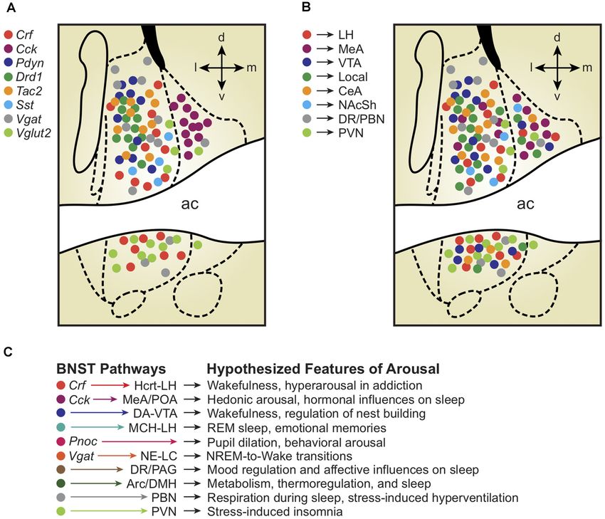
Frontiers in Behavioral Neuroscience | www.frontiersin.org 3 February 2021 | Volume 15 | Article 613025Giardino and Pomrenze BNST Neurocircuitry of Emotional Arousal FIGURE 1 | (A) Depiction of molecularly-defined bed nuclei of stria terminalis (BNST) cell types and their distribution across BNST subregions. Note the remarkable compartmentalization of some cell-types compared with others (Cck vs. Crf vs. Vglut2). (B) Depiction of projection-defined BNST cell types and their approximate distributions across BNST subregions. Interestingly, some (but not all) projection-defined cell types roughly map onto corresponding molecularly-defined subpopulations. (C) Depiction of BNST cell types, pathways, and their hypothesized relationships with distinct features of arousal and sleep/wake regulation. Colors of each hypothesized pathway reflect the molecularly- and projection-defined BNST cell types when consistent with panels (A) and (B). Distinct colors represent hypothesized BNST pathways with unknown molecular traits and spatial distributions across the BNST. Breakthrough efforts to characterize the entire genetic 2016). Aro-BNST neurons have been well-studied in contexts of diversity of the BNST and surrounding regions at the single-cell sexually dimorphic behaviors (Bayless et al., 2019), and pmBNST level provided evidence for up to 37 distinct neuronal subtypes, neurons expressing AR and PR are more numerous in males vs. although an exhaustive discussion of this data is beyond the scope females (Juntti et al., 2010; Yang et al., 2013), highlighting the of our review (Moffitt et al., 2018; Welch et al., 2019). A more importance of sex differences and hormonal interactions when recent single-cell RNA sequencing study targeted specifically studying BNST contributions to emotional arousal (Bangasser in the dorsal (d)BNST identified several neuronal clusters, and Shors, 2008; Bangasser et al., 2019). including those marked by expected genes (e.g., Pkcd, Sst, Concerning the potential wake-modulating effects of Npy), but also some surprising markers (e.g., Lmo4; Rodriguez- molecularly-defined BNST subpopulations, the reported pupil Romaguera et al., 2020). dilatory effects of Pnoc-BNST neuron stimulation (Rodriguez- Numerous BNST neurons (particularly in the posteromedial Romaguera et al., 2020) suggest that such rapid arousal responses [pm]BNST) express markers for actions of gonadal steroid may also regulate sleep/wake state transitions, but this has not hormones, including the androgen receptor (AR), progesterone been explicitly tested. Furthermore, while genetic markers in receptor (PR), estrogen receptors, and aromatase (Aro), the the BNST have thus far been primarily used to simply define the enzyme that converts androgens to estrogens (Bayless and Shah, type of cell rather than determine the role of the corresponding Frontiers in Behavioral Neuroscience | www.frontiersin.org 4 February 2021 | Volume 15 | Article 613025
Giardino and Pomrenze                                                                                    BNST Neurocircuitry of Emotional Arousal

protein product, future progress will shift toward understanding         of the VTA-GABA neuron population was found to be positively
the physiological actions of the molecule itself. For example, it        correlated with high-frequency gamma signal (30–80 Hz) during
will be essential to assess whether modern neurotechnological            wakefulness, providing another measure of physiological arousal
approaches used to stimulate CRF-expressing BNST neurons will            likely impacted by BNST→VTA projections (Eban-Rothschild
be sufficient to accurately recapitulate the sleep/wake changes          et al., 2020).
resulting from the natural release of the CRF neuropeptide from              Separate efforts on LH-projecting neurons identified a large
BNST neurons.                                                            Vgat-BNST population that preferentially targeted Vglut2-LH
                                                                         neurons downstream (Jennings et al., 2013a). Later studies
PROJECTION-DEFINED BNST CELL                                             determined that LH-projecting Vgat-BNST neurons include
TYPES                                                                    both Crf and Cck subpopulations that exhibit divergent
                                                                         preferences for downstream target cell types, with Crf -BNST
A major property of the BNST is its connections with a                   neurons displaying a particularly high level of connectivity with
plethora of downstream target brain regions, providing a useful          LH neurons containing the arousal-promoting neuropeptide
framework for conceptualizing the potential contributions of             hypocretin (Hcrt; also known as orexin; Giardino et al.,
BNST neurons to distinct features of sleep/wake arousal states           2018). Hcrt-LH neurons comprise a subset of glutamatergic
(Figure 1B). For example, by examining BNST projection                   LH neurons, consistent with the interpretation that Crf -
neurons that form synapses with cells in limbic regions regulating       BNST→Hcrt-LH connections represent a subset of the
emotional behavior, One landmark article characterized the               larger Vgat-BNST→Vglut2-LH pathway. González et al.
role of three different BNST pathways in separate features of            (2016) also investigated BNST→LH connectivity, finding
anxiety (Kim et al., 2013). The authors showed that neurons              that Vgat-BNST neurons synapse onto both Hcrt/orexin and
in the anterodorsal (ad)BNST target the lateral hypothalamus             melanin-concentrating hormone (MCH) neurons of the LH.
(LH), ventral tegmental area (VTA), and parabrachial nucleus             This discovery of BNST neurons interacting with the MCH
(PBN) to drive anxiolysis, reward, and decreased respiratory rate,       system is notable based on several studies showing that Mch-LH
respectively. Retrograde tracing determined minimal overlap              neurons promote rapid eye movement (REM) sleep (Bandaru
between cells projecting to the three different downstream               et al., 2020).
regions, collectively implying unique anxiety-relevant functions             In addition to their LH projections, Crf and Cck BNST
for different BNST cell types based in part on their projection          neurons innervate (to varying degrees) the paraventricular
targets. Given the rich literature describing roles for the LH           nucleus of the hypothalamus (PVN), medial amygdala
(Li et al., 2017, 2018), VTA (Eban-Rothschild et al., 2016,              (MeA), CeA, medial preoptic area (mPOA), NAcSh, ventral
2020), and PBN (Kaur and Saper, 2019) in various aspects                 premammillary nucleus (PMv), and ventrolateral periaqueductal
of sleep/wake regulation, we speculate that such BNST axonal             gray (vlPAG; Dabrowska et al., 2016; Giardino et al., 2018). BNST
outputs controlling separable components of emotional behavior           projections to the PVN are particularly relevant given recent
may also modulate distinct physiological aspects of sleep/wake           findings that glutamatergic neurons in this region are critical for
arousal states (Figure 1C).                                              the control of wakefulness (Liu et al., 2020). Further pursuing
    In earlier studies of BNST neuroanatomy in rodents,                  questions of BNST→hypothalamus connectivity, Barbier
VTA-projecting BNST neurons had been characterized by the                et al. (2021) traced the long-range projections of dlBNST and
Watanabe group, who identified a double-inhibitory pathway               dorsomedial (dm)BNST neurons in exquisite detail, finding that
in which GABAergic BNST neurons preferentially target                    the dlBNST projects especially to the LH and tuberomammillary
VTA-GABA neurons (Kudo et al., 2012). Kudo et al. (2012,                 nucleus (TMN; the site of wake-promoting histamine neurons;
2014) also identified VTA-projecting BNST neurons marked by              Barbier et al., 2021). In contrast to the dlBNST, the dmBNST
the glutamate transporters Vglut2 and Vglut3, as well as the             preferentially innervates the PVN, the arcuate nucleus (Arc), and
opioid peptide Penk, providing multiple diverse mechanisms for           dorsomedial hypothalamus (DMH) regions that may influence
modulating the activity of GABA and dopamine (DA) neurons in             sleep pressure via regulation of metabolic and thermoregulatory
the VTA. Jennings et al. (2013b) reported that Vgat and Vglut2           processes (Barbier et al., 2021).
neurons specifically in the ventral (v)BNST synapse onto GABA                In addition to targeted investigations, serendipitous
and DA VTA neurons, where they control distinct motivational             retrograde labeling of upstream neurons has yielded surprising
states. Numerous other studies also focused on VTA-projecting            BNST outputs and circuit motifs. For example, monosynaptic
BNST neurons, including those labeled by Crf and Pdyn (Briand            rabies tracing from either ‘‘patch’’ or ‘‘matrix’’ neurons in the
et al., 2010; Silberman et al., 2013; Marcinkiewcz et al., 2016;         dorsal striatum revealed a major input from the dBNST to patch
Pina and Cunningham, 2017; Rinker et al., 2017; Companion and            neurons that were largely absent from matrix neurons (Smith
Thiele, 2018; Dedic et al., 2018; Fellinger et al., 2020).               et al., 2016). These dBNST neurons form inhibitory synapses
    Given recent evidence that VTA-DA neurons drive                      with striatal patch neurons who in turn project to DA neurons
wakefulness and regulate nest-building (a critical sleep                 in the substantia nigra. Similarly, a GABAergic projection from
preparatory sequence in mice), BNST→VTA-DA circuits                      Sst-BNST neurons was recently characterized and shown to
may therefore be a key mechanism influencing ethologically-              form synapses with parvalbumin interneurons in the NAcSh
relevant behavioral arousal (Eban-Rothschild et al., 2016, 2017;         (Xiao et al., 2020). Therefore, in addition to directly targeting
Eban-Rothschild and de Lecea, 2017). Furthermore, the activity           the VTA, the BNST is capable of modulating mesolimbic DA

Frontiers in Behavioral Neuroscience | www.frontiersin.org           5                                February 2021 | Volume 15 | Article 613025
Giardino and Pomrenze BNST Neurocircuitry of Emotional Arousal
function through indirect mechanisms at sites of midbrain these physiological characteristics likely play a strong role in
innervation in the striatum. modulating varied forms of BNST output.
Monosynaptic rabies tracing also identified BNST neurons Regarding distinct output modes of neurophysiological
that project directly to serotonin and GABA neurons in the communication, most outgoing physiological signals from the
dorsal raphé nucleus (DR; Weissbourd et al., 2014). Interestingly, BNST have been recorded simply as GABAergic/glutamatergic
BNST→DRN neurons preferentially target GABA cells over inhibitory/excitatory postsynaptic currents. However, the
serotonin cells. While the majority of the BNST→DR cells were existence of co-expressed neuropeptides in Vgat-BNST neurons
GABAergic at mouse coordinates +0.14 mm A/P from Bregma, suggests the likelihood of multiplexed modes of signaling in
a smaller set of BNST→DR neurons were labeled by Vglut2 which single cells may influence downstream activity via a
at +0.02 mm A/P from Bregma. Separate rabies tracing studies combination of slow-acting neuropeptide release and fast-acting
also revealed BNST inputs to noradrenergic locus coeruleus classical neurotransmitter release. Indeed, a growing suite of
neurons (Schwarz et al., 2015), DA and GABA VTA neurons fluorescent sensor tools for detecting receptor signaling with the
(Beier et al., 2015), and CRF receptor 1 (CRFR1) PVN neurons cell-type resolution at rapid timescales may prove revolutionary
(Jiang et al., 2018). for elucidating the sleep/wake mechanisms of neuropeptide
Beyond its rich collection of long-range projection neurons, release and actions across synaptic and extrasynaptic sites of the
the BNST contains several species of short-range interneurons. BNST circuitry (Gizowski et al., 2016; Patriarchi et al., 2018; Sun
The Deisseroth group showed that ovBNST neurons project et al., 2018).
locally between subregions of the adBNST where they release
GABA (Kim et al., 2013). In line with this, ovBNST neurons
secrete dynorphin to inhibit excitatory basolateral amygdala FUNCTIONALLY-DEFINED BNST CELL
(BLA) fibers innervating the adBNST (Crowley et al., 2016). TYPES
Neurons in ovBNST also send dense axons to the vBNST
(Dong et al., 2001; Wang et al., 2019). Aside from inhibitory Hedonic Valence
local connections, many studies raised the possibility that The BNST is known to impact both positive and negative states
neuropeptide modulators are also released locally from within of reward and stress, and early studies relied primarily on
the BNST. For example, NPY enhances inhibitory transmission electrolytic and neurochemical lesions to generate several
in Crf -BNST neurons (Pleil et al., 2015) and although this opposing hypotheses regarding the bi-valent emotional
study did not identify the source of NPY, the BNST contains behaviors generated by activity in the BNST (Bangasser
several NPY-expressing neurons capable of local release. Another et al., 2005; Pezuk et al., 2008; Resstel et al., 2008). Access
study from the Kash group found a complex microcircuit in to molecularly-defined cell types enabled by modern
the BNST comprised of Crf interneurons that are modulated by neurotechnology has ushered in several recent advances in
serotonin and regulate the activity of separate long-range BNST understanding the sources of hedonic valence in the BNST,
projection neurons (Marcinkiewcz et al., 2016). Collectively, beginning with studies from the Stuber group showing that
these studies demonstrate the complexity of both long-range and optical stimulation of Vgat-BNST and Vglut2-BNST neurons
local connectivity in the BNST and point to another dimension produced approach and avoidance behaviors, respectively
of inherent organization arising from gene expression and (Jennings et al., 2013a,b).
neurotransmitter phenotype. Going beyond the separation of large populations of
GABAergic vs. glutamatergic neurons, additional tools for
PHYSIOLOGY-DEFINED BNST CELL recording and modulating cell-specific neural activity in vivo
TYPES revealed that dlBNST Crf neurons preferentially respond
to aversive stimuli and drive behavioral avoidance, whereas
The BNST contains multiple neuronal cell types that have been dmBNST Cck neurons are activated by rewarding stimuli
classified according to their electrophysiological properties, most and generate behavioral preference/approach (Giardino et al.,
prominently in rats by Hammack and others (Hammack et al., 2018). Mirroring this lateral/medial functional distinction,
2007; Hazra et al., 2011; Dabrowska et al., 2013; Rodríguez- Drd1 neurons in the ovBNST/dlBNST and Six3 neurons in
Sierra et al., 2013; Silberman et al., 2013; Nagano et al., 2015; the dmBNST also drive avoidance and approach, respectively
Yamauchi et al., 2018). While originally described primarily (Giardino et al., 2018). Because activation of the global Vgat-
within the ovBNST, these three types (Type I, II, and III) have BNST population promotes positive valence (Jennings et al.,
also been identified in the non-oval areas of adBNST, as well 2013a,b; Giardino et al., 2018), we hypothesize that Crf marks
as anteroventral (av)BNST. Type I neurons exhibit an Ih -like a specialized subgroup of lateral Vgat-BNST neurons with
current in response to hyperpolarizing current injection and a opposing functional properties from the larger set of combined
regular firing pattern in response to depolarizing current. Type II lateral and medial Vgat-BNST cells. In other words, global
neurons exhibit a similar Ih -like current but burst fire in response Vgat-BNST stimulation may more closely resemble activation of
to depolarizing current. Type III neurons do not exhibit an Ih - medial Vgat-BNST neurons (like Cck) rather than lateral Vgat-
like current but instead, show a fast rectification in response BNST neurons (like Crf ).
to hyperpolarizing current and exhibit a regular firing pattern It should be noted that the vast majority of data on
when depolarized. Coupled with the diverse synaptic inputs, BNST circuits driving hedonic valence has been generated only
Frontiers in Behavioral Neuroscience | www.frontiersin.org 6 February 2021 | Volume 15 | Article 613025Giardino and Pomrenze BNST Neurocircuitry of Emotional Arousal
from male mice. Given that the BNST is a major site for In 2014, an intriguing arousal-related physiological
integrating signals from centrally-circulating gonadal steroids, signature was discovered in the extended amygdala by
intense investigation of hormonal influences on the function of Haufler and Pare (2014), who recorded high-frequency
extended amygdala pathways may be key for understanding sex oscillations (HFOs; 110–160 Hz local field potentials) that
differences in the stress response and reward sensitivity. Future appeared with a significantly higher incidence in the BNST
studies may also seek to titrate levels of stressor exposure, drug and CeA relative to surrounding areas (striatum, pallidum,
consumption, and other variables to determine whether such septum; Haufler and Pare, 2014). HFOs in hippocampal,
experiential factors can influence the hedonic valence associated cortical, and subthalamic brain regions are thought to have
with mobilization of particular BNST subpopulations. functional consequences in memory-processing, epileptic
seizures, and symptoms of Parkinson’s disease, respectively.
Anxiety Whereas the role of HFOs in the BNST remains unclear, their
Despite the distinct hedonic valences associated with stimulating ability to entrain large populations of neurons (with greater
Crf and Cck BNST neurons, optogenetic or chemogenetic power during REM vs. non-REM sleep; Haufler and Pare,
activation of either Crf or Cck BNST neurons led to increased 2014) provides a fascinating example of the network-level
indices of anxiety-like behavior in multiple paradigms (Giardino changes in extended amygdala activity that might profoundly
et al., 2018), suggesting that standard measures of ‘‘anxiety’’ influence discrete states of arousal. Future endeavors will
in rodents may reflect generalized arousal states independent revolutionize understanding of how arousal state transitions
of hedonic valence per se. Consistent with this interpretation, may be aligned to physiological activity changes and/or
activation of Vgat-BNST neurons (Mazzone et al., 2018), Drd1- receptor signaling events in a BNST cell type-specific manner
ovBNST neurons (Kim et al., 2013), Pdyn-BNST→VTA neurons by implementing combinatorial approaches like EEG/EMG
(Fellinger et al., 2020), dlBNST→CeA neurons (Yamauchi sleep monitoring in tandem with next-generation neural
et al., 2018), and Crf -CeA→dlBNST neurons (Pomrenze recording technologies (fiber photometry, miniscope, and
et al., 2019b) all increased anxiety-like behaviors. Yet, separate two-photon imaging).
studies found that stimulation of adBNST→LH neurons To directly investigate the BNST as a potential node
(Kim et al., 2013), Vgat-vBNST→VTA neurons (Jennings in the arousal circuitry, Kodani et al. (2017, 2019) utilized
et al., 2013b), and Sst-BNST→NAcSh neurons (Xiao et al., optogenetic methods and reported that they could generate
2020) had the opposite effect of reducing anxiety, revealing immediate transitions from NREM sleep to wakefulness by
some unresolved issues regarding how BNST microcircuits stimulating the global GABAergic BNST population in Gad67-
and long-range pathways regulate stress-related ‘‘anxiety’’ Cre mice. Gad67-BNST arousal was associated with activation
phenotypes. Anxiogenic circumstances like fear learning and of norepinephrine neurons in the locus coeruleus (NE-LC), an
stress-induced social deficits also strongly engage the BNST established wakefulness-promoting center. Gad67-BNST arousal
(Bjorni et al., 2020; Emmons et al., 2021), providing multiple was also associated with activation of Hcrt-LH neurons and
perspectives for approaching the study of BNST in anxiety- required signaling of Hcrt receptors (Kodani et al., 2017),
related emotional arousal. consistent with the reported contributions of BNST→LH circuits
to emotional arousal and related behaviors (Giardino and
de Lecea, 2014; González et al., 2016; Giardino et al., 2018;
THE BNST IN SLEEP, WAKE, AND Barbier et al., 2021). Of course, the precise directionalities
EMOTIONAL AROUSAL of the relationships between endogenous BNST activity,
emotional behaviors, and changes in sleep and wakefulness
Only a handful of studies have explicitly investigated the remain mostly uncharacterized. For example, whereas states
BNST within the context of sleep/wake arousal states and of stress might be hypothesized to drive insomnia via
corresponding neurophysiological rhythms. Beginning in 1995, chronically elevated BNST hyperactivity, additional experiments
in vivo recordings of 63 single units in the BNST of cats are required to determine whether excess BNST activity is truly
revealed that 72% of neurons fired more frequently during a contributing factor or rather an indirect consequence of stress-
wakefulness and rapid eye movement (REM) sleep than induced arousal.
during ‘‘quiet sleep’’ (presumably non-REM/slow-wave sleep; In addition to providing outputs to wake-promoting
NREM; Terreberry et al., 1995). These findings are consistent downstream targets in the LH, VTA, and PVN, the BNST
with more recent data indicating that excitatory projections also receives inputs from arousal-promoting neurons such as
from the BNST may activate REM-active neurons in the calretinin cells of the paraventricular thalamus (PVT; Hua et al.,
sublaterodorsal tegmental nucleus (SLD) of the brainstem 2018). Hua et al. (2018) recently found that starvation promotes
(Boissard et al., 2003; Rodrigo-Angulo et al., 2008). In the a state of hyperarousal via activation of the PVT-calretinin
BNST of rats, sleep deprivation increased cFos and CRF circuit, which can be blocked by chemogenetic inhibition of the
protein expression (Duan et al., 2005; Deurveilher et al., 2008) PVT→BNST pathway. Despite this exciting progress, a complete
and M3 muscarinic acetylcholine receptor gene expression understanding of the arousal-promoting abilities of the global
(Kushida et al., 1995), suggesting possible neuropeptidergic GABAergic BNST population requires additional studies. Future
and cholinergic mechanisms for homeostatic sleep drive in experiments will aim to determine whether distinct molecularly-
the BNST. defined or pathway-specific BNST outputs are necessary and/or
Frontiers in Behavioral Neuroscience | www.frontiersin.org 7 February 2021 | Volume 15 | Article 613025Giardino and Pomrenze BNST Neurocircuitry of Emotional Arousal
sufficient for regulating various forms of behavioral arousal and revealing the neurocircuitry of mysterious phenomena like
natural sleep-to-wake transitions (see hypothesized functions in post-ingestive sleep, post-copulatory sleep, and narcolepsy
Figure 1C). with cataplexy.
On this note, it will be important to causally determine
which BNST neurocircuits have experience-dependent effects CONCLUSIONS
on sleep/wake as a function of negative vs. positive hedonic
valence. This can be accomplished by monitoring the activities Altogether, our understanding of extended amygdala circuits
of extended amygdala pathways and corresponding changes regulating affective behaviors will have direct relevance to
in arousal following emotional experiences with ethologically therapeutic strategies aimed at modulating motivation and
aversive or rewarding stimuli. For example, the discovery that sleep/wake regulation. Before the advent of tools permitting cell
reward-promoting Cck-BNST neurons are densely connected type-specific and pathway-specific labeling and manipulation,
with the medial amygdala (MeA; Giardino et al., 2018) suggests the inherent complexity of extended amygdala pathways
that these pathways powerfully influence arousal changes hindered progress in understanding their roles in emotional
linked to innately rewarding consummatory behaviors. In arousal. As BNST circuits continue to be disentangled at
contrast to the Cck-BNST→MeA circuit, Crf-BNST→Hcrt- the genetic, synaptic, and systems levels, new anatomical and
LH connections likely drive stress-induced insomnia following functional frameworks are taking shape that illustrates the
psychosocial challenges. multifaceted nature of the BNST. Data of this kind are extremely
Given the well-established role of the BNST in drug-seeking valuable for developing new therapeutic approaches for a range
behavior (Vranjkovic et al., 2017), attention should be placed of neuropsychiatric conditions, highlighting the extraordinary
on the idea that addiction-related sleep disturbances are driven relevance of emotional arousal circuits to researchers and
by specific extended amygdala pathways. Core features of clinicians across the fields of neuroscience, genetics, psychology,
drug addiction (e.g., craving, relapse, and withdrawal) are and mental health interventions. We anticipate that as studies on
each associated with maladaptive neuroplasticity in homeostatic BNST function become more precise, effective clinical treatment
circuits that regulate sleep/wake cycles and stress sensitivity, strategies will be successfully developed.
illustrating how addiction can be viewed as a condition of
pathological hyperarousal (Koob, 2013; Koob and Colrain, AUTHOR CONTRIBUTIONS
2020). Substance use disorders are highly co-morbid with
other arousal-related psychiatric conditions (e.g., insomnia, All authors listed have made a substantial, direct and intellectual
anxiety, PTSD, panic), and the BNST may be a common contribution to the work, and approved it for publication.
substrate linking dysregulated hedonic processing to chronic
sleep disruption. FUNDING
Finally, the BNST may be an important and overlooked
link in emotional arousal about the sleep disorder narcolepsy This study received funding from NIH/NIAAA
with cataplexy (in which powerful feelings of euphoria or K99/R00 AA025677 (WG) and NIH/NIDA T32
aversion can interrupt wakefulness by triggering rapid intrusion DA035165 (MP).
of a sleep-like state). Intriguingly, narcolepsy is associated
with selective loss of Hcrt neurons in the LH. Based on ACKNOWLEDGMENTS
existing evidence for BNST→LH connections driving motivated
behaviors, detailed studies are warranted on BNST circuit We thank D. W. Bayless, J. R. Knoedler, D. C. Castro, T. L. Kash,
contributions to emotionally-triggered arousal destabilization. L. R. Halladay, and others for insightful discussions.
Of course, the existence of putative sleep-promoting BNST
neurons remains to be seen. However, BNST connections SUPPLEMENTARY MATERIAL
with hypothalamic nuclei regulating the release of prolactin,
oxytocin, and vasopressin suggest that experiments linking The Supplementary Material for this article can be found
BNST activity to arousal changes following binge eating (‘‘food online at: https://www.frontiersin.org/articles/10.3389/fnbeh.
coma’’) and sexual behavior (mating) may be successful in 2021.613025/full#supplementary-material.
REFERENCES Avery, S. N., Clauss, J. A., and Blackford, J. U. (2016). The human BNST:
functional role in anxiety and addiction. Neuropsychopharmacology 41,
Adamantidis, A. R., Schmidt, M. H., Carter, M. E., Burdakov, D., Peyron, C., and 126–141. doi: 10.1038/npp.2015.185
Scammell, T. E. (2020). A circuit perspective on narcolepsy. Sleep 43:zsz296. Avery, S. N., Clauss, J. A., Winder, D. G., Woodward, N., Heckers, S., and
doi: 10.1093/sleep/zsz296 Blackford, J. U. (2014). BNST neurocircuitry in humans. NeuroImage 91,
Ahrens, S., Wu, M. V., Furlan, A., Hwang, G. R., Paik, R., Li, H., et al. (2018). 311–323. doi: 10.1016/j.neuroimage.2014.01.017
A central extended amygdala circuit that modulates anxiety. J. Neurosci. 38, Bandaru, S. S., Khanday, M. A., Ibrahim, N., Naganuma, F., and Vetrivelan, R.
5567–5583. doi: 10.1523/JNEUROSCI.0705-18.2018 (2020). Sleep-wake control by melanin-concentrating hormone (MCH)
Alheid, G. F. (2003). Extended amygdala and basal forebrain. Ann. N Y Acad. Sci. neurons: a review of recent findings. Curr. Neurol. Neurosci. Rep. 20:55.
985, 185–205. doi: 10.1111/j.1749-6632.2003.tb07082.x doi: 10.1007/s11910-020-01075-x
Frontiers in Behavioral Neuroscience | www.frontiersin.org 8 February 2021 | Volume 15 | Article 613025Giardino and Pomrenze BNST Neurocircuitry of Emotional Arousal Bangasser, D. A., Eck, S. R., and Ordoñes Sanchez, E. (2019). Sex differences in CRF-containing neurons of the rat paraventricular hypothalamus and the bed stress reactivity in arousal and attention systems. Neuropsychopharmacology 44, nucleus of the stria terminalis. Front. Neurosci. 7:156. doi: 10.3389/fnins.2013. 129–139. doi: 10.1038/s41386-018-0137-2 00156 Bangasser, D. A., Santollo, J., and Shors, T. J. (2005). The bed nucleus of the stria Dabrowska, J., Martinon, D., Moaddab, M., and Rainnie, D. G. (2016). Targeting terminalis is critically involved in enhancing associative learning after stressful corticotropin-releasing factor projections from the oval nucleus of the bed experience. Behav. Neurosci. 119, 1459–1466. doi: 10.1037/0735-7044.119. nucleus of the stria terminalis using cell-type specific neuronal tracing 6.1459 studies in mouse and rat brain. J. Neuroendocrinol. 28:12. doi: 10.1111/jne. Bangasser, D. A., and Shors, T. J. (2008). The bed nucleus of the stria terminalis 12442 modulates learning after stress in masculinized but not cycling females. De Olmos, J. S., and Ingram, W. R. (1972). The projection field of the stria J. Neurosci. 28, 6383–6387. doi: 10.1523/JNEUROSCI.0831-08.2008 terminalis in the rat brain. An experimental study. J. Comp. Neurol. 146, Barbier, M., Gonzalez, J. A., Houdayer, C., Burdakov, D., Risold, P. Y., and 303–334. doi: 10.1002/cne.901460303 Croizier, S. (2021). Projections from the dorsomedial division of the bed Dedic, N., Kühne, C., Jakovcevski, M., Hartmann, J., Genewsky, A. J., Gomes, K. S., nucleus of the stria terminalis to hypothalamic nuclei in the mouse. J. Comp. et al. (2018). Chronic CRH depletion from GABAergic, long-range projection Neurol. 529, 929–956. doi: 10.1002/cne.24988 neurons in the extended amygdala reduces dopamine release and increases Bayer, S. A., and Altman, J. (1987). Development of the preoptic area: time and site anxiety. Nat. Neurosci. 21, 803–807. doi: 10.1038/s41593-018-0151-z of origin, migratory routes and settling patterns of its neurons. J. Comp. Neurol. Deurveilher, S., Cumyn, E. M., Peers, T., Rusak, B., and Semba, K. (2008). Estradiol 265, 65–95. doi: 10.1002/cne.902650106 replacement enhances sleep deprivation-induced c-Fos immunoreactivity in Bayless, D. W., and Shah, N. M. (2016). Genetic dissection of neural circuits forebrain arousal regions of ovariectomized rats. Am. J. Physiol. Regul. Integr. underlying sexually dimorphic social behaviours. Philos. Trans. R. Soc. Lond. Comp. Physiol. 295, R1328–R1340. doi: 10.1152/ajpregu.90576.2008 B Biol. Sci. 371:20150109. doi: 10.1098/rstb.2015.0109 Dong, H., Petrovich, G. D., and Swanson, L. W. (2000). Organization of Bayless, D. W., Yang, T., Mason, M. M., Susanto, A. A. T., Lobdell, A., and projections from the juxtacapsular nucleus of the BST: a PHAL study in the Shah, N. M. (2019). Limbic neurons shape sex recognition and social behavior rat. Brain Res. 859, 1–14. doi: 10.1016/s0006-8993(99)02246-5 in sexually naive males. Cell 176, 1190.e20–1205.e20. doi: 10.1016/j.cell.2018. Dong, H.-W., Petrovich, G. D., and Swanson, L. W. (2001a). Topography of 12.041 projections from amygdala to bed nuclei of the stria terminalis. Brain Res. Rev. Beier, K. T., Steinberg, E. E., DeLoach, K. E., Xie, S., Miyamichi, K., Schwarz, L., 38, 192–246. doi: 10.1016/s0165-0173(01)00079-0 et al. (2015). Circuit architecture of VTA dopamine neurons revealed by Dong, H.-W., Petrovich, G. D., Watts, A. G., and Swanson, L. W. (2001b). Basic systematic input-output mapping. Cell 162, 622–634. doi: 10.1016/j.cell.2015. organization of projections from the oval and fusiform nuclei of the bed 07.015 nuclei of the stria terminalis in adult rat brain. J. Comp. Neurol. 436, 430–455. Beyeler, A., and Dabrowska, J. (2020). Neuronal diversity of the amygdala and doi: 10.1002/cne.1079 the bed nucleus of the stria terminalis. Handb. Behav. Neurosci. 26, 63–100. Dong, H.-W., and Swanson, L. W. (2003). Projections from the rhomboid doi: 10.1016/b978-0-12-815134-1.00003-9 nucleus of the bed nuclei of the stria terminalis: implications for cerebral Bjorni, M., Rovero, N. G., Yang, E. R., Holmes, A., and Halladay, L. R. (2020). hemisphere regulation of ingestive behaviors. J. Comp. Neurol. 463, 434–472. Phasic signaling in the bed nucleus of the stria terminalis during fear learning doi: 10.1002/cne.10758 predicts within- and across-session cued fear expression. Learn. Mem. 27, Dong, H.-W., and Swanson, L. W. (2004a). Organization of axonal projections 83–90. doi: 10.1101/lm.050807.119 from the anterolateral area of the bed nuclei of the stria terminalis. J. Comp. Boissard, R., Fort, P., Gervasoni, D., Barbagli, B., and Luppi, P. H. (2003). Neurol. 468, 277–298. doi: 10.1002/cne.10949 Localization of the GABAergic and non-GABAergic neurons projecting to the Dong, H.-W., and Swanson, L. W. (2004b). Projections from bed nuclei of the stria sublaterodorsal nucleus and potentially gating paradoxical sleep onset. Eur. terminalis, posterior division: implications for cerebral hemisphere regulation J. Neurosci. 18, 1627–1639. doi: 10.1046/j.1460-9568.2003.02861.x of defensive and reproductive behaviors. J. Comp. Neurol. 471, 396–433. Bota, M., Sporns, O., and Swanson, L. W. (2012). Neuroinformatics analysis of doi: 10.1002/cne.20002 molecular expression patterns and neuron populations in gray matter regions: Dong, H.-W., and Swanson, L. W. (2006a). Projections from bed nuclei of the rat BST as a rich exemplar. Brain Res. 1450, 174–193. doi: 10.1016/j. the stria terminalis, anteromedial area: cerebral hemisphere integration of brainres.2012.02.034 neuroendocrine, autonomic, and behavioral aspects of energy balance. J. Comp. Briand, L. A., Vassoler, F. M., Pierce, R. C., Valentino, R. J., and Blendy, J. A. Neurol. 494, 142–178. doi: 10.1002/cne.20788 (2010). Ventral tegmental afferents in stress-induced reinstatement: the role Dong, H.-W., and Swanson, L. W. (2006b). Projections from bed nuclei of the of cAMP response element-binding protein. J. Neurosci. 30, 16149–16159. stria terminalis, magnocellular nucleus: implications for cerebral hemisphere doi: 10.1523/JNEUROSCI.2827-10.2010 regulation of micturition, defecation, and penile erection. J. Comp. Neurol. 494, Chen, Y., Molet, J., Gunn, B. G., Ressler, K., and Baram, T. Z. (2015). 108–141. doi: 10.1002/cne.20789 Diversity of reporter expression patterns in transgenic mouse lines targeting Dong, H.-W., and Swanson, L. W. (2006c). Projections from bed nuclei of the corticotropin-releasing hormone-expressing neurons. Endocrinology 156, stria terminalis, dorsomedial nucleus: implications for cerebral hemisphere 4769–4780. doi: 10.1210/en.2015-1673 integration of neuroendocrine, autonomic, and drinking responses. J. Comp. Chen, A.-X., Yan, J.-J., Zhang, W., Wang, L., Yu, Z.-X., Ding, X.-J., et al. (2020). Neurol. 494, 75–107. doi: 10.1002/cne.20790 Specific hypothalamic neurons required for sensing conspecific male cues Duan, X.-L., Wang, B.-R., Yang, T.-B., Yu, H.-M., Zhu, Z.-H., Xu, Z., et al. (2005). relevant to inter-male aggression. Neuron 108, 763.e6–774.e6. doi: 10.1016/j. Preparation of polyclonal and monoclonal antibodies against CRF and change neuron.2020.08.025 of CRF in the sleep-deprived rat brain. Xi Bao Yu Fen Zi Mian Yi Xue Za Zhi Ch’ng, S., Fu, J., Brown, R. M., McDougall, S. J., and Lawrence, A. J. (2018). 21, 476–479. The intersection of stress and reward: BNST modulation of aversive and Duque-Wilckens, N., Steinman, M. Q., Busnelli, M., Chini, B., Yokoyama, S., appetitive states. Prog. Neuropsychopharmacol. Biol. Psychiatry 87, 108–125. Pham, M., et al. (2018). Oxytocin receptors in the anteromedial bed nucleus doi: 10.1016/j.pnpbp.2018.01.005 of the stria terminalis promote stress-induced social avoidance in female Companion, M. A., and Thiele, T. E. (2018). Assessment of ventral tegmental california mice. Biol. Psychiatry 83, 203–213. doi: 10.1016/j.biopsych.2017. area-projecting GABAergic neurons from the bed nucleus of the stria 08.024 terminalis in modulating binge-like ethanol intake. Eur. J. Neurosci. 48, Eban-Rothschild, A., Appelbaum, L., and de Lecea, L. (2018). Neuronal 3335–3343. doi: 10.1111/ejn.14222 mechanisms for sleep/wake regulation and modulatory drive. Crowley, N. A., Bloodgood, D. W., Hardaway, J. A., Kendra, A. M., McCall, J. G., Neuropsychopharmacology 43, 937–952. doi: 10.1038/npp.2017.294 Al-Hasani, R., et al. (2016). Dynorphin controls the gain of an amygdalar Eban-Rothschild, A., Borniger, J. C., Rothschild, G., Giardino, W. J., anxiety circuit. Cell Rep. 14, 2774–2783. doi: 10.1016/j.celrep.2016.02.069 Morrow, J. G., and de Lecea, L. (2020). Arousal state-dependent alterations Dabrowska, J., Hazra, R., Guo, J. D., Dewitt, S., and Rainnie, D. G. (2013). in VTA-GABAergic neuronal activity. eNeuro 7:ENEURO.0356-19.2020. Central CRF neurons are not created equal: phenotypic differences in doi: 10.1523/ENEURO.0356-19.2020 Frontiers in Behavioral Neuroscience | www.frontiersin.org 9 February 2021 | Volume 15 | Article 613025
Giardino and Pomrenze                                                                                                                     BNST Neurocircuitry of Emotional Arousal

Eban-Rothschild, A., and de Lecea, L. (2017). Neuronal substrates for initiation,              Hazra, R., Guo, J.-D., Ryan, S. J., Jasnow, A. M., Dabrowska, J., and Rainnie, D. G.
   maintenance and structural organization of sleep/wake states. F1000Res. 6:212.                  (2011). A transcriptomic analysis of type I–III neurons in the bed nucleus of
   doi: 10.12688/f1000research.9677.1                                                              the stria terminalis. Mol. Cell. Neurosci. 46, 699–709. doi: 10.1016/j.mcn.2011.
Eban-Rothschild, A., Giardino, W. J., and de Lecea, L. (2017). To sleep or not                     01.011
   to sleep: neuronal and ecological insights. Curr. Opin. Neurobiol. 44, 132–138.             Hostetler, C. M., Kowalczyk, A. S., Griffin, L. L., and Bales, K. L. (2011). CART
   doi: 10.1016/j.conb.2017.04.010                                                                 peptide following social novelty in the prairie vole (Microtus ochrogaster). Brain
Eban-Rothschild, A., Rothschild, G., Giardino, W. J., Jones, J. R., and de Lecea, L.               Res. 1414, 32–40. doi: 10.1016/j.brainres.2011.07.040
   (2016). VTA dopaminergic neurons regulate ethologically relevant sleep-wake                 Hua, R., Wang, X., Chen, X., Wang, X., Huang, P., Li, P., et al. (2018). Calretinin
   behaviors. Nat. Neurosci. 19, 1356–1366. doi: 10.1038/nn.4377                                   neurons in the midline thalamus modulate starvation-induced arousal. Curr.
Egli, R. E., and Winder, D. G. (2003). Dorsal and ventral distribution of excitable                Biol. 28, 3948.e4–3959.e4. doi: 10.1016/j.cub.2018.11.020
   and synaptic properties of neurons of the bed nucleus of the stria terminalis.              Jennings, J. H., Rizzi, G., Stamatakis, A. M., Ung, R. L., and Stuber, G. D. (2013a).
   J. Neurophysiol. 90, 405–414. doi: 10.1152/jn.00228.2003                                        The inhibitory circuit architecture of the lateral hypothalamus orchestrates
Emmons, R., Sadok, T., Rovero, N. G., Belnap, M. A., Henderson, H. J. M.,                          feeding. Science 341, 1517–1521. doi: 10.1126/science.1241812
   Quan, A. J., et al. (2021). Chemogenetic manipulation of the bed nucleus                    Jennings, J. H., Sparta, D. R., Stamatakis, A. M., Ung, R. L., Pleil, K. E., Kash, T. L.,
   of the stria terminalis counteracts social behavioral deficits induced by early                 et al. (2013b). Distinct extended amygdala circuits for divergent motivational
   life stress in C57BL/6J mice. J. Neurosci. Res. 99, 90–109. doi: 10.1002/jnr.                   states. Nature 496, 224–228. doi: 10.1038/nature12041
   24644                                                                                       Jiang, Z., Rajamanickam, S., and Justice, N. J. (2018). Local corticotropin-releasing
Fellinger, L., Jo, Y. S., Hunker, A. C., Soden, M. E., and Zweifel, L. S. (2020). A                factor signaling in the hypothalamic paraventricular nucleus. J. Neurosci. 38,
   midbrain dynorphin circuit promotes threat generalization. bioRxiv [Preprint].                  1874–1890. doi: 10.1523/JNEUROSCI.1492-17.2017
   doi: 10.1101/2020.04.21.053892                                                              Ju, G., and Swanson, L. W. (1989). Studies on the cellular architecture of the bed
Flook, E. A., Feola, B., Avery, S. N., Winder, D. G., Woodward, N. D., Heckers, S.,                nuclei of the stria terminalis in the rat: I. Cytoarchitecture. J. Comp. Neurol.
   et al. (2020). BNST-insula structural connectivity in humans. NeuroImage                        280, 587–602. doi: 10.1002/cne.902800409
   210:116555. doi: 10.1016/j.neuroimage.2020.116555                                           Ju, G., Swanson, L. W., and Simerly, R. B. (1989). Studies on the cellular
Fox, A. S., Oler, J. A., Tromp do, P. M., Fudge, J. L., and Kalin, N. H. (2015).                   architecture of the bed nuclei of the stria terminalis in the rat: II.
   Extending the amygdala in theories of threat processing. Trends Neurosci. 38,                   Chemoarchitecture. J. Comp. Neurol. 280, 603–621. doi: 10.1002/cne.
   319–329. doi: 10.1016/j.tins.2015.03.002                                                        902800410
Fox, A. S., and Shackman, A. J. (2019). The central extended amygdala in fear                  Juntti, S. A., Tollkuhn, J., Wu, M. V., Fraser, E. J., Soderborg, T., Tan, S., et al.
   and anxiety: closing the gap between mechanistic and neuroimaging research.                     (2010). The androgen receptor governs the execution, but not programming,
   Neurosci. Lett. 693, 58–67. doi: 10.1016/j.neulet.2017.11.056                                   of male sexual and territorial behaviors. Neuron 66, 260–272. doi: 10.1016/j.
Fudge, J. L., and Haber, S. N. (2001). Bed nucleus of the stria terminalis                         neuron.2010.03.024
   and extended amygdala inputs to dopamine subpopulations in primates.                        Kash, T. L., Pleil, K. E., Marcinkiewcz, C. A., Lowery-Gionta, E. G., Crowley, N.,
   Neuroscience 104, 807–827. doi: 10.1016/s0306-4522(01)00112-9                                   Mazzone, C., et al. (2015). Neuropeptide regulation of signaling and behavior
Fudge, J. L., Kelly, E. A., Pal, R., Bedont, J. L., Park, L., and Ho, B. (2017). Beyond            in the BNST. Mol. Cells 38, 1–13. doi: 10.14348/molcells.2015.2261
   the classic VTA: extended amygdala projections to DA-striatal paths in the                  Kaur, S., and Saper, C. B. (2019). Neural circuitry underlying waking
   primate. Neuropsychopharmacology 42, 1563–1576. doi: 10.1038/npp.2017.38                        up to hypercapnia. Front. Neurosci. 13:401. doi: 10.3389/fnins.2019.
Giardino, W. J., and de Lecea, L. (2014). Hypocretin (orexin) neuromodulation of                   00401
   stress and reward pathways. Curr. Opin. Neurobiol. 29, 103–108. doi: 10.1016/j.             Kim, S.-Y., Adhikari, A., Lee, S. Y., Marshel, J. H., Kim, C. K., Mallory, C. S., et al.
   conb.2014.07.006                                                                                (2013). Diverging neural pathways assemble a behavioral state from separable
Giardino, W. J., Eban-Rothschild, A., Christoffel, D. J., Li, S. B., Malenka, R. C.,               features in anxiety. Nature 496, 219–223. doi: 10.1038/nature12018
   and de Lecea, L. (2018). Parallel circuits from the bed nuclei of stria terminalis          Kim, B., Yoon, S., Nakajima, R., Lee, H. J., Lim, H. J., Lee, Y. K., et al. (2018).
   to the lateral hypothalamus drive opposing emotional states. Nat. Neurosci. 21,                 Dopamine D2 receptor-mediated circuit from the central amygdala to the bed
   1084–1095. doi: 10.1038/s41593-018-0198-x                                                       nucleus of the stria terminalis regulates impulsive behavior. Proc. Natl. Acad.
Girven, K. S., Aroni, S., Navarrete, J., Marino, R. A. M., McKeon, P. N.,                          Sci. U S A 115, E10730–E10739. doi: 10.1073/pnas.1811664115
   Cheer, J. F., et al. (2020). Glutamatergic input from the insula to the ventral bed         Kodani, S., Soya, S., and Sakurai, T. (2017). Excitation of GABAergic neurons
   nucleus of the stria terminalis controls reward-related behavior. Addict. Biol.                 in the bed nucleus of the stria terminalis triggers immediate transition from
   doi: 10.1111/adb.12961                                                                          non-rapid eye movement sleep to wakefulness in mice. J. Neurosci. 37,
Gizowski, C., Zaelzer, C., and Bourque, C. W. (2016). Clock-driven vasopressin                     7164–7176. doi: 10.1523/JNEUROSCI.0245-17.2017
   neurotransmission mediates anticipatory thirst prior to sleep. Nature 537,                  Kodani, S., Soya, S., and Sakurai, T. (2019). Optogenetic manipulation of neural
   685–688. doi: 10.1038/nature19756                                                               circuits during monitoring sleep/wakefulness states in mice. J. Vis. Exp. 19:148.
González, J. A., Iordanidou, P., Strom, M., Adamantidis, A., and Burdakov, D.                      doi: 10.3791/58613
   (2016). Awake dynamics and brain-wide direct inputs of hypothalamic                         Koob, G. F. (2013). Addiction is a reward deficit and stress surfeit disorder. Front.
   MCH and orexin networks. Nat. Commun. 7:11395. doi: 10.1038/ncomms                              Psychiatry 4:72. doi: 10.3389/fpsyt.2013.00072
   11395                                                                                       Koob, G. F., and Colrain, I. M. (2020). Alcohol use disorder and sleep disturbances:
Gordon-Fennell, A. G., Will, R. G., Ramachandra, V., Gordon-Fennell, L.,                           a feed-forward allostatic framework. Neuropsychopharmacology 45, 141–165.
   Dominguez, J. M., Zahm, D. S., et al. (2019). The lateral preoptic area: a novel                doi: 10.1038/s41386-019-0446-0
   regulator of reward seeking and neuronal activity in the ventral tegmental area.            Kovner, R., Fox, A. S., French, D. A., Roseboom, P. H., Oler, J. A., Fudge, J. L.,
   Front. Neurosci. 13:1433. doi: 10.3389/fnins.2019.01433                                         et al. (2019). Somatostatin gene and protein expression in the non-human
Hammack, S. E., Mania, I., and Rainnie, D. G. (2007). Differential expression                      primate central extended amygdala. Neuroscience 400, 157–168. doi: 10.1016/j.
   of intrinsic membrane currents in defined cell types of the anterolateral bed                   neuroscience.2018.12.035
   nucleus of the stria terminalis. J. Neurophysiol. 98, 638–656. doi: 10.1152/jn.             Krettek, J. E., and Price, J. L. (1978). Amygdaloid projections to subcortical
   00382.2007                                                                                      structures within the basal forebrain and brainstem in the rat and cat. J. Comp.
Hardaway, J. A., Halladay, L. R., Mazzone, C. M., Pati, D., Bloodgood, D. W.,                      Neurol. 178, 225–254. doi: 10.1002/cne.901780204
   Kim, M., et al. (2019). Central amygdala prepronociceptin-expressing                        Kudo, T., Konno, K., Uchigashima, M., Yanagawa, Y., Sora, I., Minami, M.,
   neurons mediate palatable food consumption and reward. Neuron 102:1088.                         et al. (2014). GABAergic neurons in the ventral tegmental area receive dual
   doi: 10.1016/j.neuron.2019.04.036                                                               GABA/enkephalin-mediated inhibitory inputs from the bed nucleus of the stria
Haufler, D., and Pare, D. (2014). High-frequency oscillations are prominent                        terminalis. Eur. J. Neurosci. 39, 1796–1809. doi: 10.1111/ejn.12503
   in the extended amygdala. J. Neurophysiol. 112, 110–119. doi: 10.1152/jn.00                 Kudo, T., Uchigashima, M., Miyazaki, T., Konno, K., Yamasaki, M., Yanagawa, Y.,
   107.2014                                                                                        et al. (2012). Three types of neurochemical projection from the bed nucleus of

Frontiers in Behavioral Neuroscience | www.frontiersin.org                                10                                          February 2021 | Volume 15 | Article 613025
You can also read