USH3A transcripts encode clarin-1, a four-transmembrane-domain protein with a possible role in sensory synapses
←
→
Page content transcription
If your browser does not render page correctly, please read the page content below
European Journal of Human Genetics (2002) 10, 339 – 350
                                                                        ª 2002 Nature Publishing Group   All rights reserved 1018 – 4813/02 $25.00
                                                                        www.nature.com/ejhg

    ARTICLE

USH3A transcripts encode clarin-1, a four-
transmembrane-domain protein with a possible role in
sensory synapses
Avital Adato*,1, Sarah Vreugde2, Tarja Joensuu3,4, Nili Avidan1, Riikka Hamalainen3,
Olga Belenkiy1, Tsviya Olender1, Batsheva Bonne-Tamir2, Edna Ben-Asher1,
Carmen Espinos5, José M Millán6, Anna-Elina Lehesjoki3, John G Flannery7,
Karen B Avraham2, Shmuel Pietrokovski1, Eeva-Marja Sankila3,4, Jacques S Beckmann1
and Doron Lancet*,1

1
 Department of Molecular Genetics and The Crown Human Genome Center, The Weizmann Institute of Science,
Rehovot, 76100, Israel; 2Department of Human Genetics and Molecular Medicine, Sackler School of Medicine, Tel
Aviv University, Tel Aviv 69978, Israel; 3The Folkhalsan Institute of Genetics, Biomedicum Helsinki, University of
Helsinki, Helsinki, Finland; 4Helsinki University Eye Hospital, Helsinki, Finland; 5Department of Genetics, University
of Valencia Burjassot, Spain; 6Unit of Genetics, Hospital La Fe, Valencia, Spain; 7Helen Wills Neuroscience Institute
and School of Optometry, University of California, Berkeley, California, USA

Usher syndrome type 3 (USH3) is an autosomal recessive disorder characterised by the association of
post-lingual progressive hearing loss, progressive visual loss due to retinitis pigmentosa and variable
presence of vestibular dysfunction. Because the previously defined transcripts do not account for all
USH3 cases, we performed further analysis and revealed the presence of additional exons embedded in
longer human and mouse USH3A transcripts and three novel USH3A mutations. Expression of Ush3a
transcripts was localised by whole mount in situ hybridisation to cochlear hair cells and spiral ganglion
cells. The full length USH3A transcript encodes clarin-1, a four-transmembrane-domain protein, which
defines a novel vertebrate-specific family of three paralogues. Limited sequence homology to stargazin, a
cerebellar synapse four-transmembrane-domain protein, suggests a role for clarin-1 in hair cell and
photoreceptor cell synapses, as well as a common pathophysiological pathway for different Usher
syndromes.
European Journal of Human Genetics (2002) 10, 339 – 350. doi:10.1038/sj.ejhg.5200831

Keywords: USH3; deafness; retinitis pigmentosa; clarin; four-transmembrane-domain proteins

Introduction                                                            tary disorders. Three USH types are distinguished
Combined deafness and blindness in adults is most                       clinically. Postlingual progressive hearing loss and variable
frequently caused by Usher syndromes (USH). These                       vestibular function characterise USH3 (MIM 276902).2
account for more than half of the dual sensory deficit,                 Progressive RP with variable age of onset occurs in
with a prevalence of 1/10 000 in the age group of 30 –                  all USH types. While USH1 and USH2 map to at least
50.1 Sensorineural hearing loss and retinitis pigmentosa                10 distinct loci ,3 – 11 only one locus for USH3 has
(RP) characterise this group of autosomal recessive heredi-             been reported so far.12,13 Four of the USH1 and the
                                                                        USH2A genes have been identified. Mutations in the
*Correspondence: Avital Adato, Department of Molecular Genetics, The    unconventional myosin MYO7A cause USH1B (MIM
Weizmann Institute of Science, Rehovot 76100, Israel.
                                                                        27690314) and in two unique cases also atypical
Tel: 972-8-9344121; Fax: 972-8-9344487;
E-mail: lvadato@bioinfo.weizmann.ac.il or doron.lancet@weizmann.ac.il   USH phenotype similar to that of USH3.15 Mutations
Received 24 April 2002; revised 26 April 2002; accepted 1 May 2002      in the PDZ-domain-containing protein, harmonin, cause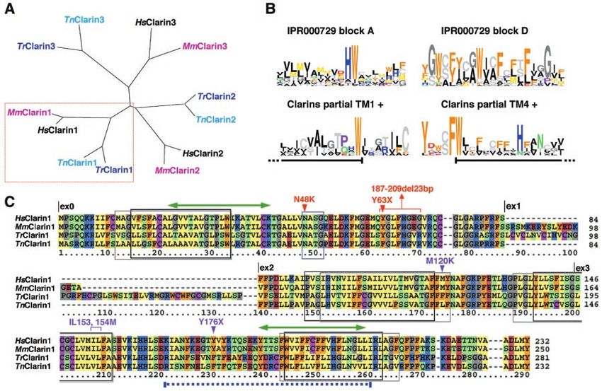
Synaptic role for USH3A gene
                                                         A Adato et al
340

      USH1C (MIM 276904)16,17. PDZ-domains are protein –                 were provided by the National Laboratory for the Genetics
      protein interaction modules that allow the binding to              of Israeli Populations. A total of 51 grandparents in families
      and clustering of specific membrane-associated proteins,           from the Centre d’Etude du Polymorphisme Humain
      such as receptors and ion channels.18 Cadherin23 and               (CEPH) were also studied as controls.
      protocadherin15 mutations have recently been shown to
      underlie USH1D (MIM 601067) and USH1F (MIM 602083),                RNA expression
      respectively.19 – 22 Most cadherins are integral membrane          The expression of Ush3a was determined by in situ hybridi-
      glycoproteins that mediate the calcium-dependent forma-            sation and reverse-transcription (RT) – PCR in mice at
      tion of cell – cell adhesion,23 while protocadherins are           embryonic day (E) 16, postnatal day (P)0, 5, 10, 15, 20,
      thought to be involved in a variety of functions, including        and 30. For RT – PCR, cochleae were dissected out of the
      neural development, neural circuit formation and forma-            temporal bones. Total RNA was prepared from cochleae
      tion of the synapse.24 Finally, mutations in Usherin, a            with TRI Reagent (Sigma). Genomic DNA was removed
      gene that encodes a basement membrane protein, cause               from all RNA samples using DNA-free (Ambion). Total
      USH2A (MIM276901)25.                                               RNA was purified using phenol-chloroform extraction
         Joensuu et al.26 have recently identified three mutations       (PCI; Gibco) and phase lock gel tubes (Eppendorf), followed
      (a missense, a nonsense and a 3 bp deletion) in exons 2            by an isopropanol precipitation. RT reactions were
      and 3 of a newly identified gene that maps to 3q24,                performed using Expand Reverse Transcriptase (Roche) with
      approximately 100 kb proximal to the previously defined            Homo-Oligomeric DNA d(T)12-18 and Random Hexamer
      USH3A linkage interval in 3q25.1. These mutations estab-           (Amersham Pharmacia Biotech). cDNA was amplified from
      lished this gene as the USH3A gene. Northern blot                  the RT reactions using primers (F) 5’-GGTCCAAGC-
      analysis and reverse-transcription PCR indicated the expres-       CATCCCCGTA-3’ AND (R) 5’- CCTCCTGCTTCTGTTATTTT-
      sion of the USH3A gene in several tissues, including retina.       CC-3’. As a control, primers spanning the last intron of
      Two predicted transmembrane domains were identified in             Myo6 (F) 5’-CTGGTGGTATGCCCATTTTGA-3’ and (R) 5’-
      its deduced protein product, which at the time did not             TCGCTTTGCATAAGGCATTTCTA-3’ were used.29
      show similarity to any known protein.26                               For in situ hybridisation, cochleae were dissected from
         In this study, we characterise new human and mouse              surrounding tissue and fixation was performed by immer-
      USH3A transcripts (AF495717; AF495718; AF495719 and                sion in 4% paraformaldehyde in PBS for 12 – 16 h at 48C.
      AF495720), identify three additional USH3 mutations,               The samples were processed without decalcification until
      analyse the USH3A expression pattern and by in situ hybri-         P10. From P10 onwards, the samples were dehydrated in
      disation localise transcripts of this gene to mouse cochlear       graded concentrations of methanol, from 25 – 100%,
      hair cells and spiral ganglion cells. We redefine the USH3A        followed by decalcification (to facilitate dissection) in 8%
      protein, name it clarin-1 and affiliate it to a new four trans-    formic acid in 100% methanol for up to 4 days. We
      membrane domain (4TM) vertebrate-specific protein family.          generated digoxygenin-labelled cRNA probes in a standard
      Furthermore, based on sequence similarities to stargazin, a        transcription reaction (Roche Molecular Biochemicals). We
      well-studied member of this hyperfamily, we suggest a role         performed whole mount in situ hybridisation as
      for clarin-1 in the hair cells synaptic junctions.                 described,30 with modifications. Finally, we visualised in
                                                                         situ reaction product by cryoprotecting whole cochleae
      Materials and Methods                                              with sucrose, embedding the tissue in OCT, and cryostat
      USH3 families and controls                                         sectioning (10 mm). Antisense and sense probes were in
      Blood samples were drawn by venipuncture after obtaining           vitro transcribed from a linearised vector, pPCR-Script
      informed consent in accordance with the guidelines of the          AMP SK(+) cloning vector (Stratagene), containing a
      Tel Aviv University Helsinki Committee. Three of the               partial mouse Ush3a cDNA (corresponding to exons 2
      Jewish Ashkenazi USH3 families included in this study,             and 3) by using either T3 or T7 RNA polymerase. No in
      were referred to us through the Center for Deaf – Blind            situ hybridisation signal was detected with the control
      Persons in Tel Aviv as part of a larger study on the genetics      sense RNA probe. Our use of animals was approved by
      of Usher syndromes in Israel. The fourth Jewish Ashkenazi          the Tel Aviv University Animal Care and Use Committee
      USH3 family living in the US has one affected and two              (11-00-65).
      unaffected siblings whose grandparents originate from East-
      ern Europe. The family was ascertained by one of us (JGF).         Mutation detection and analysis
      The USH3 Jewish Yemenite and the Spanish families have             Using the primers detailed in Table 2 we amplified frag-
      been described elsewhere.27,28 Clinical diagnosis of affected      ments encompassing USH3A exons from genomic DNA
      members in these families is compatible with the USH3              templates of USH3 patients and their family members.
      phenotype and haplotype segregation analysis does not              PCR products were gel-purified, sequenced using dye termi-
      exclude linkage to the USH3A locus. Control DNA samples            nators of Big-Dyes kits (Perkin-Elmer/Applied Biosystems)
      from unrelated Jewish Ashkenazi and Yemenite individuals,          and analysed on an ABI 3700 sequencer (Perkin-Elmer/

European Journal of Human Genetics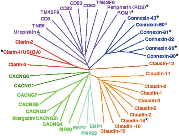
Synaptic role for USH3A gene
                                                                        A Adato et al
                                                                                                                                             341

                                    Table 1    Mutation and polymorphism in the USH3A gene
Exon/Intron        Base change                    Predicted AA change                 Frequency            Origin & No. of families

Disease causing mutations
  Ex0                143T4G                    N48K Asn4Lys (aat4aag)                   1/221        Jew. Hungarian (1), Jew. Russian (3)
  Ex0                189C4A                               Y63X                          0/135                   Spanish (1)
  Ex0            187-209del23bp      Frameshift at aa 63 and stop codon after 25 aa     0/199                Jew. Yemenite (1)

Polymorphism
  Ex0               771A4G                                UTR                           10/33          Ashkenazi Jew. (7: Russian, Polish,
                                                                                                          Hungarian, Rumanian), Jew.
                                                                                                       Moroccan (1), Jew. Yemenite (2)
  Ex0               +55A4T                        Ala19Ala (gca4gct)                    5/33         Jew. Moroccan (3), Jew. Russian (1),
                                                                                                              Jew. Yemenite (1)
  IVS1              +112a4g                              Silent                         5/18       Jew. Hungarian (1), Jew. Moroccan (1),
                                                                                                      Jew. Russian (1), Jew. Yemenite (2)
  IVS1              +135a4t                              Silent                         2/18        Jew. Moroccan (1), Jew. Yemenite (1)
  Ex3(IVS3a)    963-1002 ATnGTn                           UTR
  Ex3(IVS3a)       1013T4C                                UTR                           5/14       Jew. Hungarian (1)*, Jew. Russian (2)*,
                                                                                                            Jew. Yemenite (1)*
  Ex3(IVS3a)        1065T4C                               UTR                           5/14       Jew. Hungarian (1)*, Jew. Russian (2)*,
                                                                                                            Jew. Yemenite (1)*
Codon and nucleotide numbering starts from the codon of the first in-frame methionine of the USH3A ORF. The frequency column presents the
number of chromosomes carrying the sequence change divided by the number of all tested chromosomes. All USH3A exons from individuals
with USH3 and other family members, were amplified from leukocyte genomic DNA and screened for mutations by direct PCR sequencing. An
ATnGTn microsatellite at nucleotide positions 963 – 1002 that was identified in the 3’ UTR of USH3A exon 3 can be used as a polymorphic
genetic marker to test chromosomal segregation and linkage to the USH3 locus. *Carried on N48K chromosome.

Applied Biosystems). Sequence comparisons were performed                Results
by the use of Sequencher 4.1 software from GeneCodes                    Genomic analysis and transcripts
Corporation. The Jewish Ashkenazi N48K mutation was                     Since no mutations were identified within the described
screened by StuI digestion (16 h at 378C) followed by agar-             USH3A transcripts26 in patients from other USH3 families,
ose (2%) gel analysis. The 23 bp deletion (found in patients            we assumed that there might be additional uncharacterised
of Yemenite origin) was screened by PCR amplification of a              exons of this gene. Therefore, we assembled and recon-
227 bp fragment followed by FMC’s MetaPhor gel (4%) elec-               structed a 500 kb genomic interval spanning the
trophoresis. The Y63X mutation (found in a patient of                   published USH3A gene and the previous USH3A linkage
Spanish origin) was screened by SSCP; a 207-bp fragment                 interval, defined by D3S3413 and D3S1279 (Figure 1A).
of exon 0 was PCR amplified by primers ex0F2 (5-                        Based on gene predictions and alignment with human
ATCAAAGCCACTGTCCTCTG-3) and ex0R (5-CTGGGAA-                            and mouse ESTs, we designed primers that allowed the
GAGTCTGCCTAAA-3), and the alleles were separated on                     amplification of a new human USH3A transcript. This long-
0.76MDE gels, for 18 h at 5 W, at room temperature and                  er transcript begins with a newly identified exon 0,
were visualised by silver staining.31                                   continues with exon 2, has exons 3 and 4 transcribed
                                                                        together with their intervening intron, but does not
Computational analyses and sequence annotation                          include the previously identified exons 1 and 1b26 (Figure
Genomic sequences were retrieved from four NCBI                         1B).
(National Center for Biotechnology Information) Human                      The USH3A gene shows partial exon overlap (but no
clones: AC020636, AC036109, AC011103, AC078816. Frag-                   protein overlap) with the 5’ end of the UCRP pseudogene26
ments were assembled by Sequencher 4.1 software from                    in an opposite polarity, and in the same polarity with the
GeneCodes Corporation. Complete sequence annotation                     5’ end of a newly characterised gene, USHARF (USH3A
was performed using the GESTALT workbench.32 Sequence                   Alternative Reading Frame, ms in preparation) (Figure
similarity searches performed by the use of the BLAST33                 1C). By EST assembly and PCR amplification from mouse
and Blimps34 programs. Sequences were aligned using the                 inner ear cDNA library we also defined three alternative
BlockMaker34 and MACAW35 programs; PHYLIP ProtDist36                    transcripts of the Ush3A orthologue (Figure 1D). One of
PHYLIP – Phylogeny Inference Package (Version 3.2 Cladis-               these transcripts (AF495719) has the same exon-intron
tics 5: 164 – 166) and CLUSTAL W programs37 were used to                structure as the longest human mRNA, and its protein
compute trees and their significance (bootstrap) values. TM             product shares 88% identity with the product of the corre-
regions were predicted using the PHDhtm38 and TMHMM                     sponding human transcript (AF495717). A longer mouse
programs.39 Primers were designed using the Oligo primer                transcript (AF495718) has an extra exon coding for addi-
analysis software (http://medprobe.com/is/oligo/html).                  tional 18 amino acids (Figure 3C).

                                                                                                        European Journal of Human Genetics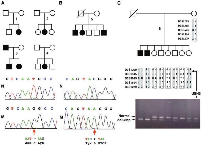
Synaptic role for USH3A gene
A Adato et al
342
Figure 1 (A) Schematic representation of physical and transcript maps covering the USH3A region comprised of partly overlapping
NCBI (National Center for Biotechnology Information) clones. Polymorphic markers and sequence-tagged sites are indicated on the bar
below the chromosome. Pink markers define the borders of the old USH3A linkage interval. The red marker is the newly identified
polymorphic intragenic microsatellite located within the USH3A’s 3’-UTR. Colored arrows below the clones indicate the position and
orientation of the UCRP pseudogene (light blue), the USH3A (red) and partially overlapping USHARF (green) genes. Below these arrows is
an enlarged view of an approximate 66 kb segment, showing the genomic structure of the USH3A gene and the first two exons of the
USHARF gene. Blue boxes and lines indicate coding exons, while grey boxes and lines indicate non-coding regions. Light blue and green
lines above the exons indicate overlapping regions between USH3A and, respectively, UCRP and USHARF. (B) Alternative transcripts of the
USH3A gene. The first two pale colored transcripts were defined previously by Joensuu et al. (2001).26 (C) The first two exons of the
USHARF gene. (D) Genomic structure of the mouse Ush3a gene and its three newly defined in frame alternative transcripts. Marked by
black arrows is the position of the primers that were used for the amplification of the (RT) – PCR and the in situ hybridisation probe.
Previously known USH3A human and mouse ESTs and mRNAs, amplified from several different tissues are listed in Table 3.
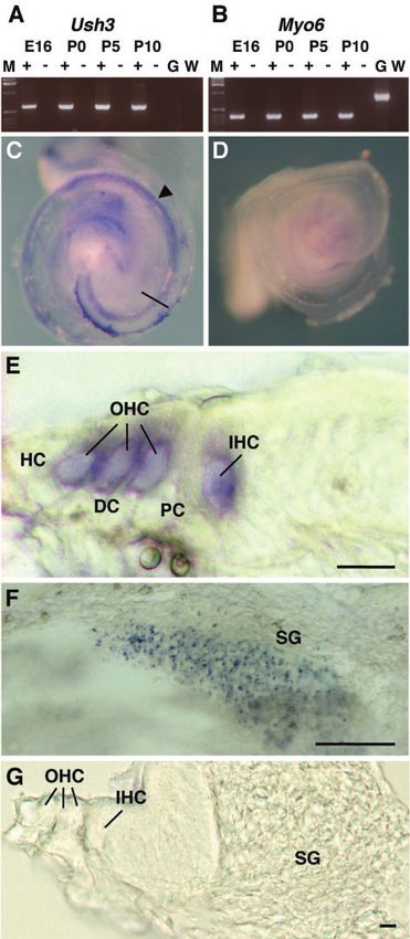
Expression patterns also demonstrate the expression of Ush3a transcripts in
By PCR amplification from cDNA or cDNA libraries we the auditory sensory organ. A developmental expression
confirmed the previously reported expression of USH3A in profile of Ush3a was performed both by PCR amplification
retina and skeletal muscle. In addition, we have amplified of mouse inner ear cDNA and whole mount in situ hybridi-
USH3A transcripts from human testis and olfactory epithe- sation on mouse cochleae. Ush3a transcripts were detected
lium cDNA. The later observation might be related to as early as E16, the earliest age tested, and at all postnatal
previously reported expression of two other Usher genes, stages examined (P0, P5, and P10) (Figure 2A,B). Hybridisa-
myosin VIIA and harmonin, in the olfactory tissue.40 – 43 tion with an Ush3a RNA probe on whole cochleae detected
Yet, despite the observation of anomalies in olfactory cilia, mRNA expression in the sensory epithelium at E16 (Figure
no chemosensory deficits have been confirmed in Usher 2C,D), P0, P5, P10, P15, P20 (n=4 for each age) (data not
patients.44 shown). Sectioning through these cochleae revealed specific
By PCR amplification of mouse inner ear cDNA and by hybridisation in the inner and outer hair cells of the organ
whole mount in situ hybridisation on mouse cochleae we of Corti (Figure 2E,G). No hybridisation was detected in the
European Journal of Human Genetics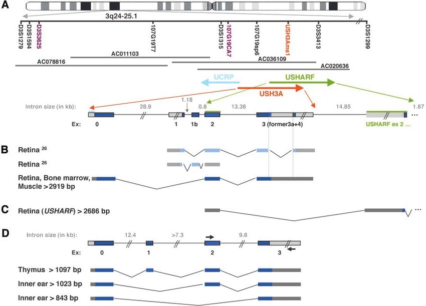
Synaptic role for USH3A gene
                                                                     A Adato et al
                                                                                                                                             343

                                                                     supporting cells of the organ of Corti, including the Deiters’
                                                                     cells, the pillar cells, and the Hensen cells. The only other
                                                                     hybridisation found in the cochlea was in the spiral gang-
                                                                     lion cells (Figure 2F) containing the primary neurons that
                                                                     innervate the cochlear sensory epithelia.

                                                                     Gene product and protein structure
                                                                     The open reading frame of the newly defined USH3A tran-
                                                                     script is expected to encode a 232 amino acid protein
                                                                     with four transmembrane domains, which we named clar-
                                                                     in-1, after the clarity of sensory perception allowed by the
                                                                     intact protein (Figure 3C). Sequence database searches by
                                                                     BLAST33 identified two additional clarin-1 human paralo-
                                                                     gues, clarin-2 and -3 (Figure 3A). The clarins were found
                                                                     to have respective orthologues in mouse as well as in fish
                                                                     (Takifugu rubripes [Fugu] and Tetraodon nigroviridis), all
                                                                     encoding small 224 – 284 amino acid proteins (Figure 3A).
                                                                     A partial clarin-1 mRNA was also identified in chicken.
                                                                     No orthologues were identified in the genomes of prokar-
                                                                     yotes, yeast, plants, nematodes and insects. Four
                                                                     transmembrane-domains (TMs), conserved sequence motifs
                                                                     and a single glycosylation consensus site between TM1 and
                                                                     TM2 characterise the clarin family members (Figure 3C).
                                                                        In a wider context the clarins appear to belong to a large
                                                                     hyperfamily of small integral membrane glycoproteins with
                                                                     four transmembrane domains. These include the tetraspan-
                                                                     nins45 (InterPro families PR00218 and PS00930) as well as
                                                                     the PMP22/EMP/MP20 and claudin family (InterPro family
                                                                     IPR000729) that perform diverse membrane transport,
                                                                     transduction and cell – cell interaction and scaffolding func-
                                                                     tions. Yet, BLAST searches did not provide a sequence
                                                                     similarity basis for a relationship between clarin-1 and
                                                                     any members of this hyperfamily. To examine this question

                                                                     mouse cochlea was done using digoxigenin-labelled sense and
                                                                     antisense riboprobes for Ush3a, followed by cryosectioning to
                                                                     confirm specific cell patterns. Expression was shown in the hair
                                                                     cells of the cochlea and in spiral ganglion cells at all ages tested.
                                                                     (C) Whole mount of mouse cochlea at E16 reveals specific
                                                                     labelling of sensory epithelium by the Ush3a antisense probe
                                                                     (arrow). Bar indicates area of cross-section for (E). (D) Absence
                                                                     of labelling with the Ush3a sense probe in a whole mount
                                                                     cochlea. (E) Cross section of cochlea in C showing a portion of
                                                                     the organ of Corti and specific labelling of the Ush3a probe only
                                                                     in the inner (IHC) and outer hair cells (OHC). Hybridisation is
                                                                     absent in the supporting cells, including the pillar cells (PC),
Figure 2 Expression of Ush3a in mouse cochlea by reverse             Deiters’ cells (DC), and Hensen’s cells (HC). Scale bar, 10 mm.
transcriptase (RT) – PCR and in situ hybridisation. (A) Expression   Left corresponds to the peripheral end of the section bar in C. (F)
of Ush3a in mouse cochlea by RT – PCR at E16 and P0, P5, and         The region containing the spiral ganglion (SG) cells is shown. No
P10. RNA derived from cochlear ducts was used to amplify a           staining was seen in any other regions of the cochlea upon
605 bp fragment spanning exons 2 – 4 of the Ush3a gene by            sectioning. Scale bar, 50 mm. (G) Cross section of cochlea
RT – PCR. Amplifications were carried out with (+) and without       showing hair cells, supporting cells, and spiral ganglion in D
(7) RT on cochlear RNA, on mouse genomic DNA (G), and a              demonstrating lack of hybridisation with sense probe. Scale bar,
water control (W). (B) A known cochlear-expressing gene,             10 mm. All experiments with mice were carried out with the
myosin VI (Myo6),64 amplified with primers spanning an intron        approval of the Tel Aviv University Animal Care and Use
from the same RNA preparation. (C – G) In situ hybridisation on      Committee (11-00-65).

                                                                                                       European Journal of Human Genetics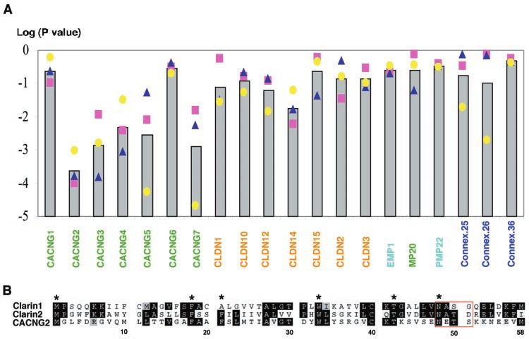
Synaptic role for USH3A gene
                                                          A Adato et al
344

      Figure 3 (A) Unrooted tree reflecting sequence relations between protein products of 12 of the 14 identified clarins. Sequence similarity
      searches revealed 14 members of the 4TM USH3A gene family, 12 of which, with the most completed sequences, are presented in this tree.
      These include HsClarin-1 (USH3A) and its two predicted paralogues HsClarin-2 (LOC166908 product of XM_068256) and HsClarin-3
      (LOC119467 product of XM_058398), that map to 4p15.33 and 10q26.2 respectively; MmClarin-1 (Ush3a) and its two predicted para-
      logues MmClarin-2, deduced from TblastN results against Celera’s mouse genomic assembly, and MmClarin-3 (product of Celera’s anno-
      tated transcript mCP9472) that map to mouse chromosomes 5 and 7 respectively; three predicted Fugu gene products, TrClarin-1
      (JGI_31106), TrClarin-2 (JGI_10159) and TrClarin-3 (JGI_29906) that was corrected according to TblastN and Block analysis results; Three
      predicted Tetraodon genes that were also reconstructed from TblastN results against Genoscope Tetraodon genomic traces and named
      TnClarin1,TnClarin-2 and TnClarin-3. Prediction of a partial mRNA of a bovine gene (BF044503) were also included in the analyses of the
      USH3 gene family but are not shown in the figure. Block multiple alignment analysis34 of the clarins identified several conserved sequence
      motifs covering about 95% of the sequence of clarin-1 and 80% or more of the sequences of other family members. Both PHYLIP and
      CLUSTAL W, used to compute trees and their significance (bootstrap) values, programs gave identical topologies. Included branch points
      have 70% and higher bootstrap values. (B) Sequence logo of the two regions (indicated in C by green arrows ) that are similar between the
      clarins and members of the IPR000729 protein family. In the logo, the height of each amino acid is scaled in bits of information and is
      proportional to its degree of conservation.34 Black lines under the logos indicate TM prediction. (C) Alignment of clarin-1 and its three
      orthologues including MmClarin1, TrClarin1 (JGI_31106) and TnClarin1, which are boxed in red on the phylogenetic tree (A). Newly
      identified and previously described26 USH3A mutations are indicated, respectively, by red and purple arrows. The starting position of each
      exon is indicated above the sequence. Black and grey boxes indicate TM positions according to, respectively, PHDhtm analysis of the four
      aligned proteins and TMHMM analysis of clarin-1. Boxed in blue is the putative N- glycosylation site. Green arrows indicate the positions of
      regions, where clarins share similarity with members of the IPR000729 protein family. Human and mouse clarin-1 harbour in their
      C-terminal region a TNV signature that might serve as a PDZ-binding motif. However this signature is not conserved among other members
      of the clarins. Gaps in the aligned sequence originate from the full alignment of all 12 clarins presented in the tree (A).

      further, it was necessary to employ more sensitive search                 looking for remote but statistically significant similarities
      routines, capable of detecting subtle sequence similarities.              between the clarin Blocks and all others. Two of the clarin
      First, we asked whether any of the clarins contained protein              Blocks, in and around the first and fourth putative TM
      motifs already included in the BLOCKS database, but the                   domains generated a hit with two of the 4TM proteins
      results were negative. Therefore, using the newly discovered              Blocks (Figure 3B,C). Pairwise Smith-Waterman align-
      group of 12 clarin sequences, we defined novel clarin-speci-              ments47 then allowed us to delineate the broad similarity
      fic Blocks motifs. These were subsequently analysed by the                relations between the clarins and representative members
      Local Alignment of Multiple Alignments (LAMA) method,46                   of several human 4TM families (Figure 5).

European Journal of Human Genetics
Synaptic role for USH3A gene
A Adato et al
345
In order to find out which of the 4TM proteins is most haplotypes on carrier chromosomes (data not shown)
highly related to the clarins, we performed further pairwise suggest the existence of a founder effect for this mutation.
alignments with accurate statistical testing (Figure 6). This A 189C4A substitution, expected to cause a Y63X nonsense
highlighted the calcium channel gamma subunit proteins mutation, was found in a homozygous state in three
(CACNG) as best clarin sequence matches. Of these affected individuals from a non-consanguineous Spanish
CACNG2 (stargazin)48,49 was the only protein showing family (Figure 4B; Table 1). A 23 bp deletion, spanning
significant matches (P50.001) to all three clarins. It is note- nucleotides 187 – 209 downstream from the first methio-
worthy that like in some USH3 cases,2 stargazin mutation nine codon, was found in a homozygous state in two
appears to affect also the inner ear vestibular function.48,49 affected individuals from a non-consanguineous family of
Yemenite Jewish origin (Figure 4C).
USH3A mutations
Three novel USH3 mutations were identified among 10 Discussion
affected individuals from six USH3A families (Table 1 and Affiliation of clarin-1 to the newly defined clarin family and
Figure 4). A 143T4G substitution expected to cause a the definition of several motifs characterizing all members
N48K missense mutation was found in six affected indivi- of this family indicate that the full-length coding sequence
duals from four unrelated families of Eastern European of the clarin-1 has now been determined. The additional
Jewish origin (Figure 4A). Shared microsatellite and SNP 18aa encoded by the longest mouse transcript as well as
Figure 4 USH3 families and mutations. (A) Four Eastern-European Jewish USH3 families. Below the pedigrees are parts of exon 0
chromatograms of DNA from non-carrier (top) and affected (bottom) individuals. Marked with an arrow is the 143T4G substitution,
which is expected to cause an N48K missense mutation. Five patients from families 2 – 4 are homozygous for the 143T4G substitution
while the patient from family 1 carries this substitution only on one allele. The second USH3A mutation of this individual was not
detected. This result might be due to mutation in unexplored control regions or to a gross deletion, which in hemizygous state is hard to
detect by the standard methods used in this work. (B) An USH3 family of Spanish origin. Below are parts of exon 0 chromatograms of
non-carrier and affected individuals. Marked with an arrow is the A 189C4A substitution that is expected to cause a Y63X nonsense
mutation. (C) An USH3 family of Yemenite Jewish origin. Below is FMC’s MetaPhor agarose-gel electrophoresis of USH3A exon 0
fragments containing the 187 – 209 23 bp, which were amplified from genomic DNA samples of all family members.
European Journal of Human GeneticsSynaptic role for USH3A gene
A Adato et al
346
Figure 5 Schematic relations of clarins to other four-transmembrane-domain proteins. Each sequence family is colour coded as follows:
clarins: red, tetraspanins: purple, connexins: blue, claudins: orange and brown, EMPs and PMP22: turquoise, MP20 and calcium
channel g subunit-like proteins (CACNGs): green, CACNG1 and CACNG6: dark green. Claudins, EMPs, PMP22, MP20 and CACNGs all
belong to the IPR000729 protein family. Marked with a black star are genes that were previously shown to be involved in forms of
deafness or retinal diseases. Distances between all sequences were calculated by aligning each sequence pair. The presented unrooted
tree was calculated from the resulting distance matrix using the Neighbor-Joining method.34 Sequence SwissProt/TREMBL accessions are
clarin-1 (USH3A): this work, clarin-2: XP_068256.1, clarin-3: XP_058398.1, CACNG1: CCG1_HUMAN, CACNG2 (stargazin):
CCG2_HUMAN, CACNG3: CCG3_HUMAN, CACNG4: CCG4_HUMAN, CACNG5: CCG5_HUMAN, CACNG6: CCG6_HUMAN, CACNG7:
CCG7_HUMAN, MP20: LMIP_HUMAN, PM22 : PM22_HUMAN, EMP1: EMP1_HUMAN, EMP2: EMP2_HUMAN, claudin 1:
CLD1_HUMAN, claudin 2: CLD2_HUMAN, claudin 3: CLD3_HUMAN, claudin 5: CLD5_HUMAN, claudin 8: CLD8_HUMAN, claudin 10:
CLDA_HUMAN, claudin 11: CLDB_HUMAN, claudin 12: CLDC_HUMAN, claudin 14: CLDE_HUMAN, claudin 15: CLDF_HUMAN,
connexin 43: CXA1_HUMAN, connexin 50: CXA8_HUMAN, connexin 32: CXB1_HUMAN, connexin 26: CXB2_HUMAN, connexin 31:
CXB3_HUMAN, connexin 30: CXB6_HUMAN, CD82: CD82_HUMAN, CD53: CD53_HUMAN, CD9: CD9_HUMAN, CD63:
CD63_HUMAN, TM4SF6: T4S6_HUMAN, TM4SF9: T4S9_HUMAN, peripherin (RDS) : RDS_HUMAN, ROM1: ROM1_HUMAN, TNE6:
TNE6_HUMAN, uroplakinA: UPKA_HUMAN.
additional protein segments encoded by the long tran- mutations likely render clarin-1 functionally inactive, and
scripts of Trclarin-1 and Hsclarin-2 elongate the putative account for the disease in most studied USH3A patients
first extracellular loop (Figure 3C). On the other hand, (Figure 4). Of the previously identified missense muta-
Trclarin-3 and Tnclarin-3 seem to be missing the C-terminal tions,26 one (M120K under the new enumeration) leads to
TM domain, indicated by a dashed blue line in Figure 3C. the replacement of a hydrophobic methionine residue by
The fact that no orthologues were identified in the a charged lysine inside or very near to the border of TM2,
genomes of prokaryotes, yeast, plants, nematodes and and another (IL153,154M) results in the shortening of
insects, suggests that this gene family is limited to verte- TM3 by one residue. Because the four TMs are rather well
brates. conserved, such mutations may also be functionally deleter-
The single missense USH3A mutation newly identified in ious.
this study, N48K, disrupts the molecule’s only N-glycosyla- USH3 is progressive, i.e. at birth both vision and audition
tion consensus site (Figure 3C), and may render clarin-1 are much less severely impaired than later in life. This
incapable of proper intracellular trafficking and plasma implies that the role played by clarin-1 in the hair cell
membrane insertion.50 Both other newly identified muta- and retina may display at least a measure of functional
tions, the Y63X and the 187 – 209del23 bp mutations, are redundancy. The best candidates for serving as substitute
expected to result in truncated proteins. Thus, these three for clarin-1 may be its two orthologues, clarin-2 and clar-
European Journal of Human GeneticsSynaptic role for USH3A gene
A Adato et al
347
Figure 6 (A) Significance values of sequence similarity scores (P values) between the three human clarin paralogues and some other
four transmembrane-domain proteins. P values were calculated using the PRSS program47 with 1000 shuffled sequences and the
program’s default parameters. Changing the shuffling method from uniform to window did not change the results significantly (not
shown). Pink squares, blue triangles and yellow circles in this figure represent P values of comparisons with clarin-1, clarin-2 and clarin-3
respectively. Bars indicate the geometric means of the three. (B) Alignment of the first 58 amino acids of clarins-1 clarin-2 and stargazin.
Boxed in red is the putative N-glycosylation site. Average positions of the inferred first extracellular loop and N-glycosylation site were
found to be more similar between the clarins and the g subunits calcium channel CACNG1-5, which include stargazin, than between the
clarins and the claudins and other members of the IPR000729 protein family (data not shown).
Table 2 USH3A Exon – Intron junctions and primers for genomic amplification of exons
Exon 5’ Junction 3’ Junction Exon length Primers for genomic amplification PCR product
0 .... CGGTTCTCATgtaagtagcaattgc 4290 bp F: CAGAAAAGGAGAAAAGCCAAG 480 bp
R: CTGGGAAGAGTCTGCCTAAA
1 .... CAGCAACCAGgtaggggtgcctgca 4390 bp F: TCACTATCTGAAACTATCTTGTTGT 910 bp
R: AAGCCCCTGAACTTTATAGG
1b tcactgagcacctacTATGTGCCAG AGCTTATAACctaatgggagaagac 87 bp F: TTGTGGCCATTTTTGGAGAT 207 bp
R: CCCCAAACATGTATCAAGTGC
2 cctttcggttctcaTTTTTTCCAGA TTCATTTCAGgtaagtacaaaattc 180 bp F: TCAGAAGGATTTTAGTGATGTTTGA 355 bp
R: TCTTTTTGACATATTGAAAAGCACA
3 tgagcttcatttcagGCTCCTGTGG .... 41638 bp F: ATGTCAATGGGGATGATGGT 1866 bp
R: AGCATCTGGAAACTCGGTGT
3a tgagcttcatttcagGCTCCTGTGG CTCATTCTGGgtcattttcttttgc 137 bp F: ATGTCAATGGGGATGATGGT 411 bp
R: GGAGCCCATTCAGAAAATGA
3b caaattgatctgcagCTGACTAAAG TGTCCTTCAAgattctttccaaata 3568 bp? F: TTCCCCTGAATTACCCATCA 339 bp
R: AGCATCTGGAAACTCGGTGT
in-3. However, it is also possible that other 4TM proteins Homepage: http://www.uia.ac.be/dnalab/hhh/). Mutations
might subserve this function. Future studies will be needed in connexin 50 cause congenital cataracts51 and mutations
to clarify the mechanism of time-dependent loss of such in human peripherin/RDS and mouse ROM were shown to
potential redundancy. disrupt photoreceptor morphogenesis, leading to retinitis
Other small 4TM proteins have previously been shown to pigmentosa.45 Also, mouse mutations in Cacng2 (stargazin)
underlie deafness and retinal diseases. Mutations in four cause defects not only in cerebellum but also in inner ear.52
different connexins (26, 30, 31, and 43) and in claudin-14 Yet, such information cannot help define more accurately
underlie several forms of deafness (Hereditary Hearing Loss the underlying mechanism for USH3, because the above
European Journal of Human GeneticsSynaptic role for USH3A gene
A Adato et al
348
Table 3 ESTs and mRNAs that align with the human and mouse USH3A transcripts
EST/mRNA Specie Align with GenBank details Organ/tissue & Dev. stage
USH3A
AF388366 Hs USH3A (USH3A) mRNA Retina
AF388368 Hs USH3A isoform b (USH3A) mRNA Retina
W27577 Hs 2066-2473 (+) cDNA randomly primed sublibrary Retina
BB591018 Mm 1-210(+) cDNA clone A830001O21 5’ 10 days neonate cortex
BB639483 Mm 64-425; 660-977 (+) cDNA clone A630099N24 5’ 3 days neonate thymus
BB638319 Mm 65-340(+) cDNA clone A630025E03 5’. 3 days neonate thymus
BB689483 Mm 66-425; 660-977 (+) cDNA clone 6820443B08 3’ 12 days mullerian duct
BB630393 Mm 238-880 (+) cDNA clone A130002D11 5’ 16 days neonate thymus
Hs - Homo sapiens; Mm - Mus musculus.
listed genes participate in diverse cellular pathways, includ- shown here to be homozygous for the 23 bp deletion in
ing the assembly and maintenance of gap junctions, tight USH3A exon 0 (Figure 4C). While the double heterozygous
junctions and synaptic junctions.53 – 55 mother and siblings are healthy, in the context of a homozy-
Because stargazin has been shown to play a key role in gous USH3A null mutation the presence of one null mutation
the shaping and maintenance of cerebellar synapses,55 our in the MYO7A tail (the out of phase one base deletion in the
protein sequence analysis is suggestive of a synaptic role complex rearrangement) mimics haploinsufficiency, and
for clarin-1 as well. The case is strengthened by corrobora- illustrates a departure from the monogenic model.
tive evidence: (a) Stargazin has been shown to interact Since MYO7A expression in the inner ear is restricted to
with PDZ domain-containing proteins of the Membrane sensory hair cells,60 – 62 including the sensory synaptic
Associated Guanylate Kinase type (MAGUK), serving as region,62 the present observation that Ush3a is expressed
intracellular anchors essential for the integrity of synaptic in mouse sensory hair cells, and the implication of a synap-
densities.55 Intriguingly, one form of the Usher syndromes tic role, are compatible with a clarin-1/MYO7A interaction.
(USH1C) is caused by mutations in another PDZ protein Furthermore, while myosin VIIA was localised only in pre-
harmonin. This may indicate that clarin-1 and harmonin synaptic cells, MyRIP, a most recently defined MYO7A-inter-
are part of the same synapse-formation pathway, despite acting protein,63 is present in both pre- and post-synaptic
the absence of a clear PDZ-binding consensus56 shared by cells, similar to clarin-1. The proposed clarin-1/MYO7A
all the clarins (Figure 3C). (b) Stargazin was shown to be interaction is also consistent with a pathophysiological
involved in controlling the expression and mobilisation of continuum between USH1 and USH3A, and is relevant to
amino-hydroxyl-methyl-isoxazole propionate (AMPA) gluta- their treatment. Access to Ush3 mutant models should
mate receptors to the post-synaptic cleft in cerebellar allow testing of these hypotheses and contributing to a
granule cells. That cochlear hair cells also utilize glutamate better understanding of the role of clarin-1 in the retina
(or a highly similar compound) as a rapid excitatory neuro- and inner ear.
transmitter57 seems relevant. (c) Stargazin and other
members of the CACNG family have been suggested to also
play a role of forming protein – protein contacts across the Acknowledgements
synaptic cleft.58 In bridging two cellular membranes they We are grateful to all patients and their family members who parti-
resemble other 4TM proteins such as the gap junction- cipated in this study. We would also like to thank Ronna Hertzano
for the preparation of the mouse inner ear cDNA. This work was
forming connexins and the tight junction-forming clau-
funded by an Infrastructure grant of the Israeli Ministry of Science
dins. That clarin-1 is expressed in inner ear, both pre- and Culture and Sports, the Crown Human Genome Center at The Weiz-
post-synaptically, is consistent with possible clarin – clarin mann Institute of Science, the Alfried Krupp Foundation and by the
homophylic interactions, which might work in the hair cell Finnish Eye and Tissue Bank Foundation, the Finnish Eye Founda-
synapse. Thus, we would like to propose that clarin-1 has a tion, the Maud Kuistila Memorial Foundation, the Oskar Oflund
role in the excitatory ribbon synapse junctions between Foundation, Finnish State grant TYH9235, the European Commis-
hair cells and cochlear ganglion cells,59 and presumably sion (QLG2-CT-1999-00988) (KB Araham) and by the Foundation
Fighting Blindness. JS Beckman holds the, Hermann Mayer professor-
also in analogous synapses within the retina.
ial chair and D Lancet holds the Ralf and Lois Silver professorial
We have previously reported a possible epistatic interac- chair.
tion between the USH3A locus and the MYO7A gene,27
whereby two USH3A haploidentical patients of a Yemenite
family showed different USH phenotypes, the patient with
the more severe, USH1-like phenotype was also found to be References
a carrier for a complex rearrangement (3260T4C and 1 Petit C: Usher syndrome: from genetics to pathogenesis. Annu Rev
3266delG) in the tail of myosin VIIA.27 Both patients are Genomics Hum Genet 2001; 2: 271 – 297.
European Journal of Human GeneticsSynaptic role for USH3A gene
A Adato et al
349
2 Pakarinen L, Karjalainen S, Simola KO, Laippala P, Kaitalo H: 24 Suzuki ST: Recent progress in protocadherin research. Exp Cell Res
Usher’s syndrome type 3 in Finland. Laryngoscope 1995; 105: 2000; 261: 13 – 18.
613 – 617. 25 Eudy JD, Weston MD, Yao S et al: Mutation of a gene encoding a
3 Kaplan J, Gerber S, Bonneau D et al: A gene for Usher syndrome protein with extracellular matrix motifs in Usher syndrome type
type I (USH1A) maps to chromosome 14q. Genomics 1992; 14: IIa. Science 1998; 280: 1753 – 1757.
979 – 987. 26 Joensuu T, Hamalainen R, Yuan B et al: Mutations in a novel gene
4 Kimberling W, Smith RJ: Gene mapping of the Usher syndromes. with transmembrane domains underlie Usher syndrome type 3.
Otolaryngol Clin North Am 1992; 25: 923 – 934. Am J Hum Genet 2001; 69: 673 – 684.
5 Smith RJ, Lee EC, Kimberling WJ et al: Localization of two genes 27 Adato A, Kalinski H, Weil D et al: Possible interaction between
for Usher syndrome type I to chromosome 11. Genomics 1992; 14: USH1B and USH3 gene products as implied by apparent digenic
995 – 1002. deafness inheritance. Am J Hum Genet 1999; 65: 261 – 265.
6 Wayne S, Der Kaloustian VM, Schloss M et al: Localization of the 28 Espinos C, Najera C, Millan JM et al: Linkage analysis in Usher
Usher syndrome type ID gene (Ush1D) to chromosome 10. Hum syndrome type I (USH1) families from Spain. J Med Genet 1998;
Mol Genet 1996; 5: 1689 – 1692. 35: 391 – 398.
7 Chaib H, Kaplan J, Gerber S et al: A newly identified locus for 29 Ahituv N, Sobe T, Robertson NG et al: Genomic structure of the
Usher syndrome type I, USH1E, maps to chromosome 21q21. human unconventional myosin VI gene. Gene 2000; 261: 269 –
Hum Mol Genet 1997; 6: 27 – 31. 275.
8 Kimberling WJ, Weston MD, Moller C et al: Localization of Usher 30 Wilkinson DG, Nieto MA: Detection of messenger RNA by in situ
syndrome type II to chromosome 1q. Genomics 1990; 7: 245 – hybridization to tissue sections and whole mounts. Methods Enzy-
249. mol 1993; 225: 361 – 373.
9 Hmani M, Ghorbel A, Boulila-Elgaied A et al: A novel locus for 31 Bassam BJ, Caetano-Anolles G, Gresshoff PM: Fast and sensitive
Usher syndrome type II, USH2B, maps to chromosome 3 at p23- silver staining of DNA in polyacrylamide gels. Anal Biochem
24.2. Eur J Hum Genet 1999; 7: 363 – 367. 1991; 196: 80 – 83.
10 Pieke-Dahl S, Moller CG, Kelley PM et al: Genetic heterogeneity of 32 Glusman G, Lancet D: GESTALT: a workbench for automatic inte-
Usher syndrome type II: localisation to chromosome 5q. J Med gration and visualization of large-scale genomic sequence
Genet 2000; 37: 256 – 262. analyses. Bioinformatics 2000; 16: 482 – 483.
11 Mustapha M, Chouery E, Torchard-Pagnez D et al: A novel locus 33 Altschul SF, Madden TL, Schaffer AA et al: Gapped BLAST and PSI-
for Usher syndrome type I, USH1G, maps to chromosome BLAST: a new generation of protein database search programs.
17q24-25. Hum Genet 2002; 110: 348 – 350. Nucleic Acids Res 1997; 25: 3389 – 3402.
12 Sankila EM, Pakarinen L, Kaariainen H et al: Assignment of an 34 Henikoff S, Henikoff JG, Alford WJ, Pietrokovski S: Automated
Usher syndrome type III (USH3) gene to chromosome 3q. Hum construction and graphical presentation of protein blocks from
Mol Genet 1995; 4: 93 – 98. unaligned sequences. Gene 1995; 163: GC17 – GC26.
13 Joensuu T, Hamalainen R, Lehesjoki AE, de la Chapelle A, Sankila 35 Schuler GD, Altschul SF, Lipman DJ: A workbench for multiple
EM: A sequence-ready map of the Usher syndrome type III critical alignment construction and analysis. Proteins 1991; 9: 180 – 190.
region on chromosome 3q. Genomics 2000; 63: 409 – 416. 36 Felsenstein J: PHYLIP – Phylogeny Inference Package(Version 3.2).
14 Weil D, Blanchard S, Kaplan J et al: Defective myosin VIIA gene Cladistics 1989: 164 – 166.
responsible for Usher syndrome type 1B. Nature 1995; 374: 60 – 37 Thompson JD, Higgins DG, Gibson TJ, Clustal W: improving the
61. sensitivity of progressive multiple sequence alignment through
15 Liu XZ, Hope C, Walsh J et al: Mutations in the myosin VIIA gene sequence weighting, position-specific gap penalties and weight
cause a wide phenotypic spectrum, including atypical Usher matrix choice. Nucleic Acids Res 1994; 22: 4673 – 4680.
syndrome. Am J Hum Genet 1998; 63: 909 – 912. 38 Rost B, Fariselli P, Casadio R: Topology prediction for helical
16 Bitner-Glindzicz M, Lindley KJ, Rutland P et al: A recessive contig- transmembrane proteins at 86% accuracy. Protein Sci 1996; 5:
uous gene deletion causing infantile hyperinsulinism, 1704 – 1718.
enteropathy and deafness identifies the Usher type 1C gene. 39 Krogh A, Larsson B, von Heijne G, Sonnhammer EL: Predicting
Nat Genet 2000; 26: 56 – 60. transmembrane protein topology with a hidden Markov model:
17 Verpy E, Leibovici M, Zwaenepoel I et al: A defect in harmonin, a application to complete genomes. J Mol Biol 2001; 305: 567 – 580.
PDZ domain-containing protein expressed in the inner ear 40 Arden GB, Fox B: Increased incidence of abnormal nasal cilia in
sensory hair cells, underlies Usher syndrome type 1C. Nat Genet patients with retinitis pigmentosa. Nature 1979; 279: 534 – 536.
2000; 26: 51 – 55. 41 Sahly I, El-Amraoui A, Abitbol M, Petit C, Dufier JL: Expression of
18 Kornau HC, Seeburg PH, Kennedy MB: Interaction of ion chan- myosin VIIA during mouse embryogenesis. Anat Embryol (Berl)
nels and receptors with PDZ domain proteins. Curr Opin 1997; 196: 159 – 170.
Neurobiol 1997; 7: 368 – 373. 42 Wolfrum U, Liu X, Schmitt A, Udovichenko IP, Williams DS:
19 Di Palma F, Holme RH, Bryda EC et al: Mutations in Cdh23, Myosin VIIa as a common component of cilia and microvilli. Cell
encoding a new type of cadherin, cause stereocilia disorganiza- Motil Cytoskeleton 1998; 40: 261 – 271.
tion in waltzer, the mouse model for Usher syndrome type 1D. 43 Marietta J, Walters KS, Burgess R et al: Usher’s syndrome type IC:
Nat Genet 2001; 27: 103 – 107. clinical studies and fine-mapping the disease locus. Ann Otol
20 Bork JM, Peters LM, Riazuddin S et al: Usher syndrome 1D and Rhinol Laryngol 1997; 106: 123 – 128.
nonsyndromic autosomal recessive deafness DFNB12 are caused 44 Seeliger M, Pfister M, Gendo K et al: Comparative study of visual,
by allelic mutations of the novel cadherin-like gene CDH23. auditory, and olfactory function in Usher syndrome. Graefes Arch
Am J Hum Genet 2001; 68: 26 – 37. Clin Exp Ophthalmol 1999; 237: 301 – 307.
21 Alagramam KN, Yuan H, Kuehn MH et al: Mutations in the novel 45 Hemler ME: Specific tetraspanin functions. J Cell Biol 2001; 155:
protocadherin PCDH15 cause Usher syndrome type 1F. Hum Mol 1103 – 1107.
Genet 2001; 10: 1709 – 1718. 46 Pietrokovski S: Searching databases of conserved sequence
22 Ahmed ZM, Riazuddin S, Bernstein SL et al: Mutations of the regions by aligning protein multiple-alignments. Nucleic Acids
protocadherin gene PCDH15 cause Usher syndrome type 1F. Am Res 1996; 24: 3836 – 3845.
J Hum Genet 2001; 69: 25 – 34. 47 Pearson WR, Lipman DJ: Improved tools for biological sequence
23 Nollet F, Kools P, van Roy F: Phylogenetic analysis of the cadherin comparison. Proc Natl Acad Sci U S A 1988; 85: 2444 – 2448.
superfamily allows identification of six major subfamilies besides
several solitary members. J Mol Biol 2000; 299: 551 – 572.
European Journal of Human GeneticsSynaptic role for USH3A gene
A Adato et al
350
48 Letts VA, Valenzuela A, Kirley JP et al: Genetic and physical maps 57 Ottersen OP, Takumi Y, Matsubara A et al: Molecular organization
of the stargazer locus on mouse chromosome 15. Genomics 1997; of a type of peripheral glutamate synapse: the afferent synapses of
43: 62 – 68. hair cells in the inner ear. Prog Neurobiol 1998; 54: 127 – 148.
49 Noebels JL, Qiao X, Bronson RT, Spencer C, Davisson MT: Starga- 58 Tomita S, Nicoll RA, Bredt DS: PDZ protein interactions regulat-
zer: a new neurological mutant on chromosome 15 in the mouse ing glutamate receptor function and plasticity. J Cell Biol 2001;
with prolonged cortical seizures. Epilepsy Res 1990; 7: 129 – 135. 153: F19 – F24.
50 van Geest M, Lolkema JS: Membrane topology and insertion of 59 Wagner HJ: Presynaptic bodies (ribbons): from ultrastructural
membrane proteins: search for topogenic signals. Microbiol Mol observations to molecular perspectives. Cell Tissue Res 1997;
Biol Rev 2000; 64: 13 – 33. 287: 435 – 446.
51 White TW, Goodenough DA, Paul DL: Targeted ablation of 60 Hasson T, Heintzelman MB, Santos-Sacchi J, Corey DP, Mooseker
connexin50 in mice results in microphthalmia and zonular MS: Expression in cochlea and retina of myosin VIIa, the gene
pulverulent cataracts. J Cell Biol 1998; 143: 815 – 825. product defective in Usher syndrome type 1B. Proc Natl Acad Sci
52 Letts VA, Felix R, Biddlecome GH et al: The mouse stargazer gene U S A 1995; 92: 9815 – 9819.
encodes a neuronal Ca2+-channel gamma subunit. Nat Genet 61 Hasson T, Gillespie PG, Garcia JA et al: Unconventional myosins
1998; 19: 340 – 347. in inner-ear sensory epithelia. J Cell Biol 1997; 137: 1287 – 1307.
53 Shibata Y, Kumai M, Nishii K, Nakamura K: Diversity and molecu- 62 El-Amraoui A, Sahly I, Picaud S et al: Human Usher 1B/mouse
lar anatomy of gap junctions. Med Electron Microsc 2001; 34: 153 – shaker-1: the retinal phenotype discrepancy explained by the
159. presence/absence of myosin VIIA in the photoreceptor cells.
54 Heiskala M, Peterson PA, Yang Y: The roles of claudin superfamily Hum Mol Genet 1996; 5: 1171 – 1178.
proteins in paracellular transport. Traffic 2001; 2: 93 – 98. 63 El-Amraoui A, Schonn JS, Kussel-Andermann P et al: MyRIP, a
55 Chen L, Chetkovich DM, Petralia RS et al: Stargazing regulates novel Rab effector, enables myosin VIIa recruitment to retinal
synaptic targeting of AMPA receptors by two distinct mechan- melanosomes. EMBO Rep 2002; 1818.
isms. Nature 2000; 408: 936 – 943. 64 64 AvrahamKB, Hasson T, Sobe T et al: Characterization of uncon-
56 Hillier BJ, Christopherson KS, Prehoda KE, Bredt DS, Lim WA: ventional MYO6, the human homologue of the gene responsible
Unexpected modes of PDZ domain scaffolding revealed by struc- for deafness in Snell’s waltzer mice. Hum Mol Genet 1997; 6:
ture of nNOS-syntrophin complex. Science 1999; 284: 812 – 815. 1225 – 1231.
European Journal of Human GeneticsYou can also read