The Value of Immune-Related Genes Signature in Osteosarcoma Based on Weighted Gene Co-expression Network Analysis - Hindawi.com
←
→
Page content transcription
If your browser does not render page correctly, please read the page content below
Hindawi
Journal of Immunology Research
Volume 2021, Article ID 9989321, 17 pages
https://doi.org/10.1155/2021/9989321

Research Article
The Value of Immune-Related Genes Signature in Osteosarcoma
Based on Weighted Gene Co-expression Network Analysis

 Xin Wang,1 Li Gan,2 Ju Ye,3 Mengjie Tang ,4 and Wei Liu 5

 1
 Department of Bone and Soft Tissue, Hunan Cancer Hospital, The Affiliated Cancer Hospital of Xiangya School of Medicine,
 Central South University, Changsha 410013, China
 2
 Department of Anesthesiology, Hunan Cancer Hospital, The Affiliated Cancer Hospital of Xiangya School of Medicine,
 Central South University, Changsha 410013, China
 3
 Department of Pharmacy, Zunyi Medical University, Zunyi 563000, China
 4
 Department of Pathology, Hunan Cancer Hospital, The Affiliated Cancer Hospital of Xiangya School of Medicine,
 Central South University, Changsha 410013, China
 5
 Department of Pharmacy, The Third Xiangya Hospital, Central South University, Changsha 410013, China

 Correspondence should be addressed to Mengjie Tang; tangmengjie@hnca.org.cn and Wei Liu; liuweixy3@csu.edu.cn

 Received 16 March 2021; Accepted 25 April 2021; Published 15 May 2021

 Academic Editor: Jialiang Liang

 Copyright © 2021 Xin Wang et al. This is an open access article distributed under the Creative Commons Attribution License,
 which permits unrestricted use, distribution, and reproduction in any medium, provided the original work is properly cited.

 Background. Osteosarcoma (OS) is a serious malignant tumor that is more common in adolescents or children under 20 years of
 age. This study is aimed at obtaining immune-related genes (IRGs) associated with the progression and prognosis of OS.
 Method. Expression profiling data and clinical data for OS were downloaded from the Therapeutically Applicable Research to
 Generate Effective Treatments (TARGET) database. ESTIMATE calculates immune scores and stromal scores of samples and
 performs the prognostic analysis. Weighted gene coexpression network analysis (WGCNA) was used to find modules correlated
 with immune and stromal scores. Cox regression analysis and least absolute shrinkage and selection operator (LASSO) analysis
 were used to explore IRGs associated with OS prognosis and construct and validate a hazard score model. Finally, we verified
 the expression and function of EVI2B in OS. Results. WGCNA selected twenty-eight IRGs, 10 of which were associated with OS
 prognosis, and LASSO further obtained three key prognostic genes. A prognostic model of EVI2B was constructed, and
 according to the risk score model, patients in the high-risk group had a worse prognosis than those in the low-risk group, and
 the prognosis was statistically significant in the high- and low-risk groups. Receiver operating characteristic (ROC) curves were
 used to assess the prognostic model’s accuracy and externally validate the independent GSE21257 cohort. The results of
 immunohistochemical staining and qPCR showed that EVI2B was a tumor suppressor gene. The differential genes in the high-
 and low-risk groups were analyzed by enrichment analysis of GO and KEGG, indicating that the EVI2B model is associated
 with immune response. Conclusion. In this study, IRG EVI2B is closely related to OS’s prognosis and can be used as a potential
 biomarker for prognosis and treatment of OS.

1. Introduction 70% for the localized disease at diagnosis [5, 6] and 20–30%
 for metastatic disease at diagnosis or recurrence [7].
Osteosarcoma is a malignant tumor that occurs more com- Although advances have been made in surgical techniques,
monly in adolescents or children under 20 years of age [1]. targeted therapy and tumor immunity, and complications
Conventional therapies for osteosarcoma include surgery, such as infection and poor survival due to limb salvage
adjuvant chemotherapy, and neoadjuvant chemotherapy surgery, there are still many issues to be addressed in
[2–4]. For patients with osteosarcoma, the use of standard the treatment of OS; therefore, there is an urgent need
multiagent chemotherapy in combination with surgical to develop new prediction methods to improve OS
resection produces a long-term survival of approximately patients’ survival [5, 8].
2 Journal of Immunology Research

 OS-related gene expression data
 were retrieved from the TARGRT
 (https://ocg.cancer.gov/programs/tar-

 +++++ +++++
 ++ + +++++
 +++ ++++++++++++++ + +++++

 OS probability
 OS probability
 +
 + ++ + + + + +++++ + + + + +
 + + + ++
 The prognosis of immune score ++++++++ + + ++++++++
 ++ +
 and stromal score was analyzed + + + +

 using the R package survival in OS.
 Time (years) Time (years)
 Immune score Stromal score

 Cluster tree Modules Interesting module
 Gene co-expression
 module was identified
 in OS using WGCNA.

 Stromal score
 Immune score Stromal score Immune score

 Univariate cox analysis and
 Multivariate cox analysis

 1 gene model

 TARGET dataset (n = 85)
 ROC curve of TARGET cohort

 GSE21257 dataset (n = 53) Model evaluation
 and validation

 Enrichment analysis
 BP
 CC

 Comparison with
 experimental validation

 Figure 1: Flowchart of the present study.
Journal of Immunology Research 3

 1.00 ++++
 ++ ++
 + +++
 ++++++ ++++ +
 ++
 0.75 ++ + + +
 +

 OS probability
 + +
 0.50 ++++
 + ++ ++ + +
 p = 0.001 + +
 0.25
 HR = 0.27
 95% CI: 0.13 − 0.56
 0.00
 0 1 2 3 4 5 6 7 8 9 10 11 12 13 14 15 16
 Time (years)
 Immune score

 Number at risk
 High (n = 43) 43 41 31 27 25 15 10 7 6 4 2 1 0 0 0 0 0
 Low (n = 42) 42 37 24 18 17 13 8 6 5 3 3 1 1 1 1 1 1
 0 1 2 3 4 5 6 7 8 9 10 11 12 13 14 15 16
 Time (years)
 Immune score
 + High (n = 43)

 + Low (n = 42)
 (a)

 1.00
 + ++++++
 + ++ +++
 ++ +++
 0.75 ++++
 + ++++++ + + + + +
 OS probability

 + +++++ ++ +++
 0.50
 ++ +

 p = 0.008 + +
 0.25
 HR = 0.36
 95% CI: 0.17 − 0.76
 0.00
 0 1 2 3 4 5 6 7 8 9 10 11 12 13 14 15 16
 Time (years)
 Stromal score

 Number at risk
 High (n = 43) 43 42 32 27 24 15 7 5 5 4 2 1 1 1 1 1 1
 Low (n = 42) 42 36 23 18 18 13 11 8 6 3 3 1 0 0 0 0 0
 0 1 2 3 4 5 6 7 8 9 10 11 12 13 14 15 16
 Time (years)
 Stromal score
 + High (n = 43)

 + Low (n = 42)
 (b)

Figure 2: The relationship between immune score and stromal score for OS prognosis. (a) Survival analysis of the correlation between
immune score and overall survival time in OS. (b) Survival analysis of the correlation between stromal score and overall survival time in
OS. Immune/stromal scores: the significance is to predict tumor purity by immune and stromal scores. Log-rank p values were less than
0.05 with statistically significant. OS: osteosarcoma.

 A large body of evidence suggests that OS has an immune tumor immunosuppressants, thereby increasing endoge-
system with multiple therapeutic targets, including receptor nous antitumor activity, and could improve therapy-
T cell therapy [9], HER2-specific immunity [10–12], and induced tumor immunogenic chemotherapy, radiotherapy,
adjuvant immune therapy with mifamurtide [13]. Immune and targeted therapy [13–17]. To date, the impact of IRGs
checkpoint inhibitors may develop immune tolerance to on OS prognosis is not well clear, so prognostic
4
 Height
 Height
 0 10000 20000 30000 40000 50000 60000
 5000 10000 15000 20000 25000 30000

 T ARGET−40− PA UXPZ−01

 Immune score
 Stromal score
 TARGET−40−PALHRL−01
 TARGET−40−PALHRL−01
 TARGET−40−PAMLKS−01
 TARGET−40−PAMLKS−01
 TARGET−40−PALFYN−01
 TARGET−40− PALFYN−01
 TARGET−40−PARFTG−01
 TARGET−40−PARFTG−01
 TARGET−40−PAPWWC−01
 TARGET−40− PAPWWC−01
 TARGET−40−PANVJJ−01
 TARGET−40−PANVJJ−01
 TARGET−40−PASFCV−01
 TARGET−40−PASFCV−01
 TARGET−40−0A4I4O−01
 TARGET−40−0A4I4O−01
 TARGET−40−P APNVD−01
 TARGET−40−PAPNVD−01
 TARGET−40−P ASSLM−01
 TARGET−40−PASSLM−01
 TARGET−40−PALKDP−01
 TARGET−40−PALKDP−01
 TARGET−40−PA UBIT−01
 TARGET−40− PAUBIT−01
 TARGET−40− PA TJVI−01
 TARGET−40−PATJVI−01
 TARGET−40−P ATA WV−01
 TARGET−40−PATAWV−01
 TARGET−40−P APFLB−01
 TARGET−40−PAPFLB−01
 TARGET−40−P ALZGU−01
 TARGET−40−PALZGU−01
 TARGET−40−P AKXLD−01
 TARGET−40−PAKXLD−01
 TARGET−40−0A4HY5−01 TARGET−40−0A4HY5−01
 TARGET−40−0A4I5B−01
 TARGET−40−0A4I5B−01
 TARGET−40−PA UUML−01
 TARGET−40−PAUUML−01
 TARGET−40− PA TKSS−01
 TARGET−40−PATKSS−01
 TARGET−40−PARDAX−01 TARGET−40−P AR D AX−01

 TARGET−40−PATMPU−01 TARGET−40−PA TMPU−01
 TARGET−40−PA TMXR−01
 TARGET−40−PATMXR−01
 TARGET−40−0A4I9K−01
 TARGET−40−0A4I9K−01
 TARGET−40−P AMJXS−01
 TARGET−40−PAMJXS−01
 TARGET−40−PARGTM−01 TARGET−40−P ARGTM−01

 TARGET−40−PATPBS−01 TARGET−40−PATPBS−01
 TARGET−40−P AMHLF−01
 TARGET−40− PAMHLF−01
 TARGET−40− PANPUM−01 TARGET−40−P ANPUM−01

 TARGET−40−0A4I0Q−01 TARGET−40−0A4I0Q−01
 TARGET−40−PAMTCM−01 TARGET−40−P AMTCM−01
 TARGET−40−0A4I42−01 TARGET−40−0A4I42−01

 TARGET−40−PANGPE−01 TARGET−40−P ANGPE−01
 TARGET−40−PASNZV−01 TARGET−40−P ASNZV−01
 TARGET−40−PARKAF−01 TARGET−40−P ARKAF−01
 TARGET−40−PALWWX−01 TARGET−40− PAL WWX−01
 TARGET−40−0A4HXS−01 TARGET−40−0A4HXS−01
 TARGET−40−PAMEKS−01 TARGET−40−P AMEKS−01
 TARGET−40−PARJXU−01 TARGET−40−P ARJXU−01

 (a)

 (b)
 TARGET−40−PAKFVX−01 TARGET−40− P AKFVX−01
 TARGET−40−0A4I0S−01 TARGET−40−0A4I0S−01
 TARGET−40−PANZHX−01 TARGET−40−P ANZHX−01
 TARGET−40−PANSEN−01 TARGET−40−P ANSEN−01
 TARGET−40−PANMIG−01 TARGET−40−P ANMIG−01
 Sample clustering to detect outliers

 TARGET−40− PAVCLP−01 TARGET−40−PA VCLP−01

Figure 3: Continued.
 TARGET−40−PASKZZ−01 TARGET−40−P ASKZZ−01
 TARGET−40−PAVECB−01 TARGET−40−PA VECB−01
 TARGET−40−PALKGN−01 TARGET−40−P ALKGN−01

 hclust (*,
 TARGET−40− PASUUH−01 TARGET−40−P ASUUH−01

 av
 TARGET−40−PAMRHD−01 TARGET−40−P AMRHD−01

 "
 TARGET−40−PAPKWD−01 TARGET−40−P APKWD−01
 Sample dendrogram and trait heatmap

 TARGET−40−PASEFS−01 TARGET−40−P ASEFS−01
 TARGET−40−0A4I48−01 TARGET−40−0A4I48−01
 TARGET−40−PASRNE−01 TARGET−40−P ASRNE−01
 TARGET−40−PAUTWB−01 TARGET−40−PA UTWB−01
 TARGET−40−PATMIF−01 TARGET−40−PATMIF−01
 TARGET−40−PATUXZ−01 TARGET−40−PA TUXZ−01

 dist(datExpr0)
 TARGET−40−PATEEM−01 TARGET−40− PA TEEM−01

 erage")
 TARGET−40−0A4I4E−01 TARGET−40−0A4I4E−01
 TARGET−40−0A4I6O−01
 TARGET−40−0A4I6O−01
 TARGET−40−PAVALD−01
 TARGET−40−PAVALD−01
 TARGET−40− PAMYYJ−01 TARGET−40−P AMYYJ−01
 TARGET−40−PASYUK−01
 TARGET−40−P ASYUK−01
 TARGET−40−PASEBY−01
 TARGET−40−P ASEBY−01
 TARGET−40−0A4HX8−01
 TARGET−40−0A4HX8−01
 TARGET−40−0A4I3S−01
 TARGET−40−0A4I3S−01
 TARGET−40−PALECC−01
 TARGET−40−P ALECC−01
 TARGET−40−PAPIJR−01
 TARGET−40−P APIJR−01
 TARGET−40−0A4I4M−01
 TARGET−40−0A4I4M−01
 TARGET−40−0A4I0W−01
 TARGET−40−0A4I0W−01
 TARGET−40−PAUYTT−01
 TARGET−40−PA UYTT−01
 TARGET−40−PAPXGT−01
 TARGET−40−P APXGT−01
 TARGET−40− PAMHYN−01
 TARGET−40−P AMHYN−01
 TARGET−40− PARBGW−01
 TARGET−40−P ARBGW−01
 TARGET−40−0A4I65−01
 TARGET−40−0A4I65−01
 TARGET−40−PAKUZU−01
 TARGET−40−PAKUZU−01
 TARGET−40− PAVDTY−01
 TARGET−40−PA VDTY−01
 TARGET−40− PAUTYB−01
 TARGET−40−PA UTYB−01
 TARGET−40−PAUVUL−01
 TARGET−40−PA UVUL−01
 TARGET−40−0A4HLD−01
 TARGET−40−0A4HLD−01
 TARGET−40−PANGRW−01
 TARGET−40−P ANG R W−01
 TARGET−40−0A4HMC−01
 TARGET−40−0A4HMC−01
 TARGET−40−PANZZJ−01
 TARGET−40−PANXSC−01 TARGET−40−P ANZZJ−01

 TARGET−40−0A4I8U−01 TARGET−40−P ANXSC−01

 TARGET−40− PAKZZK−01 TARGET−40−0A4I8U−01
 TARGET−40− P AKZZK−01
 Journal of Immunology Research
Journal of Immunology Research 5

 Scale independence Mean connectivity

 151617181920 1
 121314

 Scale free topology model fit, signed R2
 0.8 1011 1500
 9
 3 7 8
 6
 4 5

 0.6

 Mean connectivity
 1000
 0.4

 0.2 500
 2
 2
 0.0 3
 4 5 6 7
 1 0 8 9 1011121314151617181920

 5 10 15 20 5 10 15 20
 Soft threshold (power) Soft threshold (power)

 (c) (d)
 Cluster dendrogram
 1.0

 0.8

 0.6
 Height

 0.4

 0.2

 Dynamic tree cut

 Merged dynamic

 (e)
 Module membership vs. gene significance
 LILRB4
 cor = 0.024, p = 0.76 EVI2B
 RNASE6
 LAPTM5
 0.8 CD300C
 SPI1
 Gene significance for immune score

 CYTH4 SELPLG
 0.6
 CCR1
 GNA15 LCP1
 CD4 PTPRJ
 0.4 RAC2

 FERMT3
 PSTPIP1
 0.2
 PTK2B NCKAP1L CSF1R

 0.6 0.7 0.8 0.9
 Module membership in pink module
 (f) (g)

 Figure 3: Continued.
6 Journal of Immunology Research

 Module−trait relationships
 0.16 0.58
 1
 MEpink (0.1) (2e−08)
 −0.18 −0.21
 MEtan (0.1) (0.06)
 0.19 0.18
 MEblack (0.08) (0.1)
 0.083 0.18
 MEdarkred (0.4) (0.1)
 0.21 0.23
 MElightgreen (0.05) (0.03)
 0.2 0.57
 MEmidnightblue (0.06) (7e−09) 0.5
 0.017 0.11
 MEmagenta (0.9) (0.3)
 0.041 −0.17
 MEgrey60 (0.7) (0.1)
 0.065 −0.078
 MEgreenyellow (0.6) (0.5)
 −0.2 −0.33
 MElightyellow (0.06) (0.002)
 −0.059 0.038
 MEsalmon (0.6) (0.7)
 0
 −0.091 −0.15
 MEpurple (0.4) (0.2)
 −0.059 −0.071
 MElightcyan (0.6) (0.5)

 MEyellow −0.19 −0.069
 (0.08) (0.5)

 MEblue −0.24 −0.18
 (0.02) (0.1)

 MEturquoise −0.15 −0.1
 (0.2) (0.3)

 MEdarkgreen −0.021
 (0.8)
 −0.012
 (0.9)
 −0.5
 MEdarkturquoise −0.35
 (8e−04)
 −0.18
 (0.1)

 MEbrown −0.31
 (0.004)
 0.099
 (0.4)

 MEgreen −0.38
 (3e−04)
 −0.12
 (0.3)
 MEred −0.41
 (7e−05)
 −0.15
 (0.2)
 MEgrey −0.37
 (5e−04)
 −0.52
 (2e−07)
 −1
 Stromal score Immune score

 (h)

Figure 3: The samples clustering dendrogram, immune score, stromal score, and the determination of soft-thresholding power analyzed by
WGCNA. (a) Sample clustering was performed to detect outlying samples based on immune score and stromal score. The red line indicates
the cut-off point for data filtering in the data preprocessing step. (b) Clustering based on incorporating immune scores and stromal scores of
expression data from osteosarcomas with clinical data and color intensity is proportional to immune and stromal scores. (c) Analysis of the
scale-free fit index for various soft-thresholding powers (β). (d) Analysis of the mean connectivity for various soft-thresholding powers. (e)
Clustering dendrogram of genes based on a dissimilarity measure (1-TOM). (f) Pink coexpression modules based on immune scores yielded
scatter plots of gene significance (GS) versus module membership (MM). The screening conditions were the correlation between genes, and
the pink module was more significant than 0.8, and the correlation coefficient between genes and the immune score was greater than 0.5. (g)
The DEG PPI network module analysis via Cytoscape software. (h) Heatmap of the association with MES and immune score and stromal
score of OS. OS: osteosarcoma; TOM: topological overlap stromal; MES: module eigengenes; PPI: protein-protein interaction; DEGs:
differentially expressed genes.

biomarkers with IRG are clinically essential for OS prog- for prognostic risk assessment in OS patients. Finally, we val-
nosis identification. idated the prediction model’s performance and accuracy by
 A standard analysis method relates the measurement of ROC analysis. Furthermore, the prognostic significance of
these genomic covariates to patients’ survival time, which this model was validated on an independent GEO dataset.
often censored survival data [18]. A popular strategy is to To reveal the role of differential genes in these training
use these covariates to fit a Cox regression model to the cen- groups, functional and pathway enrichment analysis was per-
sored survival data and then predict new cancer prognosis formed using differential genes. The results showed that these
based on this fitted model [18–20]. Compared with Cox risk differential genes were mainly associated with immune
regression analysis, data overfitting can be solved entirely by response and inflammatory response. We also performed
the LASSO analysis [21]. LASSO variants are a popular strat- immunohistochemical staining and qPCR experimental vali-
egy to provide variable selection in regression analysis and dation, and the results were consistent with the bioinformat-
have extended to the Cox regression model [18, 22, 23]. ics results. In this study, IRG is involved in the immune
Regression with LASSO penalty is a commonly used method process to affect the prognosis of OS patients, thereby inter-
for selection in a high-dimensional variable. Still, the results vening in patients’ immune response to improve the poor
depend heavily on the value of the shrinkage setting λ [23]. prognosis and enhance the quality of life of cancer patients.
Systems biology algorithms for WGCNA have been used to
assess the association between gene sets and clinical features 2. Materials and Methods
by constructing scale-free gene coexpression networks [24–27].
 This study focused on revealing IRGs involved in OS 2.1. Data Collection and Processing. Gene expression data
prognosis. The WGCNA analysis and PPI network analysis (FPKM, fragments per kilobase million) and clinical data
were first used by constructing a Cox proportional hazards for OS obtained from the TARGET database (https://ocg
(PH) prognostic model for LASSO [18, 19, 22]. Univariate .cancer.gov/programs/target). Log2 (FPKM+1) conversion
and multivariate Cox regression analyses were performed for the expression value of the TARGET database. Clinico-
for IRGs in PPI, and the hub genes identified could be used pathological data of the corresponding patients retrieved
Journal of Immunology Research 7

 Table 1: 28 key genes selected in the pink module.

Probes Module color GS. immune score p.GS. immune score MM pink p.MM pink
PTPRJ Pink 0.601266559 7.37E-10 0.800403944 1.38E-20
SCARF1 Pink 0.527434914 1.53E-07 0.805025206 5.65E-21
CD300C Pink 0.540904961 6.35E-08 0.816077901 6.05E-22
ANPEP Pink 0.548497065 3.80E-08 0.816287387 5.79E-22
TBXAS1 Pink 0.763212372 8.47E-18 0.821317927 1.99E-22
RNASE6 Pink 0.724597637 2.13E-15 0.83463483 9.93E-24
HSD3B7 Pink 0.5189629 2.61E-07 0.835214408 8.67E-24
EVI2B Pink 0.668107494 1.56E-12 0.835636508 7.84E-24
NCKAP1L Pink 0.771339655 2.31E-18 0.840965442 2.17E-24
PTK2B Pink 0.624883531 9.88E-11 0.849623447 2.43E-25
SPI1 Pink 0.673046116 9.26E-13 0.849796638 2.32E-25
GRN Pink 0.581838777 3.41E-09 0.850541409 1.91E-25
SQOR Pink 0.656087588 5.28E-12 0.863819471 4.90E-27
GLRX Pink 0.62368047 1.10E-10 0.864140646 4.46E-27
APOBR Pink 0.600016784 8.15E-10 0.871667349 4.66E-28
GNA15 Pink 0.653638843 6.73E-12 0.881020282 2.28E-29
LILRB4 Pink 0.713838334 8.46E-15 0.882807578 1.25E-29
LAPTM5 Pink 0.630129195 6.18E-11 0.88363821 9.39E-30
CCR1 Pink 0.626964574 8.21E-11 0.894293907 1.99E-31
FERMT3 Pink 0.728808967 1.22E-15 0.898203844 4.36E-32
CYTH4 Pink 0.698812331 5.23E-14 0.905139124 2.52E-33
LCP1 Pink 0.670851401 1.17E-12 0.910716239 2.16E-34
CD4 Pink 0.646606036 1.33E-11 0.91904082 4.02E-36
CSF1R Pink 0.639120086 2.71E-11 0.9203441 2.07E-36
PSTPIP1 Pink 0.507580891 5.23E-07 0.928727774 2.19E-38
LAT2 Pink 0.580893451 3.67E-09 0.930001029 1.05E-38
SELPLG Pink 0.626714866 8.39E-11 0.935560532 3.49E-40
RAC2 Pink 0.517122434 2.92E-07 0.949814884 1.14E-44

Table 2: 10 prognostic genes for overall survival of OS patients expression dataset, numbered GSE21257 (series matrix file).
assessed by univariate regression analysis. The Ensemble IDs of the mRNAs in the TARGET database
 were extracted from the GENCODE project. The ID conver-
Gene HR z p value Lower Upper sion for the GSE21257 data set is provided by GPL10295 (Illu-
EVI2B 0.627242 -2.6835 0.007286 0.446155 0.881827 mina human-6 v2.0 expression beadchip).
GRN 0.588554 -2.57038 0.010159 0.392866 0.881716
NCKAP1L 0.570277 -2.51025 0.012065 0.367825 0.884159 2.2. Correlation between Prognosis and Stromal/Immune
SELPLG 0.653439 -2.50146 0.012368 0.46818 0.912005 Score. The ESTIMATE algorithm was applied to the normal-
LILRB4 0.625401 -2.22715 0.025937 0.413782 0.945248
 ized expression stromal [28] to estimate each osteosarcoma
 sample’s stromal and immune score. The overall survival
FERMT3 0.698225 -2.22338 0.02619 0.508714 0.958334
 deeds the primary prognostic endpoint, the immune score,
SQOR 0.601923 -2.18736 0.028716 0.381945 0.948596 and the stromal score of each sample calculated with the R
CYTH4 0.676584 -2.13248 0.032968 0.472465 0.968888 package ESTIMATE; it analyzed overall prognosis survival
GNA15 0.686282 -2.06624 0.038806 0.480192 0.980824 using the R package survival.
RNASE6 0.752659 -1.99037 0.04655 0.568959 0.995669
 2.3. Construction of Coexpression Network. TARGET’s data
 set used as the training set, the WGCNA program package
from the database, including gender, ethnicity, age, tumor [29, 30] in R software used to remove outlier samples and
location, time to recurrence, and survival information. In this find the modules related to immune score and stromal score
study, data from the GEO database were used (http://www toolkit of heat map drawn to analyze the strength of interact-
.ncbi.nlm.nih.gov/geo/) of an independent dataset for external ing. Module-trait correlations were estimated using correla-
validation, downloaded, and collected a high-throughput gene tions between module signature genes and traits (immune
8 Journal of Immunology Research

 10 10 8 5 3 2 10 10 10 10 10 9 9 8 7 5 5 4 3 3 3 3 2 2 2
 8
 11.0

 1
 0.5
 10.5

 Partial likelihood deviance
 10

 4
 Coefficients

 6
 3
 0.0
 10.0

 2 9.5
 −0.5
 5
 7
 9
 9.0
 −7 −6 −5 −4 −3 −2 −7 −6 −5 −4 −3 −2
 Log Lambda Log ( )
 (a) (b)

 1.00 1.0
 +++++
 ++++ ++
 ++ ++++++++++
 + + ++
 os.event probability

 0.75 + 0.8
 +
 + ++
 True positive rate

 0.50 +
 + +++ + ++++ ++ + + 0.6

 p = 0.016
 0.25
 HR = 2.54 0.4
 95% CI: 1.23 − 5.27
 0.00
 0 1 2 3 4 5 6 7 8 9 10 11 12 13 14 15 16 0.2
 Time (years)
 Number at risk 0.0
Risk

 High (n = 43) 43 37 25 20 19 15 8 6 6 2 2 1 0 0 0 0 0
 Low (n = 42) 42 41 30 25 23 13 10 7 5 5 3 1 1 1 1 1 1 0.0 0.2 0.4 0.6 0.8 1.0
 0 1 2 3 4 5 6 7 8 9 10 11 12 13 14 15 16 False positive rate
 Time (years) 1 year, AUC = 0.676
 3 year, AUC = 0.697
 Risk
 5 year, AUC = 0.708
 + High (n = 43)

 + Low (n = 42)
 (c) (d)

Figure 4: The results of the Cox prognostic model and verification. (a) Determine parameter Lambda in LASSO. (b) Crossvalidation
determination Lambda. (c) Survival analysis between high- and low-risk groups in TARGET. (d) The result for the ROC curve of the
TARGET cohort.

score and stromal score). The soft-threshold power of β cal- and some modules were merged. The pink module with the
culates using a scale-free topology criterion. A weighted adja- highest correlation coefficient was selected as the next
cency stromal generates, and the soft-threshold power β can research object. The conditions for the screening of IRGs in
emphasize weak and robust correlations between penalized the pink module were that the correlation between genes
genes [24, 31]. Modules of RNA were obtained using the and the pink module was more significant than 0.8, the cor-
dynamic tree cutting method. It is classifying genes with sim- relation coefficient between genes and the immune score
ilar expression profiles by modules; the average linkage was was greater than 0.5, and a total of 28 IRGs were selected.
performed to perform hierarchical clustering by TOM-
based dissimilarity [24]. Finally, the difference of module 2.4. PPI Network Analysis. Using search tools for reciprocal
eigengenes (MES) of the module dendrogram was calculated, gene/protein retrieval (https://string-db.org/) database
Journal of Immunology Research 9

 1.00
 +

 0.75 +
 ++ +

 os.event probability
 + ++++ + + + ++ + +
 +
 0.50 +
 ++

 p = 0.003 +
 0.25
 HR = 3.25
 +
 95% CI: 1.36 − 7.79
 0.00
 0 1 2 3 4 5 6 7 8 9 10 11 12 13 14 15 16 17 18 19 20
 Time (years)

 Number at risk
 23 20 16 7 4 4 4 4 4 4 3 2 2 2 2 2 1 0 0 0 0
 Risk

 High (n = 23)
 Low (n = 30) 30 29 29 24 20 20 15 12 8 6 6 5 4 4 4 4 3 2 2 1 1
 0 1 2 3 4 5 6 7 8 9 10 11 12 13 14 15 16 17 18 19 20
 Time (years)
 Risk
 + High (n = 23)

 + Low (n = 30)
 (a)

 1.0

 0.8
 True positive rate

 0.6

 0.4

 0.2

 0.0

 0.0 0.2 0.4 0.6 0.8 1.0
 False positive rate
 1 year, AUC = 0.663
 3 year, AUC = 0.69
 5 year, AUC = 0.658
 (b)

Figure 5: The results of evaluating the accuracy of the EVI2B model. (a) Survival analysis between high- and low-risk groups in GSE21257.
(b) The result for the ROC curve of the GSE21257 cohort.

(Version 10.0) [32], interactions with a composite score > 0:4 followed by screening for IRGs with significant prognostic
were considered statistically significant. PPI network results differences (p < 0:05); IRGs were further analyzed by LASSO
were visualized using Cytoscape (3.4.0); the most important to reduce genes. Finally, a multivariate Cox prognostic model
modules were identified by Cytoscape’s [33] Plugin Molecu- was built by selecting the IRGs closely related to survival. The
lar Complex Detection (MCODE) (version 1.4.2). sample was divided into two groups based on the median risk
 score. Besides, we validated the model in the training set.
2.5. Construction of Risk Assessment Model. Then, we per- External validation was performed in an independent GEO
formed univariate Cox regression analysis of 28 IRGs dataset, GSE21257.
10 Journal of Immunology Research

 p.adjust
 T cell activation
 Leukocyte cell−cell adhesion
 Leukocyte proliferation
 Regulation of leukocyte cell−cell adhesion
 Regulation of leukocyte differentiation

 BP
 Regulation of T cell activation
 Mononuclear cell proliferation
 Regulation of leukocyte proliferation
 Regulation of lymphocyte activation 5.0e−06
 Regulation of mononuclear cell proliferation

 External side of plasma membrane
 MHC class II protein complex
 Tertiary granule
 Secretory granule membrane
 MHC protein complex

 CC
 Lysosomal membrane
 1.0e−05
 Lytic vacuole membrane
 Endocytic vesicle membrane
 Endocytic vesicle
 Vacuolar membrane

 Cytokine binding
 MHC protein complex binding
 Cytokine receptor activity
 1.5e−05
 Chemokine binding
 C−C chemokine receptor activity

 MF
 C−C chemokine binding
 Amide binding
 MHC class II protein complex binding
 G protein−coupled chemoattractant
 receptor activity
 Chemokine receptor activity

 0 10 20 30 40 50

 (a)

 Figure 6: Continued.
Journal of Immunology Research 11

 p.adjust

 Osteoclast differentiation
 Staphylococcus aureus infection
 Hematopoietic cell lineage
 Rheumatoid arthritis
 Phagosome
 Leishmaniasis
 B cell receptor signaling pathway
 Tuberculosis
 Cell adhesion molecules (CAMs)
 Asthma
 Allograft rejection 1e−04

 Viral myocarditis
 Graft−versus−host disease
 Inflammatory bowel disease (IBD)
 Type I diabetes mellitus
 Antigen processing and presentation
 Toxoplasmosis
 Th17 cell differentiation
 Th1 and Th2 cell differentiation
 Autoimmune thyroid disease
 Intestinal immune network for IgA 2e−04
 production
 Natural killer cell mediated cytotoxicity
 Systemic lupus erythematosus
 Viral protein interaction with cytokine and
 cytokine receptor
 Lysosome
 Chemokine signaling pathway
 Leukocyte transendothelial migration
 Human T−cell leukemia virus 1 infection
 Epstein−Barr virus infection
 Platelet activation

 0 10 20

 (b)

Figure 6: The results of GO and KEGG enrichment analysis in OS. (a) Gene ontology analysis: biological process, cellular component and
molecular function of differentially expressed genes, respectively. (b) KEGG analysis: the top 30 significant pathways of differentially
expressed genes.

2.6. Establishment and Validation of the Prognostic Risk tein products to identify high-throughput transcriptome or
Scoring System. To generate a risk scoring system for genes, genomic data [34]. KEGG is a collection of databases dealing
we performed multivariate Cox proportional hazards regres- with genomes, diseases, biological pathways, drugs, and
sion. First, we used the survival package of R to obtain the chemicals [35]. We used the R package ClusterProfiler [36]
regression coefficients for each gene. The coefficient of each to enrich differential genes in the training set’s high- and
selected gene (parameter coefficient R) represents the esti- low-risk groups.
mated logarithm of the hazard ratio (HR, parameter exp
(coefficient R)) in R. Then, a risk score formula was estab- 2.8. Sample Collection. Osteosarcoma samples were collected
lished for all patients. ROC curve (AUC) predictions over from Hunan Cancer Hospital in 2020. OS tissues and adja-
time calculated for 1-, 3-, and 5-year survival using the sur- cent normal tissues from 20 patients were collected, immedi-
vival ROC package in R. The optimal cut-off point was ately placed in liquid nitrogen, and stored at −80°C. None of
selected as the maximum sensitivity and specificity. Accord- the OS patients received antineoplastic therapy. Both the
ing to the optimal cut-off point, patients were divided into patients and their families in this study were fully informed,
high- and low-risk groups; the survival difference between and informed consent was obtained from the participants.
the two groups were assessed with the R package survival The Hunan Cancer Hospital ethics committee approved the
analysis. study.

2.7. GO and KEGG Enrichment Analysis. Differential genes 2.9. Immunohistochemical Staining and Analysis. Immuno-
between TARGET high- and low-risk groups (R for differen- histochemistry was performed in 20 OS tissues and adjacent
tial analysis wrapped as limma package, FDR < 0:05, ∣Log normal tissues. Paraffin-embedded sections were stained to
FC ∣ >1) were subjected to enrichment analysis. GO analysis determine the expression level of EVI2B protein. Sections
is a standard method to define genes and their RNA or pro- were incubated overnight at 4°C with anti-EVI2B 1 antibody
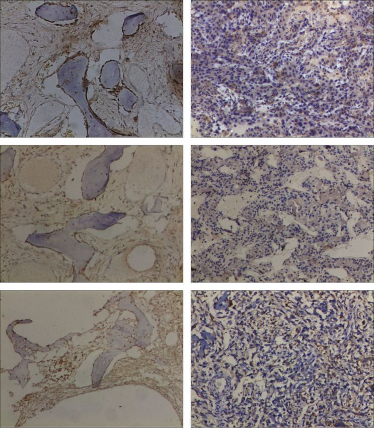
12 Journal of Immunology Research

 Case 1

 200 m 200 m

 Case 2

 200 m 200 m

 Case 3

 200 m 200 m

 Adjacent normal OS

 3
 EVI2B positive staining

 2
 (IOD/area)

 ⁎
 1

 0
 Adjacent normal OS
 (a)
 5
 (normorlized to GAPDH)
 EVI2B mRNA expression

 4

 3

 2
 ⁎⁎
 1

 0
 Adjacent normal OS
 (b)

Figure 7: The relative EVI2B expression in OS tissues and adjacent normal tissues. (a) The immunohistochemical detection results showed
the expression level of EVI2B in osteosarcoma tissues and adjacent normal tissues. (b) RT-qPCR analysis of relative EVI2B expression in OS
tissues and adjacent normal tissues.
Journal of Immunology Research 13

(Invitrogen) at a 1 : 100 dilution. After washing with those with a low stromal score (Figure 2(b)), log-rank p value
phosphate-buffered saline (PBS), the slides were incubated = 0.008. The above results indicated that the immune score
with a goat anti-mouse IgG secondary antibody conjugated and stromal score significantly correlated with OS patients’
to fluorescein isothiocyanate (ZSDB-BIO, Beijing, China) prognosis. Patients with high immune scores and the stromal
for 30 mins. They were washed with PBS and then incubated score had a good prognosis.
with an antifade reagent (Invitrogen, Carlsbad, USA).
Finally, staining was observed using an Olympus CX41 fluo- 3.2. IRGs of OS Screened with WGCNA and PPI Network
rescence microscope (Olympus, Tokyo, Japan). Positive Diagram. The sample cluster tree had an outlying sample,
staining intensity in OS and adjacent normal tissues were and the red line was the cut-off value to filter data
analyzed by integrated optical density (IOD) using Image- (Figure 3(a)). All samples were in clusters after removing
Pro Plus software (version 6.0; Media Cybernetics, United one outlying sample based on immune score and stromal
States). All images were taken using the same microscope score (Figure 3(b)). Sample dendrogram and trait heatmap
and camera set. Image-Pro Plus software was used to calcu- were drawn according to the immune score and stromal
late the mean IOD (μm2) per stained area (IOD/area) for score (Figure 3(b)). The above results were obtained by fur-
positive staining. T-test was used to analyze the results. A p ther analysis of the modules associated with immune score
value < 0.05 was considered statistically significant. and stromal score by WGCNA. In this study, R2 and connec-
 tivity were highest when the power β set at 15 (Figures 3(c)
2.10. Real-Time Quantitative PCR. Total RNA was isolated and 3(d)).
from tissues from the above patients using Trizol reagent Thus, β identifies distinct gene coexpression modules in
(TaKaRa, Japan) according to the manufacturer’s instruc- OSs. The cluster dendrogram of the selected genes was clus-
tions. One microgram of RNA was reverse transcribed into tered with an adjacency stromal, and we obtained a module
cDNA using the Revert Aid First Strand cDNA Synthesis visualization of the genes (Figure 3(e)). We also received
Kit (Thermo, USA). Quantitative RT-PCR was then per- the correlation between modules and traits (Figure 3(h)).
formed with Pro Taq HS Premix Probe qPCR Kit (Accurate, The pink module has the highest correlation coefficient with
Hunan, China). The amplification program consisted of one an immune score, with a correlation coefficient of 0.58, p <
cycle of predenaturation at 95°C for 5 mins, 37 cycles of dena- 0:05, which was statistically significant, indicating that the
turation at 95°C for 30 s, annealing at 60°C for 30 s, and immune score significantly correlated with clinical
extension at 72°C for 10 mins. The GAPDH gene was used traits—the above results were based on the WGCNA analysis
as an endogenous control gene for normalizing the expres- performed by the immune system stromal scoring. There-
sion of target genes. Each sample was analyzed in triplicate. fore, we chose this module as the next research object. We
The sequences of primers were used for RT-qPCR and screened the pink module’s IRGs with a correlation between
annealing temperature (Table S1). genes and the pink module greater than 0.8 and a correlation
 coefficient between genes and immune scores greater than
2.11. Statistical Analyses. All statistical analyses were per- 0.5 (Figure 3(f)). With conditions, we screened a total of 28
formed using the R software 3.5.0. Statistical significance set IRGs PTPRJ, SCARF1, CD300C, ANPEP, TBXAS1,
at a probability value of p < 0:05. Univariate, LASSO and RNASE6, HSD3B7, EVI2B, NCKAP1L, PTK2B, SPI1, GRN,
multivariate Cox regression analyses were applied to predict SQOR, GLRX, APOBR, GNA15, LILRB4, LAPTM5, CCR1,
the overall survival of OS patients. The effect of ROC and cal- FERMT3, CYTH4, LCP1, CD4, CSF1R, PSTPIP1, LAT2,
ibration curve on the prediction accuracy of the prognostic SELPLG, and RAC2 shown in Table 1.
model was compared. Enrichment analysis of differential A PPI regulatory network of 28 IRGs was obtained based
genes in the high- and low-risk groups in the training set on the STRING online database (https://string-db.org/). We
was performed with the R package cluster profile. found that the direct interaction between hub genes was
 through the PPI network analysis (Figure 3(g)). Nineteen
3. Results genes (PTPRJ, CD300C, RNASE6, EVI2B, NCKAP1L,
 PTK2B, SPI1, GNA15, LILRB4, LAPTM5, CCR1, FERMT3,
3.1. Relationship between Immune Score and Stromal Score CYTH4, LCP1, CD4, CSF1R, PSTPIP1, SELPLG, and
for Prognosis in OS. The flowchart of this study design is RAC2) were correlated well with immune score.
shown in Figure 1. Overall survival analysis did with R pack-
age survival analysis of the immune score’s prognosis and 3.3. Recognize IRG Prognostic Gene and Survival Analysis and
stromal score. The immune score and stromal score were cal- ROC Analysis. The 28 IRGs were subjected to univariate Cox
culated based on the ESTIMATE algorithm can promote the analysis, resulting in 10 IRGs with prognostic significance
quantification of immune and stromal components in (Table 2) (p value < 0.05). These ten genes, EVI2B, GRN,
tumors; in this algorithm, the immune and stromal score is NCKAP1L, SELPLG, LILRB4, FERMT3, SQOR, CYTH4,
calculated by analyzing the specific gene expression charac- GNA15, and RNASE6, were subjected to Lasso regression
teristics of the immune and stromal cells to predict the infil- analysis further to screen prognostic genes (Figures 4(a)
tration of nontumor cells. Patients with high immune scores and 4(b)) after 1,000 stimuli run through the crossvalidation
had better overall survival than those with low immune possibility, the optimal lambda determined, and three genes,
scores (Figure 2(a)), log-rank p value = 0.001. Patients with EVI2B, GRN, and NCKAP1L, were screened. Next, multivar-
a high stromal score had a more favorable prognosis than iate Cox analysis was performed to construct a prognostic
14 Journal of Immunology Research

model based on EVI2B, GRN, and NCKAP1L, resulting in a 3.6. IRG EVI2B Expression Is Downregulated in OS Tissues.
model containing one IRG EVI2B. EVI2B is highly expressed Finally, immunostaining analysis of IRG EVI2B showed that
in normal bone tissue controls and lowly expressed in osteo- EVI2B expression was higher in adjacent normal tissues
sarcoma (Table 2), which can be regarded as a tumor sup- (Figure 7(a)). EVI2B-positive staining was significantly dif-
pressor gene consistent with the results of a colorectal ferent between the control and OS, and positive staining
cancer study by Yuan et al. [37]. was low and statistically significant in OS (Figure 7(a)). We
 By dividing the sample into high- and low-risk groups examined IRG EVI2B expression in clinical samples from
according to the model’s median risk score, we found a sig- OS patients by qPCR. The qPCR results showed that IRG
nificant difference in survival between the high- and low- EVI2B was highly expressed at adjacent normal bone tissue
risk groups (log-rank p value = 0.016) (Figure 4(c)). ROC levels (Figure 7(b)). The above results showed that IRG
analysis showed AUC values of 0.676, 0.697, and 0.708 for EVI2B was highly expressed in adjacent normal bone tissue
ROC curves at 1, 3, and 5 years, respectively (Figure 4(d)). (control) and lowly expressed in osteosarcoma, and Yuan
All results showed that the prediction effect of the IRG EVI2B et al. included colorectal cancer study [37].
model was moderately accurate.
 4. Discussion
3.4. Evaluating the Accuracy of the IRG EVI2B Model. The
 In the past decades, traditional cancer treatments, including
time-dependent ROC curve analysis evaluated the accuracy
 surgical treatment and chemotherapy/radiotherapy, have
of the OS prediction model constructed by EVI2B. The IRG
 incredibly prolonged osteosarcoma patients’ survival time
EVI2B model evaluates in an independent GEO dataset
 [38, 39]. Although the 5-year overall survival rate is high [9,
GSE21257. By dividing GSE21257 samples into high- and
 40, 41], the survival rate of patients with metastatic disease (most
low-risk groups according to the TARGET dataset samples’
 commonly in the lung parenchyma and distal bone) is meager,
median risk score in the model, we found a significant differ-
 19-30% [39, 42]. Also, chemotherapy/radiotherapy acquired
ence in survival between the high- and low-risk groups (log-
 resistance to new antitumor drugs and severe side effects of
rank p value = 0.003) (Figure 5(a)). ROC analysis showed
 OS treatment’s drug resistance characteristics after extensive
that the AUC values for 1, 3, and 5 years of the ROC curve
 surgical resection of OS treatment reach the platform [39, 43].
were 0.663, 0.690, and 0.658, respectively (Figure 5(b)). The
 The use of targeted therapy [44–46] also benefits OS
survival rate of the low-risk group of the GSE21257 sample
 patients; for example, therapies targeting the unfolded pro-
in the validation set based on the IRG EVI2B model was
 tein response (UPR) pathway may be a feasible approach
higher than that of the high-risk group, and the prolonged
 for treating osteosarcoma. The components targeting the
survival time was consistent with the results in the training
 UPR may prove beneficial for patients refractory to conven-
set. The AUC values for the validation set GSE21257 samples
 tional chemotherapy [47]. In addition to being a marker of
were like the AUC results in the training set. The result fully
 metastatic disease, therapeutic targeting of cathepsin D
validates the IRG EVI2B prognostic model’s accuracy and
 (CTSD) may be a promising novel approach for osteosar-
illustrates that the IRG EVI2B is feasible as a prognostic indi-
 coma treatment; it may produce a good response in metasta-
cator in OS patients.
 tic disease [47]. However, targeted therapy also has some
 limitations and cannot be suitable for all types of osteosar-
3.5. GO and KEGG Enrichment Analysis. Next, to further elu- coma. In recent years, immunotherapy [48] has dramatically
cidate the molecular functions and signaling pathways in developed in cancer. Immune checkpoint-based therapy has
which differential genes between the TARGET high- and been shown to encouraging the antitumor effect by restoring
low-risk groups are involved, we performed functional immune response in the tumor microenvironment [49, 50].
enrichment analysis of all DEGs. GO results: these genes At the basement of immune checkpoint inhibitors, ipilimu-
are involved not only in T cell activation, regulation of leuko- mab (monoclonal antibody) anticytotoxic T lymphocyte
cyte differentiation, regulation of lymphocyte activation, and antigen four antibodies (mAb) (CTLA4) and monoclonal
several other biological processes but also in cytokine bind- antibody death protein 1 (PD1) or PD1 ligand (PDL1)
ing, cytokine receptor complex activity, MHC protein bind- against programmed cells show that immune checkpoints
ing, and molecular functions in other tumors. Some of the may be immune-tolerant to the tumor [13–17, 51]. A study
encoded proteins are important components of the plasma has shown that for osteosarcoma, the immune checkpoint
membrane’s external side, secretory granule membrane, inhibitor PD-L1 is negatively correlated with prognosis, and
and vacuolar membrane (Figure 6(a)). We further analyzed PD-1 is negatively correlated with overall survival [13–17,
these genes’ signaling pathways, and KEGG results showed 51]. To date, although immunotherapy [10, 11, 13, 51–53]
that these genes play a regulatory role in important has used in OS, the impact on OS prognosis is not well under-
immune-related pathways (such as the B cell receptor signal- stood, so there is an urgent need for a new biomarker that can
ing pathway and chemokine signaling pathway) use to predict the prognosis of OS patients, and our study is
(Figure 6(b)). These results suggest that the OS prognostic to affect the prognosis of cancer patients by participating in
model is significantly associated with immune response and the immune process.
inflammatory response, affecting cancer patients’ prognosis At present, the use of EVI2B [54] is still relatively rare.
by participating in immune processes, indicating the correct- The EVI2B is located in the intron of the neurofibromatosis
ness of our analysis results. type 1 (NF1) gene and transcribed in the opposite direction
Journal of Immunology Research 15

to the NF1 gene [55–58]. Still, their expression is not related, was validated and experimentally analyzed to obtain that
indicating that they are independently regulated [59]. EVI2B IRG EVI2B can significantly predict the prognosis of osteo-
is a transmembrane protein [60], while NF1 is a tumor sup- sarcoma patients.
pressor gene [61]. EVI2B is expressed in many different cell
types, including myeloid cells [62]. The EVI2B is also located Data Availability
within EVI2, a common viral integration site found in retro-
viral induced myeloid tumors [54]. It has postulated that viral The publicly available datasets were analyzed in this study;
integration on EVI2 alters EVI2B expression and that this these can be found in the TARGET and the NCBI Gene
altered expression predisposes mice to the development of Expression Omnibus (GSE21257).
bone marrow tumors [54].
 The successful construction of the IRG EVI2B prognostic Ethical Approval
model and the evaluation and validation of the accuracy of the
IRG EVI2B prognostic model with the ROC curve method and The Hunan Cancer Hospital ethics committee approved the
in the GEO dataset GSE21257 give us hope to use the IRG study.
EVI2B gene as a new biomarker and therapeutic target for pre-
dicting the prognosis of osteosarcoma patients. In the same Conflicts of Interest
study, EVI2B was found to correlate with the prognosis of
 The authors declare that the research was conducted in the
patients significantly negatively with colon cancer (p < 0:05),
 absence of any commercial or financial relationships that
and EVI2B may be involved in the prognosis of colon cancer
 could be construed as a potential conflict of interest.
patients [37, 63]. EVI2B can similarly be used as a prognosis
for osteosarcoma compared to the study by Yang et al. [64].
 EVI2B is expressed in various tumors [62] and breast Authors’ Contributions
cancer mutations [65]. EVI2B is involved in the differentia- Li Gan and Xin Wang contributed equally and are co-first
tion of melanocytes and keratinocytes [66, 67]. In authors to this work.
fibroblast-like cells derived from neurofibromas, increased
EVI2B mRNA levels were found [66]. EVI2B gene, their
related studies indicate that the EVI2B gene is a direct target
 Acknowledgments
of CCAAT/enhancer-binding protein (C/EBP). The product This study was supported by the Scientific Research Project
of this gene, the transmembrane glycoprotein EVI2B of Hunan Health Commission (Nos. 202102041763 and
(CD361) [68], shows to be abundantly expressed on the sur- 20200985), Changsha Municipal Natural Science Foundation
face of primary hematopoietic cells, reaching the highest (No. kq2014267), and Hunan Cancer Hospital Climb Plan
expression level in mature granulocytes [69]. They use (No. 2020QH001).
shRNA-mediated downregulation of EVI2B in human and
murine cell lines and primary hematopoietic stem and pro- Supplementary Materials
genitor cells (HSPC), demonstrating impaired myeloid line-
age development and altered progenitor function in EVI2B- Table S1: the sequences of primers used for RT-qPCR and
depleted cells. Its study suggests that EVI2B is an essential annealing temperature. (Supplementary Materials)
modulator of HSPC and bone marrow differentiation and
that low levels of EVI2B may contribute to the differentiation References
inhibitory profile of acute myelocytic leukemia (AML) [69].
It has shown that EVI2B appears highly expressed in patients [1] S. J. Cotterill, C. M. Wright, M. S. Pearce, and A. W. Craft,
with 11q deletion in chronic lymphocytic leukemia (CLL). It “Stature of young people with malignant bone tumors,” Pedi-
speculated that the EVI2B gene might be associated with atric Blood & Cancer, vol. 42, no. 1, pp. 59–63, 2004.
poor prognosis in patients with CLL 11q deletion [70]. It also [2] M. S. Isakoff, S. S. Bielack, P. Meltzer, and R. Gorlick, “Osteo-
studied that EVI2B was transiently expressed on cellular sarcoma: current treatment and a collaborative pathway to
 success,” Journal of Clinical Oncology, vol. 33, no. 27,
adjuvants and used to directly immunize BALB/c mice to
 pp. 3029–3035, 2015.
obtain a higher specific anti-EVI2B Ab response in immu-
 [3] A. Misaghi, A. Goldin, M. Awad, and A. A. Kulidjian, “Osteo-
nized mice than stimulated by other cellular adjuvants, which
 sarcoma: a comprehensive review,” SICOT J, vol. 4, p. 12, 2018.
used to accelerate the development of Ab drugs [71]. Our
 [4] S. S. Bielack, S. Hecker-Nolting, C. Blattmann, and L. Kager,
study also showed that EVI2B is associated with the progno-
 “Advances in the management of osteosarcoma,”
sis of osteosarcoma. These results suggest that it is feasible for F1000Research, vol. 5, 2016.
us to use EVI2B as a marker and potential target for the diag-
 [5] F. Liu, L. Xing, X. Zhang, and X. Zhang, “A four-pseudogene
nosis and treatment of osteosarcoma, but further studies classifier identified by machine learning serves as a novel prog-
should be on its possible action mechanism. nostic marker for survival of osteosarcoma,” Genes (Basel),
 vol. 10, no. 6, p. 414, 2019.
5. Conclusions [6] N. M. Bernthal, N. Federman, F. R. Eilber et al., “Long-term
 results (>25 years) of a randomized, prospective clinical trial
In conclusion, we selected IRG EVI2B by WGCNA analysis evaluating chemotherapy in patients with high-grade, operable
and constructed an IRG-based prognostic model, which osteosarcoma,” Cancer, vol. 118, no. 23, pp. 5888–5893, 2012.
16 Journal of Immunology Research

 [7] C. C. Wu, H. C. Beird, J. Andrew Livingston et al., “Immuno- [23] N. Ternes, F. Rotolo, and S. Michiels, “Empirical extensions of
 genomic landscape of osteosarcoma,” Nature Communica- the lasso penalty to reduce the false discovery rate in high-
 tions, vol. 11, no. 1, 2020. dimensional Cox regression models,” Statistics in Medicine,
 [8] A. Luetke, P. A. Meyers, I. Lewis, and H. Juergens, “Osteosar- vol. 35, no. 15, pp. 2561–2573, 2016.
 coma treatment - where do we stand? A state of the art review,” [24] P. F. Chen, F. Wang, J. Y. Nie et al., “Co-expression network
 Cancer Treatment Reviews, vol. 40, no. 4, pp. 523–532, 2014. analysis identified CDH11 in association with progression
 [9] Y. Wang, W. Yu, J. Zhu et al., “Anti-CD166/4-1BB chimeric and prognosis in gastric cancer,” OncoTargets and Therapy,
 antigen receptor T cell therapy for the treatment of osteosar- vol. Volume 11, pp. 6425–6436, 2018.
 coma,” Journal of Experimental & Clinical Cancer Research, [25] P. Langfelder and S. Horvath, “WGCNA: an R package for
 vol. 38, no. 1, 2019. weighted correlation network analysis,” BMC Bioinformatics,
[10] N. Ahmed, V. S. Brawley, M. Hegde et al., “Human epidermal vol. 9, no. 1, 2008.
 growth factor receptor 2 (HER2) -specific chimeric antigen [26] F. Wang, Y. Chang, J. Li et al., “Strong correlation between
 receptor-modified T cells for the immunotherapy of HER2- _ASPM_ gene expression and HCV cirrhosis progression
 positive sarcoma,” Journal of Clinical Oncology, vol. 33, identified by co-expression analysis,” Digestive and Liver Dis-
 no. 15, pp. 1688–1696, 2015. ease, vol. 49, no. 1, pp. 70–76, 2017.
[11] N. J. Mason, J. S. Gnanandarajah, J. B. Engiles et al., “Immuno- [27] Z. Zhou, S. Liu, M. Zhang et al., “Overexpression of topoisom-
 therapy with a HER2-targeting listeria induces HER2-specific erase 2-alpha confers a poor prognosis in pancreatic adenocar-
 immunity and demonstrates potential therapeutic effects in a cinoma identified by co-expression analysis,” Digestive
 phase I trial in canine osteosarcoma,” Clinical Cancer Diseases and Sciences, vol. 62, no. 10, pp. 2790–2800, 2017.
 Research, vol. 22, no. 17, pp. 4380–4390, 2016. [28] H. Wang, X. Wu, and Y. Chen, “Stromal-immune score-based
[12] N. Rainusso, V. S. Brawley, A. Ghazi et al., “Immunotherapy gene signature: a prognosis stratification tool in gastric can-
 targeting HER2 with genetically modified T cells eliminates cer,” Frontiers in Oncology, vol. 9, 2019.
 tumor-initiating cells in osteosarcoma,” Cancer Gene Therapy, [29] M. J. Mason, G. Fan, K. Plath, Q. Zhou, and S. Horvath,
 vol. 19, no. 3, pp. 212–217, 2012. “Signed weighted gene co-expression network analysis of tran-
[13] M. Kansara, M. W. Teng, M. J. Smyth, and D. M. Thomas, scriptional regulation in murine embryonic stem cells,” BMC
 “Translational biology of osteosarcoma,” Nature Reviews. Genomics, vol. 10, no. 1, p. 327, 2009.
 Cancer, vol. 14, no. 11, pp. 722–735, 2014. [30] X. Zhai, Q. Xue, Q. Liu, Y. Guo, and Z. Chen, “Colon cancer
[14] P. W. Kantoff, C. S. Higano, N. D. Shore et al., “Sipuleucel-T recurrence associated genes revealed by WGCNA coexpres-
 immunotherapy for castration-resistant prostate cancer,” The sion network analysis,” Molecular Medicine Reports, vol. 16,
 New England Journal of Medicine, vol. 363, no. 5, pp. 411– no. 5, pp. 6499–6505, 2017.
 422, 2010. [31] Z. Liu, M. Li, Q. Hua, Y. Li, and G. Wang, “Identification of an
[15] J. R. Brahmer, S. S. Tykodi, L. Q. Chow et al., “Safety and activ- eight-lncRNA prognostic model for breast cancer using
 ity of anti-PD-L1 antibody in patients with advanced cancer,” WGCNA network analysis and a Cox‑proportional hazards
 The New England Journal of Medicine, vol. 366, no. 26, model based on L1-penalized estimation,” International Jour-
 pp. 2455–2465, 2012. nal of Molecular Medicine, vol. 44, no. 4, pp. 1333–1343, 2019.
[16] A. D. Garg and P. Agostinis, “Cell death and immunity in can- [32] D. Szklarczyk, A. Franceschini, S. Wyder et al., “STRING v10:
 cer: from danger signals to mimicry of pathogen defense protein-protein interaction networks, integrated over the tree
 responses,” Immunological Reviews, vol. 280, no. 1, pp. 126– of life,” Nucleic Acids Research, vol. 43, no. D1, pp. D447–
 148, 2017. D452, 2015.
[17] G. Kroemer, L. Galluzzi, O. Kepp, and L. Zitvogel, “Immuno- [33] P. Shannon, A. Markiel, O. Ozier et al., “Cytoscape: a software
 genic cell death in cancer therapy,” Annual Review of Immu- environment for integrated models of biomolecular interac-
 nology, vol. 31, no. 1, pp. 51–72, 2013. tion networks,” Genome Research, vol. 13, no. 11, pp. 2498–
[18] C. Liu, X. Wang, G. Z. Genchev, and H. Lu, “Multi-omics facil- 2504, 2003.
 itated variable selection in Cox-regression model for cancer [34] M. Ashburner, C. A. Ball, J. A. Blake et al., “Gene Ontology:
 prognosis prediction,” Methods, vol. 124, pp. 100–107, 2017. tool for the unification of biology,” Nature Genetics, vol. 25,
[19] Y. Zhang, H. Li, W. Zhang, Y. Che, W. Bai, and G. Huang, no. 1, pp. 25–29, 2000.
 “LASSO‑based Cox‑PH model identifies an 11‑lncRNA signa- [35] D. W. Huang, B. T. Sherman, and R. A. Lempicki, “Systematic
 ture for prognosis prediction in gastric cancer,” Molecular and integrative analysis of large gene lists using DAVID bioinfor-
 Medicine Reports, vol. 18, no. 6, pp. 5579–5593, 2018. matics resources,” Nature Protocols, vol. 4, no. 1, pp. 44–57, 2009.
[20] M. Bagnoli, S. Canevari, D. Califano et al., “Development and [36] G. Yu, L. G. Wang, Y. Han, and Q. Y. He, “clusterProfiler: an R
 validation of a microRNA-based signature (MiROvaR) to pre- package for comparing biological themes among gene clus-
 dict early relapse or progression of epithelial ovarian cancer: a ters,” OMICS, vol. 16, no. 5, pp. 284–287, 2012.
 cohort study,” Lancet Oncology, vol. 17, no. 8, pp. 1137–1146, [37] Y. Yuan, J. Chen, J. Wang et al., “Identification hub genes in
 2016. colorectal cancer by integrating weighted gene co-expression
[21] Y. Xiong, L. Yuan, J. Xiong et al., “An outcome model for network analysis and clinical validation in vivo and vitro,”
 human bladder cancer: a comprehensive study based on Frontiers in Oncology, vol. 10, 2020.
 weighted gene co-expression network analysis,” Journal of Cellu- [38] P. A. Meyers, C. L. Schwartz, M. Krailo et al., “Osteosarcoma: a
 lar and Molecular Medicine, vol. 24, no. 3, pp. 2342–2355, 2020. randomized, prospective trial of the addition of ifosfamide
[22] R. Tibshirani, “The lasso method for variable selection in the and/or muramyl tripeptide to cisplatin, doxorubicin, and
 Cox model,” Statistics in Medicine, vol. 16, no. 4, pp. 385– high-dose methotrexate,” Journal of Clinical Oncology,
 395, 1997. vol. 23, no. 9, pp. 2004–2011, 2005.
You can also read