Convex probe endobronchial ultrasound: historical, contemporary, and cutting-edge applications - Journal of Thoracic ...
←
→
Page content transcription
If your browser does not render page correctly, please read the page content below
Editorial Section of Interventional Pulmonology

Convex probe endobronchial ultrasound: historical, contemporary,
and cutting-edge applications
Sameer K. Avasarala, Carlos Aravena, Francisco A. Almeida

Respiratory Institute, Cleveland Clinic, Cleveland, OH, USA
Contributions: (I) Conception and design: All authors; (II) Administrative support: None; (III) Provision of study materials or patients: None; (IV)
Collection and assembly of data: All authors; (V) Data analysis and interpretation: All authors; (VI) Manuscript writing: All authors; (VII) Final
approval of manuscript: All authors.
Correspondence to: Francisco A. Almeida, MD, MS. Respiratory Institute, Cleveland Clinic, 9500 Euclid Avenue, Cleveland, OH 44195, USA.
Email: almeidf@ccf.org.

                Abstract: The use of convex-probe endobronchial ultrasound (CP-EBUS) has revolutionized
                bronchoscopy. It has provided the option of a relatively safe, minimally invasive approach for the assessment
                of various intrathoracic diseases. In current practice, its most dramatic impact has been on the diagnosing
                and staging of lung cancer. It has served as an invaluable tool that has replaced mediastinoscopy in a variety
                of clinical scenarios. Many pulmonologists and thoracic surgeons consider CP-EBUS the most significant
                milestone in bronchoscopy after the development of the flexible bronchoscope itself. In this review, we
                summarize the historical aspects, current indications, technical approach, and future direction of CP-EBUS.

                Keywords: Bronchoscopy; lung cancer; lymphadenopathy; endobronchial ultrasound-guided transbronchial
                needle aspiration (EBUS-TBNA)

                Submitted Jun 21, 2019. Accepted for publication Oct 24, 2019.
                doi: 10.21037/jtd.2019.10.76
                View this article at: http://dx.doi.org/10.21037/jtd.2019.10.76

Introduction                                                                 technology to assist with imaging structures that are beyond
                                                                             the field of visualization of white light bronchoscopy. A
Since the inception of endobronchial ultrasound, its
                                                                             convex ultrasound transducer is incorporated into the tip of
use within thoracic medicine has become widespread
                                                                             the flexible bronchoscope; the transducer forms a grey-scale
in most developed countries. It has allowed for the use
                                                                             image that is parallel to the insertion of the bronchoscope
of a minimally invasive approach for the management
of intrathoracic lymphadenopathy and lung lesions.                           (Figure 2) (1). A saline-filled balloon tip can be used
Radial probe endobronchial ultrasound (RP-EBUS) and                          to improve surface contact and enhance image quality.
convex-probe EBUS (CP-EBUS) are two modalities of                            Doppler imaging can also be used to delineate vascular
endobronchial ultrasound use within the tracheobronchial                     structures as well. The key differences between RP-EBUS
tree. In contemporary practice, RP-EBUS is performed                         and CP-EBUS are summarized in Table 1. One of the
with a radial probe (with or without a balloon),                             main advantages of CP-EBUS is its ability to provide real-
(Figure 1), which is advanced through the working channel                    time guidance when obtaining samples. When combined
of a bronchoscope. After processing, the ultrasound                          with concurrent transbronchial needle aspiration (TBNA),
waves are translated into a 360-degree grey scale image                      it has gained popularity in the diagnosis of intrathoracic
(Figure 1). RP-EBUS is most commonly used for the                            lymphadenopathy as well as in the diagnosis and staging of
evaluation of pulmonary nodules. Although it does allow                      lung cancer. Excluding the development of the fiberoptic
real-time imaging, it unfortunately does not allow real-                     bronchoscope itself, many consider endobronchial
time guidance. Convex-probe EBUS also uses ultrasound                        ultrasound-guided transbronchial needle aspiration

© Journal of Thoracic Disease. All rights reserved.                  J Thorac Dis 2020;12(3):1085-1099 | http://dx.doi.org/10.21037/jtd.2019.10.76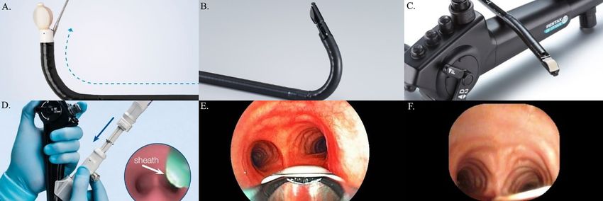
1086 Avasarala et al. A review of CP-EBUS
A B
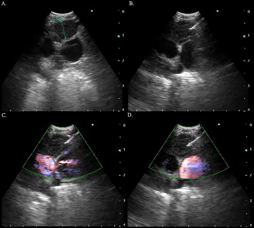
Figure 1 Key features of RP-EBUS. (A) A RP-EBUS probe can be advanced through the working channel of the bronchoscope to obtain
and a 360° greyscale image of parenchymal lesions, such as a lung nodule (B). RP-EBUS, radial probe endobronchial ultrasound.
A B
C D
Figure 2 Key features of CP-EBUS. (A) The CP-EBUS is an excellent tool for visualizing and sampling (B,C,D) intrathoracic lymph nodes.
Color Doppler can be used to assess for vascularity prior to and during sampling (C,D). CP-EBUS, convex probe endobronchial ultrasound.
© Journal of Thoracic Disease. All rights reserved. J Thorac Dis 2020;12(3):1085-1099 | http://dx.doi.org/10.21037/jtd.2019.10.76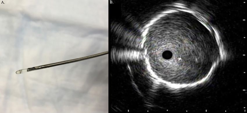
Journal of Thoracic Disease, Vol 12, No 3 March 2020                                                                                           1087

 Table 1 Key differences between RP-EBUS and CP-EBUS
 Characteristic                                       RP-RBUS                                         CP-EBUS

 Angle of view                                        360°                                            90°

 Penetration                                          4–5 cm                                          >5 cm

 Resolution                                           Comparatively worse                             Comparatively better

 Airway wall visualization                            Yes, with balloon inflation                     Yes, not as well studied

 Doppler                                              No                                              Yes

 Real-time sampling                                   No                                              Yes

 RP-EBUS, radial probe endobronchial ultrasound; CP-EBUS, convex probe endobronchial ultrasound.

(EBUS-TBNA) as the greatest milestone in bronchoscopy                               Because the convex-probe EBUS has been used for the
history (2). In a number of clinical scenarios, it has                          staging of lung cancer from its infancy its accuracy is often
either replaced or decreased significantly the use of                           compared to what is seen with mediastinoscopy (9-11).
mediastinoscopy. Convex-probe EBUS-TBNA is a safe                               The latter has been regarded as the gold standard for the
procedure and is usually performed by pulmonologists or                         mediastinal staging of non-small cell lung cancer (NSCLC).
thoracic surgeons. Its benefits include its minimally invasive                  It typically allows access to the upper paratracheal (stations
approach and ability to be performed under conscious                            2R and 2L), lower paratracheal (stations 4R and 4L)
sedation. In settings where rapid on-site evaluation (ROSE)                     and subcarinal (station 7) lymph nodes (12). Overall,
is used, patients typically receive a preliminary diagnosis                     it is safe; the reported complication rate is up to 2.5%
after the procedure. Even when general anesthesia is used,                      (pneumothorax, infection, and injury to nearby structures
most cases can be conducted in an outpatient setting.                           [vessels, nerves, bronchi, and esophagus]), mortality rate
A tool that was initially developed for evaluation of                           is estimated to be 0.08% (mostly being driven by injury
intrathoracic lymphadenopathy has proven to be useful in                        to major vascular structures) (13). Due to the use of more
the management of a number of other thoracic diseases (3).                      minimally invasive techniques, rates of mediastinoscopy
It has been a research niche for thousands of people, the                       have been falling in the United States. One study indicated
number publications describing EBUS-TBNA has risen                              that the median center rate for mediastinoscopy being
exponentially (4). In this review, we discuss the history of                    performed dropped from 21.6% to 10% between 2006 and
CP-EBUS, its current application, and its future direction.                     2010 (14). When compared to EBUS-TBNA, standard
                                                                                cervical mediastinoscopy has more complications,
                                                                                unequivocally requires general anesthesia and a small
Past
                                                                                incision at/above the suprasternal notch, is more expensive,
Endoscopic ultrasound was first used within the field                           and has a higher risk and rate of complications (15). In
of gastroenterology for staging esophageal and gastric                          addition, data shows endosonographic assessment of
malignancies (5). Later, the use of ultrasound within the                       lymphadenopathy [EBUS-TBNA/endoscopic ultrasound
airways was developed in the early 1990s. These small                           fine needle aspiration (EUS-FNA)] has a similar yield
ultrasound probes, also known as “miniprobes” were the                          but with lower complication rates when compared to
first generation of radial probe EBUS (RP-EBUS) (6). In                         mediastinoscopy for the mediastinal staging of NSCLC (16).
2002, Herth et al. published the first report of using RP-                          It is the culmination of these important factors that set
EBUS to guide transbronchial biopsies for the assessment of                     the foundation for CP-EBUS to rise in popularity and
lung nodules (7). Although RP-EBUS was able to provide                          achieve widespread use.
real-time images for biopsy planning, the actual biopsy was
blind and had to be guided with imaging modalities such as
                                                                                Present
fluoroscopy. As the importance of real-time biopsy guidance
rose, the CP-EBUS was subsequently developed in 2002 (8).                       At present, there are three companies [Olympus America©

© Journal of Thoracic Disease. All rights reserved.                     J Thorac Dis 2020;12(3):1085-1099 | http://dx.doi.org/10.21037/jtd.2019.10.76
1088 Avasarala et al. A review of CP-EBUS
A B C
D E F
Figure 3 There are three CP-EBUS bronchoscopes currently available on the market: (A) Olympus©, (B) Fujifilm©, and (C) Pentax©. The
most significant difference between the bronchoscopes is the viewing angle: (D) Olympus©, (E) Fujifilm©, and (F) Pentax©. CP-EBUS,
convex probe endobronchial ultrasound.
Table 2 Salient features of CP-EBUS bronchoscopes currently available on the market
Characteristic Olympus© Pentax© Fujifilm©
Model(s) BF-UC160-OL8, BF-UC180F EB-1970UK EB-530US
Scanning frequency* 5, 6, 7.5, 8, 10, or 12 MHz 5, 7.5, or 10 MHz 5, 7.5, 10, or 12 MHz
Distal outer diameter 6.9 mm 6.3 mm 6.7 mm
Working channel size 2.0 or 2.2 mm 2.0 mm 2.0 mm
Direction of view 30° forward oblique 45° forward oblique 10° forward oblique
*, availability of scanning frequency is dependent on the processor the bronchoscope is paired with. CP-EBUS, convex probe
endobronchial ultrasound.
(Center Valley, PA, United States), Pentax Ricoh Imaging© that diagnosis and staging be performed during the same
(Tokyo, Japan), and Fujifilm Holdings © (Tokyo, Japan)] procedure (i.e., EBUS-TBNA), especially in patients with
that produce the CP-EBUS bronchoscopes that are in any adenopathy. Strict attempts to promote this process
current use (Figure 3). The bronchoscopes share a similar appear to protect the patient from unnecessary diagnostic
design and have a few unique features. The salient features procedures (i.e., CT-guided fine needle aspiration in
are described in Table 2. In current practice, CP-EBUS is the setting of obvious nodal disease) and result in more
mainly used for the diagnosis and/or staging of NSCLC. rapid and accurate staging (17,18). In fact, current expert
In addition, there is a body of literature to support its panel recommendations state suspicious mediastinal
use in diagnosing various diseases that may present with lymphadenopathy should undergo staging evaluation by
intrathoracic adenopathy such as lymphoma, sarcoidosis, a needle technique such as EBUS-TBNA, EUS-FNA,
and infectious diseases. or a combined approach (19,20). In addition, these same
guidelines recommend the least number of procedures
should be performed for both the stating and diagnosis
Diagnosis and staging of NSCLC
of suspected NSCLC (21). Unfortunately, studies have
An important tenant within the evaluation of NSCLC is shown that failing to follow guideline-consistent care
© Journal of Thoracic Disease. All rights reserved. J Thorac Dis 2020;12(3):1085-1099 | http://dx.doi.org/10.21037/jtd.2019.10.76Journal of Thoracic Disease, Vol 12, No 3 March 2020 1089
is not uncommon (18,22). A large retrospective cohort positivity (30). In the current landscape of lung cancer therapy,
study of over 15,000 patients with lung cancer showed immunotherapy is considered the cornerstone of management
that an exceedingly high number of patients with NSCLC in advanced stages that can, ultimately, extend survival (31).
(44%) did not have the mediastinum sampled and relied After appropriate diagnosis, staging and therapy, EBUS-
on imaging only (22). When compared to chest CT TBNA still maintains an important role in lung cancer
(sensitivity 76.9%/specificity 55.3%) or PET (80%/70.1%), management. It is the preferred modality for re-biopsy to
EBUS-TBNA (92.3%/100%) is more accurate in staging assess for relapse after therapy or response after neoadjuvant
intrathoracic lymph nodes (23). In the same study, the treatment (32,33). These re-biopsies are useful in detecting
diagnostic accuracy of EBUS-TBNA was also higher (98%) specific new mutations, which may have therapeutic
when compared to CT (60.8%) or PET (72.5%). implications (the use of osimertinib for T790M mutation
In addition to superior diagnostic performance when for instance) (34).
compared to imaging modalities alone, other minimally
invasive methods can be used to augment EBUS-TBNA. In
Lymphoma
general, EBUS-TBNA can access several nodal stations: 2,
3P, 4, 7, 10, 11 and 12. Endoscopic ultrasound fine-needle Endobronchial ultrasound-TBNA has also been shown
aspiration typically allows access to stations 4L, 7, 8, and useful in the diagnosis of lymphoma, albeit, with limitations.
9 (24). Specifically, the addition of EUS-FNA allows access A study has shown that the sensitivity of EBUS-TBNA
to stations 8 and 9, which are not accessible by EBUS- for the detection of lymphoma was 76%; however, the
TBNA. In a meta-analysis, the sensitivity of an EBUS- sensitivity for the definitive diagnosis of lymphoma was
TBNA and EUS-FNA approach is estimated to be 90% (25). only 57% (35). Lymphoma detection by EBUS-TBNA in
The combination of which may provide the best option another study was 65% (diagnostic sensitivity) and 96.1%
for a minimally invasive approach to stage lung cancer. (negative predictive value) (36). A more recent study quoted
In addition to intrathoracic adenopathy, there is data that an overall sensitivity that was 85%, but 91% for detection
demonstrates that EBUS-TBNA can be useful in sampling of recurrence (37). Overall, these were small studies; the
the left adrenal gland in patients with lung cancer (26). body of medical literature suggests that the diagnostic
In recent history, the molecular characterization of lung accuracy of EBUS-TBNA for lymphoma is significantly
cancer has gained clinical significance. Newer treatment lower than it is for lung cancer staging. The small volume of
modalities target molecular pathways that are essential specimens obtained poses difficulties for confirming specific
to the survival of malignant cells: epidermal growth subtypes, which is key for formulating a treatment plan.
factor receptor (gefitinib), anaplastic lymphoma kinase Even with this potential limitation, it may be reasonable to
(certinib), ROS-1 (crizotinib), programmed death-ligand 1 consider using EBUS-TBNA in a patient suspected to have
(PD-1/PD-L1) (nivolumab), etc. The most novel of these lymphoma with isolated mediastinal lymphadenopathy, in
targets is PD-1/PD-L1. PD-L1 positivity appears to be order to avoid a surgical biopsy.
higher in certain patients with NSCLC: males, smokers, At present, the American College of Chest Physicians
advanced pathologic stages, positive vessel invasion, and Expert Panel states that EBUS-TBNA is an acceptable
positive lymphatic invasion (27). Tissue specimens derived initial, minimally invasive diagnostic test for a patient with
from EBUS-TBNA has been found to be reliable to suspected lymphoma (38). It is important to keep in mind
perform molecular bio-marker analysis (28). However, that this is an ungraded, consensus-based statement; there
this is dependent on the cutoff value used for a sample is not an agreement amongst international societies. The
to be considered PD-L1 positive (29). A study by Sakata British Thoracic Society states that “at present, there is
et al. showed that concordance rates between EBUS-TBNA insufficient evidence to recommend EBUS-TBNA for
derived samples and surgical lung biopsy samples range routine use in the diagnosis of lymphoma” (39).
between 82–87%. The concordance rates for PD-L1 ≥1%
and ≥50% were 87% and 82%, respectively (29). Sakakibara
Sarcoidosis
et al. also showed good concordance between EBUS-TBNA
and the corresponding primary tumor in regard to PD-L1 Endobronchial ultrasound TBNA has been proven
© Journal of Thoracic Disease. All rights reserved. J Thorac Dis 2020;12(3):1085-1099 | http://dx.doi.org/10.21037/jtd.2019.10.761090 Avasarala et al. A review of CP-EBUS to be useful for evaluating benign causes of thoracic geographic variability is importance since as prevalence of lymphadenopathy as well. It provides diagnostic information the disease may vary based on geographic location. in patients with early stage sarcoidosis that have adenopathy, EBUS-TBNA has also been found to be useful in but minimal changes to the lung parenchyma (40). It should identifying the cause of granulomatous inflammation be kept in mind that a tissue diagnosis is not necessary for within intra-thoracic lymph nodes. In a study of 210 most patients with stage I (bilateral hilar lymphadenopathy) patients that underwent EBUS-TBNA, 56 were found sarcoidosis and that clinical/radiological findings may to have granulomatous inflammation on histocytological be specific enough to secure a diagnosis (41-43). When evaluation (50). Twenty of these patients were found to have compared to the conventional transbronchial biopsy, the caseating granulomas of varying etiologies: histoplasma, yield of EBUS-TBNA in this clinical setting is higher. Blastomyces, or mycobacterium TB. Gupta et al. suggest that EBUS-TBNA should be combined There are useful applications amongst immunocompromised with transbronchial lung biopsies to optimize yield for hosts as well. In patients with HIV, EBUS-TBNA itself had sarcoidosis (44). A significant criticism of this study was the a diagnostic yield of 60.5%. A diagnostic accuracy of 97.7% lack of ROSE in either arm (45). was present with the combination of transbronchial biopsy and Based on the results of the GRANULOMA study, EBUS-TBNA. The most common infectious organism in this endosonographic (EUS-FNA or EBUS-TBNA) the cohort was TB (51). diagnosis of lower stage (I and II) of sarcoidosis was Akin to other etiologies of thoracic lymphadenopathy, higher when compared to conventional bronchoscopy the data for EBUS-TBNA in diagnosing an infectious biopsy (transbronchial or endobronchial biopsy) (46). etiology is not as unequivocally proven as it is for lung and The diagnostic yield of granuloma detection was 80% other solid cancers (52-54). In a study of 82 patients, the in the endosonographic group vs. 53% in the standard results suggested that EBUS-TBNA may not be sufficiently bronchoscopy group. sensitive to rule out infectious causes of adenopathy. Only Unfortunately the latest consensus statement regarding five percent of patients in this cohort had HIV. Only two the diagnosis sarcoidosis by a multi-organizational group patients had a bacterial culture from the EBUS-TBNA (American Thoracic Society, European Respiratory Society, that was positive for an organism that could potentially be and the World Association of Sarcoidosis and Other pathogenic. In addition, two more patients had a positive Granulomatous Disorders) was made several years prior to Mycobacterial culture (both for Mycobacterium avium- the development of EBUS-TBNA (47). intracellulare) (55). Infectious diseases Mediastinal lesions There are several infectious diseases that can present In contemporary practice, EBUS-TBNA can be used as a with intrathoracic lymphadenopathy. For example, both therapeutic intervention as well. If clinically indicated, a primary tuberculosis (TB) and post-primary TB can variety of mediastinal lesions can be accessed or sampled. have intrathoracic lymphadenopathy. In fact, thoracic In the case of a cyst in the mediastinum, full drainage lymphadenopathy is an important radiographic feature may also be possible with EBUS-TBNA (56). Prior to of primary TB and is seen in 40% of adult cases and procedural planning, a careful review of the chest CT can 90–95% of pediatric cases (48). In a meta-analysis of help formulate a differential diagnosis of what the lesion eight studies involving 809 patients with TB, the pooled may be. Location of the mass in relation to the mediastinal sensitivity and specificity of EBUS-TBNA for the compartment and its Hounsfield Units are useful clues. In a diagnosis of intrathoracic TB was 80% and 100% (49). retrospective study of 140 patients with mediastinal masses In a similar light, the pooled sensitivity of EBUS-TBNA of unknown etiology and no evidence of lung cancer or for diagnostic intrathoracic TB lymphadenitis was 87%. other pulmonary malignancy, EBUS-TBNA was diagnostic The overall diagnostic sensitivity for intrathoracic TB was in 131 (93.6%) of patients (57). It is unclear how many of 80%, sensitivity for TB lymphadenitis was 87% (49). The these mediastinal masses could have been large intrathoracic studies that were included in this meta-analysis were from lymph nodes. There are several case reports outlining the China, Korea, Turkey, United Kingdom and Ireland. The use of CP-EBUS for draining mediastinal lesions such as © Journal of Thoracic Disease. All rights reserved. J Thorac Dis 2020;12(3):1085-1099 | http://dx.doi.org/10.21037/jtd.2019.10.76
Journal of Thoracic Disease, Vol 12, No 3 March 2020                                                                                   1091

                    A                                                    B

Figure 4 Convex probe endobronchial ultrasound is useful in the management of a variety of mediastinal pathology. (A) A middle
mediastinal mass noted on contrast-enhanced chest CT; (B) it was visualized and sampled successfully with the use of CP-EBUS; the
cytological results were consistent with an esophageal duplication cyst. CP-EBUS, convex probe endobronchial ultrasound.

esophageal duplications cysts (Figure 4), bronchogenic cysts,          to identify different strain/stiffness in the tissue (61-63).
and pericardial effusions (58-60). It is important to note that        Several dedicated aspiration needles are available allowing
these therapeutic applications of CP-EBUS are yet to be                real-time EBUS-TBNA. For optimization of the ultrasound
supported by robust data. In our own experience, complete              (US) imaging, a latex balloon should be attached to the tip
therapeutic success is uncommon.                                       of the EBUS scope with a dedicated applicator forceps and
                                                                       be filled with 0.3 to 0.5 mL of saline if it is necessary during
                                                                       the procedure (63,64).
Performing EBUS-TBNA in present day

The success of EBUS-TBNA is predicated on a variety                    Insertion of the EBUS bronchoscope
of factors: patient selection, available equipment, and the            It is essential to know that the tip of the CP-EBUS scope
management of specimens (acquisition, processing, and                  may not be visible and the field of view depends on the type
interpretation). The procedural steps of EBUS-TBNA                     of bronchoscope used (Table 2). The quality of the image is
are outlined below. There are a lot of nuances within the              lower than a conventional bronchoscope. In order to obtain
described technique; there may be subtle differences in                a straight view, the tip of the scope needs to be flexed down
approach between any two bronchoscopists. The procedure                (thumb up). Therefore, when the scope is advanced, the
can be performed under moderate sedation or with general               visualization must be slightly anterior, and in this way do
anesthesia. Two commonly used airway devices include                   not traumatize the posterior structures of the upper and
the endotracheal tube and the laryngeal mask airway. The               lower airways.
type of anesthesia and the airway used can vary between                   If an endotracheal tube is not used, the scope must be
bronchoscopists and centers.                                           passed throughout the vocal cords visualizing the anterior
                                                                       angle of the glottis. Never force the scope into the glottis,
                                                                       because the non-visible tip may cause dislocation of the
EBUS-TBNA procedural steps
                                                                       cartilage (63). Passing through the vocal cords should occur
Equipment                                                              with the tip of the bronchoscope slightly flexed anteriorly
The CP-EBUS bronchoscope is connected to an ultrasound                 (thumb down). While it is advanced into the proximal
processor that allows to record the patient information,               airway the thumb should progressively push the control
measure the lesion, label, adjust the scanning range (the              lever up, as it passes downwards, the upper trachea follows
typical default 4 cm range permits to visualize best most              the curvature of the spine going slightly backward. Thus,
of the hilar, mediastinal and vascular structures), regulate           the scope must follow its curvature to prevent any trauma;
the gain to improve imaging, use of color/power flow to                callousness in this step can lead to tears of the tracheal wall
help identify vascular structures, or perform elastography             (Video 1). Posteriorly, the tip of the scope is adjusted to

© Journal of Thoracic Disease. All rights reserved.             J Thorac Dis 2020;12(3):1085-1099 | http://dx.doi.org/10.21037/jtd.2019.10.76
1092 Avasarala et al. A review of CP-EBUS
favor anterior visualization until it is positioned in the distal use does not improve diagnostic yield in CP-EBUS (67).
trachea. The suction system could be attached if is necessary and the
needle is moved up and down 5 to 15 times into the target
Finding the lymph node from its upper to the lower edge (capsule to capsule in a
Once the EBUS scope is in the mid to distal trachea, the lymph node). Then, if the suction was attached, it must be
balloon is inflated with saline to optimize the US probe turned off. The needle can be removed moving the sliding
contact, wave transmission, and imaging. Visualization of needle upwards until a click is felt for most commonly
areas such as the hilar and interlobar lymph nodes may used needles; the needle adjuster is pushed up back into
only need a small amount of saline in the balloon (less position and is locked. The bronchoscope must be in
than 0.5 mL) or no balloon at all. Most other areas usually neutral position, the connecting slider is unlocked, and the
require use of the balloon with more than 0.5 mL of saline aspiration needle can be withdrawn.
for best visualization. Due to the angulation of the airway,
station 4L can be difficult area for the EBUS scope to make Optimization and troubleshooting
contact (63,65,66). Airway anatomic landmarks and vascular During EBUS-TBNA, penetration of the needle through
ultrasound imaging are used to identify the different the airway may push the airway away from the probe, which
stations (65,66). will result in loss of ultrasound imaging of the target. It
When an EBUS-TBNA staging is performed, to avoid can be resolved by asking the assistant to hold and push the
contamination and upstaging, the starting station should bronchoscope lightly while the bronchoscopist introduces
be the contralateral hilum or mediastinum of the primary the needle into the lesion. When several hyperechoic
suspicious or known lung cancer location. This should be parallel lines are observed in the US image, it means the
followed by staging of the lymph nodes of the contralateral US probe/balloon does not have enough contact with the
mediastinum (if starting on the contralateral hilum), central/ airway wall. Re-positioning the EBUS probe or increasing
ipsilateral mediastinum, and finally, the ipsilateral hilum. the size of the balloon should resolve this problem.
If evaluating a right lung lesion, the staging should start at In case of latex allergy, the procedure must be performed
station 11L (or 4L/2L) and typically conclude with station without the balloon (63). If imaging is not ideal, some
11R. A summary of the endoscopic locations of the various authors recommend the use of a water-based lubricant to
stations can be found in Table 3. improve US visualization (64). For improvement of the
suctioning, the working channel may be covered with
Sampling technique a finger while the suction button is pressed. When the
The aspiration needle is inserted through the instrument camera has a clot and cannot be suctioned, the balloon can
channel while the control lever is in a neutral position to be deflated, and the scope should be advanced to distal
avoid channel damage. The aspiration needle is secured in airway. The scope must be pulled up rubbing the walls
the scope, locking the connecting slider to the instrument while suctioning to clean the camera. When the insertion
channel port. The sheet adjuster knob is released, the of the needle is difficult, sharpen the needle pulling up the
adjuster is pushed down until the sheet is visible and the stylet a millimeter cold help to ease the needle insertion.
knob is tightened. After locating and positioning the target If a scant sample was obtained without suction, a vacuum
(most commonly) to the left of the US image (this can pressure syringe could be used to improve the sample
be changed based on operator’s preference), the stopper size. If suctioning was used and the ROSE showed a non-
is adjusted or removed, the needle adjuster is unlocked, diagnostic bloody sample, next pass should be done without
and the needle slider is pushed down until the needle is suctioning.
advanced into the target. The needle will appear in the right
upper corner of the US image and will advance obliquely Procedural billing
down and to the left. When the needle is in the target, most Unfortunately, reimbursement within the United States has
operators will move the stylet up and down at least once to recently been reduced for EBUS (68). Due to federal re-
presumably eliminate the bronchial epithelium that passed evaluation, reimbursement for an EBUS was lowered in the
into the needle, and the stylet is then pulled back about beginning of 2016. Based on an equation published in this
15 cm or completely remove it. Data has shown that stylet study, it is estimated that almost $5,000 in total revenue
© Journal of Thoracic Disease. All rights reserved. J Thorac Dis 2020;12(3):1085-1099 | http://dx.doi.org/10.21037/jtd.2019.10.76Journal of Thoracic Disease, Vol 12, No 3 March 2020 1093
Table 3 Endoscopic locations of lymph nodes accessible by CP-EBUS
Station Anatomic location Important points
2L Superior to the upper margin of the aortic arch (AA) in The left lateral border of the trachea separates the left from the right
the left lateral border of the trachea stations. Lymph nodes located immediately anterior to the trachea,
are right paratracheal nodes. This is important when differentiating
ipsilateral from contralateral involvement
2R Superior to intersection of caudal margin of the –
innominate vein with the trachea (or a horizontal line
traced from the upper border of the AA)
4L Nine o'clock position in the distal trachea. The upper Identified by advancing the scope to the distal left main stem
border of station 4 is the transverse plane of the AA. bronchus (LMSB), the scope is slightly rotated to the left if needed
The lower border is the superior margin of the left in order to position the left upper at the 12 o’clock position in the
pulmonary artery (PA) airway screen. The scope is then flexed up at which point the
PA should be visualized. The scope is pulled back until the next
vascular structure is visualized, the AA. Nodes identified between
these two vessels belong to station 4L
4R Between 12 and 3 o'clock in the distal trachea about Place the scope in the right main stem bronchus (RMSB) just
1-2 cm above the main carina. The inferior border of proximal to the right upper lobe orifice positioned at 12 o’clock of
the 4R node is the inferior margin of the azygos vein the airway view. The scope is then flexed up and pulled back until
(AV). Often, you will see the node right on top of the the AV is identified. Lymph nodes located between lower border of
superior vena cava the AV and the intersection of caudal margin of the innominate vein
with the trachea (or a horizontal line traced from the upper border
of the AA) will be station 4R. Identification of the AV is critical as it
separates 4R (N2) from 10R (N1) for ipsilateral tumors
7 Between the carina of the trachea and the upper Can be located by placing the scope in the RMSB and identifying
border of the lower lobe bronchus on the left (before the PA at 12 o’clock and rotating the scope to 9 o’clock. It also can
takeoff of left lower and left upper lobes) and the be located on the right or left main stem bronchi medial wall starting
lower border of the bronchus intermedius (before from the main carina. Caution should be placed as the node must
takeoff of the right middle lobe) on the right be clearly identified from the esophagus, which is more commonly
identified when US used in the left main stem bronchi (LMSB)
10L Typically found in the LMSB around the 10 o’clock –
position in the area between the upper rim of the PA
and the left interlobar space
10R Located in the RMSB under the lower rim of the AV It can be localized using the same strategy of localizing the 4R. But
and above the plane extended from the interlobar instead of moving proximally after identifying the AV, the operator
region moves distally
11L Situated in the interlobar area of the left side in the Advance the EBUS scope to the opening of left lower lobe. Place
proximal portion of the left lower lobe the transducer on the lower surface of the interlobar carina and
typically rotating the scope slightly to the left
11Rs Located between the upper lobe bronchus and the –
bronchus intermedius on the right lateral wall
11Ri Located between the middle and lower lobe bronchus –
AA, aortic arch; LMSB, left main stem bronchus; PA, pulmonary artery; AV, azygos vein; RMSB, right main stem bronchus.
© Journal of Thoracic Disease. All rights reserved. J Thorac Dis 2020;12(3):1085-1099 | http://dx.doi.org/10.21037/jtd.2019.10.761094 Avasarala et al. A review of CP-EBUS
(unadjusted for geographic practice cost index) would also been used to objectively assess EBUS-TBNA skills
be lost for every 100 patients that underwent an EBUS (76,77). However, these require the purchase of expensive
procedure. When extrapolated to a high-volume center, simulation machines.
this can equate to thousands of dollars of lost revenue
every year.
Cutting edge horizon
The application of CP-EBUS continues to expand in
Future
breadth and depth. Recently, EBUS-TBNA has been shown
The future of CP-EBUS looks bright. Its current to be effective in sampling intrapulmonary lesions, with a
applications are well established; new indications are diagnostic yield close to 90% amongst 108 procedures (78).
currently being evaluated. It is equally important to Endobronchial ultrasound has also become useful in
standardize EBUS training for the next generation of imaging the pulmonary artery. There are multiple case
bronchoscopists. reports of pulmonary artery embolisms being diagnosed
in patients who were undergoing CP-EBUS (79-81). In
conventional teaching, one of the main advantages of
Standardization of training
CP-EBUS is the real time visualization and avoidance of
No two pulmonary training programs are alike. This has adjacent vascular structures during a needle aspiration.
led to a significant degree of variance of a trainee’s exposure In recent history, CP-EBUS has been used to visualize
to procedures such as CP-EBUS. At present the American and diagnose primary and metastatic pulmonary artery
Thoracic Society, American College of Chest Physicians, tumors (82,83).
and European Respiratory Society recommend that a The field of lung cancer chemotherapy is also reaping
minimum of 40 to 50 supervised EBUS bronchoscopies are the benefits of CP-EBUS. Currently there is ongoing
performed, before a trainee is ready for independent practice research into the effectiveness of EBUS transbronchial
(69,70). To maintain competency, it is recommended that at bronchial needle injection delivery of chemotherapy to treat
least 20 procedures are performed every year. The debate locoregional recurrence of lung cancer (84).
of the value of volume based competency for EBUS-TBNA The next generation of CP-EBUS is still under
is ongoing (71-73). It is suspected that most institutions do development. It is noted to be smaller, have sharper
not track trainee EBUS-TBNA volume and do not follow needle angles, decreased forward oblique view, and more
ATS or CHEST recommendations (73). This has the flexible angulation range (85). Thin CP-EBUS probes
potential of causing significant bottlenecks in the training of are hopefully going to allow the operator to assess distal
competent advanced diagnostic bronchoscopists in the years N1 nodes and more distal intrapulmonary lesions. When
to come. these bronchoscopes were used among five ex vivo human
Recently, EBUS training has relied heavily on virtual lungs that were declined for lung transplantation, they
simulators. There is data to support that bronchoscopy demonstrated a 22.1 mm greater maximum reach and 10.3
skills improve with early use of high-fidelity virtual further endoscopic visibility range when compared to the
simulators (74). The American College of Chest Physicians current, standard size CP-EBUS (86). Subsegmental lymph
recommends that simulators be incorporated (low or high nodes could be accessed. The lymph nodes in this study
fidelity) in the training of EBUS-TBNA operators (Grade were sampled via the use of a prototype 25 G aspiration
2C) (38). They also recommend objective validated EBUS needle.
skills assessment tests be used to assess skill (ungraded,
consensus-based statement) (38). The EBUS Skills and Task
Conclusions
Assessment Tool (EBUS-STAT) has been validated as an
objective assessment of skill level; it is the probably the most Convex probe EBUS is an essential tool for advanced
commonly used assessment tool (75). Other virtual reality- diagnostic pulmonologists, interventional pulmonologists,
based assessment tools [Bronch Mentor™ (3D Systems, and thoracic surgeons with applications that have become
Littleton, Colorado, U.S.) and EndoVR™ Interventional more versatile since its inception. Many consider it one
Simulator (CAE Healthcare, Sarasota, Florida, U.S.)] have of the major landmarks in the history of bronchoscopy. In
© Journal of Thoracic Disease. All rights reserved. J Thorac Dis 2020;12(3):1085-1099 | http://dx.doi.org/10.21037/jtd.2019.10.76Journal of Thoracic Disease, Vol 12, No 3 March 2020 1095
this current day and age, it is rare that mediastinoscopy ultrasonography: current status and future directions. J
is performed for evaluation of thoracic lymphadenopathy Thorac Oncol 2007;2:970-9.
where EBUS is available. In fact, mediastinoscopy in 2. Bade B, Furukawa B, Tanner NT. Convex probe
lung cancer staging may become obsolete in the years to endobronchial ultrasound. Semin Respir Crit Care Med
come. There are current, ongoing trials to assess whether 2014;35:636-44.
mediastinoscopy should even be considered after a negative 3. Yasufuku K, Chiyo M, Sekine Y, et al. Real-time
endosonographic evaluation of intrathoracic lymph nodes endobronchial ultrasound-guided transbronchial needle
for the staging of patients with NSCLC (87). aspiration of mediastinal and hilar lymph nodes. Chest
In summary, studies over the past two decades have 2004;126:122-8.
proven EBUS’s usefulness in the diagnosis and staging 4. Fernandez-Villar A, Mouronte-Roibas C, Botana-Rial
of lung cancer as well as the diagnosis of other causes of M, et al. Ten Years of Linear Endobronchial Ultrasound:
mediastinal and hilar lymphadenopathy. The procedure has Evidence of Efficacy, Safety and Cost-effectiveness. Arch
been proven to be efficacious, cost-effective, and safe. With Bronconeumol 2016;52:96-102.
the continued advancement of biomedical engineering and 5. Gompelmann D, Eberhardt R, Herth FJ. Endobronchial
imaging technology, we anticipate more applications to ultrasound. Endosc Ultrasound 2012;1:69-74.
come to light. 6. Schuhmann M, Eberhardt R, Herth FJF. Endobronchial
ultrasound for peripheral lesions: a review. Endosc
Ultrasound 2013;2:3-6.
Acknowledgments
7. Herth FJ, Ernst A, Becker HD. Endobronchial
Funding: None. ultrasound-guided transbronchial lung biopsy in solitary
pulmonary nodules and peripheral lesions. Eur Respir J
2002;20:972-4.
Footnote
8. Yang H, Zhang Y, Wang KP, et al. Transbronchial needle
Provenance and Peer Review: This article was commissioned aspiration: development history, current status and future
by the Guest Editor (Kassem Harris) for the series perspective. J Thorac Dis 2015;7:S279-86.
“Interventional Pulmonology” published in Journal of 9. Adams K, Shah PL, Edmonds L, et al. Test performance
Thoracic Disease. The article was sent for external peer of endobronchial ultrasound and transbronchial needle
review organized by the Guest Editor and the editorial aspiration biopsy for mediastinal staging in patients with
office. lung cancer: systematic review and meta-analysis. Thorax
2009;64:757-62.
Conflicts of Interest: The series “Interventional Pulmonology” 10. Varela-Lema L, Fernandez-Villar A, Ruano-Ravina A.
was commissioned by the editorial office without any Effectiveness and safety of endobronchial ultrasound-
funding or sponsorship. The authors have no other conflicts transbronchial needle aspiration: a systematic review. Eur
of interest to declare. Respir J 2009;33:1156-64.
11. Gu P, Zhao YZ, Jiang LY, et al. Endobronchial ultrasound-
Open Access Statement: This is an Open Access article guided transbronchial needle aspiration for staging of
distributed in accordance with the Creative Commons lung cancer: a systematic review and meta-analysis. Eur J
Attribution-NonCommercial-NoDerivs 4.0 International Cancer 2009;45:1389-96.
License (CC BY-NC-ND 4.0), which permits the non- 12. Czarnecka-Kujawa K, Yasufuku K. The role of
commercial replication and distribution of the article with endobronchial ultrasound versus mediastinoscopy for non-
the strict proviso that no changes or edits are made and the small cell lung cancer. J Thorac Dis 2017;9:S83-S97.
original work is properly cited (including links to both the 13. Toloza EM, Harpole L, Detterbeck F, et al. Invasive
formal publication through the relevant DOI and the license). staging of non-small cell lung cancer: a review of the
See: https://creativecommons.org/licenses/by-nc-nd/4.0/. current evidence. Chest 2003;123:157s-66s.
14. Vyas KS, Davenport DL, Ferraris VA, et al.
Mediastinoscopy: trends and practice patterns in the
References
United States. South Med J 2013;106:539-44.
1. Yasufuku K, Nakajima T, Chiyo M, et al. Endobronchial 15. Hegde PV, Liberman M. Mediastinal Staging:
© Journal of Thoracic Disease. All rights reserved. J Thorac Dis 2020;12(3):1085-1099 | http://dx.doi.org/10.21037/jtd.2019.10.761096 Avasarala et al. A review of CP-EBUS
Endosonographic Ultrasound Lymph Node Biopsy or 27. Miyazawa T, Marushima H, Saji H, et al. PD-L1
Mediastinoscopy. Thorac Surg Clin 2016;26:243-9. Expression in Non-Small-Cell Lung Cancer Including
16. Sehgal IS, Dhooria S, Aggarwal AN, et al. Various Adenocarcinoma Subtypes. Ann Thorac
Endosonography Versus Mediastinoscopy in Mediastinal Cardiovasc Surg 2018.
Staging of Lung Cancer: Systematic Review and Meta- 28. Minami D, Ozeki T, Okawa S, et al. Comparing the
Analysis. Ann Thorac Surg 2016;102:1747-55. Clinical Performance of the New 19-G ViziShot FLEX
17. Almeida FA, Uzbeck M, Ost D. Initial evaluation of the and 21- or 22-G ViziShot 2 Endobronchial Ultrasound-
nonsmall cell lung cancer patient: diagnosis and staging. guided Transbronchial Needle Aspiration Needles. Intern
Curr Opin Pulm Med 2010;16:307-14. Med 2018.
18. Almeida FA, Casal RF, Jimenez CA, et al. Quality gaps 29. Sakata KK, Midthun DE, Mullon JJ, et al. Comparison
and comparative effectiveness in lung cancer staging: of Programmed Death Ligand-1 Immunohistochemical
the impact of test sequencing on outcomes. Chest Staining Between Endobronchial Ultrasound
2013;144:1776-82. Transbronchial Needle Aspiration and Resected Lung
19. De Leyn P, Dooms C, Kuzdzal J, et al. Revised ESTS Cancer Specimens. Chest 2018;154:827-37.
guidelines for preoperative mediastinal lymph node staging 30. Sakakibara R, Inamura K, Tambo Y, et al. EBUS-TBNA
for non-small-cell lung cancer. Eur J Cardiothorac Surg as a Promising Method for the Evaluation of Tumor
2014;45:787-98. PD-L1 Expression in Lung Cancer. Clin Lung Cancer
20. Detterbeck FC, Lewis SZ, Diekemper R, et al. 2017;18:527-34.e1.
Executive Summary: Diagnosis and management of lung 31. Qin H, Wang F, Liu H, et al. New advances in
cancer, 3rd ed: American College of Chest Physicians immunotherapy for non-small cell lung cancer. Am J
evidence-based clinical practice guidelines. Chest Transl Res 2018;10:2234-45.
2013;143:7s-37s. 32. Czarnecka-Kujawa K, Yasufuku K. Molecular alterations
21. Rivera MP, Mehta AC, Wahidi MM. Establishing the in non-small-cell lung cancer: perspective for targeted
diagnosis of lung cancer: Diagnosis and management of therapy and specimen management for the bronchoscopist.
lung cancer, 3rd ed: American College of Chest Physicians Respirology 2014;19:1117-25.
evidence-based clinical practice guidelines. Chest 33. Anraku M, Pierre AF, Nakajima T, et al. Endobronchial
2013;143:e142S-e65S. ultrasound-guided transbronchial needle aspiration in
22. Ost DE, Niu J, Elting LS, et al. Quality gaps and the management of previously treated lung cancer. Ann
comparative effectiveness in lung cancer staging and Thorac Surg 2011;92:251-5; discussion 255.
diagnosis. Chest 2014;145:331-45. 34. Tuzi A, Bolzacchini E, Suter MB, et al. Biopsy and re-
23. Yasufuku K, Nakajima T, Motoori K, et al. Comparison of biopsy in lung cancer: the oncologist requests and the
endobronchial ultrasound, positron emission tomography, role of endobronchial ultrasounds transbronchial needle
and CT for lymph node staging of lung cancer. Chest aspiration. J Thorac Dis 2017;9:S405-S9.
2006;130:710-8. 35. Steinfort DP, Conron M, Tsui A, et al. Endobronchial
24. Nasir B, Cerfolio RJ, Bryant AS. Endobronchial ultrasound-guided transbronchial needle aspiration for
ultrasound (EBUS) with tranbronchial needle aspiration the evaluation of suspected lymphoma. J Thorac Oncol
(TBNA) versus mediastinoscopy for mediastinal staging 2010;5:804-9.
in non-small cell lung cancer (NSCLC) thoracic cancer. 36. Erer OF, Erol S, Anar C, et al. Diagnostic yield of EBUS-
Thorac Cancer 2012;3:131-8. TBNA for lymphoma and review of the literature. Endosc
25. Dhooria S, Aggarwal AN, Gupta D, et al. Utility and Ultrasound 2017;6:317-22.
Safety of Endoscopic Ultrasound With Bronchoscope- 37. Gandotra S, Dotson T, Lamar Z, et al. Endobronchial
Guided Fine-Needle Aspiration in Mediastinal Lymph Ultrasound Transbronchial Needle Aspiration for the
Node Sampling: Systematic Review and Meta-Analysis. Diagnosis of Lymphoma. J Bronchology Interv Pulmonol
Respir Care 2015;60:1040-50. 2018;25:97-102.
26. Crombag LM, Annema JT. Left Adrenal Gland Analysis 38. Wahidi MM, Herth F, Yasufuku K, et al. Technical Aspects
in Lung Cancer Patients Using the Endobronchial of Endobronchial Ultrasound-Guided Transbronchial
Ultrasound Scope: A Feasibility Trial. Respiration Needle Aspiration: CHEST Guideline and Expert Panel
2016;91:235-40. Report. Chest 2016;149:816-35.
© Journal of Thoracic Disease. All rights reserved. J Thorac Dis 2020;12(3):1085-1099 | http://dx.doi.org/10.21037/jtd.2019.10.76Journal of Thoracic Disease, Vol 12, No 3 March 2020 1097
39. Du Rand IA, Barber PV, Goldring J, et al. British Thoracic in Patients with Human Immunodeficiency Virus
Society guideline for advanced diagnostic and therapeutic Infection and Mediastinal Lymphadenopathy. Respiration
flexible bronchoscopy in adults. Thorax 2011;66 Suppl 2017;93:424-9.
3:iii1-21. 52. Tertemiz KC, Alpaydin AO, Karacam V. The role
40. Nakajima T, Yasufuku K, Fujiwara T, et al. Recent advances of endobronchial ultrasonography for mediastinal
in endobronchial ultrasound-guided transbronchial needle lymphadenopathy in cases with extrathoracic malignancy.
aspiration. Respir Investig 2016;54:230-6. Surg Endosc 2017;31:2829-36.
41. Deshwal H, Avasarala SK, Ghosh S, et al. Forbearance 53. Tournoy KG, Govaerts E, Malfait T, et al. Endobronchial
With Bronchoscopy: A Review of Gratuitous Indications. ultrasound-guided transbronchial needle biopsy for
Chest 2019;155:834-47. M1 staging of extrathoracic malignancies. Ann Oncol
42. Mehta AC, Almeida FA. Choose wisely: endobronchial 2011;22:127-31.
ultrasound-guided transbronchial needle aspiration for 54. Navani N, Nankivell M, Woolhouse I, et al. Endobronchial
sarcoidosis. Chest 2014;146:530-2. ultrasound-guided transbronchial needle aspiration for the
43. Trisolini R, Baughman RP, Spagnolo P, et al. diagnosis of intrathoracic lymphadenopathy in patients
Endobronchial ultrasound-guided transbronchial needle with extrathoracic malignancy: a multicenter study. J
aspiration in sarcoidosis: Beyond the diagnostic yield. Thorac Oncol 2011;6:1505-9.
Respirology 2019;24:531-42. 55. Harris RM, Arnaout R, Koziel H, et al. Utility of
44. Gupta D, Dadhwal DS, Agarwal R, et al. Endobronchial microbiological testing of thoracic lymph nodes sampled
ultrasound-guided transbronchial needle aspiration vs by endobronchial ultrasound-guided transbronchial needle
conventional transbronchial needle aspiration in the aspiration (EBUS-TBNA) in patients with mediastinal
diagnosis of sarcoidosis. Chest 2014;146:547-56. lymphadenopathy. Diagnostic Microbiology and Infectious
45. Kumar S, Chandra S. A "ROSE" in every "EBUS" keeps Disease 2016;84:170-4.
transbronchial lung biopsy away. Chest 2014;146:e97. 56. Maturu VN, Dhooria S, Agarwal R. Efficacy and Safety
46. von Bartheld MB, Dekkers OM, Szlubowski A, et al. of Transbronchial Needle Aspiration in Diagnosis and
Endosonography vs conventional bronchoscopy for the Treatment of Mediastinal Bronchogenic Cysts: Systematic
diagnosis of sarcoidosis: the GRANULOMA randomized Review of Case Reports. J Bronchology Interv Pulmonol
clinical trial. Jama 2013;309:2457-64. 2015;22:195-203.
47. Statement on sarcoidosis. Joint Statement of the 57. Yasufuku K, Nakajima T, Fujiwara T, et al. Utility of
American Thoracic Society (ATS), the European endobronchial ultrasound-guided transbronchial needle
Respiratory Society (ERS) and the World Association aspiration in the diagnosis of mediastinal masses of
of Sarcoidosis and Other Granulomatous Disorders unknown etiology. Ann Thorac Surg 2011;91:831-6.
(WASOG) adopted by the ATS Board of Directors and 58. Rosenblum MK, Wang SX, Seeley EJ. A mass that has no
by the ERS Executive Committee, February 1999. Am J (EBUS) echo. Respir Med Case Rep 2017;23:18-20.
Respir Crit Care Med 1999;160:736-55. 59. Gella V, Ghana S, Srinivas U. Concurrent diagnostic
48. Jeong YJ, Lee KS. Pulmonary tuberculosis: up-to- pericardiocentesis and subcarinal mediastinal lymph node
date imaging and management. AJR Am J Roentgenol aspiration by EBUS TBNA. European Respiratory Journal
2008;191:834-44. 2015;46:PA782.
49. Ye W, Zhang R, Xu X, et al. Diagnostic Efficacy and Safety 60. Hohenforst-Schmidt W ZP, Steinheimer M, Rupprecht
of Endobronchial Ultrasound-Guided Transbronchial H, Vogl T, Turner JF, Browning R, Tsakiridis K, Huang
Needle Aspiration in Intrathoracic Tuberculosis: A Meta- H. A New Endobronchial Ultrasound (EBUS) Application
analysis. J Ultrasound Med 2015;34:1645-50. for Benign and Malignant Pericardial Effusion (PE)
50. Berger J, Zamora F, Podgaetz E, et al. Usefulness of Aspiration: Transbronchial Pericardial Effusion Aspiration
lymphoid granulomatous inflammation culture obtained (TPEA) with a Regular EBUS Transbronchial (TBNA)
by endobronchial ultrasound-guided transbronchial needle Needle under Apneic Nasal Jet-Catheter Ventilation.
aspiration in a fungal endemic area. Endosc Ultrasound Journal of Biomedicine 2016;1:9-25.
2016;5:243-7. 61. Fournier C, Dhalluin X, Wallyn F, et al. Performance
51. Sanchez-Cabral O, Martinez-Mendoza D, Fernandez- of Endobronchial Ultrasound Elastography in the
Bussy S, et al. Usefulness of Endobronchial Ultrasound Differentiation of Malignant and Benign Mediastinal
© Journal of Thoracic Disease. All rights reserved. J Thorac Dis 2020;12(3):1085-1099 | http://dx.doi.org/10.21037/jtd.2019.10.761098 Avasarala et al. A review of CP-EBUS
Lymph Nodes: Results in Real-life Practice. J Bronchology College of Chest Physicians. 2017. Available online:
Interv Pulmonol 2019;26:193-8. https://www.mdedge.com/chestphysician/article/135227/
62. Hernandez Roca M, Perez Pallares J, Prieto Merino D, et society-news/pulmonary-perspectives-ensuring-quality-
al. Diagnostic Value of Elastography and Endobronchial ebus-bronchoscopy. Accessed 02/07/2019 2019.
Ultrasound in the Study of Hilar and Mediastinal Lymph 74. Unroe MA, Shofer SL, Wahidi MM. Training for
Nodes. J Bronchology Interv Pulmonol 2019;26:184-92. endobronchial ultrasound: methods for proper training
63. Nakajima T, Yasufuku K. The techniques of endobronchial in new bronchoscopic techniques. Curr Opin Pulm Med
ultrasound-guided transbronchial needle aspiration. 2010;16:295-300.
Innovations (Phila) 2011;6:57-64. 75. Davoudi M, Colt HG, Osann KE, et al. Endobronchial
64. Harris K, Dhillon SS. Enhancing Endobronchial ultrasound skills and tasks assessment tool: assessing the
Ultrasound Images Using a Water-based Lubricant validity evidence for a test of endobronchial ultrasound-
Technique. Ann Am Thorac Soc 2015;12:1734-6. guided transbronchial needle aspiration operator skill. Am
65. Rusch VW, Asamura H, Watanabe H, et al. The IASLC J Respir Crit Care Med 2012;186:773-9.
lung cancer staging project: a proposal for a new 76. Stather DR, Maceachern P, Rimmer K, et al. Validation
international lymph node map in the forthcoming seventh of an endobronchial ultrasound simulator: differentiating
edition of the TNM classification for lung cancer. J operator skill level. Respiration 2011;81:325-32.
Thorac Oncol 2009;4:568-77. 77. Konge L, Annema J, Clementsen P, et al. Using
66. Yasufuku K. Linear Endobronchial Ultrasound. In: Virtual-Reality Simulation to Assess Performance in
Ernst A, Herth FJF. editors. Principles and Practice of Endobronchial Ultrasound. Respiration 2013;86:59-65.
Interventional Pulmonology. New York, NY: Springer 78. Almeida FA, Salam S, Mehta AC, et al. Sampling Utility
New York, 2013:185-95. of the Convex Probe Endobronchial Ultrasound Visible
67. Scholten EL, Semaan R, Illei P, et al. Stylet Use Does Intrapulmonary Lesion. J Bronchology Interv Pulmonol
Not Improve Diagnostic Outcomes in Endobronchial 2018;25:290-9.
Ultrasonographic Transbronchial Needle Aspiration: A 79. Erer OF, Cimen P, Unlu M, et al. Four cases of pulmonary
Randomized Clinical Trial. Chest 2017;151:636-42. thromboembolism diagnosed by endobronchial ultrasound.
68. Gildea TR, Nicolacakis K. Endobronchial Ultrasound: J Clin Ultrasound 2017;45:441-4.
Clinical Uses and Professional Reimbursements. Chest 80. Sariaydin M, Gunay S, Gunay E, et al. Endobronchial
2016;150:1387-93. ultrasound: an unusual diagnostic tool for pulmonary
69. Ernst A, Silvestri GA, Johnstone D. Interventional embolism. Am J Emerg Med 2016;34:684.e1-2.
pulmonary procedures: Guidelines from the American 81. Mahajan AK, Ibrahim O, Shostak E, et al. EBUS-TBNA
College of Chest Physicians. Chest 2003;123:1693-717. of pulmonary artery clot. J Bronchology Interv Pulmonol
70. Bolliger CT, Mathur PN, Beamis JF, et al. ERS/ATS 2014;21:371-3.
statement on interventional pulmonology. European 82. Harris K, Modi K, Kumar A, et al. Endobronchial
Respiratory Society/American Thoracic Society. Eur ultrasound-guided transbronchial needle aspiration of
Respir J 2002;19:356-73. pulmonary artery tumors: A systematic review (with video).
71. Kinsey CM, Channick CL. Counterpoint: are >50 Endosc Ultrasound 2015;4:191-7.
supervised procedures required to develop competency 83. Al-Saffar F, Ibrahim S, Seeram V, et al. Use of
in performing endobronchial ultrasound-guided endobronchial ultrasound to evaluate nonthrombotic
transbronchial needle aspiration for lung cancer staging? endovascular lesions in pulmonary arteries: a systematic
No. Chest 2013;143:891-3. review. J Bronchology Interv Pulmonol 2015;22:28-32.
72. Folch E, Majid A. Point: are >50 supervised procedures 84. Kinsey CM. Endobronchial ultrasound-guided-
required to develop competency in performing transbronchial needle injection for direct therapy of lung
endobronchial ultrasound-guided transbronchial cancer. AME Med J 2018;3:74.
needle aspiration for mediastinal staging? Yes. Chest 85. Fujino K, Ujiie H, Kinoshita T, et al. First Evaluation of
2013;143:888-91. the Next Generation Endobronchial Ultrasound System in
73. Mahajan AKK, Sandeep J, Folch E. Pulmonary preclinical models. Ann Thorac Surg 2019;107:1464-71.
Perspectives®: Ensuring quality for EBUS bronchoscopy 86. Patel P, Wada H, Hu HP, et al. First Evaluation of the
with varying levels of practitioner experience. American New Thin Convex Probe Endobronchial Ultrasound
© Journal of Thoracic Disease. All rights reserved. J Thorac Dis 2020;12(3):1085-1099 | http://dx.doi.org/10.21037/jtd.2019.10.76Journal of Thoracic Disease, Vol 12, No 3 March 2020 1099
Scope: A Human Ex Vivo Lung Study. Ann Thorac Surg by endobronchial and endoscopic ultrasonography
2017;103:1158-64. with or without additional surgical mediastinoscopy
87. Bousema JE, Dijkgraaf MGW, Papen-Botterhuis NE, et (MEDIASTrial): study protocol of a multicenter
al. MEDIASTinal staging of non-small cell lung cancer randomised controlled trial. BMC Surg 2018;18:27.
Cite this article as: Avasarala SK, Aravena C, Almeida
FA. Convex probe endobronchial ultrasound: historical,
contemporary, and cutting-edge applications. J Thorac Dis
2020;12(3):1085-1099. doi: 10.21037/jtd.2019.10.76
© Journal of Thoracic Disease. All rights reserved. J Thorac Dis 2020;12(3):1085-1099 | http://dx.doi.org/10.21037/jtd.2019.10.76You can also read