Neuronal delivery of nanoparticles via nerve fibres in the skin - Nature
←
→
Page content transcription
If your browser does not render page correctly, please read the page content below
www.nature.com/scientificreports
OPEN Neuronal delivery of nanoparticles
via nerve fibres in the skin
Neeraj Katiyar, Gayathri Raju, Pallavi Madhusudanan, Vignesh Gopalakrishnan‑Prema &
Sahadev A. Shankarappa*
Accessing the peripheral nervous system (PNS) by topically applied nanoparticles is a simple and
novel approach with clinical applications in several PNS disorders. Skin is richly innervated by long
peripheral axons that arise from cell bodies located distally within ganglia. In this study we attempt to
target dorsal root ganglia (DRG) neurons, via their axons by topical application of lectin-functionalized
gold nanoparticles (IB4-AuNP). In vitro, 140.2 ± 1.9 nm IB4-AuNP were found to bind both axons and
cell bodies of DRG neurons, and AuNP applied at the axonal terminals were found to translocate to
the cell bodies. Topical application of IB4-AuNP on rat hind-paw resulted in accumulation of three to
fourfold higher AuNP in lumbar DRG than in contralateral control DRGs. Results from this study clearly
suggest that topically applied nanoparticles with neurotropic targeting ligands can be utilized for
delivering nanoparticles to neuronal cell bodies via axonal transport mechanisms.
Neurons of the peripheral nervous system are involved in the processing and propagation of electrical signals to
and from the central nervous system. Strategies to deliver therapeutic molecules to the peripheral neurons have
the potential to be used in several clinical conditions, such as neuropathic p ain1, neuropathies2, nerve i njury3,
and regeneration4,5. The cell bodies of peripheral neurons give out axons that form distinct bundles within
nerves, and innervate limbs, organs, and other tissues. Although axons are an important therapeutic target for
various nerve disorders, the neuronal cell body itself can also be potentially targeted for alteration of gene and
protein expression.
Attempts to deliver drug molecules to the peripheral neurons has been difficult, primarily because of the
restrictive neuroanatomical distribution of the peripheral nerves6, and also due to the presence of the tight
blood-nerve barrier that protects the nerve endoneurial microenvironment. Systemic route of drug delivery is
associated with high drug concentration related off-target effects, while local delivery around a nerve, as used
in regional and nerve block anaesthesia, are quite limited in their a pplication7.
To circumvent many of these limitations related to nerve-drug delivery, nanoparticles have been utilized
to specifically target neural tissue8,9. Attempts to deliver specialized nanoparticles of various sizes and surface
decorations to neural tissue have yielded mixed r esults10,11. Though nanoparticles are small enough to be pas-
sively transported through most cell junctions12, the blood-nerve barrier still limits nanoparticle entry13. Direct
administration of nanoparticles locally around a peripheral nerve has been shown to result in partial entry of
nanoparticles into the nerve substance14, though their distribution specifically within axons and cell body is not
known. Additionally, direct application of nanoparticles around the nerve requires expertise in neuroanatomical
location of the nerve and the procedure itself can be technically challenging. In the midst of these limitations,
a possible alternative strategy to access peripheral axons and consequently the neuronal cell bodies, could be
from the skin. The skin is innervated by a vast number of axonal terminals, which are the branched endings of
peripheral nerves. Several neurotropic viruses, such as the rabies virus, are known to utilize peripheral axons to
gain access into individual neurons and translocate themselves to neuronal cell bodies, and even to the brain15–18.
In this study, we took motivation from such neurotropic viruses and attempted to deliver gold nanoparticles via
skin nerve endings to neuronal cell bodies. We and others have previously reported that topically applied metallic
nanoparticles not only penetrate skin layers, but also tend to aggregate in the nerve fibre rich, epidermal-dermal
junction19–21. Here we demonstrate that the epidermal-dermal fibres of sensory neurons can be used to deliver
nanoparticles to their respective cell bodies, which potentially has wide applications in treating conditions such
as neuropathic pain and other neuropathies.
Center for Nanosciences and Molecular Medicine, Amrita Institute of Medical Sciences and Research Center, Amrita
Vishwa Vidyapeetham University, Kochi 682041, Kerala, India. *email: sahadevs@icloud.com
Scientific Reports | (2021) 11:2566 | https://doi.org/10.1038/s41598-021-81995-x 1
Vol.:(0123456789)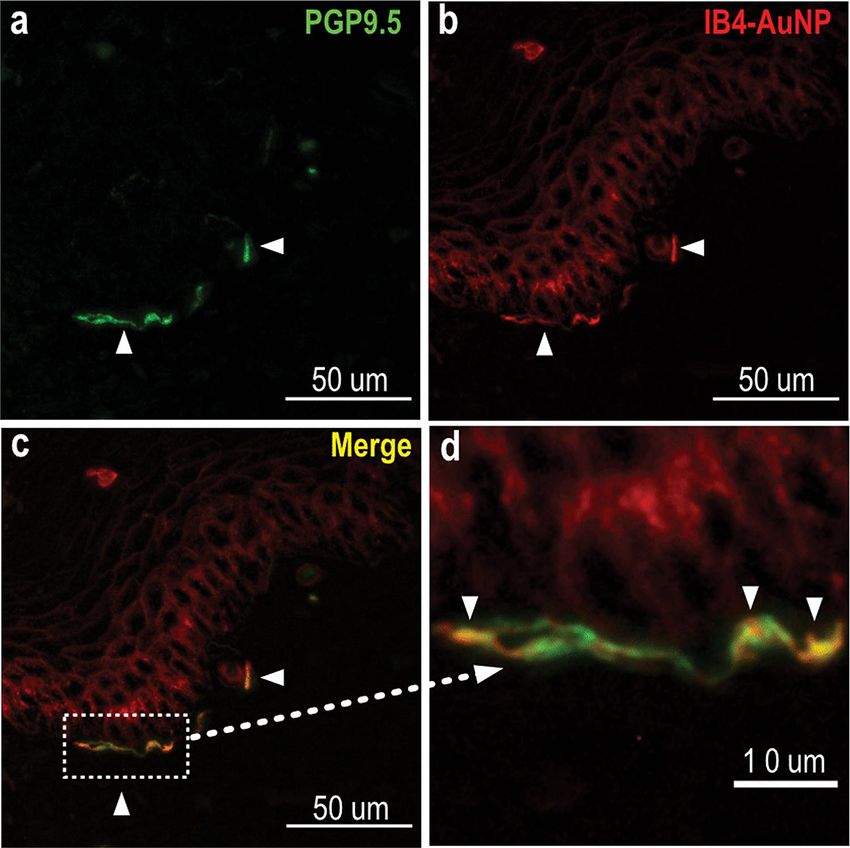
www.nature.com/scientificreports/
Figure 1. Surface functionalization of AuNP with IB4. Representative transmission electron microscopy (TEM)
images of ‘as-synthesized’ citrate capped AuNP (a) and IB4-AuNP (b). Arrows indicate a corona on AuNP
surface (inset), suggestive of IB4 adsorption. Histogram (c) depicting AuNP diameter quantified from 15 non-
overlapping TEM micrographs (n = 105 particles).
Results
Surface functionalization and characterization of gold nanoparticles. Gold nanoparticles
(AuNP) were synthesized by kinetically controlled step-wise seeded growth method, and surface functional-
ized with isolectin B4 (IB4) through ionic interaction. High resolution TEM images of AuNP showed spherical
shaped particles, with an average size of 111.3 ± 14.9 nm (Fig. 1a,c). IB4 functionalized AuNP showed a distinct
peripheral corona, suggestive of IB4 adsorption on the surface (Fig. 1b). Hydrodynamic size of AuNP, as meas-
ured using dynamic light scattering was 105.3 ± 1.0 nm, and was consistent with the particle size measurement
obtained using TEM. Surface adsorption of IB4 on AuNP resulted in an increase in the hydrodynamic size to
140.2 ± 1.9 nm, and decreased the surface charge of AuNP from − 43.3 ± 0.8 mV, to − 24.9 ± 1.9 mV. In addition,
IB4-AuNP demonstrated a small red-shift (~ 4 nm) in the surface plasmon resonance (SPR) absorption peak
(Table 1), as compared to the peak obtained from non-functionalized AuNP (Supplementary figure S1), further
confirming IB4 surface adsorption. Further, estimation of IB4 content on IB4-AuNPs yielded results that sug-
gested conjugation of 7–9 IB4 molecules per AuNP (Supplementary figure S2).
IB4‑AuNP bind to cell bodies of dissociated sensory neurons. To determine neuronal binding and
uptake of IB4-AuNP within cell bodies of sensory neurons, dissociated rat dorsal root ganglion (DRG) cell cul-
tures were exposed to varying dilutions of IB4-AuNP. We utilized the SPR-enhanced light scattering property of
AuNP to detect optical reflective intensity signals under a laser scanning confocal microscope22. Approximately
70–80% of DRG neurons demonstrated strong IB4 binding that was quite distinctly visible on neuronal cell
membrane (Supplementary figure S3). In addition, out of the total IB4 positive neurons, 80% were small-diam-
eter neurons, while the remaining were medium and large diameter neurons (Supplementary figure S3). These
binding characteristics, to a large extent, determined our choice to use IB4 as the neurotropic ligand. DRG cells
Scientific Reports | (2021) 11:2566 | https://doi.org/10.1038/s41598-021-81995-x 2
Vol:.(1234567890)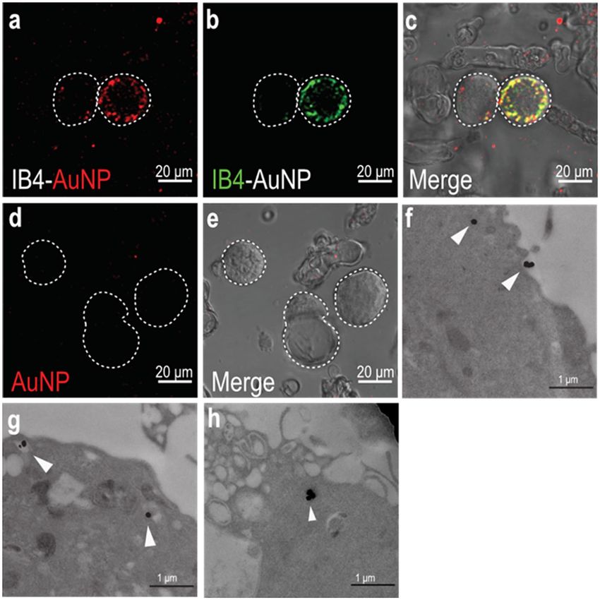
www.nature.com/scientificreports/

                                                                                AuNP                     IB4-AuNP
                                   Hydrodynamic size (nm)                       105.3 ± 1                140.2 ± 1.9
                                   Poly dispersity index                        0.1                      0.2
                                   Zeta potential (− mV)                        43.3 ± 0.8               24.5 ± 3.4
                                   SPR (nm)                                     562.8 ± 0.6              566.2 ± 0.2

                                  Table 1.  Gold nanoparticle characterization. Data obtained from dynamic light scattering and
                                  spectrophotometric analysis of non-functionalized and IB4 functionalized AuNP. Data shown are
                                  mean ± standard deviation (S.D.), from three experimental replicates.

                                  Figure 2.  IB4-AuNP bind and enter cell bodies of dissociated sensory neurons. Representative confocal
                                  microscopy images of dissociated rat DRG neurons treated with FITC-tagged IB4-AuNPs (a–c) and non-
                                  functionalized AuNPs (d,e). AuNP associated laser excited light-scattering signals (a) and FITC-IB4 associated
                                  signals (b), were seen only in specific neurons, while absent in others, indicating IB4 selectivity. Representative
                                  TEM images of gold nanoparticles (f) binding to DRG cell membrane, and undergoing endocytosis, with
                                  nanoparticles localized in well-formed endosome (g,h arrows).

                                  exposed to AuNP that were functionalized with FITC-IB4, demonstrated colocalized signals that were clearly
                                  observed on the plasma membrane and within the DRG neuronal cell body (Fig. 2a–c), suggestive of bind-
                                  ing and uptake of IB4-AuNP. Expectedly, few DRG neurons exhibited no AuNP associated signals (Fig. 2a–c),
                                  that was strongly suggestive of IB4-AuNP selectivity. The reflective intensity signals from IB4-AuNP appeared

Scientific Reports |   (2021) 11:2566 |                     https://doi.org/10.1038/s41598-021-81995-x                                                  3

                                                                                                                                                Vol.:(0123456789)
www.nature.com/scientificreports/
clustered and were not uniformly distributed within the cells. In contrast, cells treated with non-functionalized
AuNP (at matched concentration of IB4-AuNP) demonstrated no intracellular AuNP signals (Fig. 2d,e). How-
ever, non-functionalized AuNP at higher concentrations show distinct non-specific binding (Supplementary
figure S4). To further confirm IB4-AuNP uptake, we obtained TEM images of DRG cells treated with IB4-AuNP.
Images obtained after 1 h of IB4-AuNP exposure demonstrated clear surface binding and internalization of the
nanoparticles into DRG neurons. Distinct endosomal vacuoles around the internalized nanoparticles were also
observed (Fig. 2f–h), suggestive of nanoparticle endocytosis.
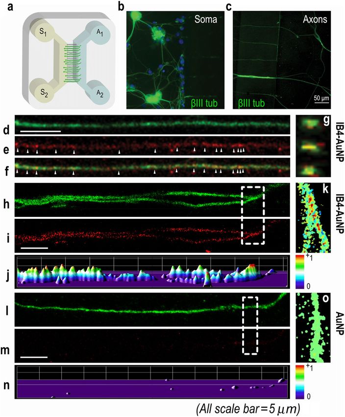
IB4‑AuNP bind to spatially separated axons of dissociated sensory neurons. To determine
axonal binding of IB4-AuNP, neurons cultured in microfluidic system were utilized. Neuronal microfluidic cul-
ture system (Fig. 3a) enables the separation of DRG cell soma and axons such that the dissociated DRG cells
seeded in the soma compartment extend axons through the microgrooves which emerge in the axonal compart-
ment in 3–4 days. The axonal compartments were then treated with IB4-AuNP in 4-day cultures, and selected
axons were imaged using a laser scanning confocal microscope (Fig. 3b,c). All neurons were stained with the
neuronal marker, β-III tubulin. Differential fluid levels maintained between the axonal and the soma compart-
ments ensured fluidic isolation within the system and prevented nanoparticle leakage into the microfluidic
grooves and the soma compartment (Supplementary figure S5). High resolution images of axons treated with
IB4-AuNP demonstrated discontinuous reflective intensity signals along the length of axons, which colocalized
well with β-III tubulin signals (Fig. 3d–f). Optical cross-sections of IB4-AuNP treated axons showed strong
colocalizing signals in the inner regions of the axons suggesting that the nanoparticles were located within the
axons and not just on the surface (Fig. 3g). In addition, the co-localization colour map analysis of specific seg-
ments of IB4-AuNP treated axons indicated strong reflective signals from the axons (Fig. 3h–k). In contrast,
no signals were detected from axons cultured in microfluidic chambers treated with non-functionalized AuNP
(Fig. 3l–o), suggesting that IB4 functionalization is necessary for the uptake of AuNP into sensory axons.
Axonal transport of IB4‑AuNP to the cell body of sensory neurons. To further determine whether
axon bound IB4-AuNP are indeed taken up and transported through axons, we performed time-lapse experi-
ments where change in cell soma fluorescence intensity was measured in the soma compartment after addition
of IB4-AuNP in the axonal compartment. Interestingly, we noticed a gradual increase in the DRG soma fluores-
cence after addition of FITC tagged IB4-AuNP in the axonal compartment (Fig. 4a). The change in fluorescence
intensity was less than 20% in the first two hours, but increased drastically after the 3rd hour. Curiously, inhibi-
tion of the axonal transport system by colchicine, an inhibitor of microtubule polymerization, prevented the
change in soma fluorescence, suggesting that IB4-AuNP translocation to the cell body from the axon, occurs via
the axonal retrograde transport system. Naïve neurons showed no changes in cell fluorescence. We further con-
firmed the translocation of IB4-AuNP by isolating and subjecting the cells from the soma compartment to TEM
imaging (Fig. 4b). TEM images of DRG cell bodies showed the presence of IB4-AuNP within the cytoplasm.
Taken together, results from these experiments suggest that IB4-AuNP bind to axons of cultured DRG cells and
are translocated to the cell body via the microtubule-based retrograde axonal transport system.
Binding of IB4‑AuNP to peripheral nerve fibres in the skin. To determine binding of IB4-AuNP to
terminal axonal endings in skin, we exposed rat hind-paw skin sections to AuNP that were surface functional-
ized with fluorescently labelled IB4. Skin nerve fibres were identified using the PGP 9.5 protein, which is widely
expressed in all nerve tissue. We observed tortuous peripheral nerve fibres, measuring approximately 1–2 µm in
diameter, that were distributed within the dermal and epidermal layers of skin (Fig. 5a). Interestingly, fluorescent
signals arising from IB4-AuNP colocalized with the dermal and epidermal nerve fibres. Magnified microscopic
images distinctly showed that the IB4-AuNP binds to skin nerve fibres in a discontinuous manner, giving an
appearance of varicosities along the length of the fibre (Fig. 5a–d).
Topical application of IB4‑AuNP to the rat hind paw. Binding, uptake, and translocation of topically
applied IB4-AuNP in vivo was determined by analysing tissue gold content using inductively coupled plasma
mass spectrometer (ICP-MS). The plantar surface of rat hind paw was immersed in solutions containing either
IB4 functionalized, or non-functionalized AuNP, at concentrations measuring 187.97 µg/mL and 195.84 µg/mL
respectively. The total amount of AuNP that penetrated across skin after 3 h of topical exposure was significantly
larger in rats exposed to non-functionalized AuNP, as compared to those exposed to IB4-AuNP (Fig. 6a). Over-
all, 44.1 ± 4.9% of AuNP were able to penetrate the skin of rats exposed to non-functionalized AuNP in compari-
son to 12.4 ± 3.4% in rats exposed to IB4-AuNP. Surprisingly, even though the total absorption of nanoparticles
was lower in IB4-AuNP treated rats (Fig. 6b), the number of nanoparticles that accumulated in the ipsilateral
DRGs of IB4-AuNP group, was much higher compared to the non-functionalized AuNP group (Fig. 6c,d). The
contralateral DRGs harvested from IB4-AuNP exposed rats, exhibited nanoparticle numbers that were similar to
those harvested from control rats, strongly supporting the possibility that the uptake of nanoparticles observed
in the ipsilateral DRG harvested from IB4-AuNP group most likely occurred due to axonal transport from the
skin. There was no difference in the gold content measured in sciatic nerve segments harvested from any of the
groups.
Peripheral nerve function of rats exposed to topical AuNP. Considering the observation that IB4-
AuNP were found to undergo axonal transport, nerve electrophysiological studies were performed to assess
nerve function and viability. The conduction velocities recorded from sciatic nerves of rats treated with non-
functionalized, or IB4-AuNP were comparable (Fig. 7a,b). Similarly, the obtained peak compound muscle action
Scientific Reports | (2021) 11:2566 | https://doi.org/10.1038/s41598-021-81995-x 4
Vol:.(1234567890)www.nature.com/scientificreports/
Figure 3. IB4-AuNP binds to axons of dissociated sensory neurons. Schematic representation of a microfluidic
device (a) showing the soma (s1–2) and axonal (a1–2) compartments, with microgrooves in between. Confocal
microscopy images of β-III tubulin stained dissociated rat DRG neurons cultured in the soma compartment (b)
showing glial cells (blue) and sprouted axons that traversed the microgrooves and were observed in the axonal
compartment (c). Representative images of β-III tubulin stained axons (d) treated with IB4-AuNP, showing
light scattering from AuNP (e). Both signals from (d) and (e), co-localized (arrows) at various locations on the
axon (f). Optical sections from (f), showing co-localized signals (g) from within the axon. Colocalization colour
map analysis of randomly selected axonal segments from IB4-AuNP treated devices (h–k) showing strong light
scatter signals from within axons, while devices with non-functionalized AuNP (i–o) showed no light scatter.
Experiments were performed in 3–4 devices per group.
Scientific Reports | (2021) 11:2566 | https://doi.org/10.1038/s41598-021-81995-x 5
Vol.:(0123456789)www.nature.com/scientificreports/
Figure 4. Axonal transport of IB4-AuNP to the cell body of sensory neurons. Graph indicating the time-
dependent change in fluorescent intensity from cell bodies of dissociated rat DRG neurons within microfluidic
devices, where AuNP conjugated with FITC tagged IB4 were treated in the axonal compartment (a). Data shown
are mean ± S.D, * P < 0.05, IB4-AuNP vs Naïve, and # P < 0.05, IB4-AuNP vs IB4-AuNP with colchicine, one-way
ANOVA with Sidak’s multiple comparison tests. Representative TEM image of DRG cells (b) collected from
experiments in (a), after 6 h, showing intracellular localization of gold nanoparticles.
potential ratio recorded from two different anatomical landmarks in the vicinity of the sciatic nerve did not
exhibit any difference between the non-functionalized and IB4-AuNP groups. This suggests that the topically
applied IB4-AuNP in the concentration tested did not interfere with peripheral nerve function.
Discussion
Peripheral nerve fibres innervating the skin are arguably the least explored targets for delivering therapeutic
molecules and their carriers. Barring topical anaesthetics that are used for blocking nociceptive receptor activ-
ity on peripheral a xons23,24, clinically there are almost no other drug molecules or delivery systems that target
peripheral nerve fibres. In this study, 105.3 ± 1.0 nm sized AuNP were functionalized with IB4 to target axonal
terminals and promote binding and uptake. Surface functionalization of AuNP with IB4 resulted in the reduc-
tion of total surface charge by almost − 20 mV, and an SPR peak shift that was most likely due to the positively
charged lysine and tryptophan rich sites on I B425.
IB4-AuNP were found to bind sensory neurons dissociated from rat DRGs. It is worth noting that IB4 was
selected from a list of known neurotropic ligands, including rabies virus glycoprotein (RVG)26, tetanus toxin C27,
and wheat germ a gglutinin28,29. Though RVG29 has been used to target Neuro 2A cells and mouse brain cells
in culture30, we were unable to detect RVG29 binding and uptake in DRG derived sensory neurons (data not
shown). IB4 was selected based on its selectivity in binding sensory neurons31, and for possible future application
in pain mitigation. In addition, IB4 has been reported to bind almost 70% of small diameter sensory neurons32,
which are known to predominately carry nociceptive signals and express pain associated proteins such as Nav1.8,
Nav1.9, and TRPV family of r eceptors33. In our study, we observed IB4-AuNP bound to cell membrane of sen-
sory neurons, and additionally found AuNP excitation scatter within neurons, suggestive of uptake. Here, it is
important to note that we have utilized the intrinsic property of AuNP to scatter light, for direct visualization
under a confocal microscope. It is well-known that at specific wavelengths of incident light, the surface plasmon
resonance phenomena of 100 nm sized AuNP causes light to be reflected in the 650–700 nm r ange22,34.
IB4-AuNP were found bound to axonal fibres in the dermal-epidermal regions, and also to a few intra-
epidermal nerve fibres in thin skin sections. Individual nerve fibres in the skin measure about 1–4 microns in
diameter35,36, which allows for the possibility of nanoparticles to bind and enter terminal axons. Previously, we
have showed that gold nanoparticles in the size ranges of 20 nm to 180 nm, easily penetrate all layers of thick
skin, and appear in the blood stream within 4 days19. In addition, topically applied gold nanoparticles accumulate
in the nerve fibre rich, epidermal-dermal regions (Supplementary figure S6), thereby significantly increasing
the chances of nanoparticle-nerve fibre contact. Our in vitro studies very clearly showed that IB4-AuNP bind
axonal terminals and undergo retrograde axonal transport to the cell bodies. Previous in vitro studies show
that quantum dots and iron oxide nanoparticles undergo endosome associated transport within axons via the
dynein-microtubule system37,38. Our results agree quite well with this observation since disruption of microtu-
bule polymerization by colchicine, prevented the axonal transport of AuNP. Interestingly our in vitro data also
shows a slight decrease in fluorescence intensity at the 4th hour time-point, and an increase in data variability
at subsequent time points. This is curious, because it has been well reported that metallic nanoparticles undergo
cellular exocytosis from various cell-lines39–41, though such phenomenon has not been reported in primary
Scientific Reports | (2021) 11:2566 | https://doi.org/10.1038/s41598-021-81995-x 6
Vol:.(1234567890)www.nature.com/scientificreports/
Figure 5. IB4-AuNP binding to peripheral nerve fibres in the skin. Representative fluorescent images of rat
hind-paw sections treated with AuNP conjugated with DyLight 594 tagged IB4, and co-stained with anti-PGP
9.5, showing skin nerve fibres (a), and AuNP signals (b), that were found to co-localize (c,d), suggestive of
AuNP binding to skin nerve fibres. (n = 3 sections).
sensory neurons. It is plausible that the reduction in fluorescence intensity at the soma could be due to differ-
ences in the rate of nanoparticle transport and nanoparticle exocytosis. More focused studies are warranted to
better explore this phenomenon in differentiated neurons.
Further, our in vivo experiments clearly showed a larger amount of topically applied IB4-AuNP accumulat-
ing in the ipsilateral lumbar DRG as compared to the contralateral control. This observation was quite similar
to what was observed in cultured axons, where IB4-AuNP accumulated in the cell soma after application at the
axonal end. Interestingly, we also observed nanoparticles in sciatic and DRG tissue harvested from rats that were
topically exposed to AuNP, albeit in very low amount. Taken together, this data strongly supports the hypothesis
that topically applied IB4-AuNP accumulate in the DRG, in large part, by utilizing the axonal innervation from
the topical site. Since DRG neurons synapse with second order neurons in the dorsal horn of the spinal cord, it
is plausible that topically applied AuNP may translocate to the spinal cord, but this was not checked in this study.
Considering our observation that AuNP were detected in contralateral DRG and sciatic nerve, it is possible that
part of the total nanoparticles detected in the ipsilateral DRG of rats exposed to IB4-AuNP could also be due to
implantation from the systemic circulation. It has been shown that nanoparticles as large as 180 nm can penetrate
across intact rat hind-paw skin and appear in the blood stream 4 days l ater19.
Another alternative possibility for IB4-AuNP accumulating in the ipsilateral DRG, could be due to the local-
ized distribution of nanoparticles in the ipsilateral limb, resulting in axonal entry and nanoparticle translocation
via nerve fibres innervating the muscle42. However, the predominant nerve fibres innervating the muscle tend to
be motor with nerve terminals ending in the spinal cord, thus making this route of AuNP delivery less probable.
It is also interesting to note that even though rats were exposed to an almost similar amount of topical AuNP
in the IB4-functionalized and non-IB4 functionalized groups, the total amount of IB4-AuNP that were absorbed
through the skin was much lower compared to non-functionalized AuNP. This is of much relevance because
IB4 functionalization seemed to reduce the number of particles actually crossing the skin, even though the total
amount of AuNP accumulating in the ipsilateral DRG was much higher. One probable explanation for reduced
permeability of IB4-AuNP compared to citrate capped AuNP could be the uneven surface distribution of lectin
Scientific Reports | (2021) 11:2566 | https://doi.org/10.1038/s41598-021-81995-x 7
Vol.:(0123456789)www.nature.com/scientificreports/
Figure 6. Topical application of IB4-AuNP to the rat hind paw. Bar graphs indicating the amount (a) and
percent (b) of gold nanoparticles that penetrate across the rat hind-paw skin after topical application of non-
functionalized AuNP or IB4-AuNP. Data shown are mean ± S.D., with n = 4 rats per group. * P < 0.05, Students
t-test. Box-whisker plots indicating the amount of gold nanoparticles detected in DRG and sciatic nerve tissue
harvested from rats that were topically treated with non-functionalized AuNP (c), or IB4-AuNP (d). Data
shown are measurements obtained from n = 2–4 animal sets per group, with each set comprising of pooled tissue
from 4 rats. (i = ipsilateral; c = contralateral).
on the IB4-AuNP surface. Simulation studies suggest that nanoparticles with homogeneously distributed binding
moieties on their surface, similar to our citrate-capped AuNP, dramatically enhance permeability, compared to
nanoparticles with random or heterogeneously distributed l igands43.
Furthermore, the presence of AuNP within the sciatic nerve in amounts as seen in this study, did not affect
nerve function, suggesting the benign nature of this approach. We also did not find any aberrant behavioural
alterations that could indicate any sensory dysfunction. However, further safety studies need to be performed
to determine functional and cellular effects of nanoparticles within axons.
In conclusion, we have demonstrated that metallic nanoparticles can be specifically translocated to neuronal
cell bodies using axonal terminals located in the skin via simple topical application.
Methods
Reagents and antibodies. Materials were acquired from the following sources: Gold chloride, poly (eth-
ylene glycol) (PEG), poly-D-lysine (PDL), bovine serum albumin (BSA), colchicine and paraformaldehyde
(PFA): Sigma-Aldrich, USA; Trisodium citrate dihydrate, papain, nitric acid, hydrogen peroxide, hydrochlo-
ric acid, TRIS-buffer, triton X-100, di-sodium hydrogen phosphate and potassium dihydrogen phosphate:
Merck Chemicals, USA; Collagenase-II, dispase-II, fetal bovine serum (FBS), penicillin–streptomycin mix and
trypsin–EDTA: Gibco, USA; Hank’s buffered saline solution (HBSS), Ham’s F-12 medium and Dulbecco’s modi-
fied Eagle’s medium (DMEM): Lonza Chemicals, USA; Isolectin B4, DyLight 594-IB4, FITC-IB4, DyLight 488
horse anti-mouse IgG antibody and Vectashield hardset mounting medium: Vector Laboratories, USA; Glutar-
aldehyde (EM grade), osmium tetroxide (OsO4), sodium cacodylate trihydrate and EMBed-812 embedding kit:
Electron Microscopy Sciences, USA; Hoechst 33342: Santa Cruz Biotechnology, Inc. USA; Anti β-III tubulin
monoclonal antibody: Cell Signaling Technology, Inc USA; Anti PGP9.5 antibody: Abcam Plc UK; Microfluidic
devices (SND 150): Xona Microfluidics, LLC, USA.
Scientific Reports | (2021) 11:2566 | https://doi.org/10.1038/s41598-021-81995-x 8
Vol:.(1234567890)www.nature.com/scientificreports/
Figure 7. Peripheral sciatic nerve function of rats exposed to topical AuNP. Representative electrophysiological
tracings obtained from the plantaris muscle by stimulating the sciatic nerve at the ankle and sciatic notch from
AuNP (a) and IB4-AuNP treated rats. Conduction velocity and signal amplitudes are shown in (b). Data shown
are mean ± S.D., n = 4. (ips = ipsilateral; con = contralateral).
Gold nanoparticle synthesis. Citrate capped AuNP were synthesized using the kinetically controlled
seeded growth method as reported earlier44. Briefly, 150 ml of trisodium citrate (2.2 mM) was heated to 100 ∘C
followed by the addition of 1 mL of gold chloride (25 mM). The solution was maintained at 100 ∘C for 10 min,
and then stepped down to 90 ∘C, followed by the addition of 1 mL of 25 mM gold chloride. Additional 1 mL gold
chloride (25 mM) was added to the reaction mixture after 30 min. This step was repeated 15–17 times where
55 mL of the reaction mixture was replaced by fresh 1 mL gold chloride (25 mM) and 55 mL of trisodium citrate
(2.18 mM) each time, until desired particle size was attained.
Gold nanoparticle characterization. The hydrodynamic size, dispersity index, and surface charge of
synthesized AuNP were determined using Zetasizer Nano ZS (Malvern, UK). UV–vis absorption spectra were
measured using Synergy H1 multi-mode plate-reader (Biotek, USA). High-resolution transmission electron
microscopy (TEM) images were obtained using the Tecnai G2 F20 TEM (FEI, USA).
Surface functionalization of gold nanoparticles. IB4 was surface adsorbed on gold nanoparticles as
reported earlier25. Briefly, 15 µL of IB4 or fluorophore (FITC or DyLight 594) tagged IB4 (1 mg/mL) was sus-
pended in 1 mL of 10 mM phosphate buffer (pH 6.5) containing AuNP, under stirring condition at 4 ∘C. 1%
PEG (w/v) was added to the solution, followed by high-speed centrifugation at 400g for 30 min and washed
thrice. Pelleted AuNP were resuspended in 10 mM phosphate buffer saline (PBS) (pH 7.4) containing 1% PEG
Scientific Reports | (2021) 11:2566 | https://doi.org/10.1038/s41598-021-81995-x 9
Vol.:(0123456789)www.nature.com/scientificreports/
(w/v). Surface functionalization was confirmed using absorption peak shift, dynamic light scattering and TEM.
It was also observed that non-functionalized AuNPs could be stored stably at 4 °C for up to 6 months, while IB4-
AuNPs aggregate within 5–7 days. Hence, only freshly prepared IB4-AuNPs were used in this study.
Animal care. Adult, female Sprague Dawley (SD) rats weighing 200–250 g were housed in pairs, allowed
standard rat diet and water ad libitum, and maintained on 10 h/14 h light/dark cycle. This study was conducted
under protocols approved by the Institutional Animal Ethical Committee (IAEC), Amrita Institute of Medical
Sciences, Kochi, India (Approval number IAEC/2013/3/5) in accordance with guidelines set forth by the Com-
mittee for Control and Supervision of Experiments on Animals (CPCSEA), Government of India.
DRG neuron isolation and culture. Rats were euthanized by CO2 asphyxiation, laminectomy per-
formed and dorsal root ganglia extracted in ice-cold HBSS. DRG neurons were isolated and cultured as reported
earlier45. Briefly, harvested DRG’s were incubated in papain (> 16 units/mg) for 30 min at 37 ∘C. DRG’s were then
transferred to a cocktail of collagenase (4 mg/mL), and dispase (4.66 mg/mL), for 30 min at 37 ∘C, followed by
mechanical trituration and suspension in DMEM media containing Ham’s F-12 medium (10% v/v), FBS (10%
v/v), and penicillin–streptomycin (1% v/v). For studies involving nanoparticle binding and uptake in cell bodies,
freshly isolated DRG cells were exposed to media containing AuNP (10% or 100% v/v) for 20 min, followed by
wash, prior to imaging.
DRG cultures in microfluidic devices. The microfluidic devices were primed as per manufacturer’s
instructions. DRG neurons were extracted and dissociated as described above. Approximately 1.5 × 10⁶ cells
were seeded in the soma compartment of the microfluidic device. Fluidic isolation was achieved by maintaining
a higher volume of media in the soma compartment, as compared to the axonal compartment. Sprouting axons
from the soma compartment were observed in the axonal compartment on the 5th day. Nanoparticles were
added into the axonal compartment (10 µL, 10% v/v, at 37 ∘C), and washed twice after 20 min before subjecting
the devices to imaging studies. In experiments to block retrograde axonal transport, colchicine (10 µM) was
added in the axonal compartment for 1 h, prior to nanoparticle treatment.
Immunostaining studies. Cells in microfluidic devices were washed with PBS (100 mM, pH 7.4) and
fixed in PFA (4% w/v) for 25 min, washed and permeabilized with triton X-100 (0.2% v/v in PBS) for 30 min
with gentle shaking. Cells were then treated with a blocking solution (BSA, 3% w/v in PBS) for 30 min and incu-
bated overnight at 4 ∘C with mouse anti-β-III Tubulin (1:200) antibody. DyLight 488 tagged horse anti-mouse
IgG antibodies were used to detect primary antibody binding, followed by Hoechst nuclear counterstain, and
visualized under an inverted confocal laser scanning microscope (DMI6000 CS, Leica Microsystems GmbH,
Germany). Non-specific IgG primary antibody was used as controls. Further, immunohistochemical studies
were performed on 4% PFA fixed skin sections to determine skin nerve fibre binding of IB4-AuNP. Briefly, 5 µm
thin skin sections were permeabilized using Tris-buffer (pH 7.5), and blocked with BSA (1% w/v), for 2 h at RT.
Sections were then co-treated with mouse anti-PGP9.5 antibody (1:500), and DyLight 594 tagged IB4-AuNP,
overnight at 4 ∘C. Sections were then counterstained with DyLight 488 horse anti-mouse secondary antibody,
mounted and visualized under an inverted fluorescence microscope (DMI3000 B, Leica Microsystems, Ger-
many).
Image acquisition and analysis. For visualizing axonal binding of AuNP, a laser wavelength of 514 nm
(argon, 30%) was used to excite the SPR of AuNP under confocal microscopy22. All acquisition parameters were
maintained at the same settings for all images, and data collected sequentially as multiple image stack files.
Light scatter signals from AuNP, along with fluorescent signals from fluorophore tagged antibodies were plotted
using Fiji plugin Interactive 3D surface plot v2.4.1. and co-localized as a color map. A randomly selected axonal
segment was used to demonstrate level of colocalization, where hot colors represent colocalization of AuNP
within axons. For time-lapse imaging experiments, DRG cells in the soma compartment of microfluidic devices
were visualized within a live-cell platform. Intensity data log from soma regions were acquired every hour and
analysed by Metamorph microscopy automation software (Molecular Devices, USA). Background noise level
threshold correction was applied from intensity signals obtained from images acquired at t = 0.
High‑resolution transmission electron microscopy. DRG cells treated with nanoparticles were
trypsinized and centrifuged to obtain a pellet, which was fixed with pre-warmed (37 °C) glutaraldehyde (2.5%
sO4 (2% v/v)46. Cells in the microfluidic devices
v/v) in 0.05 M cacodylate buffer (pH 7.4), and post-fixed with O
were gently scrapped off from the soma compartment and collected by centrifugation. After dehydration, epoxy
resin embedded ultra-thin Sections (110–120 nm) were obtained using ultramicrotome (PowerTome PC, RMC
Boeckeler Instruments, Inc. USA) and imaged under HR-TEM.
Topical application of AuNP and distribution. Female SD rats were anesthetized by intraperitoneal
administration of ketamine (100 mg/kg) and xylazine (5 mg/kg). The left hind-paw was placed in a modified
tube containing nanoparticle solution, such that the plantar area was maximally in contact with the nanoparticle
solution. After 3 h, the hind-paw was washed with Milli-Q water (Direct-Q 5 UV, Merck-Millipore, Germany)
to remove surface adherent nanoparticles. Care was taken to collect all wash fluid for calculating nanoparticle
penetration across skin. Further, we utilized the following equation to determine the amount of nanoparticles
(Np) that penetrated across hind-paw skin upon topical application ( AT).
Scientific Reports | (2021) 11:2566 | https://doi.org/10.1038/s41598-021-81995-x 10
Vol:.(1234567890)www.nature.com/scientificreports/
AT = Total Np amount in solution before topical application − (Amount of Np remaining after topical applica-
tion + Amount of Np in wash fluid).
Animals were euthanized by CO2 inhalation on day 3, and sciatic nerve and lumbar dorsal root ganglion from
both sides harvested, and processed for ICP-MS. Tissue samples from 2–3 pooled animal sets per experimental
group (each pool containing 4 animals each) were used in the study. Harvested tissue were cut into small pieces
and suspended in nitric acid at 80 ∘C for 3 h, and then transferred to a 1:1 mixture of hydrogen peroxide and
hydrochloric acid for 1 h. The samples were then diluted with Milli-Q water and analysed using ICP-MS (Thermo
Fisher Scientific, Germany).
Electrophysiological studies. Electrophysiological studies were performed on isoflurane anesthetized
rats, 3 days after topical application of IB4-AuNP. Amplitude and latencies obtained from the direct motor
response (M wave) and the mono-synaptically evoked H reflex (H wave) were recorded by stimulating the sci-
atic nerve at the sciatic notch (hip) and the tibial nerve at the ankle. Stimulations were performed via electrodes
using a square pulse (5 V, 0.5 ms, 1 Hz) (MP36, BIOPAC Systems, Inc, USA). The recording electrodes were
placed in the plantaris muscle of the feet and a reference electrode was placed in the middle digit, and the
ground electrode was placed in the inner thigh. Evoked responses were recorded and nerve conduction velocity
was calculated by dividing the distance from ankle to notch with the difference in notch—ankle latency values.
Statistics. All data are shown as mean ± standard deviation (S.D). Difference in values of mean among vari-
ous experimental groups were statistically tested using appropriate tests as stated in relevant figure legends.
Statistical analyses were performed using GraphPad Prism version 7.0.2 for Mac (GraphPad Software Inc, USA).
Data availability
The datasets generated during and/or analysed during the current study are available in the (Mendeley Data)
repository (http://dx.doi.org/10.17632/6cj84pw4hn.1).
Received: 8 May 2020; Accepted: 13 January 2021
References
1. Shankarappa, S. A. et al. Prolonged nerve blockade delays the onset of neuropathic pain. Proc. Natl. Acad. Sci. USA. 109, 17555–
17560 (2012).
2. Wolfe, D., Mata, M. & Fink, D. J. Targeted drug delivery to the peripheral nervous system using gene therapy. Neurosci. Lett. 527,
85–89 (2012).
3. Liu, J. et al. Peripherally delivered glutamic acid decarboxylase gene therapy for spinal cord injury pain. Mol. Ther. 10, 57–66 (2004).
4. Tajdaran, K., Gordon, T., Wood, M. D., Shoichet, M. S. & Borschel, G. H. An engineered biocompatible drug delivery system
enhances nerve regeneration after delayed repair. J. Biomed. Mater. Res. Part A 104, 367–376 (2016).
5. Fang, Z. et al. Enhancement of sciatic nerve regeneration with dual delivery of vascular endothelial growth factor and nerve growth
factor genes. J. Nanobiotechnol. 18, 46 (2020).
6. Liu, Q., Wang, X. & Yi, S. Pathophysiological changes of physical barriers of peripheral nerves after injury. Front. Neurosci. 12, 597
(2018).
7. Shankarappa, S. A. et al. Duration and local toxicity of sciatic nerve blockade with coinjected site 1 sodium-channel blockers and
quaternary lidocaine derivatives. Reg. Anesth. Pain Med. 37, 483–489 (2012).
8. Saraiva, C. et al. Nanoparticle-mediated brain drug delivery: Overcoming blood-brain barrier to treat neurodegenerative diseases.
J. Control. Release 235, 34–47 (2016).
9. Dong, X. Current strategies for brain drug delivery. Theranostics 8, 1481–1493 (2018).
10. Surnar, B. et al. Nanotechnology-mediated crossing of two impermeable membranes to modulate the stars of the neurovascular
unit for neuroprotection. Proc. Natl. Acad. Sci. 115, E12333–E12342 (2018).
11. Gonzalez-Carter, D. et al. Targeting nanoparticles to the brain by exploiting the blood brain barrier impermeability to selectively
label the brain endothelium. Proc. Natl. Acad. Sci. USA. 117, 19141–19150 (2020).
12. Geiser, M. et al. Ultrafine particles cross cellular membranes by nonphagocytic mechanisms in lungs and in cultured cells. Environ.
Health Perspect. 113, 1555–1560 (2005).
13. Abram, S. E., Yi, J., Fuchs, A. & Hogan, Q. H. Permeability of injured and intact peripheral nerves and dorsal root ganglia. Anes-
thesiology 105, 146–153 (2006).
14. Liu, Q. et al. Hollow silica nanoparticles penetrate the peripheral nerve and enhance the nerve blockade from tetrodotoxin. Nano
Lett. 18, 32–37 (2018).
15. Finke, S. & Conzelmann, K.-K.K. Replication strategies of rabies virus. Virus Res. 111, 120–131 (2005).
16. Diefenbach, R. J., Miranda-Saksena, M., Douglas, M. W. & Cunningham, A. L. Transport and egress of herpes simplex virus in
neurons. Rev. Med. Virol. 18, 35–51 (2008).
17. Jang, H. et al. Highly pathogenic H5N1 influenza virus can enter the central nervous system and induce neuroinflammation and
neurodegeneration. Proc. Natl. Acad. Sci. 106, 14063–14068 (2009).
18. Racaniello, V. R. One hundred years of poliovirus pathogenesis. Virology 344, 9–16 (2006).
19. Raju, G., Katiyar, N., Vadukumpully, S. & Shankarappa, S. A. Penetration of gold nanoparticles across the stratum corneum layer
of thick-Skin. J. Dermatol. Sci. 89, 146–154 (2018).
20. Zvyagin, A. V. et al. Imaging of zinc oxide nanoparticle penetration in human skin in vitro and in vivo. J. Biomed. Opt. 13, 064031
(2008).
21. Filipe, P. et al. Stratum corneum is an effective barrier to TiO2 and ZnO nanoparticle percutaneous absorption. Skin Pharmacol.
Physiol. 22, 266–275 (2009).
22. Klein, S., Petersen, S., Taylor, U., Rath, D. & Barcikowski, S. Quantitative visualization of colloidal and intracellular gold nanopar-
ticles by confocal microscopy. J. Biomed. Opt. 15, 036015 (2010).
23. Wang, L. et al. Topical drug formulations for prolonged corneal anesthesia. Cornea 32, 1040–1045 (2013).
24. Rowbotham, M. C., Davies, P. S. & Fields, H. L. Topical lidocaine gel relieves postherpetic neuralgia. Ann. Neurol. 37, 246–253
(1995).
25. Hermanson, G. T. Bioconjugate Tech. Second Edn. (Academic Press) https://doi.org/10.1016/C2009-0-64240-9 (2008).
Scientific Reports | (2021) 11:2566 | https://doi.org/10.1038/s41598-021-81995-x 11
Vol.:(0123456789)www.nature.com/scientificreports/
26. Gluska, S. et al. Rabies virus hijacks and accelerates the p75NTR retrograde axonal transport machinery. PLoS Pathog. 10, e1004348
(2014).
27. Lalli, G., Bohnert, S., Deinhardt, K., Verastegui, C. & Schiavo, G. The journey of tetanus and botulinum neurotoxins in neurons.
Trends Microbiol. 11, 431–437 (2003).
28. Gonatas, N. K., Harper, C., Mizutani, T. & Gonatas, J. O. Superior sensitivity of conjugates of horseradish peroxidase with wheat
germ agglutinin for studies of retrograde axonal transport. J. Histochem. Cytochem. 27, 728–734 (1979).
29. Butowt, R. & Von Bartheld, C. S. Connecting the dots: trafficking of neurotrophins, lectins and diverse pathogens by binding to
the neurotrophin receptor p75 NTR. Eur. J. Neurosci. 17, 673–680 (2003).
30. Kumar, P. et al. Transvascular delivery of small interfering RNA to the central nervous system. Nature 448, 39–43 (2007).
31. Fullmer, J. M., Riedl, M. S., Higgins, L. & Elde, R. Identification of some lectin IB4 binding proteins in rat dorsal root ganglia.
NeuroReport 15, 1705–1709 (2004).
32. Fang, X. et al. Intense isolectin-B4 binding in rat dorsal root ganglion neurons distinguishes C-fiber nociceptors with broad action
potentials and high Nav1.9 expression. J. Neurosci. 26, 7281–7292 (2006).
33. Cummins, T. R., Sheets, P. L. & Waxman, S. G. The roles of sodium channels in nociception: Implications for mechanisms of pain.
Pain 131, 243–257 (2007).
34. Link, S. & El-Sayed, M. A. Optical properties and ultrafast dynamics of metallic nanocrystals. Annu. Rev. Phys. Chem. 54, 331–366
(2003).
35. Engelstad, J. K. et al. Epidermal nerve fibers: Confidence intervals and continuous measures with nerve conduction. Neurology
79, 2187–2193 (2012).
36. Myers, M. I., Peltier, A. C. & Li, J. Evaluating dermal myelinated nerve fibers in skin biopsy. Muscle Nerve 47, 1–11 (2013).
37. Chowdary, P. D. et al. Nanoparticle-assisted optical tethering of endosomes reveals the cooperative function of dyneins in retrograde
axonal transport. Sci. Rep. 5, 18059 (2016).
38. Lesniak, A. et al. Rapid growth cone uptake and dynein-mediated axonal retrograde transport of negatively charged nanoparticles
in neurons is dependent on size and cell type. Small 15, 1803758 (2019).
39. Slowing, I. I. et al. Exocytosis of mesoporous silica nanoparticles from mammalian cells: From asymmetric cell-to-cell transfer to
protein harvesting. Small 7, 1526–1532 (2011).
40. BD, C., Chithrani, B. D. & Chan, W. C. W. Elucidating the mechanism of cellular uptake and removal of protein-coated gold
nanoparticles of different sizes and shapes. Nano Lett. (2007).
41. Wang, Y. et al. A quantitative study of exocytosis of titanium dioxide nanoparticles from neural stem cells. Nanoscale 5, 4737 (2013).
42. Zampieri, N., Jessell, T. M. & Murray, A. J. Mapping sensory circuits by anterograde transsynaptic transfer of recombinant rabies
virus. Neuron 81, 766–778 (2014).
43. Gkeka, P., Sarkisov, L. & Angelikopoulos, P. Homogeneous hydrophobic-hydrophilic surface patterns enhance permeation of
nanoparticles through lipid membranes. J. Phys. Chem. Lett. 4, 1907–1912 (2013).
44. Bastús, N. G., Comenge, J. & Puntes, V. Kinetically controlled seeded growth synthesis of citrate-stabilized gold nanoparticles of
up to 200 nm: Size focusing versus Ostwald ripening. Langmuir 27, 11098–11105 (2011).
45. Shankarappa, S. A., Piedras-Rentería, E. S. & Stubbs, E. B. Forced-exercise delays neuropathic pain in experimental diabetes: Effects
on voltage-activated calcium channels. J. Neurochem. 118, 224–236 (2011).
46. Binoy, A. et al. Plumbagin induces paraptosis in cancer cells by disrupting the sulfhydryl homeostasis and proteasomal function.
Chem. Biol. Interact. 310, 108733 (2019).
Acknowledgements
We thank the Indian Council of Medical Research, Government of India for SRF fellowship to Mr. Neeraj Katiyar,
and the Department of Biotechnology, Government of India for the Ramalingaswamy fellowship, and Nano-
mission, Department of Science and Technology, Government of India for grants to Dr. Sahadev Shankarappa.
We thank Sophisticated Analytical Instrument Facility (SAIF), Indian Institute of Technology (IIT-B) Bombay,
India, for ICP-MS analysis. We thank Mr. Sarath S. and Mr. Dennis Mathew for technical help with confocal
microscopy. We sincerely thank Dr. Shivanee Shah for her valuable editorial support.
Author contributions
N.K.: Methodology, Investigation, Formal analysis, Data curation, Visualization, Writing—original draft prepa-
ration; G.R.: Investigation, Formal analysis; P.M.: Investigation, Formal analysis; V.G.P.: Investigation, Formal
analysis; S.A.S.: Conceptualization, Supervision, Project administration, Funding acquisition, Writing—Review
& Editing, Formal analysis. All authors reviewed the manuscript.
Funding
Senior research fellowship (ICMR, Government of India) to NK, CSIR fellowship (DST, Govt. of India) to GR
and PM, and grant SR/NM/NS-1153/2013 (G) (Nanomission, DST, Govt. of India), and the Ramalingaswami
fellowship (DBT, Govt. of India) to SS.
Competing interests
The authors declare no competing interests.
Additional information
Supplementary Information The online version contains supplementary material available at https://doi.
org/10.1038/s41598-021-81995-x.
Correspondence and requests for materials should be addressed to S.A.S.
Reprints and permissions information is available at www.nature.com/reprints.
Publisher’s note Springer Nature remains neutral with regard to jurisdictional claims in published maps and
institutional affiliations.
Scientific Reports | (2021) 11:2566 | https://doi.org/10.1038/s41598-021-81995-x 12
Vol:.(1234567890)www.nature.com/scientificreports/
Open Access This article is licensed under a Creative Commons Attribution 4.0 International
License, which permits use, sharing, adaptation, distribution and reproduction in any medium or
format, as long as you give appropriate credit to the original author(s) and the source, provide a link to the
Creative Commons licence, and indicate if changes were made. The images or other third party material in this
article are included in the article’s Creative Commons licence, unless indicated otherwise in a credit line to the
material. If material is not included in the article’s Creative Commons licence and your intended use is not
permitted by statutory regulation or exceeds the permitted use, you will need to obtain permission directly from
the copyright holder. To view a copy of this licence, visit http://creativecommons.org/licenses/by/4.0/.
© The Author(s) 2021
Scientific Reports | (2021) 11:2566 | https://doi.org/10.1038/s41598-021-81995-x 13
Vol.:(0123456789)You can also read