THE UNUSUAL STRUCTURE OF THE PIGGYMAC CYSTEINE-RICH DOMAIN REVEALS ZINC FINGER DIVERSITY IN PIGGYBAC-RELATED TRANSPOSASES
←
→
Page content transcription
If your browser does not render page correctly, please read the page content below
Guérineau et al. Mobile DNA      (2021) 12:12
https://doi.org/10.1186/s13100-021-00240-4

 RESEARCH                                                                                                                                            Open Access

The unusual structure of the PiggyMac
cysteine-rich domain reveals zinc finger
diversity in PiggyBac-related transposases
Marc Guérineau1†, Luiza Bessa2,3†, Séverine Moriau2†, Ewen Lescop2, François Bontems2, Nathalie Mathy1,4,
Eric Guittet2, Julien Bischerour1, Mireille Bétermier1* and Nelly Morellet2*

  Abstract
  Background: Transposons are mobile genetic elements that colonize genomes and drive their plasticity in all
  organisms. DNA transposon-encoded transposases bind to the ends of their cognate transposons and catalyze their
  movement. In some cases, exaptation of transposon genes has allowed novel cellular functions to emerge. The
  PiggyMac (Pgm) endonuclease of the ciliate Paramecium tetraurelia is a domesticated transposase from the
  PiggyBac family. It carries a core catalytic domain typical of PiggyBac-related transposases and a short cysteine-rich
  domain (CRD), flanked by N- and C-terminal extensions. During sexual processes Pgm catalyzes programmed
  genome rearrangements (PGR) that eliminate ~ 30% of germline DNA from the somatic genome at each
  generation. How Pgm recognizes its DNA cleavage sites in chromatin is unclear and the structure-function
  relationships of its different domains have remained elusive.
  Results: We provide insight into Pgm structure by determining the fold adopted by its CRD, an essential domain
  required for PGR. Using Nuclear Magnetic Resonance, we show that the Pgm CRD binds two Zn2+ ions and forms
  an unusual binuclear cross-brace zinc finger, with a circularly permutated treble-clef fold flanked by two flexible
  arms. The Pgm CRD structure clearly differs from that of several other PiggyBac-related transposases, among which
  is the well-studied PB transposase from Trichoplusia ni. Instead, the arrangement of cysteines and histidines in the
  primary sequence of the Pgm CRD resembles that of active transposases from piggyBac-like elements found in
  other species and of human PiggyBac-derived domesticated transposases. We show that, unlike the PB CRD, the
  Pgm CRD does not bind DNA. Instead, it interacts weakly with the N-terminus of histone H3, whatever its lysine
  methylation state.
  (Continued on next page)

* Correspondence: mireille.betermier@i2bc.paris-saclay.fr;
nelly.morellet@cnrs.fr
†
 Marc Guérineau, Luiza Bessa and Séverine Moriau contributed equally to
this work.
1
 Université Paris-Saclay, CEA, CNRS, Institute for Integrative Biology of the
Cell (I2BC), 1 Avenue de la Terrasse, 91198 Gif sur Yvette Cedex, France
2
 Université Paris-Saclay, CNRS, Institut de Chimie des Substances Naturelles,
UPR 2301, 1 Avenue de la Terrasse, 91198 Gif sur Yvette Cedex, France
Full list of author information is available at the end of the article

                                         © The Author(s). 2021 Open Access This article is licensed under a Creative Commons Attribution 4.0 International License,
                                         which permits use, sharing, adaptation, distribution and reproduction in any medium or format, as long as you give
                                         appropriate credit to the original author(s) and the source, provide a link to the Creative Commons licence, and indicate if
                                         changes were made. The images or other third party material in this article are included in the article's Creative Commons
                                         licence, unless indicated otherwise in a credit line to the material. If material is not included in the article's Creative Commons
                                         licence and your intended use is not permitted by statutory regulation or exceeds the permitted use, you will need to obtain
                                         permission directly from the copyright holder. To view a copy of this licence, visit http://creativecommons.org/licenses/by/4.0/.
                                         The Creative Commons Public Domain Dedication waiver (http://creativecommons.org/publicdomain/zero/1.0/) applies to the
                                         data made available in this article, unless otherwise stated in a credit line to the data.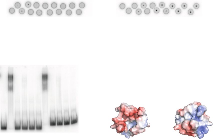
Guérineau et al. Mobile DNA       (2021) 12:12                                                             Page 2 of 22

 (Continued from previous page)
 Conclusions: The present study points to the structural diversity of the CRD among transposases from the
 PiggyBac family and their domesticated derivatives, and highlights the diverse interactions this domain may
 establish with chromatin, from sequence-specific DNA binding to contacts with histone tails. Our data suggest that
 the Pgm CRD fold, whose unusual arrangement of cysteines and histidines is found in all PiggyBac-related
 domesticated transposases from Paramecium and Tetrahymena, was already present in the ancestral active
 transposase that gave rise to ciliate domesticated proteins.
 Keywords: Domesticated transposase, Zinc finger structure, Genome rearrangements, Ciliates, Histones

Background                                                  species, including five in human, but their cellular
Transposons or transposable elements (TEs) are mo-          function has remained unclear in normal tissues [13,
bile genetic elements that have been shown to               14]. The most ancient, Pgbd5, is active in transpos-
colonize the genome of organisms from all kingdoms          ition [15] and promotes genomic rearrangements in
of life [1]. TEs are divided into two major classes:        solid tumors [16], but no catalytic function has been
class I TEs or retrotransposons, which use an RNA           attributed to the other four (Pgbd1 to Pgbd4) [17].
intermediate for their “copy-and-paste” transposition,      Intriguing instances of domesticated PGBD transpo-
and class II TEs, also called DNA transposons, which        sases playing an essential role during development
transpose through a DNA intermediate [2]. A sub-            were reported in the ciliates Paramecium tetraurelia
class of DNA transposons follow a “cut-and-paste”           (PiggyMac and its PiggyMac-like partners) [18, 19]
transposition mechanism, in which they are excised          and Tetrahymena thermophila (Tpb1, Tpb2, Tpb6
from their donor site before being inserted into a          and Lia5) [20–23].
new target locus. Transposases are enzymes encoded             Ciliates are unicellular eukaryotes, in which two
by DNA transposons [3]. They generally bind to ter-         functionally distinct types of nuclei coexist in the
minal inverted repeats (TIRs) at transposon ends and        same cytoplasm (reviewed in [24, 25]). The diploid
catalyze their movement from one genomic position           micronucleus (MIC) transmits the germline genome
to another. TEs can have harmful consequences when          to sexual progeny during reproduction. The highly
they invade coding or regulatory regions, inactivating      polyploid somatic macronucleus (MAC), derived from
or altering the regulation of host genes, but this may      the MIC and responsible for gene transcription, is
sometimes set up novel regulatory networks beneficial       destroyed at each sexual cycle. In the new developing
to the host [4]. The genome sequences of many spe-          MAC of P. tetraurelia, the genome undergoes amplifi-
cies have also revealed a number of previously              cation from 2n to 800n, while extensive programmed
unrecognized TE-derived genes. In some cases, trans-        genome rearrangements take place. These consist in
posase genes have been coopted by their hosts during        the precise elimination of ~ 45,000 single-copy, non-
evolution to create new cellular genes, conferring an       coding and short Internal Eliminated Sequences
adaptive benefit to their host [5, 6]. This is called       (IESs), representing ~ 3% of all germline DNA, and
transposase “domestication”.                                the heterogeneous removal of regions encompassing
  The piggyBac transposon that was originally isolated      TEs and other DNA repeats, which altogether consti-
from Trichoplusia ni (T. ni) [7] is a well-studied          tute ~ 25% of the germline genome [26, 27]. Because
DNA transposon with efficient transposition activity        ~ 50% of P. tetraurelia genes are interrupted by at
in many insect and mammalian species (reviewed in           least one IES in the MIC, precise IES excision is es-
[8]). It inserts almost exclusively into TTAA target        sential for the assembly of functional genes in the
sites and restores the original TTAA sequence after         new MAC. The conserved TA dinucleotides that flank
excision, leaving no footprint at its donor site [9]. PB,   each IES are targeted for DNA cleavage by the Piggy-
the PiggyBac transposase encoded by the T.ni piggy-         Mac (Pgm) endonuclease associated with its
Bac transposon, catalyzes the DNA strand breakage           PiggyMac-like (PgmL) partners [18, 19] and a single
and rejoining reactions that take place during trans-       TA is retained at the excision site in the rearranged
position [10]. PiggyBac-like elements (PBLE), some of       MAC genome. Developmentally regulated deposition
which were shown to be active, have been found in           of H3K9me3 and H3K27me3 heterochromatin marks
the genomes of numerous organisms including fungi,          by the Ezl1 histone methyltransferase is required for
plants and a wide array of metazoans [11–13]. In            the elimination of all TEs and ~ 70% of IESs [28, 29].
addition, piggyBac transposable element-derived             Similarly, T. thermophila uses Tpb2 [20], a Pgm
(PGBD) transposases are present in several eukaryotic       ortholog, to eliminate about a third of its germline
Guérineau et al. Mobile DNA   (2021) 12:12                                                                     Page 3 of 22

genome through the heterochromatin-driven impre-               We further show that the Pgm CRD, unlike the PB
cise excision of ~ 12,000 TE-related IESs [30–33].             CRD, does not bind DNA in vitro, but interacts weakly
   Pgm, a 1065-amino acid protein, plays an essential          with the N-terminal tail of histone H3 (residues 1–19),
catalytic role in DNA cleavage at IES ends [34], while its     independently of the methylation state of Lys9. Our
PgmL partners are likely architectural subunits organiz-       work opens new perspectives on the structural and func-
ing the excision complex [19]. A comparison with the           tional diversity of the CRD of PBLE transposases and
PB transposase indicates that Pgm is composed of four          their domesticated derivatives.
distinct domains [19, 34]: (i) a 220-amino acid N-
terminal domain; (ii) a 424-amino acid core domain typ-        Results
ical of transposases from the PiggyBac family, which in-       Variability of the CRD of PB transposases and
cludes the three conserved aspartic acids (D401D491D609)       domesticated PGBD proteins
that are essential for IES excision; (iii) a short cysteine-   Previous protein sequence alignments highlighted differ-
rich domain (CRD) also found, with some variations in          ent Cys/His arrangements among the CRDs located C-
the number and order of its cysteine and histidine resi-       terminal to the core domain of PBLE transposases and
dues, in PBLE transposases and domesticated PGBD               most PGBD domesticated transposases [13, 19]. The
proteins from other organisms [13, 19]; and (iv) a pre-        CRD of the T. ni PB transposase has a CxxC-CxxC-
dicted C-terminal coiled-coil structure (CC) encompass-        CxxH-CxxC motif (where C, H and x respectively de-
ing the last 307 residues, which appears to be an              note cysteine, histidine and any other residue), also
innovation of ciliate domesticated PGBDs. Previously,          present in a subset of PBLE transposases and PGBD pro-
we demonstrated that PgmΔCRD, a deletion mutant lack-          teins (Fig. 1), which forms a PHD-like zinc finger coord-
ing the CRD, is unable to support IES excision during          inating two Zn2+ ions [35]. The putative variant motif
MAC development, showing that the CRD is essential             (CxxC-CxxC-CH-CxxxH) found in the Pgm CRD was
for Pgm activity in vivo, although its exact role has          initially proposed to adopt a similar fold, in spite of the
remained unclear [34]. The PB CRD, essential for trans-        different number and position of its Cys and His resi-
position, adopts a PHD-like cross-brace zinc finger fold       dues [18]. However, primary sequence alignments have
(i.e. a zinc finger, in which the structural cores of the      revealed that His738, adjacent to the fifth cysteine of the
two zinc ions overlap) and binds specifically to a re-         variant motif in Pgm, is not conserved in the CRDs of
peated DNA sequence motif present at piggyBac trans-           other ciliate PGBD domesticated transposases nor in
poson ends [35]. Recent cryo-electron microscopy data          PBLE transposases harboring an otherwise similar ar-
indicated that the PB transposase assembles as a dimer         rangement of their Cys and His residues (Fig. 1), calling
within a synaptic complex composed of two piggyBac             into question the actual involvement of His738 in the
left ends, with the two PB CRDs binding together to a          folding of the Pgm CRD. We noticed instead that a histi-
single end, introducing asymmetry within the complex           dine residue is systematically present at a variable dis-
[36]. P. tetraurelia IESs, intriguingly, do not carry a con-   tance upstream of the first conserved cysteine doublet in
served motif that may serve as a sequence-specific rec-        Pgm (His701) and other CRDs carrying the variant Cys/
ognition site for Pgm and, compared with PB, the               His arrangement. The observed differences in the pri-
primary sequence of the Pgm CRD exhibits a different           mary sequence features of the Pgm and PB CRDs sug-
arrangement of its potentially zinc-coordinating residues      gested that they might adopt different folds. This
[19]. Taken together, these observations have raised two       prompted us to solve the structure of the Pgm(692–768)
questions: whether the Pgm CRD adopts the same struc-          variant domain.
tural fold as the PB CRD and whether it also interacts
with DNA.                                                      Zn2+ coordination mode of His701, His738 and His749
   In the present study, we have addressed the two issues      The 1H-15N HSQC spectrum of Pgm(692–768)* ob-
through a structure/function analysis. We used nuclear         tained using NMR spectroscopy (Fig. 2a) exhibits
magnetic resonance (NMR) spectroscopy to determine             marked dispersion of the 1H resonance chemical shifts
the structure of Pgm(692–768), which contains the              with most cross-peaks being in the 6.6–11 ppm range,
CRD, and found that it binds two zinc ions with two dis-       reflecting the presence of a well-folded structure. We
tinct coordination modes (His-Cys2-His (ZF1) and Cys4          found that zinc ions play a critical role in maintaining
(ZF2)), in a cross-brace zinc finger motif quite unusual       the structural integrity of Pgm(692–768)*. Indeed, in the
for a domain that was previously proposed to interact          presence of excess EDTA (10 mM) complexing zinc ions,
with chromatin [24]. Pgm(692–768) adopts a circularly          the 1H 1D spectrum gives evidence of a random coil ap-
permutated binuclear treble-clef fold [37], similar to the     pearance, suggesting unfolding of the domain, since a
fold observed only in the C1 cross-brace zinc finger           significant decrease in the chemical shift dispersion was
motif of protein kinase C (PKC) superfamily members.           observed (Fig. S1). Histidine ligands can bind Zn2+ using
Guérineau et al. Mobile DNA        (2021) 12:12                                                                                              Page 4 of 22

 Fig. 1 Sequence alignment of the CRDs of PBLE transposases and PGBD domesticated proteins. All accession numbers can be found in File S1.
 PBLE transposases: Ago (Aphis gossypii); Bmo (Bombyx mori); Cag (Ctenoplusia agnate); Har (Helicoverpa armigera); Hvi (Heliothis virescens); PB-Tni
 (Trichoplusia ni); Mlu (PiggyBat from Myotis lucifugus); PLE-wu (Spodoptera frugiperda). Domesticated PGBD transposases: Oni (Oreochromis
 niloticus); Pny (Pundamilia nyererei); Lia5, Tpb1, Tpb2, Tpb6 and Tpb7 (Tetrahymena thermophila); Pgm, PgmL1, PgmL2, PgmL3a/b/c, PgmL4a/b,
 PgmL5a/b (Paramecium tetraurelia); Tru (Takifugu rubripes); Pgbd2, Pgbd3 and Pgbd4 (Homo sapiens). In the sequence of Pgm(692–768), the
 coordinates of histidine residues are indicated, with His701 and His738 in red. In all sequences, the upstream histidine corresponding to Pgm
 His701 is highlighted in grey, when present. Conserved residues specific for the PB CRD are highlighted in yellow, those specific for the Pgm CRD
 are highlighted in green. For each peptide, isoelectric points (on the right: in blue or red for basic or acidic peptides, respectively) were calculated
 for the sequences highlighted in orange, using the ExPASy Compute pI/MW tool (https://web.expasy.org/compute_pi/)

two different coordination modes involving either of                           the chemical shift of Nε2 and Nδ1, which are inverted in
their endo-cyclic nitrogens (Nδ1 or Nε2). The Zn2+ co-                         the H-Nε2 tautomer compared to the H-Nδ1 tautomer.
ordination mode is related to the tautomeric form of                           Whatever the tautomer, two strong correlations are ob-
histidine: when the deprotonated Nδ1 binds Zn2+, Nε2                           served between Nε2 and both H-Cε1 and H-Cδ2, and
is protonated and vice-versa (Fig. 2b). So, at first, we                       only one between Nδ1 and H-Cε1. The pattern observed
used NMR to determine the protonation states of histi-                         for His701 and His738 has three connectivities (Hε1-
dine residues in Pgm(692–768)*. As shown previously                            Nδ1, Hε1-Nε2 and Hδ2-Nε2) (Fig. 2d), consistent with
[38], the Cδ2 and Cε1 chemical shifts of histidine can be                      the formation of the H-Nε2 tautomer [39]. For His749,
used as a signature of the coordination mode of histi-                         the observed pattern is constituted of four connectivities
dines in proteins. Using 1H-13C HSQC (Fig. 2c) we ob-                          (HCε1-Nε2, HCδ2-Nε2, HCε1-Nδ1 and HCδ2-Nδ1)
tained the Cδ2 and Cε1 chemical shifts of His749 (126.0                        compatible with the formation of the H-Nδ1 tautomer
ppm and 140.0 ppm respectively), which clearly indicate                        (Fig. 2d). So His701 and His738 Nδ1 on the one hand
that His749 has an Nε2 coordination mode [38]. How-                            and His749 Nε2 on the other are potentially available to
ever the Cδ2 and Cε1 chemical shifts of His701 (120.2                          interact with Zn2+. Comparison of the differences be-
ppm and 139.0 ppm, respectively) and His738 (118.4                             tween the 15Nδ1 and 15Nε2 chemical shifts of His701
ppm and 138.0 ppm, respectively) did not allow us to                           (45.7 ppm), His738 (88.4 ppm) and His749 (46.0 ppm), to
distinguish between coordinated and non-coordinated                            that observed for a non-Zn2+-interacting histidine resi-
histidines. We could only conclude that His701 and                             due (about 80 ppm on average) allows us to propose that
His738 are in the H-Nε2 tautomeric form.                                       His701 and His749, but not His738, are coordinated to
  We also used long-range 1H-15N HSQC to determine                             Zn2+.
the different forms of the three histidine residues of                           Zn2+ coordination was further confirmed for His701
Pgm(692–768). The intensity of the cross-peaks (which                          by atomic emission spectroscopy (Table S1). Mutating
depend on J-coupling) makes it possible to determine                           His701 to a serine residue caused a decrease in zinc
Guérineau et al. Mobile DNA      (2021) 12:12   Page 5 of 22

 Fig. 2 (See legend on next page.)
Guérineau et al. Mobile DNA       (2021) 12:12                                                                                              Page 6 of 22

 (See figure on previous page.)
 Fig. 2 Determination of the histidine residues implicated in Zn2+ coordination. a 1H-15N HSQC spectrum of 250 μM 15N- and 13C-labeled
 Pgm(692–768)* at 800 MHz, in the presence of 5 mM Hepes pH 6.8, 25 mM NaCl at 20 °C. Pgm(692–768)* corresponds to residues 692–768 plus
 the first seven linker residues (in blue, see also panel e) left after PreScission cleavage of the GST-Pgm(692–768) fusion. b Schematic
 representation of the two tautomeric states of histidine able to complex zinc ions. The Nδ1 coordination form is associated to the H-Nε2
 tautomeric form and the Nε2 coordination form is associated to the H-Nδ1 tautomeric form. Zinc ions are represented as red spheres. c 1H-13C
 HSQC (20 °C, pH 7.5) used to compare the Cδ2, Hδ2 and Cε1, Hε1 chemical shifts of His701, His738 and His749. d 1H-15N HSQC (20 °C, pH 7.5)
 used to compare the Nδ1 and Nε2 chemical shifts of His701, His738 and His749 e Sequence of the Pgm(692–768)* peptide used for NMR
 experiments. The Zn2+ ligands are highlighted in red. TALOS+ secondary structure predictions based on the experimental chemical shift
 information and those found in the 3D NMR structures of Pgm(692–768) are shown, with β-strands in green (703–705, 710–712, 718–719, 722–
 724) and α-helices in orange (725–730 and 745–751). The additional mini-helix observed in the final structures is highlighted in blue. The cross-
 brace arrangement of the two zinc-binding motifs is indicated by blue and pink arrows, each corresponding to one zinc-binding motif

emission compared to wild type Pgm(692–786). How-                            Analysis of 15N relaxation highlights chemical exchanges
ever mutation of His738 did not significantly affect zinc                    involving the N- and C-terminal regions of Pgm(692–768)
emission. Combining our NMR and atomic emission                              We observed heterogeneity in the line widths of the
spectroscopy results, we can unambiguously discern that                      peaks in the 1H-15N HSQC, some of them showing a
His701 Nδ1 and His749 Nε2 are involved in the coord-                         significantly weaker intensity (Fig. 2a). To determine if
ination of Zn2+ while His738 is not.                                         heterogeneity results from an exchange process between
                                                                             different conformations, we monitored the dynamics of
                                                                             the Pgm(692–768)* backbone using NMR relaxation
Structures of the Pgm(692–768) domain                                        measurements at 950 MHz 1H frequency and at 20 °C
A TALOS+ analysis [40] was performed to obtain esti-                         (Fig. 4). NMR 15N relaxation measurements are a power-
mations of the Pgm(692–768)* secondary structure and                         ful tool to characterize dynamic processes for proteins in
the phi and psi backbone torsion angles using the HN,                        solution, over a wide range of time scales. 15N relaxation
Hα, Cα, Cβ, CO and N chemical shifts. The TALOS+ re-
sults reveal that Pgm(692–768)* is constituted of two α-                     Table 1 NMR and refinement statistics for Pgm(692-768)*
helices encompassing residues 724–730 and 744–751                            structures generated with CYANA
and four β-strands composed of residues 703–705, 709–                        Experimental restraints
710, 720–723 and 735–737 (Fig. 2e).                                              Total number of restraints                              1066
  CYANA structure calculations were performed
                                                                                 Intra-residual and sequential                           736
using distance and angle restraints that imposed
tetrahedral coordination of two Zn2+ ions: the first                             Medium range (1 < |i_j| < 5)                            108
one with the His701 Nδ1, Cys723 S, Cys726 S and                                  Long range                                              222
His749 Nε2 atoms and the other one with the                                      TALOS+ Dihedral angles                                  116
Cys712 S, Cys715 S, Cys737 S and Cys745 S atoms.                             CYANA target function                                       0.55 ± 0.09
Fifteen structures were selected for structural ana-                         Rmsd from experimental restraintsa
lysis (Table 1) and are superimposed in Fig. 3a and
                                                                                 Upper limits (Å)                                        0.006 ± 0.003
S2. The structure of Pgm(692–768) reveals a well-
defined globular domain for residues His701 to                                   Dihedral angles (deg)                                   0.392 ± 0.094
                                                                                                                 a
His749, with a root mean square deviation (rmsd) of                          Rmsd from idealized geometry
0.54 ± 0.19 Å from the average structure on the back-                            Bonds (Å)                                               0.0012 ± 0.0008
bone atoms, and a cross-brace arrangement of the                                 Angles (deg)                                            0.53 ± 0.50
two zinc-binding motifs (Figs. 2e and 3b). The CRD                           Rmsd from mean structurea
structure is composed of two anti-parallel β-sheets
                                                                                 Backbone atoms (Å)                                      0.54 ± 0.19 Å
and two α-helices, plus a mini helix composed of
four residues (residues 738–741). The first β-sheet                              Heavy atoms (Å)                                         1.03 ± 0.19 Å
contains two strands, β1 (residues 703–705) and β4                           Ramachandran plotb
(residues 721–723). The second β-sheet consists of                               Most favored                                            90.0%
two strands, β2 (residues 710–712) and β3 (716–                                  Additionally allowed                                    10.0%
719) and is part of a long bent hairpin extending                                Generously allowed                                      0.0%
from residues 706 to 720, positioned above the core
                                                                                 Disallowed                                              0.0%
formed by the two helices (Fig. 3a and b). A high                            a
                                                                              Calculated over residues 701-749 of the 15 selected structures
degree of flexibility is observed for the N- and C-                          b
                                                                              Calculated over ordered residues 698-714, 717-750, 762-764 of the 15
terminal regions (Fig. S2).                                                  selected structures
Guérineau et al. Mobile DNA (2021) 12:12 Page 7 of 22
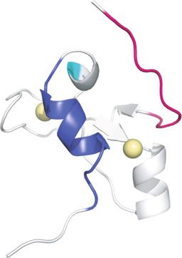
Fig. 3 Structure of the Pgm CRD. a Backbone superposition on residues (701–751) of the best 15 structures generated with CYANA, with the two
anti-parallel β-sheets in pink and blue, the two longest α-helices in orange (the secondary elements are highlighted according to the BMRB
protein structure validation suite result report) and Zn2+ ions in purple. The same color code is used in all panels. b The cysteine and histidine
residues involved in the coordination of Zn2+ ions are shown as sticks and colored by atom types: His701, Cys723, Cys726, His749 (blue) and
Cys712, Cys715, Cys737, Cys745 (pink) are respectively involved in the formation of each of the two zinc-binding motifs. Atoms from His738,
which is not implicated in Zn2+ coordination, are colored in green. c Topology diagram of Pgm(692–768). d Topology diagram of the C1 domain
of Rho-associated protein kinase 2 (ROCK II) (PDB id 2ROW). e Structure of the ROCK II C1 domain. f Topology diagram of the typical PHD
domain. In panels c, d and f, Zn2+ coordination is highlighted by dotted lines. Only Zn2+-coordinating histidine residues are indicated (H, in
green), all others ligands are cysteine residues (not indicated). β-strands are represented as arrows, helices as cylinders and Zinc ions as
purple spheres
results from the motion of the 1H-15N bond vector, the R2 transverse relaxation rate. They can be described
which represents the combination of the global move- by measuring the excess contribution (Rex) to R2.
ments of the protein (diffusion of a rigid body) and the The results show that 15N relaxation rates are rela-
movements of internal vectors (atomic bonds). Fast mo- tively homogeneous over the region encompassing resi-
tions on a picosecond-nanosecond (ps-ns) scale can be dues 701 to 737, with averaged R1, R2, hetNOE values of
characterized by heteronuclear 15N longitudinal relax- 1.07 ± 0.03 s− 1, 13.00 ± 0.56 s− 1 and 0.85 ± 0.09, respect-
ation rate (R1), transverse relaxation rate (R2) and 15N- ively (Fig. 4a-c). In contrast, the N- and C-terminal ex-
{1H} heteronuclear nuclear Overhauser effect (hetNOE) tremities (residues 687–700 and 752–768) show overall
of amide group resonances. Chemical exchange mecha- higher 15N R1 and lower 15N R2 and hetNOE values, in-
nisms are involved in general movements on the dicative of an elevated mobility of the segments flanking
microsecond-millisecond (μs-ms) scale and contribute to the cross-brace zinc finger. The order parameter S2Guérineau et al. Mobile DNA (2021) 12:12 Page 8 of 22
Fig. 4 Relaxation rates and backbone dynamics of Pgm(692–768)*. Plots of the 15N R1 a, R2 b relaxation and heteronuclear {1H}-15N NOE c
parameters obtained at 950 MHz 1H and 20 °C, using 0.5 mM of the 13C-15N-labeled Pgm(692–768)* domain, as a function of residue number and
solution secondary structure. β-sheets are represented as full arrows and α-helices as full rectangles. d Backbone order parameter S2, derived from
the chemical shifts, was generated by TALOS+ [40]. e Plot of the local backbone rmsd calculated using the CcpNmr software [41] on the 15
structures (aligned on residues 685–768), as a function of residue number and solution secondary structure. Dynamic parameters were extracted
from the 15N relaxation data using the model-free formalism of Lipari-Szabo with an isotropic reorientation model: f amplitude of the picosecond
(ps) to nanosecond (ns) time scale motion (S2), g internal correlation time (τε) and h exchange contributions on the microsecond (μs) to
millisecond (ms) timescale (Rex). i The structure of the Pgm CRD highlights residues with conformational exchange: (695–702) in pink, (741–755)
in dark blue and 8 amino acids from the structured region in light blue. Zinc ions are represented as yellow spheres
describes the amplitude of the internal motion of an H- N- and C-terminal flexible regions, with average values
N bond within the frame of the structure and is close to around 20 Å (residues 685–700) and 15 Å (residues
1 for rigid bonds and 0 for flexible bonds. The regions 756–768), compared with an rmsd of 2.5 Å for residues
with increased flexibility are also highlighted by the 701–755 (Fig. 4e).
backbone order parameter S2 estimated by TALOS+ We noted that the Pgm regions encompassing residues
[40] (and derived from the chemical shifts), since the corresponding to the flexible N-terminal domain (695–
average TALOS-predicted S2 values are 0.55 and 0.63 699) on one hand, and the 741–744 loop, the α2 helix
for residues 685–700 and 756–768 respectively, com- (745–751) and several residues (752–755) from the flex-
pared to 0.86 for residues 701–755 from the well- ible C-terminal domain on the other hand, showed
structured domain (Fig. 4d). This is in agreement with higher R2 values than the average R2 calculated for the
the rmsd per residue, which is particularly high for the first three quarters of the structured part of the CRDGuérineau et al. Mobile DNA (2021) 12:12 Page 9 of 22
(residues 701–737) (Fig. 4b). This may be due to the with either Pgm(692–768) or the CRD of any PgmL pro-
contribution of μs-ms conformational or chemical ex- tein. We found, however, that MBP fusions to full-
change. Model-free analysis [42, 43] of 15N NMR relax- length Pgm or its different variants – including MBP-
ation data provides information in terms of internal PgmΔCRD (in which the CRD was deleted), MBP-
mobility and the time scale of molecular motions. Be- PgmΔCC (carrying a deletion of the C-terminal exten-
yond the global molecular motion described by the cor- sion) [34] and MBP-PgmD401A (in which the first aspar-
relation time (τc), the description of internal motions is tic acid of the catalytic triad was replaced by an alanine)
reflected by the squared order parameter (S2), the in- – all bind DNA (Fig. 5a), indicating that the CRD is dis-
ternal correlation time (τε), and the chemical exchange pensable for the binding of full-length Pgm to DNA,
contribution (Rex) to the transverse relaxation (R2). The even though it is essential for Pgm activity in vivo [34].
overall correlation time (τc) estimated from the R2/R1 In parallel to DRaCALA, we performed Electrophoretic
ratios (6.62 ± 0.04 ns), is consistent with Pgm(692–768)* Mobility Shift Assays (EMSA) and detected no DNA
being in a monomeric state [44]. The mean S2 (0.92 ± binding activity for purified GST fusions with Pgm(692–
0.03) calculated for residues 701–755 is consistent with 768), PgmL2(540–614) or PgmL4(856–931), following
a rigid and well structured domain (Fig. 4f). Residues incubation with double-strand DNA substrates carrying
from the N- and C-terminal regions showed a mean S2 IES 51A1835 or the left end of IES 51A4404 (Fig. 5b and
of 0.38 ± 0.06 and 0.32 ± 0.03 respectively: internal cor- S3). We conclude from these experiments that the Pgm
relation times (τε) corresponding to motions in the CRD has no DNA binding activity in vitro by itself.
nanosecond time-scale had to be introduced for these To complete this analysis, we tested by NMR whether
residues to fit the relaxation data (Fig. 4g), consistent the Pgm CRD is capable of binding double-stranded
with the N- and C-terminal regions being very flexible. DNA oligonucleotides carrying IES 51A1835 and its left
As a result of model-free analysis, significant chemical flanking sequences, or the MAC-destined sequence
exchange contributions were introduced for two regions flanking IES 51A4404 (Fig. S3). Cross-peaks correspond-
(residues 695–702 and 741–755) and for eight amino ing to Pgm(692–768)* in 1H-1H NOESY spectra re-
acids scattered inside the structured region (Fig. 4h). corded in the absence and presence of either DNA
These exchanges may result from the long-range tertiary displayed neither chemical shift nor intensity changes
folding of the Pgm CRD, which brings the N- and C- (Fig. 5c). This again stands in contrast to what we re-
terminal regions close to each other, allowing them to cently described for the PB CRD [35]. According to the
interact in a transient manner (Fig. 4i). electrostatic charge distribution calculated on one NMR
structure of Pgm(692–768)*, the global surface charge of
No detectable binding of the Pgm CRD to DNA the Pgm cross-brace zinc finger (Pgm(701–749)) is nega-
We showed previously that the PB CRD is a double- tive (Fig. 5d), consistent with its calculated isoelectric
strand DNA binding domain that specifically recognizes point of 5.48 (Fig. 1). One face of the cross-brace zinc
a conserved sequence motif (5′-TGCGT-3′/3′-ACGCA- finger presents an even more negatively charged distri-
5′) at the ends of the piggyBac transposon from T. ni bution than the other, suggesting that the two faces of
[35]. To examine whether the Pgm CRD is also a DNA Pgm(701–749) are accessible for electrostatic interac-
binding domain, we performed DRaCALA experiments tions with positively charged molecules, rather than with
(Differential Radial Capillary Action of Ligand Assay negatively charged molecules such as DNA.
[45], see Materials and Methods), in which we compared Altogether, our results indicate that the Pgm CRD, un-
the binding of purified GST-Pgm(692–768) and GST- like the PB CRD, does not bind DNA in vitro, in agree-
PB(538–594) to a double-strand DNA carrying an IES, ment with a globally negatively charged pattern. Of note,
in the absence of competitor DNA to allow detection of no DNA binding activity was detected for the CRDs of
non-specific DNA binding (Fig. 5a). We first chose IES any PgmL protein, which all exhibit a similar arrange-
51A1835 as a substrate (Fig. S3) because it is among the ment of His and Cys residues relative to the Pgm CRD
30% of IESs, whose excision only depends upon the core (Fig. 1).
Pgm/PgmL machinery and not on the deposition of epi-
genetic chromatin marks [46, 47]. We observed reprodu- Interaction of the Pgm CRD with histone H3
cible binding of GST-PB(538–594) to this DNA In P. tetraurelia, the depletion of Ezl1, the histone methyl-
substrate, while no binding was detected in the absence transferase responsible for the trimethylation of both
of protein or following incubation with GST alone. Be- H3K9 and H3K27 [29], inhibits the elimination of TEs
cause IES 51A1835 is unrelated to the natural specific and a large fraction of IESs [28]. This suggests that
substrate of the PB CRD, this confirms that the DRa- H3K9me3 and H3K27me3 modifications are involved in
CALA assay can reveal non-specific protein-DNA inter- targeting the Pgm-associated complex to heterochromatin
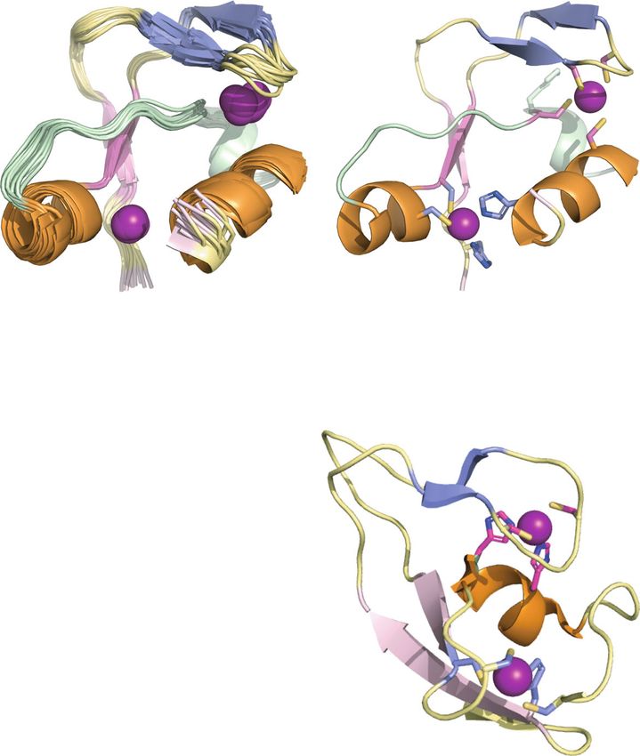
actions. In contrast, we did not detect DNA binding regions and drive their elimination. The primary sequenceGuérineau et al. Mobile DNA (2021) 12:12 Page 10 of 22 Fig. 5 The Pgm and PgmL CRDs have no DNA binding activity. a DRaCALA assays using a radiolabeled 80-bp double-strand substrate carrying IES 51A1835 (28 bp) and purified GST or MBP N-terminal protein fusions. Loading onto the membrane was performed in duplicate for all complexes, except for the controls incubated without protein (no prot) or, on the left panel, with the GST tag alone, which were loaded only once. b EMSA assays were performed with a radiolabeled 70-bp double-strand substrate carrying 31 bp from the left end of IES 51A4404 (77 bp) and its left flanking MAC-destined sequences (left), or the same substrate as in panel a for IES 51A1835 (right). c NMR evidence for the lack of an interaction between the Pgm CRD and DNA. Aromatic regions of the 1H-1H NOESY spectra of Pgm(692–768) (black) are not perturbed in the presence of double-stranded DNA (red) from IES 1835 (left) or the left flanking region of IES 51A4404 (right). The sequences of the double-strand DNA substrates used in panels a, b and c are shown in Fig. S3. d Electrostatic potential calculated on one structure of Pgm(701–749). The red and blue colors in surface representations highlight negative and positive charges, respectively of the Pgm CRD shows a similar arrangement of its His able to determine whether histones H2A/B or H4 were and Cys residues compared with the CRD of Tpb2, the also precipitated, since no specific Paramecium histone Tetrahymena orthologous domesticated transposase (Fig. antibodies were available. 1) that was shown to interact with heterochromatin, in To gain further insight into the involvement of histone particular with the trimethylated N-terminal tail of histone N-terminal tail methylation in the interaction of the Pgm H3 [48]. Since the Pgm CRD does not bind DNA, we ex- CRD with H3, we performed Enzyme-Linked Immuno- amined whether it has histone-binding capability, similar Sorbent Assays (ELISA) using purified MBP-Pgm(692– to the Tpb2p CRD. We first performed in vitro pulldown 768) and synthetic C-terminally biotinylated Paramecium assays of purified P. tetraurelia histones (Fig. S4) with H3(1–19) and H3(16–35) peptides. We used H3(1–19) GST-Pgm(692–768) and MBP-Pgm(692–768), and found peptides, either non-methylated or tri-methylated on Lys4 that each fusion protein precipitates endogenous H3 or 9, as well as two scrambled peptides (Scrambled 1 and (Fig. 6a-b). Little or no precipitation was detected with the Scrambled 2) containing the same amino acids as H3(1– GST or MBP tags alone, demonstrating that the inter- 19) in a different sequential order (Fig. 6c). We also used action is mediated by Pgm(692–768) itself. We were not two H3(16–35) peptides: a non-methylated version and a
Guérineau et al. Mobile DNA      (2021) 12:12                                                                                       Page 11 of 22

 Fig. 6 Interaction of the Pgm CRD with histone H3. a In vitro pulldown assay of P. tetraurelia histones with GST or GST-Pgm(692–768) (GST-CRD).
 b In vitro pulldown assay of P. tetraurelia histones with MBP or MBP-Pgm(692–768) (MBP-CRD). MBP fusion proteins and histone H3 were revealed
 on western blots using α-MBP-HRP and α-H3 antibodies, respectively (see Fig. S4 for the control of histone preparations and full-size blots with
 molecular weight markers). c Amino acid composition of the synthetic peptides used in this study. Methylated lysines are underlined.
 Hydrophobic residues are in red, residues with positively charged side chains in blue, residues with uncharged polar side chains in purple. C-
 terminally biotinylated peptides (−b) used in ELISA assays are indicated. d ELISA assays with 100 nM MBP or MBP-Pgm(692–768) (MBP-CRD)
 against unmodified H3(1–19), trimethylated H3(1–19)K4me3 and H3(1–19)K9m3, control peptides Scrambled 1 (*: TECAN absorbance detection at
 450 nm close to saturation) and Scrambled 2, unmodified H3(16–35) and trimethylated H3(16–35)K27me3. p-values were calculated using the
 Wilcoxon-Mann-Whitney test calculator (https://ccb-compute2.cs.uni-saarland.de/wtest/); sample sizes m = 3, n = 3 and test variant H1: (H3(1–
 19)K4me3 signal) < (H3(1–19) signal) and H1: (H3(1–19)K4me3 signal) < (H3(1–19)K9me3 signal). e ELISA assays with MBP alone or MBP-Pgm(692–
 768) (MBP-CRD) against the H3(1–19) peptide. One hundred nM of each MBP fusion protein was loaded into H3(1–19)-coated wells either in
 absence (left panel) or in presence of 10 mM EDTA (right panel). Error bars represent the standard deviation between technical triplicates

modified variant carrying trimethylated Lys27. H3(1–19),                   MBP-Pgm(692–768), but interaction was also detected
H3(1–19)K4me3 and H3(1–19)K9me3 all gave an inter-                         with H3(1–19)K4me3. This suggests that the Pgm CRD is
action signal with MBP-Pgm(692–768), while no signal                       able to interact with the N-terminal tail of histone H3 in-
was detected with MBP alone (Fig. 6d). Only a back-                        dependently of the methylation state of Lys4 and 9. Simi-
ground signal was detected for H3(16–35) and H3(16–                        lar interaction signals were obtained with all PgmL CRDs
35)K27me3 in the presence of MBP-Pgm(692–768), indi-                       in the presence of the different H3 peptides (Fig. S5).
cating that the Pgm CRD does not interact with H3(16–                        In order to monitor whether correct folding of the
35), whether Lys27 is methylated or not. Among all tested                  Pgm CRD is essential for its interaction with H3(1–19),
H3(1–19) peptides, unmethylated H3(1–19) and H3(1–                         we repeated the ELISA assay and incubated the MBP-
19)K9me3 displayed the strongest interaction signals with                  Pgm(692–768) fusion with H3(1–19) in the presence of
Guérineau et al. Mobile DNA (2021) 12:12 Page 12 of 22
10 mM EDTA, an excess concentration that triggers with Glu739 displaying the largest chemical shift
unfolding of the domain (Fig. S1). We observed a strong changes. Interestingly, these two regions in contact with
decrease of the interaction signal under these conditions the H3 peptide correspond to those for which we ob-
(Fig. 6e), indicating that the cross-brace zinc finger fold tained evidence for conformational exchanges (Fig. 4f),
of the Pgm CRD is essential for its interaction with possibly resulting from a transient interaction between
H3(1–19). Surprisingly, we found that two mutant ver- each other (Fig. 7e). This experiment allowed us to cal-
sions of the Pgm CRD still interact with H3, both in his- culate an apparent dissociation constant (KD) of 289 ±
tone pulldown experiments and ELISA assays (Fig. S6): a 70 μM (Fig. 7e), assuming a 1:1 binding, indicating that
single mutant carrying a C712S substitution in one of its the interaction between the Pgm CRD and H3(1–19) is
zinc-coordinating motifs, which reduces the overall Zn2+ rather weak.
load to ~ 20% (Table S1), and a double mutant carrying Taken together, our results indicate that the inter-
the C712S and H701S substitutions expected to disrupt action of the Pgm CRD with H3(1–19) is weak, does not
both zinc-coordinating motifs. Excess EDTA abolished depend on Lys4 and Lys9 methylation and mostly relies
the interaction signal with the double mutant (Fig. S6d), on electrostatic interactions, with acidic residues of
suggesting that the mutant CRD H701S + C712S still co- Pgm(692–768) contacting the positively charged N-
ordinates enough Zn2+ ions to maintain a structured fold terminus of H3.
and interact with H3(1–19).
Discussion
Analysis of the contacts between Pgm(692–768) and The Pgm CRD forms an unusual zinc finger structure
H3(1–19) Programmed genome rearrangements in P. tetraurelia
We observed that the two scrambled H3(1–19) peptides provide a nice illustration of the impact of transposons
behaved differently in the presence of the Pgm CRD on their host genome plasticity and evolution. Pgm is an
(Fig. 6d and S7a). Scrambled 1 gave an even greater example of a catalytically functional domesticated trans-
interaction signal with MBP-Pgm(692–768) than H3(1– posase [18]. Yet little is known about the structure-
19), while only a background signal was observed with function relationships of its different domains. In this
Scrambled 2. A comparison of peptide sequences high- study, we provide the first insight into Pgm structure
lights that, like H3(1–19), Scrambled 1 has an N- and demonstrate that its CRD, previously shown to be
terminal alanine followed by five basic residues, while essential for activity in vivo, binds two Zn2+, resulting in
Scrambled 2 carries an uncharged STQ sequence at its the formation of a cross-brace zinc finger. Additionally,
N-terminus, suggesting the importance of a positive N- we show that the Pgm CRD does not bind DNA but is
terminal charge for interacting with the Pgm CRD (Fig. capable of binding the N-terminal residues of histone
6c). The 30% reduced interaction signal of MBP- H3 in vitro – although with no specific affinity for meth-
Pgm(692–768) with H3(1–19)K4me3 relative to un- ylated lysines.
modified H3(1–19) seems consistent with positive Zinc fingers are generally part of larger proteins
charges near the N-terminus of H3 being important for known to play a role in almost all cellular processes.
the interaction. Of note, a TOCSY experiment revealed These domains, characterized either by their zinc-
chemical shift perturbation of the resonances of the first binding residues and their respective entanglement or by
two residues of H3 (Ala1 and Arg2) in the presence of the fold of their secondary structure, have been classified
the Pgm CRD (Fig. S7b), confirming that the N-terminal into 40 families [37], among which are the most-
residues of H3 are involved in the interaction. populated and experimentally well-characterized Really
To further characterize the contacts between Interesting New Gene (RING) [49, 50], Plant Homeo-
Pgm(692–768) and H3(1–19), we again used NMR spec- Domain (PHD) [51] and FYVE families [52, 53]. Even if
troscopy. The effect of adding H3(1–19) to 15N-labeled the RING, FYVE and PHD domains share a common
Pgm(692–768)* was monitored by 1H-15N HSQC (Fig. 7a structural fold and binding mode of two Zn2+ ions (Fig.
and b). We observed both differential broadening of a 3f), they display significant diversity in the selection of
subset of signals and chemical shift perturbations in the their targets. PHD fingers [51], for instance, are found in
presence of the histone peptide (Fig. 7c). Mapping of known chromatin-modifying proteins and in many chro-
these perturbations highlighted two regions (Fig. 7d). matin regulatory factors. They generally function as epi-
The first one (695–702) is located within the flexible N- genetic effectors or readers that interact with modified
terminal extension upstream of the globular zinc finger or unmodified histone H3 tails [54, 55]. Upon inter-
structure (Fig. 7a). The second region (739–753) in- action with a PHD domain, the H3 tail generally adopts
cludes several acidic residues from the structured fold an extended β strand-like conformation and aligns with
(Glu739 and Asp741 from the linker between helices α1 the β sheet of the PHD zinc finger as a third antiparallel
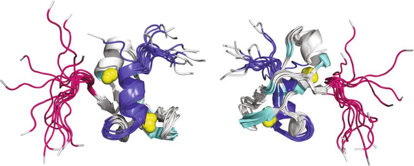
and α2 and Glu750 from helix α2 (743–753)) (Fig. 7b), β strand (Fig. S8) [56]. We show here that the Pgm CRDGuérineau et al. Mobile DNA (2021) 12:12 Page 13 of 22 Fig. 7 Pgm(692–768)* interacts with histone H3(1–19). a and b Superimposition of the 1H-15N HSQC spectra recorded at 800 MHz and 20 °C of 100 μM 15N-Pgm(692–768) in the absence (black) and presence of 0.5 (red), 1 (orange), 2 (yellow), 4 (green), 6 (blue) and 10 (purple) molar equivalents of H3(1–19). The panels show resonances from residues from the N-terminal a and the α2 helix b regions that display significant chemical shift perturbations and/or broadening upon addition of H3(1–19). The residues that do not undergo chemical shift variations are annotated in grey and the others in black. c Relative intensities and, below, chemical shift perturbations of the resonances of Pgm(692–768)* in the presence of a 10-fold excess of H3(1–19). d One of the ensemble structures of Pgm(692–768)* showing the most affected residues in the presence of H3(1–19) in dark blue and pink. These are located in two main regions: the flexible N-terminal extension (695–702) and the α2 helix (743–753), in addition to Glu739 (light blue) in the preceding loop. Zn2+ ions are represented as spheres. e Determination of a dissociation constant (KD) of 289 ± 70 μM using the chemical shift perturbations for residue E739 does not fold into any of the canonical RING, FYVE or Pgm(701–749) does not exhibit the hydrophobic pocket PHD structures, contrary to what we observed for the found in a typical C1 domain targeted by DAG/phorbol PB CRD [35]. It adopts a topology analogous to the C1 ester (Fig. S10). cross-brace zinc finger motif, which is highly conserved among the protein kinase C (PKC) superfamily members The Pgm and PB CRDs interact with different substrates that share a common requirement for phospholipids for We observed that full-length Pgm binds DNA, as ex- their kinase activity [57], however with a different num- pected for an active endonuclease, whose catalytic site is ber of β strands and α helices (Fig. 3d-e and S9). Despite responsible for the cleavage of IES boundaries (Fig. 5). this similar fold, the long accessible bent hairpin of However, contrary to what we reported for the PB CRD
Guérineau et al. Mobile DNA   (2021) 12:12                                                                  Page 14 of 22

[35], we detected no DNA binding activity for the Pgm        Lys27. Taken together, our results rather suggest that
CRD in DRaCALA, EMSA or NMR assays, nor did we               the observed weak interaction of the Pgm CRD with the
find DNA binding activity of the CRDs of its PgmL part-      N-terminal tail of H3 is mediated through electrostatic
ners. The lack of DNA binding activity of the Pgm CRD        charges and that we may not have identified a specific
may reflect important differences between cut-and-paste      substrate of this domain. Another component of the
transposition and Pgm-mediated IES elimination. In-          Pgm-associated complex, which remains to be identified,
deed, even though Paramecium IESs are, at least for a        may carry a specific H3K9me3 and/or H3K27me3 recog-
large fraction, remnants of ancestral Tc1/mariner trans-     nition domain and position the excision complex at
posons [26], their excision is mediated by PiggyBac-         Ezl1-dependent IES excision sites.
related domesticated transposases, which belong to a
distinct family of class II transposons [18, 19]. In         Structure-function variability of the CRDs of PBLE
addition, IESs carry no conserved motif that may serve       transposases and domesticated PGBD proteins
as a specific recognition sequence for the excision com-     We recently identified five groups of paralogous Pgm-
plex. Available experimental evidence indicates that IES     like (PgmL) proteins in P. tetraurelia (PgmL1 – PgmL5)
recognition is maternally controlled through the com-        [19], none of which contains an intact DD(D/E) active
bined action of non-coding RNAs and epigenetic chro-         site, suggesting that PgmLs are not catalytically active.
matin marks [58]. Our results suggest that, in contrast      All PgmLs carry a CRD, in which the zinc-binding Cys/
to the PB transposase, what drives Pgm to Paramecium         His are conserved with respect to Pgm, suggesting that
IESs is not the direct binding of its CRD to IES DNA.        the PgmL CRDs may share the same tertiary fold in spite
   We observed instead that the Pgm CRD interacts with       of otherwise divergent primary sequences (Fig. 1). Each
histone H3 with a weak binding affinity. The histone- ra-    PgmL is capable of forming complexes with Pgm, plays
ther than DNA-binding activity of the Pgm CRD is con-        an essential role in IES excision and is required for the
sistent with the charge distribution on the surface of the   correct completion of autogamy. Due to their variant
folded domain, which is mainly negatively charged (Fig.      primary sequences, the PgmL CRDs have very different
5). In comparison, the PB CRD, which does bind DNA,          isoelectric points, with only the PgmL4 CRD exhibiting
is globally positively charged [35]. We mapped the inter-    an acidic isoelectric point close to that of the Pgm CRD
action to two distinct regions of the Pgm CRD: one           (Fig. 1). Despite these differences, all PgmL CRDs
mainly hydrophobic region of the flanking N-terminal         present comparable interaction signals with unmethy-
part (residues 695–702) and a second region (743–753)        lated or methylated H3(1–19) peptides in ELISA assays,
harboring acidic residues and corresponding approxi-         while they interact neither with H3(16–35) nor H3(16–
mately to the last α-helix (Figs. 4 and 7). When the         35)K27me3 (Fig. S5). We conclude that the Pgm and
cross-brace zinc finger fold is disrupted following          PgmL CRDs share similar binding properties to the N-
addition of excess EDTA, these two regions may not           terminal tail of H3, independently of the methylation
properly fold together, leading to a strong reduction of     state of Lys9 and 27. Future studies should address
the contact with H3 (Fig. 6). Of note, the Pgm CRD har-      whether cooperative interaction between Pgm and PgmL
bors two β-sheets that are potentially accessible for an     CRDs may provide higher affinity and/or specificity to
interaction with the N-terminal tail of H3 in an ex-         the recognition of histone tails by the Pgm-associated
tended β strand-like conformation, as observed for PHD       complex.
domains, but they do not appear to take part in the             In the distantly related ciliate T. thermophila, the
interaction with H3(1–19) (Fig. S8).                         Tpb2        domesticated        transposase       catalyzes
   For the histone H3 tail, several lines of evidence have   heterochromatin-driven DNA elimination during sexual
pointed to positive charges at the N-terminus of H3 be-      reproduction [20]. The Tpb2 CRD is capable of binding
ing involved in contacting the Pgm CRD (Fig. 6 and S7).      heterochromatin and interacts with the N-terminal tail
The lack of specificity for tri-methylated Lys9 and Lys27    of histone H3 with a preference for tri-methylated pro-
was somewhat unexpected, given the essential role of         teins on Lys9 or Lys27 [48]. The fold of the Tpb2p CRD
the histone methyltransferase Ezl1, which catalyzes both     has not been characterized and seven histidine and cyst-
H3K9 and H3K27 tri-methylation, during programmed            eine residues were previously proposed to participate in
DNA elimination in P. tetraurelia [28, 29]. It was hy-       the folding of a PHD zinc finger [48]. Based on sequence
pothesized previously that H3K9me3 and/or H3K27me3           alignments (Fig. 1), we show here that the primary se-
drive the Pgm complex to Ezl1-dependent IESs and             quences of the Tpb2 and Pgm CRDs have a similar
other heterochromatin-associated regions and target          organization, with one histidine and seven cysteine resi-
their elimination. We show here that the Pgm CRD in-         dues, suggesting that both CRDs may adopt a similar
teracts with H3(1–19) independently of Lys9 methyla-         cross-brace zinc finger structure. The weak interaction
tion and does not interact with the region encompassing      of the Pgm CRD with the N-terminal tail of H3 appears
Guérineau et al. Mobile DNA   (2021) 12:12                                                                  Page 15 of 22

to require the coordination of Zn2+ ions involved in          originated from active PBLE transposases carrying the
folding of the CRD (Fig. 6). Likewise, a mutant Tpb2          Pgm CRD fold. Currently available data reveal no
CRD with two of its potentially Zn2+-binding cysteine/        straightforward correlation between the DNA sequence
histidine residues changed to alanine loses its ability to    organization at PBLE transposon ends and the putative
bind H3 N-terminal peptides in pulldown assays [48]. As       fold of the CRDs of their respective transposases. Even
a result, the full-length mutant Tpb2 fails to trigger het-   within the fourth group of PBLEs, for instance, the PLE-
erochromatin body formation and is inactive for DNA           wu CRD may adopt a similar structure to the Pgm CRD
elimination in vivo, demonstrating the importance of the      fold (Fig. 1). Whether the PLE-wu CRD, like the PB
Tpb2 putative fold in interacting with heterochromatin        CRD, binds sequence-specifically to DNA repeats or as-
[48]. In spite of their structural similarity, the Pgm and    sociates with histones has not been established. More
Tpb2 CRDs have different isoelectric points and abilities     generally, the presence of similar structures does not
to discriminate between methylated and non-methylated         allow us to predict the preferred substrate of the CRDs
histone tails, which may reflect major differences in the     of PBLE transposases or their domesticated PGBD coun-
mechanism and/or control of programmed DNA elimin-            terparts, emphasizing the versatility of these domains
ation between the two ciliates. Indeed, Paramecium IESs       and the diversity of the interactions in which they may
are all excised precisely from genes and non-coding re-       be involved.
gions, while only a subset depend upon H3 trimethyla-
tion to be excised. In contrast, all Tetrahymena IESs,        Materials and methods
which are all intergenic, require heterochromatin forma-      Sequence analysis
tion for their imprecise elimination.                         Protein sequence alignment was performed using
   PBLEs are found in numerous organisms, such as             MUSCLE 3.8 (https://www.ebi.ac.uk/Tools/msa/muscle/)
fungi, plants, insects, fishes and mammals [13] and some      and adjusted manually.
of them are active in transposition [11, 12, 59, 60]. A
large-scale survey of PBLE transposons allowed the def-       Peptides
inition of four structural groups, based on the differen-     Untagged and biotinylated synthetic Paramecium tetra-
tial organization of DNA repeats at their ends [13]. In       urelia histone H3 peptides (Fig. 6c) were synthesized by
particular, the piggyBat transposon from the bat Myotis       Proteogenix (Schiltigheim, France) and used without fur-
lucifugus, which belongs to the first group, carries simple   ther purification.
15-bp TIRs at its ends [11], while piggyBac from T. ni
and PLE-wu from the insect Spodoptera frugipeda [12],         Expression and purification of GST-tagged CRDs
representatives of the fourth group, carry complex TIRs       Synthetic DNA fragments (Integrated DNA Technolo-
with multiple internal repeats and may require complex        gies) encoding Pgm(692–768) and its C712S and
interactions for the correct conformation of their trans-     H701S + C712S mutant derivatives, PB(538–594),
pososome [36]. While all PBLE transposases harbor a           PgmL1(463–539), PgmL2(540–614), PgmL3a(471–550),
characteristic conserved domain (PF13843, or DDE_             PgmL4a(856–931) and PgmL5a(761–840) were PCR
Tnp_1_7), which includes their catalytic site, the study      amplified and cloned between the EcoRI and XhoI re-
by Bouallègue et al. [13] has highlighted the diversity of    striction sites of plasmid pGEX6p1 (resulting plasmid se-
the Cys/His motifs found in their C-terminal CRDs. Our        quences in File S2).
previous [35] and present studies indicate that different       For NMR spectroscopy, BL21-Gold (DE3) E. coli cells
Cys/His motifs may adopt distinct structural folds. Se-       expressing GST-Pgm(692–768) were grown at 37 °C in
quence alignment of two active PBLE transposases (PLE-        M9 minimal medium with 15NH4Cl (1 g/L) (Cambridge
wu [12] and PiggyBat-Mlu [11]) shows that they share a        Isotope Laboratories, USA) and either D-glucose-13C6
conserved His (aligned with Pgm His701) followed by           (Cambridge Isotope Laboratories, USA) or unlabeled D-
seven Cys or His residues (Fig. 1), suggesting that their     glucose (3 g/L) for expression of 13C/15N- or 15N-labeled
CRDs may adopt the Pgm CRD fold. Thus, the variant            Pgm CRD, respectively. At OD600 = 0.6 to 0.8, cells were
fold of the Pgm CRD, also possibly found in other ciliate     induced with 0.2 mM Isopropyl β-D-1-thiogalactopyran-
domesticated transposases (PgmL or Tpb proteins), may         oside (IPTG) for 3 h30 in the presence of 1 mM ZnSO4
have been inherited from an ancestral active ciliate PBLE     and were then collected and suspended in buffer A (25
transposase quite distinct from the T. ni PB transposase,     mM Tris–HCl pH 7.5, 0.15 M NaCl, 10% glycerol, 10
rather than evolved during the domestication process. In      mM 2-Mercaptoethanol) supplemented with 0.5 mM
human, while Pgbd1 and 5 do not carry a recognizable          phenylmethane sulfonyl fluoride (PMSF). Cells were
CRD, Pgbd2, 3 and 4 exhibit a Pgm-like arrangement of         lysed with a French press and the cleared supernatant
His and Cys residues in the primary sequence of their         was filtered through a 0.45 μm syringe filter before load-
CRDs (Fig. 1), again suggesting that they may have            ing onto 2.5 ml of Glutathion Sepharose™ 4B resin (GE
You can also read