Comparative Validation of Machine Learning Algorithms for Surgical Workflow and Skill Analysis with
←
→
Page content transcription
If your browser does not render page correctly, please read the page content below
Comparative Validation of Machine
Learning Algorithms for Surgical Workflow
and Skill Analysis with the HeiChole
Benchmark
Authors

Martin Wagner MDa,b, Beat-Peter Müller-Stich MDa,b, Anna Kisilenkoa,b, Duc Trana,b, Patrick
Heger MDa, Lars Mündermann PhDc, David M Lubotsky BSca,b, Benjamin Müllera,b, Tornike
Davitashvilia,b, Manuela Capeka,b, Annika Reinked,e,f, Tong Yu MScg,h, Armine Vardazaryan
MScg,h, Chinedu Innocent Nwoye MScg,h, Nicolas Padoy PhDg,h, Xinyang Liui, Eung-Joo Leej,
Constantin Dischk, Hans Meine PhDk,l, Tong Xia BScm, Fucang Jia PhDm, Satoshi Kondo
PhDn,1, Wolfgang Reitero, Yueming Jinp, Yonghao Longp, Meirui Jiangp, Qi Doup, Pheng Ann
Hengp, Isabell Twickq, Kadir Kirtacq, Enes Hosgorq, Jon Lindström Bolmgrenq, Michael
Stenzelq, Björn von Siemensq, Hannes G. Kenngott MD MSca, Felix Nickel MD MMEa, Moritz
von Frankenberg MDr, Franziska Mathis-Ullrich PhDs, Lena Maier-Hein PhDd,e,f,t, Stefanie
Speidel PhD*u,v, Sebastian Bodenstedt PhD*u,v

Authors affiliations
a
 Department for General, Visceral and Transplantation Surgery, Heidelberg University
Hospital, Im Neuenheimer Feld 420, 69120 Heidelberg, Germany

b
 National Center for Tumor Diseases (NCT) Heidelberg, Im Neuenheimer Feld 460, 69120
Heidelberg, Germany

c
 Data Assisted Solutions, Corporate Research & Technology, KARL STORZ SE & Co. KG,
Dr. Karl-Storz-Str. 34, 78332 Tuttlingen

d
 Div. Computer Assisted Medical Interventions, German Cancer Research Center (DKFZ), Im
Neuenheimer Feld 223, 69120 Heidelberg Germany

e
 HIP Helmholtz Imaging Platform, German Cancer Research Center (DKFZ), Im Neuenheimer
Feld 223, 69120 Heidelberg Germany

 1
f
 Faculty of Mathematics and Computer Science, Heidelberg University, Im Neuenheimer Feld
205, 69120 Heidelberg

g
 ICube, University of Strasbourg, CNRS, France. 300 bd Sébastien Brant - CS 10413 - F-
67412 Illkirch Cedex, France

h
 IHU Strasbourg, France. 1 Place de l’hôpital, 67000 Strasbourg, France

i
 Sheikh Zayed Institute for Pediatric Surgical Innovation, Children’s National Hospital, 111
Michigan Ave NW, Washington, DC 20010, USA

j
 University of Maryland, College Park, 2405 A V Williams Building, College Park, MD 20742

k
 Fraunhofer Institute for Digital Medicine MEVIS,Max-von-Laue-Str. 2, 28359 Bremen,
Germany

l
 University of Bremen, FB3, Medical Image Computing Group, ℅ Fraunhofer MEVIS, Am
Fallturm 1, 28359 Bremen, Germany

m
 Lab for Medical Imaging and Digital Surgery, Shenzhen Institute of Advanced Technology,
Chinese Academy of Sciences, Shenzhen 518055, China

n
 Konika Minolta, Inc., JP TOWER, 2-7-2 Marunouchi, Chiyoda-ku, Tokyo 100-7015, Japan

o
 Wintegral GmbH, Ehrenbreitsteiner Str. 36, 80993 München, Germany

p
 Department of Computer Science and Engineering, Ho Sin-Hang Engineering Building, The
Chinese University of Hong Kong, Sha Tin, NT, Hong Kong

q
 caresyntax GmbH, Komturstr. 18A, 12099 Berlin, Germany

r
 Department of Surgery, Salem Hospital of the Evangelische Stadtmission Heidelberg,
Zeppelinstrasse 11-33, 69121 Heidelberg, Germany

s
 Health Robotics and Automation Laboratory, Institute for Anthropomatics and Robotics,
Karlsruhe Institute of Technology, Geb. 40.28, KIT Campus Süd, Engler-Bunte-Ring 8, 76131
Karlsruhe, Germany

t
 Medical Faculty, Heidelberg University, Im Neuenheimer Feld 672, 69120 Heidelberg

u
 Div. Translational Surgical Oncology, National Center for Tumor Diseases Dresden,
Fetscherstraße 74, 01307 Dresden, Germany

 2
v
 Cluster of Excellence “Centre for Tactile Internet with Human-in-the-Loop” (CeTI) of
Technische Universität Dresden, 01062 Dresden, Germany

1
 Present address: Muroran Institute of Technology, Hokkaido, 050-8585, Japan.

*Stefanie Speidel and Sebastian Bodenstedt contributed equally to this work

Corresponding author

Martin Wagner MD

e-mail: martin.wagner@med.uni-heidelberg.de

Postal address:
Universitätsklinikum Heidelberg
Chirurgische Klinik
Im Neuenheimer Feld 420,
69120 Heidelberg
Germany

 3
Abstract
PURPOSE: Surgical workflow and skill analysis are key technologies for the next generation
of cognitive surgical assistance systems. These systems could increase the safety of the
operation through context-sensitive warnings and semi-autonomous robotic assistance or
improve training of surgeons via data-driven feedback. In surgical workflow analysis up to 91%
average precision has been reported for phase recognition on an open data single-center
video dataset. In this work we investigated the generalizability of phase recognition algorithms
in a multi-center setting including more difficult recognition tasks such as surgical action and
surgical skill.
METHODS: To achieve this goal, a dataset with 33 laparoscopic cholecystectomy videos from
three surgical centers with a total operation time of 22 hours was created. Labels included
framewise annotation of seven surgical phases with 250 phase transitions, 5514 occurences
of four surgical actions, 6980 occurences of 21 surgical instruments from seven instrument
categories and 495 skill classifications in five skill dimensions. The dataset was used in the
2019 international Endoscopic Vision challenge, sub-challenge for surgical workflow and skill
analysis. Here, 12 research teams trained and submitted their machine learning algorithms for
recognition of phase, action, instrument and/or skill assessment.
RESULTS: F1-scores were achieved for phase recognition between 23.9% and 67.7% (n=9
teams), for instrument presence detection between 38.5% and 63.8% (n=8 teams), but for
action recognition only between 21.8% and 23.3% (n=5 teams). The average absolute error
for skill assessment was 0.78 (n=1 team).
CONCLUSION: Surgical workflow and skill analysis are promising technologies to support the
surgical team, but are not solved yet, as shown by our comparison of machine learning
algorithms. This novel HeiChole benchmark can be used for comparable evaluation and
validation of future work. In future studies, it is of utmost importance to create more open, high-
quality datasets in order to allow the development of artificial intelligence and cognitive
robotics in surgery.

Keywords
Surgical Workflow Analysis, Endoscopic Vision, Surgical Data Science, Laparoscopic
Cholecystectomy

 4
1. Introduction
Surgical workflow and skill analysis are key technologies for the development and
seemingless integration of artificial intelligence systems (AI) in the operating room (OR).
These systems may increase the safety and efficiency of the operation through early context-
sensitive warnings (Katić et al., 2013), OR management (Tanzi et al., 2020) and procedure
time prediction (Aksamentov et al., 2017; Bodenstedt et al., 2019b), continuing surgical
education (Maier-Hein et al., 2017) and professional development (Greenberg et al., 2018) by
objective assessment of surgical skill and competency (Hashimoto et al., 2019; Vedula et al.,
2017) as well as semi-autonomous assistance (Lalys and Jannin, 2014; Vercauteren et al.,
2020). Furthermore, if today's robotic telemanipulators are to become cognitive assistance
systems, which perceive their environment, interpret it according to previous experience and
perform a context-aware action in a semi-autonomous way, they will have to understand the
surgical workflow and learn from the skilled surgeons (Bihlmaier, 2016; Maier-Hein et al.,
2017). Thus, surgical workflow and skill analysis are a prerequisite for the next generation of
surgical robotics.
Given that aim, the data-driven investigation of laparoscopic surgeries is most promising.
Already, the analysis of laparoscopic video data attracts increasing attention for workflow
analysis (Loukas, 2018; Ward et al., 2020) as well as surgical skill assessment (Azari et al.,
2019; Curtis et al., 2020). This is because laparoscopic surgery offers many advantages for
the patient (Greene et al., 2007), thus being the gold standard in some routine operations,
such as cholecystectomy. In consequence, this medical procedure is one of the most common
operations in the United States, with approximately 300,000 operations per year (Hassler and
Jones, 2020). At the same time, laparoscopy is a challenge for surgeons and assistants in
their hand-eye coordination and requires a lot of training inside and outside the OR
(Grantcharov et al., 2004; Schijven et al., 2005). The vascular supply of the gallbladder, for
example, may be very diverse, thus confusing less experienced surgeons. In a review on the
variability of the cystic artery, it has been found that in 8.9% of cases more than one artery
was present and in 18.5% of cases the artery did not lie within Calot's triangle (Andall et al.,
2016). In addition, there is still no conclusive solution for preventing bile duct injury (Madani et
al., 2020; Mascagni et al., 2021, 2020), the most common severe complication of
cholecystectomy (Michael Brunt et al., 2020). When creating AI in surgery to solve these
clinical challenges, a great obstacle to train the underlying machine learning algorithms is the
lack of high quality annotated data sets (Maier-Hein et al. 2021). The use of machine learning
techniques has been successfully researched on the basis of annotated medical data (Topol,
2019), for example through the segmentation and recognition of tumours and metastases

 5
(Ehteshami Bejnordi et al., 2017; Golden, 2017; Heller et al., 2019), diseased atrial structures
in magnetic resonance imaging (Xiong et al., 2021), pathologies in chest X-rays (Li et al.,
2018; Nam et al., 2019; Singh et al., 2018; Wang et al., 2017), as well as healthy abdomen
segmentation of CT and MR data (Kavur et al., 2020), or medical sensors such as for the
interpretation of electrocardiograms (Strodthoff and Strodthoff, 2019; Willems et al., 1991). At
the same time, research on surgical videos is comparatively underrepresented, even though
the availability of surgical video data is increasing thanks to time-efficient and cost-effective
recording and storage of laparoscopic surgery videos. A reason may be that the process of
video annotation with meaningful information for machine learning algorithms is time-
consuming and laborious. As a result, only few surgical video data sets are openly available
for research, even though the publication rate for the analysis of surgical procedures has
increased in recent years (Loukas, 2018). For instance, the Cholec80 data set contains videos
of 80 laparoscopic cholecystectomies from a single center and annotation of surgical phase
and instrument presence with a phase recognition average precision of up to 91% (Twinanda
et al., 2017). Similar results have been reported on a larger multicenter data set of 1243
laparoscopic cholecystectomies, but this data is not openly available (Bar et al., 2020). The
utilization of the Cholec80 data set in studies on the automatic prediction of procedure duration
(Bodenstedt et al., 2019b) and autonomous annotation assistance of surgical workflow
(Bodenstedt et al., 2019a; Lecuyer et al., 2020) underlines the importance and usefulness of
surgical workflow recognition through machine learning. Another open data set is the
Heidelberg Colorectal data set. It comprises 30 videos of three different types of laparoscopic
colorectal surgeries, corresponding sensor data from medical devices in the OR and pixel-
wise semantic segmentation of surgical instruments (Maier-Hein et al., 2021). The successful
use of the Heidelberg Colorectal data set during the Endoscopic Vision (EndoVis) challenges
2017 (https://endovissub2017-workflow.grand-challenge.org/) and 2019
(https://robustmis2019.grand-challenge.org/) is an example for the comparative validation of
machine learning algorithms to explore an optimal solution for surgical problems. Apart from
laparoscopy, surgical workflow has been investigated in ophthalmology. The CATARACTS
Challenge presented successful results on instrument presence detection during cataract
surgery using computer vision algorithms (Al Hajj et al., 2019). Though all three data sets used
clinical patient videos as the basis for their annotations, they are limited in their transferability,
because they do not sufficiently reflect the diversity of clinical data in a multi-center setting
limiting their representativeness. Furthermore, they focus on a limited variety of annotated
features.
In contrast, the preclinical JIGSAWS data set for gesture and skill analysis contains kinematic
and video data of surgical tasks with detailed action and skill annotation (Ahmidi et al., 2017).
Surgical skill has a significant impact on the long-term survival of patients (Brajcich et al.,

 6
2020) and a higher skill level is associated with fewer postoperative complications and better
patient outcome (Birkmeyer et al., 2013; Stulberg et al., 2020). The usage of the JIGSAWS
data set in studies on the automatic assessment of technical and surgical skills (Funke et al.,
2019; Ismail Fawaz et al., 2019; Wang and Majewicz Fey, 2018) emphasizes the importance
of annotated data sets for the improvement of surgical training through personalized feedback
(Zia and Essa, 2018). However, it does not contain real patient data.
Due to their achievements, EndoVis (https://endovis.grand-challenge.org/) (28%), Cholec80
(21%), and JIGSAWS (17%) were mentioned as the most useful publicly available datasets
for surgical data science (Maier-Hein et al., 2020a), but there is still an urgent demand in the
scientific community and medical device industry for high-quality data sets from laparoscopic
surgery that allow a comparison of machine learning algorithms (Maier-Hein et al., 2020a).
Apart from this, it is generally important to view medical recognition challenges with caution
due to the lack of standardized and evidence-based quality control. For example, it is possible
for later participating teams to boost their performance if the test data has been published
(Maier-Hein et al., 2018). Moreover, standardized phase definitions are missing in the existing
data sets, especially for laparoscopic cholecystectomy (Garrow et al., 2020; Meireles et al.,
2021). Since the reproducibility of research results is an important element of science, a
standardized benchmark for comparing such results is of great importance.
To counteract this deficiency and investigate the generalizability of algorithms in a multi-center
setting, we present a new data set of 33 annotated laparoscopic cholecystectomies from three
different surgical centers that were used in the sub-challenge “Surgical Workflow and Skill
Analysis” of the international EndoVis challenge 2019 (https://endovissub-
workflowandskill.grand-challenge.org/). The purpose of this study was to establish an open
benchmark for surgical workflow and skill analysis by providing a state of the art comparison
of machine learning algorithms on a novel and publicly accessible data set that improves
representativeness with multi-centric data clinical data. Thus, our study aimed at answering
the following research questions:

 (1) Can the previously reported performance in recognition of surgical phase and surgical
 instrument be reproduced on this data set by independent researchers?
 (2) What performance can be achieved for recognition tasks more difficult than phase
 recognition such as surgical action (often brief and subtle) and surgical skill (holistic
 assessment of the whole video)?

 7
2. Data set
The structure of this paper follows the BIAS statement for transparent reporting of biomedical
image analysis challenges (Maier-Hein et al., 2020b) and includes the structured challenge
design and the reviewer checklist in the appendix.The generation of the challenge dataset is
described including annotations for surgical phase, action, instrument and skill. Then, the
challenge design and a description of the competing machine learning algorithms are
described.

2.1. Data collection
The data set contains n=33 videos of laparoscopic cholecystectomies from three surgical
centers in Germany with a total video length of 22 hours. The total number of cases was
chosen based on annotation capacity. The operation videos at Heidelberg University Hospital
(n=15) were recorded with a laparoscopic 2D camera (Karl Storz SE & Co KG, Tuttlingen
Germany) with 30° optics, a resolution of 960x540 pixels and 25 frames per second. The
operation videos at Salem Hospital (n=15) and the GRN-hospital Sinsheim (n=3) were
recorded with the laparoscopic 2D camera ENDOCAM Logic HD (Richard Wolf GmbH,
Knittlingen, Germany) with 30° optics, a resolution of 1920x1080 pixels and for the greater
part 50 frames per second. Three operations at Salem Hospital were recorded with a
resolution of 720x576 pixels and 25 frames per second. Every video starts at the first insertion
of the laparoscopic camera into the patient’s abdomen and ends with the last removal of the
laparoscopic camera.
The videos were split into the training (n=24) and test (n=9) data set. In the training dataset,
videos from Heidelberg University Hospital and Salem Hospital are equally represented (n=12
each). In the test data set, all three centres are equally represented (n=3 each). Assignment
to training or test data set was performed with stratified random assignment. The total number
of test cases was chosen to maximize the ability to generalize and evaluate while maintaining
a large enough training set.
To comply with ethical standards and the general data protection regulation of the European
Union, routinely collected data was used and anonymized. To this end, scenes outside the
abdominal cavity, for example when the camera was pulled out for cleaning purposes, were
manually censored (frames replaced were replaced with white frames) and files were renamed
anonymously (HeiChole-1, HeiChole-2 etc.).

 8
2.2. Data annotation
The anonymized video data were annotated with surgical knowledge by medical experts
following annotation rules in the appendix. Annotation, i.e. labelling of each video frame with
information about what is depicted in this frame, was performed using the video annotation
research tool Anvil (Kipp, 2014). The annotation included framewise annotation of surgical
phases, actions and instruments as well as skill classification for procedures and selected
phases. Thus, different perspectives of the surgical activity were annotated. According to
Neumuth et al. a surgical activity consists of five components (Neumuth et al., 2009), which
are the functional, organizational, operational, spatial and behavioral perspectives. In this
study, all perspectives exempt from the spatial were annotated. The performed action
describes what is done (functional, e.g. “grasp”, see paragraph “Action”) and is defined as a
sequence of related gestures. The performer of the action (organizational, e.g. “left hand of
the surgeon”) and the surgical instrument used (operational, e.g. “atraumatic grasper”, see
paragraph “Instrument”) were annotated in relation to the exact time (behavioral, framewise
annotation of the video). An example of a comprehensive annotation would be “the left hand
of the surgeon performs the grasping and holding action with the atraumatic grasper at 10
minutes and 15 seconds after start of the operation”.
To ensure standardization and reproducibility of annotation as well as to minimize sources of
error, explicit rules were formulated for phase, action and instrument annotation. An identical
procedure was followed for both training and test cases. The annotation rules are enclosed in
the appendix. In order to increase the reliability of the annotation, the phases were annotated
independently by three specifically instructed medical students and the surgical skill by two
specifically instructed medical students. Possible error sources occurred with disagreement
on the beginning or end of a phase or the skill level. Deviations were discussed and resolved
by consensus between the same students.
According to the BIAS statement, a case in our data set encompassed all data for which the
algorithm(s) participating in a specific challenge task produced one result. One case
comprises three videos, a full laparoscopic cholecystectomy, all frames of the phase calot
triangle dissection (P1) and all frames of the phase gallbladder dissection (P3), respectively.
Annotations for surgical phase were one value per frame. Annotations for action were a 4D
binary vector per frame indicating if the corresponding action is being performed (1) or not
(0)). Annotations for instrument category were a 21D binary vector per frame, consisting of 7
instrument categories used during the EndoVis challenge, one undefined instrument shaft plus
14 unused categories reserved for future additions like further grasping instruments, with each
entry indicating if the corresponding instrument category is visible (1) or not (0)). Annotations
for instruments were a 31D binary vector per frame, consisting of 21 instruments used during

 9
the EndoVis challenge, one undefined instrument shaft plus 9 unused instruments reserved
for future additions, with each entry indicating if the corresponding instrument category is
visible (1) or not (0)). Surgical skill was annotated in five different dimensions, each ranked
with integer values between 1 and 5, for each of the three videos full operation, P1 and P3.

2.2.1. Phase

For the surgical phases, one of seven surgical phases was assigned to each individual frame,
analogous to the Cholec80 data set (Twinanda et al., 2017) following the annotation protocol
in the appendix. The seven phases were preparation (P0), calot triangle dissection (P1),
clipping and cutting (P2), gallbladder dissection (P3), gallbladder packaging (P4), cleaning
and coagulation (P5) and gallbladder retraction (P6). The phases did not necessarily occur in
a fixed order.

2.2.2. Action
The surgical action is the functional component of the activity a surgeon performs within a
phase. Action was annotated as performed following the annotation protocol in the appendix,
if any of the four actions grasp (A0), hold (A1), cut (A2) or clip (A3) occurred. Additionally, the
performer of the action (organizational component) was annotated as the left hand of the
surgeon, right hand of the surgeon or hand of the assistant.

2.2.3. Instrument
Instrument presence detection is important for surgical workflow analysis because it correlates
with the current surgical phase. A total of 21 instruments (plus “undefined instrument shaft”)
of different types were annotated and additionally grouped into the seven categories grasper
(IC0), clipper (IC1), coagulation instruments (IC2), scissors (IC3), suction-irrigation (IC4),
specimen bag (IC5), and stapler (IC6). Furthermore, in different surgical centers, instruments
by different vendors were used, which increases the representativeness of this data set. The
stapler was not present in the test data set. For instrument presence, an instrument was
annotated visible as soon as its characteristic instrument tip appeared in the image. The
annotation continued when the tip disappeared later and only the shaft of the instrument
remained visible. If the instrument shaft entered the field of view of the camera without its tip
having been visible before, it was referred to as the "undefined instrument shaft", because
even a human annotator would not be able to recognize a particular instrument due to the
identically looking shafts. Three exceptions to this rule were the suction-irrigation, stapler and
the clipper categories, as these instruments have characteristic shafts. Figure 1 shows sample
images of the instruments from the data set.

 10
Figure 1 - Instruments in the HeiChole benchmark. Examples of all 21 surgical
 instruments plus undefined instrument shaft present in the HeiChole benchmark arranged
 according to the eight categories grasper (IC0), clipper (IC1), coagulation instruments (IC2),
 scissors (IC3), suction-irrigation (IC4), specimen bag (IC5), staper (IC6), undefined
 instrument shaft (IC20).
 Instruments are curved atraumatic grasper (I0), toothed grasper (I1), fenestrated toothed
 grasper (I2), atraumatic grasper (I3), overholt (I4), LigaSure (I5), electric hook (I6), scissors
 (I7), clip-applier metal (I8), clip-applier Hem-O-Lok (I9), swab grasper (I10), Argon beamer
 (I11), suction-irrigation (I12), specimen bag (I13), tiger mouth forceps (I14), claw forceps
 (I15), atraumatic grasper short (I16), crocodile grasper (I17), flat grasper (I18), pointed
 grasper (I19), stapler (I20) and undefined instrument shaft (I30). Numbers I21 to I29 have
 been reserved for future additions.

2.2.4. Skill

To assess the surgical skill, the videos were scored using the modified Global Operative
Assessment of Laparoscopic Skills (GOALS). It has been validated for video assessment of
laparoscopic skills, including the five domains depth perception (S1), bimanual dexterity (S2),
efficiency (S3), tissue handling (S4) and autonomy (Vassiliou et al., 2005). The item
"autonomy" was omitted in our study, because a valid assessment based solely on
intraabdominal video alone is not possible without information about what was spoken during
the operation or how much assistance was provided by a senior surgeon. The difficulty of the
operation (S5) was additionally annotated based on Chang’s adaptation of the GOALS-score

 11
(Chang et al., 2007). Here, parameters such as inflammatory signs, adhesions and individual
anatomical conditions were used to objectify the assessment of the skill. Thus, the skill
assessment in this study included five ranking components. Skill was annotated for the
complete operation and additionally for phases calot triangle dissection (P1) and gallbladder
dissection (P3).

3. Methods

3.1 EndoVis challenge 2019
Based on our data set, 12 international teams trained and evaluated their algorithms during
the EndoVis challenge 2019 (EndoVis challenge, 2021) within the sub-challenge for “Surgical
Workflow and Skill Analysis” hosted in conjunction with the Medical Image Computing and
Computer Assisted Intervention conference (MICCAI). The aim of this sub-challenge was to
investigate the current state of the art on surgical workflow analysis and skill assessment in
laparoscopic cholecystectomy on one comprehensive dataset. Specifically, the aims were (1)
surgical phase recognition with high precision and recall, (2) surgical action recognition with
high precision and recall, (3) surgical instrument presence detection with high precision and
recall as well as (4) surgical skill assessment with a low mean absolute error. Before
acceptance as a MICCAI challenge, the challenge protocol underwent a peer review process.
Participants were invited to submit a Docker image and a description of the used method(s).
For the submission process, participants had to register for the challenge on Synapse
(https://www.synapse.org/#!Synapse:syn18824884/wiki/591922), upload their Docker
container(s) to that project and then submit the appropriate version(s) to the challenge. The
container(s) had to implement an interface that took a video as input, computed the
appropriate challenge results and output these in CSV file(s). Only full submissions, i.e. with
no results missing, for each task were considered.
The Docker images were not and will not be shared by the organizers. Each team could
choose to provide their source code, though they were not required to. Only automatic
methods were accepted. Participants were encouraged to provide results for recognition of
surgical phase, action and instrument as well as skill assessment, but it was not required to
submit in all categories and participants were free to provide results for a subset. To reduce
the complexity of the challenge, not every annotation of the data set described above was
used for the challenge. The action recognition did not include the recognition of the performer
of an action. The instrument presence detection did not include the category undefined
instrument shaft (IC20).

 12
Participants were free to use third party public data to augment the provided training data.
Submissions were “online analysis only”, i.e. methods were not allowed to use information
from future frames. However, in the case of skill assessment, an entire video could be used
as input.
The challenge committee members were M. Wagner, S. Bodenstedt, A. Kisilenko, H.
Kenngott, L. Maier-Hein, S. Speidel and B. Müller-Stich with affiliations as stated in the authors
list. The award policy was to award a prize to each winner of one of the four tasks, if at least
three participants entered a submission. Members of the organizing institutes were allowed to
participate, but were not eligible for awards.
On May 31st 2019, the first part of the training dataset consisting of 12 videos was published,
followed by the second part, also consisting of 12 videos, on August 15th 2019. With the
second part, the organisers' evaluation software scripts for computing metrics and rankings
were provided. The evaluation and submission period of the Docker containers was between
October 1st and 7th 2019. The challenge day was on October 13th 2019. On this day, the
results of all teams were presented during the EndoVis challenge meeting at the MICCAI in
Shenzhen, China. Before publication of the joined paper, no results were allowed to be
published.

3.2. Participating teams
The following sections provide a detailed description of the algorithms of the participating
teams in alphabetical order. In addition, table 1 gives an overview of the different methods.

3.2.1. Team CAMI-SIAT (Phase)

The CAMI-SIAT team proposed a method for determining surgical phases based on Pseudo-
3D residual networks (ResNet) (Qiu et al., 2017). Through the usage of a 3D convolutional
network, temporal information from previous frames in an operation could be utilized directly
to determine the current surgical phase. Further, they hypothesized that fusing the predictions
of the Pseudo-3D residual network with prior knowledge would improve performance. For this,
they determined the probability of occurrence of each phase at a given time point in the
operations from the training data and applied them to the output of the network for the final
prediction. No additional data was used for pre-training.

3.2.2. Team CAMMA (Phase & Action)

For the phase recognition, the CAMMA team utilized two different methods for image feature
extraction in parallel: Inflated-3D (I3D) (Carreira and Zisserman, 2018), a 3D, and an

 13
Inception-ResNet (Szegedy et al., 2016), a 2D convolutional network. The aim of mixing 3D
and 2D convolutions was to capture fine-grained temporal dynamics from the data. The
convolutional networks were followed by 3 long-short-term memory (LSTM) units, one for each
preceding feature extractor and one for the combined features. The predictions of the 3 LSTMs
were then merged via majority-voting. During training, the binary instrument data was also
used as an additional task for enhancing the results of the workflow recognition. ImageNet
and Kinetics were used for pre-training.

For the action recognition, team CAMMA built on a ResNet (He et al., 2015) for feature
extraction, which was extended with a convolutional LSTM to take temporal information into
account (Nwoye et al., 2019). Following the LSTM, a combination of a convolutional layer and
a fully-connected layer were utilized for higher-level reasoning on the spatio-temporal features
for surgical action recognition. ResNet was pretrained on ImageNet.

3.2.3. Team CareSyntax (Instrument & Skill)

For the instrument presence detection, team CareSyntax utilized the approach outlined in
(Vardazaryan et al., 2018), which combined a ResNet (He et al., 2015) with additional
convolutional layers, so called localization maps, that helped to map features corresponding
to a classification to their spatial coordinates. A spatial pooling was then utilized to determine
which instrument classes were currently in use. ImageNet and Cholec80 were used for pre-
training.

For the skill assessment team CareSyntax utilized a method based on (Funke et al., 2019).
The method relied on a 3D-ResNet (Wang et al., 2016) for feature extraction. The method
divided a given video into multiple segments, each segment was then fed into the 3D-ResNet.
To concatenate the results of the segments, team CareSyntax used a fully-connected layer.
The final scores were then computed by rounding the output of the network to the nearest
integer. Kinetics was used for pre-training.

3.2.4. Team CUHK (Phase & Instrument)

To recognize phase and instrument, team CUHK implemented a multitask approach. A
ResNet (Qiu et al., 2017) was used to extract visual features from a given laparoscopic image.
The features were used to a) determine which laparoscopic instruments were currently visible
via a fully-connected layer and b) to determine the current surgical phase via a LSTM. For the
phase recognition task, the elapsed time, normalized via the average duration of all surgeries
in the training data set, was concatenated to the feature representation. The output of the

 14
network was then post-processed: a median filter was applied to phase predictions with low
probability and the PKI strategy (Jin et al., 2018), which takes phase order and consistency
into account, was utilized to detect and correct impossible changes in phases. ResNet was
pretrained on ImageNet, all networks were pretrained with Cholec80.

3.2.5. Team HIKVision (Phase & Instrument)

Team HIKVision utilized a ResNet (Qiu et al., 2017) for feature restriction. The ResNet was
first trained for multi-task recognition of phase and instruments. The fully-connected layers for
these two tasks were then replaced by a LSTM and again two output layers for phase and
instrument recognition. To take phase order and consistency into consideration, PKI (Jin et
al., 2018) was used to post-process the output. ResNet was pretrained on ImageNet.

3.2.6. Team IGITech (Phase & Instrument)

A ResNet (Qiu et al., 2017) was used by team IGITech to extract visual features from a given
laparoscopic frame. A fully-connected layer was used to determine which laparoscopic
instruments were located in a video frame and a support vector machine was used to
recognize the current phase. It is unclear whether data was used for pre-training.

3.2.7. Team Konica Minolta (Action & Instrument)

Team Konica Minolta used a 101-layer ResNeXt (Xie et al., 2017) with Squeeze-and-
Excitation block (Hu et al., 2019) as their base network. They attached a fully-connected layer
for the instrument presence detection and one for the action recognition to the network.
Furthermore, a third layer was added for detecting instrument super-classes. Here, one super-
class generally consisted of a combination of instruments that regularly occur together, e.g.
grasper and scissors. ResNeXt was pretrained on ImageNet.

3.2.8. Team MEVIS (Phase)

Team MEVIS used a ResNet (Qiu et al., 2017) for extracting visual features from the
laparoscopic video frames. A LSTM was used to incorporate information from past frames.
For each task, a fully-connected layer was added to the LSTM. During training, frames at the
beginning of a video clip were given a weight of zero to allow the network a “warm-up phase”.
ResNet was pretrained on ImageNet and Cholec80. In addition, video-clips from 16 Cholec80
videos were added during training to increase the data set and video-clips were sampled
stratified based on the phase-labels.

 15
3.2.9. Team NCT (Phase, Action & Instrument)

Team NCT used a multi-task approach for determining from a laparoscopic video the phase,
which instruments were visible and what actions were being performed. The approach used a
ResNet (Qiu et al., 2017) for feature extraction, a LSTM was used to propagate information
from past frames along the temporal axis and used the information for predicting current
frames. For each task, i.e. phase, action and instrument presence detection) a fully-connected
layer was added to the LSTM. ResNet was pretrained on ImageNet, all networks were
pretrained with Cholec80.

3.2.10. Team VIE-PKU (Phase, Action & Instrument)

The approach of team VIE-PKU combined two approaches for feature extraction. A 2D
architecture, ResNet (Qiu et al., 2017), and a 3D architecture, 3D (Carreira and Zisserman,
2018), were run in parallel to capture both spatial and temporal features. Starting from the
features, three separate branches emerged, one branch built upon the features to predict the
current surgical action, a second branch predicted which surgical instruments were currently
visible. A third branch combined the features with the action and instrument predictions to
finally determine the current surgical phase. The phase results were then post-processed
using PKI (Jin et al., 2018) and also using learned intra- and inter-class probabilities. ResNet
and I3D were pretrained on ImageNet, I3D was also pretrained on Kinetics.

3.2.11. Team Wintegral (Phase, Action & Instrument)

Team Wintegral combined several different types of models. They trained single-task models
for each task, three multi-task models that combined phase recognition with instrument type
detection, instrument category presence detection and action recognition respectively, as well
as binary one-vs-all models that focused on a single class. For all models, a ResNet (Qiu et
al., 2017) was used as a basis. The results of the different models were then aggregated and
one predictor for each task was used to compute the final result. All models were pretrained
on ImageNet.

 16
Basic (Additional)
 Multi- architect temporal Output
Team Task(s) Task ure component component Post-processing Pretraining Data augmentation Loss function(s) Optimizer
 Pseudo- Output of the
 3D network is
 Residual fused with the
 Network prior probability RGB shift, brightness
CAMI- (Qiu et None (3D of each surgical and contrast changes, Adam (Kingma and Ba,
SIAT Phase No al., 2017) architecture) phase None None drop-out of frames Binary Crossentropy-loss 2017)
 Parallel
 I3D(Carre
 ira and
 Zisserma
 Yes n, 2018)
 (Informat and
 ion on Inception- Majority voting
 instrume ResNet 3 LSTMs to aggregate
CAMMA nts used (Szegedy (192 , 512 the outputs of
 during et al., and 512 the three ImageNet RMSProp (Hinton et al.,
 Phase training) 2016) units) LSTMs None and Kinetics None Binary cross-entropy loss 2012)

 A combination
 of convolutional
 layer and fully-
 ResNet- Convolutiona connected layer ResNet
 50 (He et l LSTM (512 connected to pretrained
 Action No al., 2015) units) the LSTM None on ImageNet None Binary Crossentropy-loss Momentum Optimizer

 Convolutional
 ResNet- layers, spatial ImageNet
 None
 Instrume 18 (He et pooling, fully and Rotation and horizontal SGD (Kiefer and
CareSyn nt No al., 2015) None connected layer Cholec80 flip Weighted cross-entropy loss Wolfowitz, 1952)
tax Concatenation
 3D
 None (3D of results using Resizing with central
 ResNet- None
 architecture) fully-connected cropping, random SGD (Kiefer and
 18
 Skill No layer Kinetics horizontal flip Mean squared error Wolfowitz, 1952)

 ResNet- LSTM (512 Fully-connected Median filter on ResNet
 Cropping, flipping, SGD (Kiefer and
CUHK Yes 50 (He et units), layers preceding frames, pretrained unknown
 mirroring and color jitter Wolfowitz, 1952)
 Phase al., 2015) elapsed time connected to Prior Knowledge on

 17
as input LSTM and Inference (Jin et ImageNet,all
 instrument al., 2018) pretrained
 output with
 Phase & Action: Cholec80
 Fully-connected
 layers
 connected to
 LSTM

 Instrume Fully-connected
 nt None layer

 Phase Fully-connected
 ResNet- Prior Knowledge ResNet
HIKVisio layer per task, Random crop, rotation SGD (Kiefer and
 Yes 50 (He et LSTM Inference (Jin et pretrained unknown
n Instrume connected to and flip Wolfowitz, 1952)
 al., 2015) al., 2018) on ImageNet
 nt the LSTM

 Support Vector
 Phase ResNet- Machine
IGITech No 50 (He et None None unknown unknown unknown unknown
 Instrume al., 2015) Fully-connected
 nt layer

 Action ResNeXt-
 101 (Xie
 et al.,
 2017)
 Random translation,
 with ResNeXt
Konica Fully-connected rotation, resizing, SGD (Kiefer and
 Yes Squeeze- None None pretrained Binary Crossentropy-loss
Minolta layer per task horizontal flip and Wolfowitz, 1952)
 and- on ImageNet
 contrast changes
 Excitation
 block (Hu
 Instrume et al.,
 nt 2019)

 ResNet Categorical Crossentropy-
 Fully-connected
 pretrained loss
 ResNet- LSTM (512 layer per task, Random crop and Adam (Kingma and Ba,
MEVIS Yes None on ImageNet
 50 [5] units) connected to horizontal flip 2017)
 and Binary Crossentropy-loss
 the LSTM
 Phase Cholec80

 ResNet- LSTM (512 Fully-connected ResNet Categorical Crossentropy- Adam (Kingma and Ba,
NCT Yes None None
 Phase 50 [5] units) layer per task, pretrained loss 2017)

 18
Action connected to on
 the LSTM ImageNet,
 all pretrained Dice-loss
 Instrume with
 nt Cholec80

 Parallel
 ResNet- ResNet and Binary Crossentropy-loss
 Phase 101 (He I3D
 Prior Knowledge
 et al., pretrained
 Fully-connected Inference (Jin et
VIE- 2015) and None (3D on
 Action Yes layer connected al., 2018), Inter- None unknown
PKU I3D architecture) ImageNet,
 to features and intra-task Weighted Binary
 (Carreira I3D
 correlation Crossentropy-loss
 and pretrained
 Instrume Zisserma on Kinetics
 nt n, 2018)

 Categorical Crossentropy-
 Yes
 Phase Results from loss
 (Single
 ResNet- different models ResNet
Wintegr Action and Contrast changes, color Adam (Kingma and Ba,
 50 (He et None are aggregated, None pretrained
al multi- jitter, center crop 2017)
 al., 2015) one regressor on ImageNet Binary Crossentropy-loss
 task
 Instrume per task
 nt models)

Table 1 - Overview of algorithms. The submissions of the teams participating in the EndoVis challenge are presented with components of their
machine learning methods for the specific tasks.

 19
3.3. Assessment Method
The properties of the dataset were analyzed with descriptive statistics mean and standard
deviation (SD). To calculate results of the EndoVis challenge for the tasks of phase, action
 ⋅
and instrument recognition, the F1-score 1 = 2 ⋅ over all classes was
 +

computed. The final ranking was calculated by averaging the F1-scores per video. We
selected the F1-score as it takes both false positives and negatives into account. We decided
to average over all classes and all videos so that each class and video would have the same
weight in the final score regardless of occurence rate and length.
 1
For skill assessment, the mean absolute error = ∑ ̂ − | over all criteria was
 |
 =1

evaluated and ranked. We decided to use the MAE for skill assessment, as it gives a good
impression on how far predictions are on average from the reference.
To analyze the ranking stability, bootstrapping methods were applied as suggested in (Maier-
Hein et al., 2018). The rankings were recalculated over 1,000 bootstrap datasets drawn from
the test set to assess the variability in ranks against small perturbations. Kendall’s tau
(Kendall, 1938) correlation coefficient was used to quantify the changes between the original
and the bootstrap rankings for all three tasks. It is ranged between -1 and 1, where a value of
1 indicates an unchanged ranking and -1 a reversed ranking. Finally, pairwise Wilcoxon signed
rank tests with a 5% alpha level and adjustment according to Holm for multiple testing were
computed between each algorithm pair. The tests were used to check for algorithms that were
significantly superior in terms of performance compared to their competitors. The statistical
analysis described above was performed using the challengeR toolkit, version 1.0.1, that was
presented in (Wiesenfarth et al., 2021).

4. Results
In this section we will give a quantitative overview of the data set including the annotations,
followed by the results of the EndoVis challenge 2019. Together they build the foundation for
the HeiChole benchmark that will serve as a validated reference benchmark.

4.1. Data set for Surgical Workflow and Skill Analysis
We introduced a novel data set with 33 laparoscopic cholecystectomy videos with a total
operation time of 22 hours (40.04 min ± 18.06 min per video) from three surgical centers
including annotation of 250 transitions between seven surgical phases, 5514 occurences of

 20
four surgical actions, 6980 occurences of 21 surgical instruments from seven categories and
495 skill classifications in five dimensions.

4.1.1. Phase

There was a high variability of the individual phase lengths and occurrences (figure 2). In 28
videos (84.8%), all seven phases occurred. Each operation began with phase preparation (P0)
and ended with phase gallbladder retraction (P6, 84.8 %) or cleaning and coagulation (P5,
15.2%). Phase cleaning and coagulation (P5) was omitted in five videos (15.1%). The average
number of phase transitions per video was 7.6 (± 1.8 SD). The mean phase length in the data
set ranged from 1.4 min ± 1.2 min (gallbladder retraction, P6) to 17.2 min ± 9.6 min (calot
triangle dissection, P3).

 Figure 2 - Phase distribution in the training data set. Operation duration is normalized
 to 100% and phase annotations are displayed in increments of 1%. Phases displayed are:
 preparation (P0), calot triangle dissection (P1), clipping and cutting (P2), gallbladder
 dissection (P3), gallbladder packaging (P4), cleaning and coagulation (P5) and gallbladder
 retraction (P6).

 21
4.1.2. Action
A total of 5514 actions were annotated, with an average of 167.1 ± 93.6 actions per operation.
Grasp (A0) was by number of occurrences the most common action (n=2830 in total, n=28.6
± 34.6 per operation, mean total action length per operation 9.9 s ± 11.3 s), followed by hold
(A1) less often, but much longer in total (n=2124 in total, n=21.5 ± 25.9 per operation, mean
total action length per operation 974.2 s ± 879.0 s). Less common as well as shorter in time
were cut (A2, n=315 in total, n=9.6 ± 9.1 per operation, mean total action length per operation
7.7 s ± 10.6 s) and clip (A3, n=245 in total, n=7.4 ± 3.3 per operation, mean total action length
per operation 9.6 s ± 5.4 s). Whereas the actions grasp (A0) and hold (A1) occurred throughout
the complete operation, the actions clip (A3) and cut (A2) were mainly conducted during phase
clipping and cutting (P2). The left hand of the surgeon, as well as the assistant hand performed
only grasp (A0) and hold (A1). Cut (A2) and clip (A3) were performed by the surgeons right
hand only. Mean total action length per procedure of hold (A1) was 1497.8 s ± 620.6 s (left
hand of the surgeon), 68.6 s ± 56.6 s (right hand of the surgeon) and 1356.3 s ± 830.1 s
(assistant hand). However, the performer (i.e. surgeon right or left hand, assistant hand) was
annotated, but not published as part of the dataset.

4.1.3. Instrument

We found a total of 6980 instrument occurrences, with an average of 211.5 ± 134.0 instrument
occurences per operation. The average instrument category presence per operation was
highest for the grasper category (IC0) with 25.1 min ± 12.3 min, followed by the coagulation
instruments category (IC2) with 18.1 min ± 8.4 min, specimen bag (IC5) with 4.2 min ± 3.4
min, the suction-irrigation category (IC4) with 3.8 min ± 6.7 min, the clipper category (IC1) with
1.5 min ± 0.9 min, scissors category (IC3) with 1.1 min ± 1.4 min and stapler category (IC6)
with 0.03 min ± 0.16 min. Figure 8 illustrates the variation in instrument category presence
depending on the progress of the operation for the test data set. For example, the grasper
category (IC0) is almost continuously present in the test data set, followed by the coagulation
instruments category (IC2). The categories of clipper (IC1) and scissors (IC3) are with a small
proportion in the middle third of the surgeries, which corresponds with the phase clipping and
cutting (P2). In contrast, the category specimen bag (IC5) has a high presence in the last third
of each operation, which corresponds to the phase gallbladder packaging (P4). In the
beginning of the operation, no instruments are visible, which corresponds with the preparation
phase (P0), as this only includes trocar insertion and visual inspection of the abdomen.
Furthermore, according to the annotation rules, the appearance of the first instrument marks
the beginning of the next phase.

 22
4.1.4. Skill

The skill assessment based on the ranking components ranged between the medium grade
3 and the best grade 5 (figure 3). The grades 1 and 2 have never been given. The data set
contains surgeries of each level of difficulty.

 Figure 3 - Distribution of annotated grades for surgical skill and
 operation difficulty in training data set. The histograms present
 the annotated reference distribution of surgical skill grades within
 the training data set of the HeiChole benchmark. Displayed are the
 skill components depth perception (S1), bimanual dexterity (S2),
 efficiency (S3), tissue handling (S4) as well as the degree of difficulty
 (S5) of the operation

4.1.5. Differences between centers

For the reference annotation all phases and actions occurred in all the centers. The instrument
presence differed per phase depending on the center, in which the operation was performed.
For example, the argon beamer (I11, mean instrument presence per operation 29.83 s ± 61.05
s) was used exclusively at Heidelberg University Hospital, whereas the crocodile forceps (I17,

 23
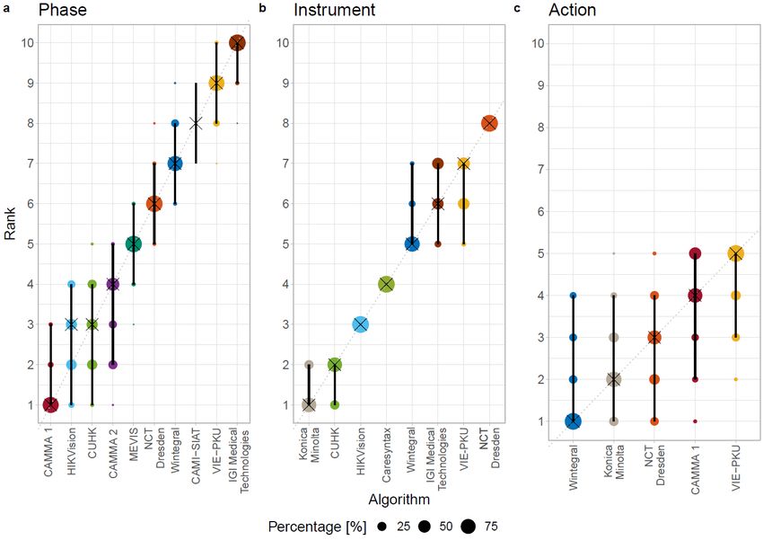
mean instrument presence per surgery 29.72 s ± 43.85 s) was used only at Salem Hospital
and GRN-hospital Sinsheim.
The cases analysed at Heidelberg University Hospital were rated to be more difficult than the
cases at the smaller hospitals. There were 2 of 15 cases at Heidelberg University Hospital
rated with the highest difficulty grade 5, another 2 cases with a grade 4. Only 3 cases were
rated with the easiest difficulty 1. In contrast, grade 5 was not assigned to cases from either
Salem Hospital or GRN-hospital Sinsheim. 8 of Salem's 15 cases were given a grade 1, as
were all 3 cases from Sinsheim. Only one case from Salem was rated 4.

4.2. EndoVis challenge 2019
A comparison of the performance of the algorithms in the individual recognition tasks is
presented in figure 4. A ranking of the algorithms together with ranking uncertainty is
presented in figure 5. Both figures include results for teams CAMMA 1 and 2, which submitted
after the challenge deadline and were thus not considered for the challenge awards.

 Figure 4 - Visualization of raw metric values. Dot and box plots representing the F1-scores
 for each algorithm for the phase, instrument and action task. The scores are shown separately

 24
for each hospital from which the videos were taken. Salem is blue, Heidelberg University
 Hospital (UKHD) is red, GRN-hospital Sinsheim is yellow.

 Figure 5 - Ranking uncertainty. Blob plots visualizing ranking uncertainty for (a) the phase, (b)
 the instrument and (c) the action task. The radius of the blobs represents the frequency of a rank
 achieved by an algorithm for 1,000 bootstrap samples. The median rank is displayed by a black
 cross. The 95% intervals are shown by a black line.

4.2.1. Phase

The results achieved in the task of phase recognition are depicted in figure 4. For phase
recognition the algorithms of HIKVision and CUHK, the winning teams participating in the
EndoVis challenge, achieved a F1-score up to 65.4% and 65.0%. Since the difference
between the two best methods of HIKVision and CUHK was so small and the second best

 25
team achieved superior detection results in most of the videos, both were declared the winner
of the challenge and received the prize. As an example, figure 6 shows a confusion matrix of
the different phases for HIKVision and all participants. The highest recognition rates were
achieved for phase preparation (P0, 79.4%) and calot triangle dissection (P1, 82.8%). The
lowest recognition rates were achieved for phases of gallbladder packaging (P4, 50.2%) and
cleaning and coagulation (P5, 54.8%).

When including submissions that were received after the challenge deadline (CAMMA 1 and
2), the best performing team CAMMA 1 and the runner-up HIKVision achieved significantly
superior results to the algorithms from rank 5 to 10. No significant superiority was found for
them compared to algorithms CUHK (rank 3) and CAMMA 2 (rank 4). This is also reflected by
the ranking uncertainty analysis performed by using bootstrapping strategies. The frequency
of ranks for each algorithm achieved across 1,000 bootstrap samples is illustrated in figure 5.
It can be seen that the ranking is relatively stable, with the second and third algorithm being
very close together. This is further supported by the mean Kendall’s tau value for all bootstrap
samples of 0.93 (median: 0.91, interquartile range (IQR): (0.91,0.96)).

 Figure 6 - Phase recognition results. Left: Phase recognition results for one of the
 challenge winners HIKVision for all test videos. Right: Results of all algorithms for all test
 videos. Due to statistical propagation of uncertainty, the values of the confusion matrices
 are only sufficiently approximated so that the cumulated lines do not add up to 100%.

 26
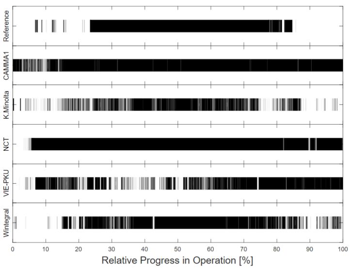
4.2.2. Action
The task of action recognition was completed with the lowest recognition rates (figure 4). Only
an F1 score of 23.3% was reached by the best team Wintegral. The action grasp (A0) occured
during 1.2% (mean of all operations) of an operation, whereas the participants recognized it
during 1.1% (mean recognition of all teams) of the operation. In comparison, the action hold
(A1) occured during 73.3% of an operation, whereas the participants recognized it during
83.2% of the operation. The actions cut (A2) and clip (A3) occurred least frequently, each
during 0.4% of an operation. Cut (A2) was recognized during 1.0% and clip (A3) during 0.3%
of the operation. It is apparent that all challenge algorithms almost exclusively recognize hold
(A1), whether it occurred or not. Figure 7 shows the exemplary results for the recognition of
the action hold (A1) of all teams frame by frame.
None of the algorithms showed significant differences in the performance metric scores.
Analyzing the ranking uncertainty revealed that the rankings were not as clearly separable as
for the phase task (figure 5). The first three algorithms were closely together, therefore often
exchanging their ranks. This is also reflected in a lower mean Kendall’s tau of 0.66 (median:
0.60, IQR: (0.60,0.80)).

 Figure 7 – Action recognition results. Comparison
 of all challenge algorithm results for recognition of
 action hold (A1) for test video HeiChole-25. The x-axis
 represents the relative time progress in the operation.

 27
4.2.3. Instrument
Konica Minolta, the best team in the instrument presence detection task, achieved a F1-score
of 63.8%. Their algorithm recognized 51872 instrument occurrences in the test data set, with
an average of 5763.56 ± 4310.01 instrument occurences per operation. Compared to the
reference, the algorithm detected a similar relative instrument presence within the operation
progress, except for minor differences. For example, noticeable differences are within 10-35%
of the operation progress, where the scissors (IC3) and specimen bag (IC5) were detected
less frequently (figure 8). The mean results of all participants also showed considerable
differences, especially in the presence detection of scissors (IC3) and specimen bag (IC5).
Specimen bag (IC5) was detected continuously during the operation progress, whereas
scissors (IC3) were detected less frequently than in the reference.
For the instrument task, the rankings were very stable across all bootstrap samples (figure 5).
Ranks 6 and 7 were often interchanged and the first two algorithms were close together.
Besides this, the ranking was quite clear with a mean Kendall’s tau of 0.93 (median: 0.93,
IQR: (0.93,1.00)). This was further shown by the statistical analysis. The winning team (Konica
Minolta) and the runner-up (CUHK) both were superior over all other algorithms. The same
trend could be seen for the other algorithms, most of them showing significant effects for their
following ranks.

 Figure 8 - Instrument presence detection results. For the test data set this figure
 compares reference annotation, averaged results of all challenge algorithms and the best
 challenge algorithm (Konica Minolta) for the different instrument categories IC0 to IC5 (IC6
 was not present in the test data set). The horizontal axis of the graphs represent the relative
 time progress in the operation.

 28
4.2.4. Skill
The average absolute error for skill assessment of the only participating team CareSyntax was
0.78 (n=1 team). For each surgical video, a score of 4 was given for the ranking components
depth perception (S1), bimanual dexterity (S2) and efficiency, and a score of 3 for tissue
handling (S4). The difficulty (S5) was rated with a score of 2 for each video.

4.3. HeiChole benchmark

The results of the EndoVis challenge demonstrate a comparative validation of algorithms
based on our comprehensive multi task data set. New methods can be compared to these
results by testing their algorithms with our data set to allow for a validated reference
benchmark. The training data set will be published together with this publication. However, we
decided not to publish the test data set to avoid fraud (Maier-Hein et al., 2018). To use the
data set for future testing of novel algorithms against this benchmark, the following approach
was implemented. To have their algorithm tested, a team must:

 1. register on Synapse to register for the challenge
 (https://www.synapse.org/#!Synapse:syn18824884/wiki/59192). As an option, a team
 can be created or an existing team can be joined.
 2. access the data.
 3. train their algorithm on the provided training data set.
 4. submit the trained algorithm.

We will then evaluate the algorithm using the test data set and report the results to the team
and publish the results in an online leaderboard upon request of the submitting team. To
compute a performance rank the entries will be sorted, as each task consists of a scalar metric.
The current leaderboard as of the publication of this study is shown in figure 9.

 29
You can also read