Candidate Enzymes for Saffron Crocin Biosynthesis Are Localized in Multiple Cellular Compartments1 OPEN
←
→
Page content transcription
If your browser does not render page correctly, please read the page content below
Candidate Enzymes for Saffron Crocin Biosynthesis Are
Localized in Multiple Cellular Compartments1[OPEN]

Olivia Costantina Demurtas,a Sarah Frusciante,a Paola Ferrante,a Gianfranco Diretto,a
Noraddin Hosseinpour Azad,b Marco Pietrella,a,c Giuseppe Aprea,a Anna Rita Taddei,d Elena Romano,e
Jianing Mi,f Salim Al-Babili,f Lorenzo Frigerio,g and Giovanni Giulianoa,2
a
  Italian National Agency for New Technologies, Energy and Sustainable Economic Development, 00123 Rome,
Italy
b
  Department of Medicinal Plant and Plant Production, University of Mohaghegh Ardabili, Ardabil, Iran 56199-
11367
c
  Council for Agricultural Research and Economics, Research Center for Olive, Citrus, and Tree Fruit, 47121 Forli,
Italy
d
  Center of Large Equipment, Section of Electron Microscopy, University of Tuscia, Largo dell’Università, 01100
Viterbo, Italy
e
  Department of Biology, University of Rome Tor Vergata, 00133 Rome, Italy
f
 King Abdullah University of Science and Technology, Biological and Environmental Sciences and Engineering
Division, The Bioactives Lab, Thuwal 23955-6900, Kingdom of Saudi Arabia
g
  School of Life Sciences, University of Warwick, Coventry CV4 7AL, United Kingdom
ORCID IDs: 0000‑0003‑3344‑5754 (O.C.D.); 0000‑0001‑8161‑3797 (S.F.); 0000‑0003‑3071‑2212 (P.F.); 0000‑0002‑6459‑1490 (N.H.A.);
0000‑0002‑8789‑6860 (M.P.); 0000‑0003‑4969‑2696 (G.A.); 0000‑0001‑6063‑9939 (A.R.T.); 0000‑0001‑7846‑9472 (J.M.); 0000‑0003‑4100‑6022 (L.F.);
0000‑0002‑2486‑0510 (G.G.)

Saffron is the dried stigmas of Crocus sativus and is the most expensive spice in the world. Its red color is due to crocins, which are
apocarotenoid glycosides that accumulate in the vacuole to a level up to 10% of the stigma dry weight. Previously, we character-
ized the first dedicated enzyme in the crocin biosynthetic pathway, carotenoid cleavage dioxygenase2 (CsCCD2), which cleaves
zeaxanthin to yield crocetin dialdehyde. In this work, we identified six putative aldehyde dehydrogenase (ALDH) genes expressed
in C. sativus stigmas. Heterologous expression in Escherichia coli showed that only one of corresponding proteins (CsALDH3I1)
was able to convert crocetin dialdehyde into the crocin precursor crocetin. CsALDH3I1 carries a carboxyl-terminal hydrophobic
domain, similar to that of the Neurospora crassa membrane-associated apocarotenoid dehydrogenase YLO-1. We also charac-
terized the UDP-glycosyltransferase CsUGT74AD1, which converts crocetin to crocins 1 and 2′. In vitro assays revealed high
specificity of CsALDH3I1 for crocetin dialdehyde and long-chain apocarotenals and of CsUGT74AD1 for crocetin. Following
extract fractionation, CsCCD2, CsALDH3I1, and CsUGT74AD1 were found in the insoluble fraction, suggesting their associ-
ation with membranes or large insoluble complexes. Analysis of protein localization in both C. sativus stigmas and following
transgene expression in Nicotiana benthamiana leaves revealed that CsCCD2, CsALDH3I, and CsUGT74AD1 were localized to
the plastids, the endoplasmic reticulum, and the cytoplasm, respectively, in association with cytoskeleton-like structures. Based
on these findings and current literature, we propose that the endoplasmic reticulum and cytoplasm function as transit centers
for metabolites whose biosynthesis starts in the plastid and are accumulated in the vacuole.

   One gram of dried saffron is composed of the stig-                       2014). Due to their complex structure and abundance
mas of approximately 150 Crocus sativus flowers,                            of chiral centers, crocins cannot be synthesized chem-
picked by hand. This makes saffron the most expen-                          ically, and there is a strong industrial interest in their
sive spice in the world, with prices ranging from 2 to                      biotechnological production.
10 euros g−1. The main organoleptic properties of the                          In C. sativus stigmas, crocin biosynthesis starts with
stigmas are due to the accumulation of three apocarot-                      symmetric oxidative cleavage of the C7,C8 and C7′,C8′
enoid classes: crocins (composed of different glucosyl                      double bonds in zeaxanthin, which is catalyzed by the
and gentiobiosyl esters of crocetin), picrocrocin, and                      enzyme carotenoid cleavage dioxygenase2 (CsCCD2),
safranal (Tarantilis et al., 1995), conferring to saffron                   producing crocetin dialdehyde (Fig. 1; Frusciante et al.,
its red color, bitter taste, and pungent aroma, respec-                     2014). Like many aldehydes, crocetin dialdehyde is
tively. Crocins make up to 10% of stigma dry weight                         highly reactive and is converted into the diacid cro-
and, by virtue of their glycosylation, are water soluble.                   cetin by yet unidentified ALDH enzymes. ALDHs are
They have applications as textile colorants and histo-                      NAD(P)+-dependent oxidoreductases that generally
chemical stains (Bathaie et al., 2014), as antioxidants                     contribute to different processes, such as cytoplasmic
(Alavizadeh and Hosseinzadeh, 2014), and in the pre-                        male sterility, plant defense, and abiotic stress response
vention of age-related macular degeneration (Bisti et al.,                  (Kirch et al., 2004). The final step of crocin biosynthesis
                      ®
990   Plant Physiology , July 2018, Vol. 177, pp. 990–1006, www.plantphysiol.org © 2018 American Society of Plant Biologists. All Rights Reserved.
                                    Downloaded on April 29, 2021. - Published by https://plantphysiol.org
                                  Copyright (c) 2020 American Society of Plant Biologists. All rights reserved.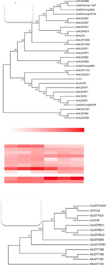
Compartmentation of Saffron Crocin Biosynthesis
involves the glycosylation of crocetin. This reaction
is usually catalyzed by uridine diphosphate glycosy
ltransferase (UGTs) that mediate the glycosylation of
secondary metabolites, xenobiotics, and hormones
(Wang, 2009). In Gardenia jasminoides, two crocetin
UGTs have been characterized (Nagatoshi et al., 2012):
UGT75L6 is responsible for the primary glycosylation
of crocetin, producing crocetin monoglucosyl and di-
glucosyl esters, whereas UGT94E5 is responsible for the
secondary glycosylation of the Glc groups, leading to
the formation of one or two gentiobiose groups (Fig. 1).
Contrasting data have been reported on crocin forma-
tion in saffron: a crocetin UGT purified from C. sativus
stigmas has been shown to catalyze only primary gly-
cosylation (Cote et al., 2001), whereas a cloned UGT
(UGTCs2) has been reported to mediate both types of
glycosylation (Moraga et al., 2004).
The crocin biosynthetic pathway encompasses mul-
tiple subcellular compartments: the initial precursor,
zeaxanthin, is localized in plastids, whereas the final
products, crocins, accumulate in vacuoles (Bouvier et al.,
2003b; Gómez-Gómez et al., 2017), like many other
plant glycosylated metabolites (Martinoia et al., 2007).
We have identified the carotenoid cleavage dioxygen-
ase responsible for the first dedicated step in C. sativus
carotenoid biosynthesis, CsCCD2. It has been shown
that CsCCD2, the enzyme that catalyzes the cleavage
of zeaxanthin in crocin biosynthesis (Frusciante et al.,
2014), is localized to the plastid (Ahrazem et al., 2016).
In contrast, the subcellular localization of the further
intermediates in crocin biosynthesis and the corre-
sponding enzymes remains elusive. There are other
examples of glycosylated metabolites, such as steviol
(Brandle and Telmer, 2007) and abscisic acid glucosyl
ester (ABA-GE) (Nambara and Marion-Poll, 2005),
which originate from plastid-localized precursors and
accumulate in the vacuole. For these, plastid-derived
metabolites are modified and glycosylated in the cyto- Figure 1. Crocin biosynthesis in C. sativus stigmas. A, C. sativus flower
plasm, forming final glycosylated products that are at anthesis with red stigmas. B, Proposed scheme of crocin biosyn-
transported into vacuoles via tonoplast transport- thesis (Frusciante et al., 2014). The CsCCD2 enzyme cleaves zeaxan-
ers (Martinoia et al., 2007). This compartmentation is thin at the 7,8 and 7′,8′ positions, producing 3-OH-β-cyclocitral and
crocetin dialdehyde. Crocetin dialdehyde is oxidized to crocetin by
an aldehyde dehydrogenase (ALDH), then glycosylated to crocins by
1
This work was supported by the European Union (From discov- UGT enzymes. C, Sugar moieties of the four most abundant C. sativus
ery to products: A next generation pipeline for the sustainable gen- crocins (crocins 1–4).
eration of high-value plant products, FP7 Contract 613153) and by
baseline funding given to S.A.-B. from King Abdullah University of
Science and Technology, and benefited from the networking activ-
ities within the European Cooperation in Science and Technology consistent with the assumed cytoplasmic localization
Action CA15136 (EUROCAROTEN). of the majority of plant UGTs (Ross et al., 2001). A dif-
2
Address correspondence to giovanni.giuliano@enea.it. ferent model for crocin biosynthesis and sequestra-
The author responsible for distribution of materials integral to tion was proposed based only on proteomic data and
the findings presented in this article in accordance with the policy microscopic observation (Gómez-Gómez et al., 2017).
described in the Instructions for Authors (www.plantphysiol.org) is: This model proposed that crocins are biosynthesized
Giovanni Giuliano (giovanni.giuliano@enea.it). entirely in the plastid, prior to accumulation in plas-
O.C.D., S.F., P.F., G.D., N.H.A., A.R.T., E.R., J.M., S.A.-B., and L.F.
tid-localized vesicles and final transport to the vacu-
produced data; O.C.D., M.P., G.A., S.A.-B., L.F., and G.G. analyzed
data; G.G. coordinated the study; O.C.D. and G.G. wrote the article;
ole, where crocins are delivered and stored. A crocetin
all authors reviewed the results and approved the final version of UGT (UGTCs2) has been identified in C. sativus stig-
the article. mas (Moraga et al., 2004) and, on the basis of proteom-
[OPEN]
Articles can be viewed without a subscription. ic data, has been proposed to reside in C. sativus stigma
www.plantphysiol.org/cgi/doi/10.1104/pp.17.01815 chromoplasts (Gómez-Gómez et al., 2017). The ALDH
Plant Physiol. Vol. 177, 2018 991
Downloaded on April 29, 2021. - Published by https://plantphysiol.org
Copyright (c) 2020 American Society of Plant Biologists. All rights reserved.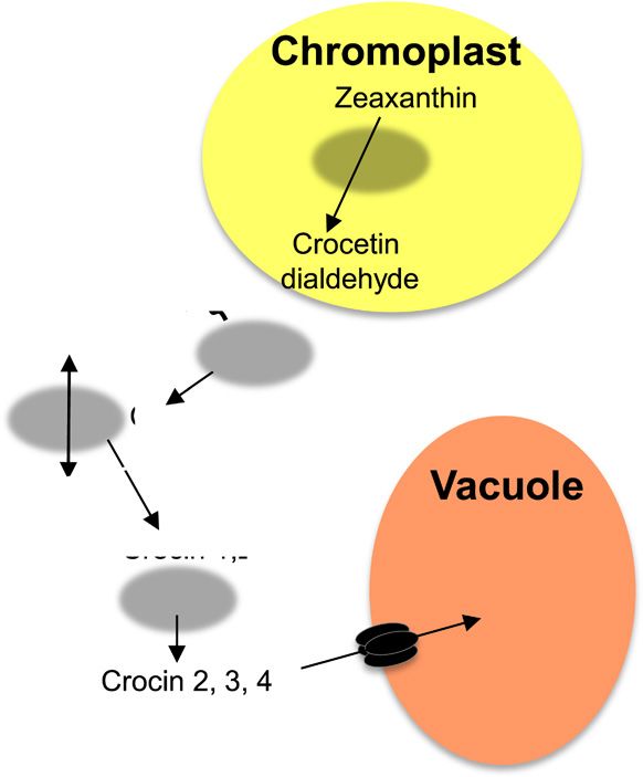
Demurtas et al.

responsible for the second step in crocin biosynthesis            and CsALDH7B4 (family 7; EC 1.2.1.31), comprising
has not been identified so far.                                   Δ1-piperideine-6-carboxylate dehydrogenases and
   To further elucidate the crocin biosynthetic pathway           α-aminoadipic semialdehyde dehydrogenases (Brock-
and to determine its compartmentation, we searched                er et al., 2013), is a close homolog of AtALDH7B4
a C. sativus stigma transcriptome for putative ALD-               encoded by an osmotic stress-inducible ALDH gene
Hs and UGTs and characterized their activity using                (Kotchoni et al., 2006).
Escherichia coli as an expression system. We identified              The CsALDH5F1 and CsALDH7B4 amino acid se-
an ALDH and a UGT, which together convert croce-                  quences indicate the presence of tetramerization inter-
tin dialdehyde to crocins. In addition, we determined             faces (Supplemental Fig. S1), whereas CsALDH3I1 and
the localization of these two enzymes by immunogold               YLO-1 are equipped with a C-terminal hydrophobic
electron microscopy and by analyzing the distribution             domain (Supplemental Fig. S2) flanked by short re-
of GFP fusions in Nicotiana benthamiana cells using               gions of positively charged amino acids and preceded
confocal microscopy. Based on the data obtained, we               by an unstructured domain (Supplemental Fig. S1). A
propose a model for crocin biosynthesis/compartmen-               similar structure has been found in a rat microsome-
tation in C. sativus stigmas.                                     localized ALDH and has been shown to mediate its
                                                                  localization to the endoplasmic reticulum (ER; Masaki
                                                                  et al., 1994). Indeed, YLO-1 is a cytosolic, membrane-
                                                                  localized enzyme, although its exact localization is un-
RESULTS                                                           known (Estrada et al., 2008). This C-terminal domain
Identification and Characterization of ALDH Candidate             has been lost in CsADHComp54788, which represents
Transcripts in C. sativus Stigmas                                 a truncated form of CsALDH3I1, due to an early stop
                                                                  codon. The subcellular localization of the identified
   To identify the enzyme(s) involved in the conversion           CsALDHs was predicted using TargetP (Supplemen-
of crocetin dialdehyde into crocetin, an in-house tran-           tal Table S1; Emanuelsson et al., 2000). None of the en-
scriptome assembly obtained from RNA sequencing                   zymes contained a plastid transit peptide, excluding
data from developing C. sativus stigmas (Frusciante               localization within this organelle, whereas CsALD-
et al., 2014) was searched for homologs of transcripts            H2B4, CsALDH6B2, and CsALDH5F1 are predicted to
encoding apocarotenoid ALDH enzymes from differ-                  be mitochondrial. All CsALDH genes were expressed
ent organisms: (1) Neurospora crassa YLO-1, a member              throughout stigma development (Fig. 2B).
of family 3 ALDH (EC 1.2.1.5) responsible for the de-
hydrogenation of β-apo-4′-carotenal into neurospo-
                                                                  CsALDH3I1 Catalyzes the Dehydrogenation of Crocetin
raxanthin (Estrada et al., 2008) and, together with its
                                                                  Dialdehyde into Crocetin
Arabidopsis (Arabidopsis thaliana) homologs ALDH3I1
and ALDH3H1 (Stiti et al., 2011a,b), responsible for the             To check the ability of the different CsALDH en-
oxidation of medium-chain fatty aldehydes; (2) Bixa               zymes to convert crocetin dialdehyde to crocetin, we
orellana ALDH (BoALDH), a family 2 ALDH member                    expressed them as thioredoxin fusion proteins in E. coli
(EC 1.2.1.3), which has been claimed to convert the               accumulating crocetin dialdehyde (see “Materials and
apocarotenoid bixin dialdehyde into norbixin (Bouvier             Methods”). Briefly, a strain of E. coli carrying a plasmid
et al., 2003a); (3) Synechocystis sp. PCC6803 ALDH (Syn-          for zeaxanthin biosynthesis (Kanr) was cotransformed
ALDH), which converts a wide range of apocarotenals               with the pTHIO-CsCCD2 (Cmr) vector harboring the
and alkanals into the corresponding carboxylic acids              CsCCD2 enzyme that converts zeaxanthin to cro-
(Trautmann et al., 2013); and five ALDHs claimed,                 cetin dialdehyde (Frusciante et al., 2014) and a third
based on proteomic data, to reside within stigma chro-            pTHIO-ALDH vector (Ampr) containing one of the six
moplasts (CsADHComp; Gómez-Gómez et al., 2017).                   CsALDH cDNAs or the SynALDH gene (Supplemental
Using this approach, we identified six candidate genes            Fig. S3A; Trautmann et al., 2013). Immunoblot analysis
expressed in C. sativus stigmas and encoding putative             using an anti-6×His antibody confirmed the expres-
ALDH enzymes (Fig. 2A). CsALDH2C4, a member of                    sion of CsCCD2-thioredoxin and ALDH-thioredoxin
family 2, is the closest homolog of BoALDH and At-                fusion proteins at the expected molecular masses of
ALDH2C4; a second family 2 member, CsALDH2B4,                     81.9 and 69.8 to 76.9 kD, respectively. The intensities
corresponds to CsADHComp11367 and shows identity                  of the bands corresponding to ALDH-thioredoxin fu-
of 97.2% and 85.2% to CsADHComp2946 and CsADH-                    sion proteins were similar, indicating that all ALDH
Comp20158, respectively; CsALDH3I1 (family 3) is a                proteins were expressed at comparable levels upon in-
close homolog of AtALDH3I1, AtALDH3H1, YLO-1,                     duction (Supplemental Fig. S3B).
SynALDH, and CsADHComp54788; CsALDH5F1,                              We quantified zeaxanthin, crocetin dialdehyde,
which belongs to family 5, comprising succinic semi-              and crocetin levels in induced bacterial cells harbor-
aldehyde dehydrogenases (EC 1.2.1.24; Brocker et al.,             ing the various constructs. Nonpolar and polar me-
2013), is a close homolog of AtALDH5F1, involved                  tabolites were analyzed by HPLC-photodiode array
in plant defense against reactive oxygen species (Stiti           detection-high resolution mass spectrometry (HPLC-
et al., 2011b); CsALDH6B2 is a family 6 member and                PDA-HRMS) using a Q-Exactive mass spectrometer.
a homolog of AtALDH6B2 and CsADHComp3893;                         The analysis revealed a decrease of zeaxanthin in all
992                                                                                                       Plant Physiol. Vol. 177, 2018
                             Downloaded on April 29, 2021. - Published by https://plantphysiol.org
                           Copyright (c) 2020 American Society of Plant Biologists. All rights reserved.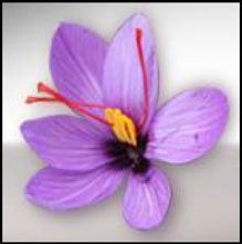
Compartmentation of Saffron Crocin Biosynthesis

                                                                            clones expressing CsCCD2 due to the activity of the
                                                                            CsCCD2 enzyme (Fig. 3, A and D). An ion with the
                                                                            mass of crocetin dialdehyde and comigrating with a
                                                                            crocetin dialdehyde authentic standard was detected
                                                                            in all clones expressing CsCCD2, with the exception
                                                                            of the CsCCD2/ALDH3I1 clone, where this metabo-
                                                                            lite was almost absent (Fig. 3, B and E). Trace levels of
                                                                            an ion with the accurate mass of the ALDH product,
                                                                            crocetin, were observed in all clones expressing CsC-
                                                                            CD2, possibly due to the action of an endogenous E.
                                                                            coli ALDH or to the nonenzymatic dehydrogenation
                                                                            of crocetin dialdehyde (Fig. 3, C and F). We observed
                                                                            a different pattern in cells coexpressing CsCCD2 and
                                                                            CsALDH3I1. Compared with all other cells, we de-
                                                                            tected a much higher reduction in crocetin dialdehyde
                                                                            and, accordingly, a higher increase in crocetin content
                                                                            (Fig. 3, E and F). Thus, since all ALDHs were expressed
                                                                            at similar levels (Supplemental Fig. S3B), we assumed
                                                                            that CsALDH3I1 is the enzyme that converts crocetin
                                                                            dialdehyde into crocetin with high efficiency. The C.
                                                                            sativus ortholog of BoALDH, CsALDH2C4, as well as
                                                                            SynALDH did not show any activity above the E. coli
                                                                            background.
                                                                               Crocetin was resolved by our chromatographic sys-
                                                                            tem into two different peaks with identical mass but
                                                                            different chromatographic mobilities (peaks 1 and
                                                                            2 in Fig. 3C). The commercial standard of crocetin is
                                                                            composed mostly of an all-trans-isomer, which cor-
                                                                            responds to the fastest migrating peak (peak 1 in Fig.
                                                                            3C). Hence, we assume that the two peaks correspond
                                                                            to all-trans- and cis-crocetin with absorption maxima
                                                                            at 426 and 420 nm, respectively, and with an addition-
                                                                            al absorption peak (cis-peak) at 318 nm (Supplemental
                                                                            Fig. S4; Rubio-Moraga et al., 2010; Chryssanthi et al.,

                                                                            all described in this article, indicated by black arrows); AtALDHs (Stiti
                                                                            et al., 2011b); BoALDH (AJ548846; Bouvier et al., 2003a); SynALDH
                                                                            (ALJ68758.1; Trautmann et al., 2013); YLO-1 (XP_011394899.1; Es-
                                                                            trada et al., 2008); and CsADHComp2946 (AQM36713.1), CsADH-
                                                                            Comp11367 (AQM36717.1), CsADHComp20158 (AQM36716.1),
                                                                            CsADHComp3893            (AQM36715.1),        and    CsADHComp54788
                                                                            (AQM36714.1; Gómez-Gómez et al., 2017). B, Expression of C. sativus
                                                                            CCD2, ALDH, and UGT enzymes in stigma tissue at different devel-
                                                                            opmental stages. Data are expressed as log2 of fragments per kilobase
                                                                            per million. Developmental stages are as follows: yellow, orange, red,
                                                                            2 d before anthesis (−2dA), day of anthesis (0dA), and 2 d after anthe-
                                                                            sis (+2dA). C, Phylogenetic relationships between UGTs of C. sativus
                                                                            (Cs), Arabidopsis (At), G. jasminoides (Gj), and M. truncatula (Mt), in-
                                                                            ferred using the neighbor-joining method. Accession numbers are as
                                                                            follows: CsUGT74AD1 (MF596166; described in this article); UGTCs2
                                                                            (AAP94878.1; Moraga et al., 2004); CsGT45 (ACM66950.1; Moraga
                                                                            et al., 2009); CsUGT85V1 (AIF76150.1), CsUGT85U1 (AIF76152.1),
Figure 2. Candidate genes for crocin biosynthesis in C. sativus stig-       and CsUGT85U2 (AIF76151.1; Ahrazem et al., 2015; Gómez-
mas. A, Phylogenetic relationships between ALDHs of C. sativus (Cs),        Gómez et al., 2017); CsUGT707B1 (CCG85331.1; Trapero et al., 2012);
Arabidopsis (At), B. orellana (Bo), Synechocystis spp. (Syn), and N.        AtUGT71B6 (NP_188815.1), AtUGT71B7 (NP_188816.1), and
crassa (YLO-1), inferred using the neighbor-joining method. The sub-        AtUGT71B8 (NP_188817.1; Dong and Hwang, 2014); GjUGT94E5
cellular localizations of ALDH enzymes, experimentally demonstrated         (F8WKW8.1) and GjUGT75L6 (F8WKW0.1; Nagatoshi et al., 2012);
by Stiti et al. (2011b) or predicted by TargetP, are indicated by colored   and MtUGT71G1 (AAW56092.1; Shao et al., 2005). The biosynthetic
dots. Accession numbers are as follows: CsALDH2B4 (MG672523),               products of the various UGTs are represented by colored triangles. In A
CsALDH2C4 (MF596160), CsALDH3I1 (MF596165), CsALDH5F1                       and C, the percentages of replicate trees that clustered together in the
(MF596161), CsALDH6B2 (MG672524), and CsALDH7B4 (MF596162;                  bootstrap test are indicated to the left of the branches.

Plant Physiol. Vol. 177, 2018                                                                                                                   993
                                   Downloaded on April 29, 2021. - Published by https://plantphysiol.org
                                 Copyright (c) 2020 American Society of Plant Biologists. All rights reserved.
Demurtas et al.
Figure 3. CsALDH3I1 mediates crocetin biosynthesis in E. coli. A to C, Accurate mass chromatograms of zeaxanthin (A), cro-
cetin dialdehyde (B), and crocetin (C) extracted from bacterial clones harboring the pTHIO empty vector or overexpressing
CsCCD2 alone or in combination with CsALDH3I1 or CsALDH2C4. Polar and nonpolar fractions were extracted from bacterial
cells and run on an HPLC-PDA-HRMS system alongside authentic standards. The metabolites have an accurate mass and a
chromatographic mobility identical to those of the authentic standard. Two peaks were detected of crocetin (1, trans-isomer,
and 2, cis-isomer; Supplemental Fig. S4). D to F, Relative quantities of zeaxanthin (D), crocetin dialdehyde (E), and crocetin (F)
detected by HPLC-atmospheric pressure chemical ionization-HRMS (D and E) and HPLC-electrospray ionization (ESI)-HRMS
(F) in recombinant clones. Data are means ± sd of three independent growth batches. Asterisks indicate statistically significant
differences (Student’s t test, P < 0.01) compared with CsCCD2 values. Fold IS, Fold internal standard.
2011). Interestingly, the all-trans- and cis-isomers pro- dialdehyde into crocetin in a time-dependent manner,
duced in E. coli were present in approximately equi- with an almost complete conversion after 120 min of
molar amounts. Because our chromatographic system incubation (Fig. 4). We also assayed the CsALDH3I1
does not separate the trans- and cis-isomers of croce- activity on different apocarotenals and nonapocarot-
tin dialdehyde, we are not able to assess whether the enoid aldehydes. As shown in Supplemental Table
cis-configuration is already present in crocetin dialde- S2, CsALDH3I1 displayed a strong preference for
hyde or is generated during the dehydrogenation re- long-chain apocarotenals and for diapocarotenoids,
action. showing after 60 min conversion rates of 93.3%, 76.4%,
To further characterize the substrate specificity of and 3.6% for β-apo-8′-carotenal (C30), crocetin dialde-
CsALDH3I1, we introduced the pTHIO-ALDH vector hyde (C20), and retinal (C20), respectively. In contrast,
in an E. coli strain harboring the groES-groEL-chaperones the activity of the enzyme was very low or even not
system to maximize the amount of correctly folded detectable upon incubation with nonapocarotenoid
protein (Nishihara et al., 1998). Proteins were solubi- (fatty) aldehydes, such as dodecanal, hexanal, or 4-OH-
lized using Triton X-100 (Supplemental Fig. S5) and benzaldehyde.
utilized in an in vitro assay with different aldehydes
according to Trautmann et al. (2013). The control ex-
CsUGT74AD1 Catalyzes the Glycosylation of Crocetin
tract from E. coli cell transformed with the void plasmid
into Crocins 1 and 2′
showed negligible (1.3%) oxidation of crocetin dialde-
hyde into crocetin (Supplemental Fig. S6), whereas To check whether the previously identified UGT en-
the extract containing CsALDH3I1 converted crocetin zyme (UGTCs2; GenBank accession number AY262037.1;
994 Plant Physiol. Vol. 177, 2018
Downloaded on April 29, 2021. - Published by https://plantphysiol.org
Copyright (c) 2020 American Society of Plant Biologists. All rights reserved.Compartmentation of Saffron Crocin Biosynthesis
et al., 2017), G. jasminoides UGTs involved in crocin
biosynthesis (Nagatoshi et al., 2012), and Arabidopsis
and M. truncatula UGTs (Shao et al., 2005; Dong and
Hwang, 2014) is shown in Figure 2C. Three clusters
are observed: one comprising the three C. sativus genes
putatively involved in auxin-GE biosynthesis (Ahrazem
et al., 2015); a second one comprising CsUGT74AD1,
GjUGT75L6, involved in crocetin primary glycosyla-
tion (Nagatoshi et al., 2012), and CsGT45, involved
in flavonoid glycosylation (Moraga et al., 2009); and
a third one comprising Arabidopsis ABA UGTs.
GjUGT94E5, involved in crocetin secondary glycosyla-
tion (Nagatoshi et al., 2012), and CsUGT707B1, puta-
tively involved in flavonoid glycosylation (Trapero
et al., 2012), did not cluster with any other sequence.
None of the described CsUGTs contains transmem-
brane domains (Supplemental Fig. S2) or chloroplast
transit peptides (Supplemental Table S1).
To verify the function of CsUGT74AD1, we cloned
the corresponding cDNA in the bacterial expres-
sion vector pTHIO (Spectr). As expected, coexpres-
Figure 4. CsALDH3I1 mediates crocetin biosynthesis in vitro. A, HPLC sion of pTHIO-CsCCD2, pTHIO-CsALDH3I1, and
chromatograms (absorbance at 440 nm) of in vitro assays performed pTHIO-CsUGT74AD1 in a zeaxanthin-accumulating
for 120 min in the absence (THIO) and presence (CsALDH3I1:THIO) E. coli strain did not result in the accumulation of cro-
of CsALDH3I1. STANDARD = crocetin dialdehyde authentic standard. cin, probably due to insufficient levels of UDP-Glc
In the presence of CsALDH3I1, the crocetin dialdehyde is converted in E. coli, which is required for UGT activity (Ross et
into crocetin (1, trans-isomer, and 2, cis-isomer; Supplemental Fig. S4). al., 2001). Thus, we performed an in vitro assay using
B, Time course of crocetin dialdehyde conversion in the presence of CsUGT74AD1 produced in the E. coli strain harboring
CsALDH3I1. Data are averages ± sd of three biological replicates and
the groES-groEL-chaperones system (Nishihara et al.,
expressed as fold internal standard (Fold IS).
1998). Immunoblotting with the anti-His-6 antibody
revealed a band corresponding to the CsUGT74AD1-
thioredoxin fusion protein (69.7 kD) in soluble extracts
of arabinose-induced cells (Supplemental Fig. S8).
Moraga et al., 2004) is involved in crocin biosynthesis We performed an in vitro assay using 40 µg of solu-
and how many sugars it adds, we searched our tran- ble E. coli extract, encapsulated crocetin, and UDP-Glc
scriptome, as well as that of Jain et al. (2016), for the as substrates (Moraga et al., 2004). The reaction was
presence of the corresponding transcript. However, we incubated at 30°C for increasing periods of time, fol-
could not find an identical sequence. The most simi- lowed by analysis of semipolar and polar metabolites
lar UGT transcript present in these transcriptomes by HPLC-PDA-HRMS. In the assay performed with
was that of CsUGT74AD1, which encodes a version of extracts of cells transformed with the empty pTHIO
UGTCs2 (Moraga et al., 2004), showing a four-amino vector, we only detected two peaks with a maximum
acid extension at the N terminus, lacking seven amino A440, corresponding to trans- and cis-crocetin. On the
acids at the C terminus, and differing in 44 amino ac- contrary, in the presence of CsUGT74AD1, we detect-
ids (Supplemental Fig. S7). The plant secondary prod- ed two additional peaks (Fig. 5A) corresponding to the
uct glycosyltransferase motif, which is responsible for monoglucosyl and diglucosyl esters of crocetin (crocin
binding the sugar donor UDP-Glc (Gachon et al., 2005), 1 and 2′, respectively; Fig. 5B). A time course of the re-
is conserved in CsUGT74AD1, UGTCs2, and G. jasm- action is shown in Figure 5C: the monoglucosyl ester is
inoides UGTs involved in crocin formation (Nagatoshi a reaction intermediate reaching a maximum between
et al., 2012), whereas some differences were observed 10 and 20 min, whereas an almost complete conversion
in other catalytic residues. Specifically, both UGTCs2 of crocetin into the diglucosyl ester, crocin 2′, was de-
and GjUGT75L6 lack the Asp-121 residue (Supple- tected in 60 min. Although we incubated the reaction
mental Fig. S7), which has been shown to be necessary, for up to 120 min and repeated the assay several times,
together with His-22, for the catalytic activity of Med- we could not detect the formation of gentiobiosyl es-
icago truncatula UGT71G1 (Shao et al., 2005). Similar ters of crocetin, which are the most abundant forms
to CsCCD2, CsUGT74AD1 is highly expressed in early of crocins in mature stigmas (Fig. 5A). Thus, in our
stigma development, with transcript levels reaching hands, the CsUGT74AD1 enzyme performed only the
a maximum at the red stage and declining thereafter first step of crocetin glycosylation (the addition of Glc
(Fig. 2B). A dendrogram that includes CsUGT74AD1, moieties to crocetin), in agreement with previous data
other described C. sativus UGTs (Moraga et al., 2009; obtained in G. jasminoides (Nagatoshi et al., 2012) and
Trapero et al., 2012; Ahrazem et al., 2015; Gómez-Gómez C. sativus (Cote et al., 2001).
Plant Physiol. Vol. 177, 2018 995
Downloaded on April 29, 2021. - Published by https://plantphysiol.org
Copyright (c) 2020 American Society of Plant Biologists. All rights reserved.Demurtas et al.
Figure 5. CsUGT74AD1 mediates the synthesis of crocins 1 and 2′ in vitro. A, HPLC chromatograms (absorbance at 440 nm) of
in vitro assay reactions performed for 60 min in the absence (THIO) and presence (CsUGT74AD1:THIO) of the CsUGT74AD1
enzyme or extracted from mature saffron (saffron extract). t = trans and c = cis. B, HPLC-ESI-HRMS chromatograms (extracted
accurate masses) of trans-crocin 1 and trans-crocin 2′ accumulated in the bacterial clone overexpressing the CsUGT74AD1 en-
zyme. Mass and PDA spectra of peaks are shown in the right and left boxes, respectively. C, Time course of crocetin conversion
into trans-crocin 1 and trans-crocin 2′ in the presence of UGT enzyme. Data are averages ± sd of three biological replicates and
expressed as fold internal standard (Fold IS).
The activity of CsUGT74AD1 also was tested on a CsCCD2, CsALDH3I1, and CsUGT74AD1 Tissue
broad range of different substrates known to undergo Specificity, Solubility, and Subcellular Localization
glycosylation in planta, using UDP-Glc or UDP-Gal as
To analyze the tissue-specific expression pattern
sugar donors. We observed that the enzyme is high-
of the CsCCD2, CsALDH3I1, and CsUGT74AD1 en-
ly specific, showing a high activity with crocetin and
UDP-Glc (81.3% conversion in 30 min; Supplemental zymes in flowers of C. sativus, we raised polyclonal
Table S3), although we detected a lower activity with antibodies against immunogenic peptides from the
UDP-Gal as a sugar donor (35.5% conversion in 30 three proteins (see “Materials and Methods”) and used
min). The enzyme also displayed low levels of activity, them in immunoblot analysis on protein extracts from
with UDP-Glc as sugar donor, on flavonoids (i.e. quer- C. sativus stigmas, stamens, and tepals at anthesis (Fig.
cetin and naringenin) and on the phenolic acid cin- 6A). The antibodies raised against CsCCD2 recognized
namic acid, whereas it did not show any activity with a single band corresponding to the molecular mass of
ABA or indole 3-acetic acid (Supplemental Table S3). the enzyme (63 kD after the removal of the transit pep-
These results suggest that CsUGT74AD1 is the enzyme tide), which was only present in the stigma fraction.
responsible for the primary glycosylation of crocetin. The anti-CsALDH3I1 antibodies recognized a major
996 Plant Physiol. Vol. 177, 2018
Downloaded on April 29, 2021. - Published by https://plantphysiol.org
Copyright (c) 2020 American Society of Plant Biologists. All rights reserved.Compartmentation of Saffron Crocin Biosynthesis
band of 53 kD, corresponding to the molecular mass of
the protein, which was highly expressed in stigmas but
also present at much lower levels in stamens and te-
pals. Anti-CsALDH3I1 antibodies also recognized sev-
eral fainter bands, likely due to cross-reactivity with
other ALDH enzymes. The antibodies raised against
CsUGT74AD1 recognized a band of 51 kD, which
corresponds to the molecular mass of CsUGT74AD1,
and two additional bands with molecular masses of 69
and 29 kD. The 29-kD band is too small to represent a
functional UGT (Gachon et al., 2005) and corresponds
probably to a proteolytic product of CsUGT74AD1.
In agreement with this hypothesis, the 51- and 29-kD
bands are present together in the stigma fraction and
are absent in other tissues. In contrast, we detected the
69-kD band in all three tissues, indicating that it rep-
resents an unrelated UGT or a different protein.
The substrates of CsCCD2 and CsALDH3I1 (zea-
xanthin and crocetin dialdehyde, respectively) are hy-
drophobic molecules that are presumably membrane
associated in vivo. Accordingly, both CsCCD2 and
CsALDH3I1 contain a transmembrane domain (Sup-
plemental Fig. S2), which in CsALDH3I1 is reminiscent
of an ER localization sequence (Masaki et al., 1994). To
determine the solubility of the three enzymes involved
in crocin biosynthesis, we homogenized C. sativus stig-
mas in PBS buffer to lyse plastids and then sequentially
fractionated the extracts into soluble proteins, extrinsic
membrane proteins, and insoluble proteins (lanes 1, 2,
and 3, respectively, in Fig. 6B). Interestingly, all three
Figure 6. Tissue specificity and solubility of enzymes involved in
enzymes were found in the insoluble fraction (lane 3
crocin biosynthesis. A, Immunoblot analysis performed on total pro-
in Fig. 6B). This result was expected for CsCCD2 and teins (10 µg) extracted from C. sativus stigmas, stamens, and tepals,
CsALDH3I1, which contain a transmembrane domain, performed with rabbit polyclonal antibodies raised against CsCCD2,
but not for CsUGT74AD1, which, like other CsUGTs, CsALDH3I1, and CsUGT74AD1 peptides. B, Immunoblot analysis
does not have any hydrophobic domain (Supplemen- performed on total protein extract from C. sativus stigmas. Lane 1, Sol-
tal Fig. S2). uble proteins extracted with PB buffer; lane 2, extrinsic membrane
To determine the localization of the three enzymes proteins extracted with PB buffer + 200 mm NaCl; lane 3, insoluble
within stigma cells, we performed immunogold elec- proteins extracted with SDS loading buffer (for details, see “Materials
tron microscopy on mature stigmas. Figure 7, A and and Methods”).
B, each shows a scanning electron micrograph of a C.
sativus stigma. Figure 7C shows an immunoelectron mi-
crograph of a crocin-accumulating cell, decorated with elements (Fig. 7G). A cytoskeletal association of UGTs
a secondary antibody coupled to colloidal gold. The was reported previously for the maize (Zea mays) UDP-
various organelles (i.e. nucleus, chromoplast, cell wall, Glc starch glycosyltransferase (Azama et al., 2003). The
and vacuole) are clearly visible without any gold par- membrane or cytoskeletal association of CsUGT74AD1
ticles. Figure 7, D and E, show low- and high-magni- could explain its insolubility. Unfortunately, IEM per-
fication immunoelectron micrographs, respectively, of formed using two different anti-CsALDH3I1 antibodies
cells immunodecorated using the primary anti-CsCCD2 gave no signal, probably due to the fact that the epitopes
antibodies followed by the secondary antibody cou- recognized by these antibodies are not accessible in the
pled to colloidal gold. The gold particles are localized folded protein.
to the chromoplasts, in agreement with the reported To confirm the data obtained by IEM on CsCCD2
plastid localization of a CCD2 from Crocus ancyrensis and CsUGT74AD1 and to determine the subcellular lo-
(Ahrazem et al., 2016) and the predicted presence of a calization of CsALDH3I1, we fused the three proteins
plastid transit peptide for this enzyme (Supplemental to enhanced GFP (eGFP; Cinelli et al., 2000) and ex-
Table S1). The CsCCD2 signal was homogenously dis- pressed them in N. benthamiana leaves through infiltra-
tributed in the organelles. IEM with anti-CsUGT74AD1 tion with Agrobacterium tumefaciens (strain C58C1). The
antibodies gave a clear signal in the cytoplasm (Fig. 7, F confocal microscopy results indicate that CsCCD2:eG-
and G). At the higher magnification, the gold particles FP is localized on chloroplast-associated speckles (Fig.
showed an association with electron-dense filamentous 8A), further confirming the plastid localization demon-
structures reminiscent of membranes or cytoskeletal strated by IEM. In contrast, CsUGT74AD1 showed
Plant Physiol. Vol. 177, 2018 997
Downloaded on April 29, 2021. - Published by https://plantphysiol.org
Copyright (c) 2020 American Society of Plant Biologists. All rights reserved.Demurtas et al.
Figure 7. Subcellular localization of CsCCD2 and CsUGT74AD1 in C. sativus stigmas. A and B, Scanning electron microscopy
images of C. sativus stigmas. C to G, Immunoelectron microscopy (IEM) of C. sativus stigmas. IEM reactions were performed
with an anti-CsCCD2 antibody (D and E) and an anti-CsUGT74AD1 antibody (F and G) followed by an anti-rabbit secondary
antibody conjugated to 20-nm gold particles. As a control, labeling was performed omitting primary antibodies (C). For both
proteins, a general view (bars = 2 µm) and a 4× detail (bars = 500 nm) are shown. CsCCD2 was detected only in chromoplasts
(D and E), while CsUGT74AD1 was detected in the cytoplasm (F), in association with filamentous structures (membranes or
cytoskeleton-like structures; G). C, Chromoplast; CS, cytoskeleton; CW, cell wall; M, mitochondrion; N, nucleus; V, vacuole.
a cytosolic localization (Fig. 8A), which was further C-terminal retention signal (KKRK; Supplemental Fig.
confirmed by coexpression with cytosolic mCherry S1; Benghezal et al., 2000). Coexpression of CsALDH3I1
(Fig. 8C). Finally, CsALDH3I1 labeled a reticulate with the ER marker RFP-HDEL showed the colocaliza-
membrane network that is likely identical with the ER tion of both proteins, thus confirming that CsALDH3I1
(Fig. 8A). Indeed, CsALDH3I1 contains a canonical is an ER-associated enzyme (Fig. 8B).
998 Plant Physiol. Vol. 177, 2018
Downloaded on April 29, 2021. - Published by https://plantphysiol.org
Copyright (c) 2020 American Society of Plant Biologists. All rights reserved.Compartmentation of Saffron Crocin Biosynthesis
Figure 8. Subcellular localization of CsCCD2, CsALDH3I1, and CsUGT74AD1 expressed in N. benthamiana leaves. A, For
each construct, red (chlorophyll fluorescence), green (GFP fluorescence), merged (overlap of chlorophyll and GFP fluores-
cence), and a 2.5× zoom of merged are shown. GFP shows atypical cytoplasmic-nuclear localization; CsCCD2 localizes to
plastid-associated speckles; CsALDH3I1 shows a cytoplasmic, reticulate localization; CsUGT74AD1 shows a cytoplasmic
localization. B, Colocalization of RFP:HDEL (ER marker) and CsALDH3I1:eGFP. C, Colocalization of mCherry (cytoplasmic
marker) and CsUGT74AD1:eGFP is cytosol localized. Bars = 10 μm.
Plant Physiol. Vol. 177, 2018 999
Downloaded on April 29, 2021. - Published by https://plantphysiol.org
Copyright (c) 2020 American Society of Plant Biologists. All rights reserved.Demurtas et al.
All other CsALDH proteins investigated in this and 7 enzymes, respectively, reported to oxidize suc-
study showed a cytosolic localization, as judged by cinic semialdehydes and Δ1-piperideine-6-carboxyl-
confocal fluorescence microscopy, with the exception ate and α-aminoadipic semialdehydes, respectively
of CsALDH5F1, which may be a mitochondrial en- (Brocker et al., 2013).
zyme (Supplemental Fig. S9; Supplemental Table S1). By expression in E. coli, we demonstrated that, of the
six tested ALDH enzymes whose transcripts are high-
ly expressed in C. sativus stigmas, only CsALDH3I1 is
DISCUSSION
able to catalyze the conversion of crocetin dialdehyde
into a mixture of trans- and cis-crocetin, whereas all
In this and in a previous work (Frusciante et al., other ALDHs, including SynALDH, did not show any
2014), we have isolated and characterized transcripts appreciable activity above the endogenous E. coli back-
expressed in C. sativus stigmas, encoding CCD, ALDH, ground. In vitro assays confirmed the high activity of
and UGT enzymes that catalyze the first, second, and CsALDH3I1 in converting crocetin dialdehyde into
third steps, respectively, in C. sativus crocin biosynthe- crocetin (Fig. 4) and indicated a strong preference for
sis in E. coli and/or in vitro. carotenoid-derived substrates, particularly crocetin al-
dehyde and long-chain apocarotenals (Supplemental
The First Step: Cleavage of Zeaxanthin Table S2). The CsALDH3I1 protein is expressed much
more in stigmas, the tissue of crocin biosynthesis,
The C. sativus enzyme CsCCD2 has been shown pre- than in anthers and tepals. Taken together, these data
viously to catalyze the first dedicated step in crocin strongly suggest that CsALDH3I1 is the enzyme cata-
biosynthesis (i.e. the cleavage of zeaxanthin to crocetin lyzing the dehydrogenation of crocetin dialdehyde in
dialdehyde; Frusciante et al., 2014). A CCD2 from the C. sativus stigmas. Interestingly, both CsALDH3I1 and
spring crocus C. ancyrensis, which synthesizes crocins the fungal apocarotenal dehydrogenase YLO-1 contain
in tepals, is localized to plastids when overexpressed a C-terminal transmembrane domain, which is flanked
in tobacco leaves (Ahrazem et al., 2016). In this work, by sequences rich in basic amino acids and linked to
both immunoelectron microscopy on C. sativus stigmas the ALDH core through an unstructured domain (Sup-
and confocal fluorescence microscopy in N. benthami- plemental Fig. S1; Estrada et al., 2008). It has been
ana leaves confirmed the plastidial localization of CsC-
suggested that this domain mediates the YLO-1 asso-
CD2, indicating that this localization probably extends
ciation with intracellular membranes (Estrada et al.,
to the whole Crocus genus, irrespective of the organ
2008). Indeed, a rat microsomal ALDH contains very
where crocins are synthesized. CsCCD2 is homoge-
similar C-terminal domain that is responsible for post-
nously distributed in C. sativus stigma chromoplasts
translational targeting to the ER (Masaki et al., 1994).
and, upon extract fractionation, behaves as an insol-
We were unable to obtain a specific IEM reaction us-
uble enzyme, consistent with the presence of a large
internal transmembrane domain. ing two distinct anti-CsALDH3I1 antibodies. However,
confocal fluorescence in N. benthamiana leaves showed
a clear localization in a cytoplasmic reticular structure,
The Second Step: Dehydrogenation of Crocetin similar to that observed for ER-localized proteins har-
Dialdehyde boring a C-terminal KKXX signal (Benghezal et al.,
We used an approach similar to that of Frusciante et 2000), also found in CsALDH3I1. CsALDH3I1 clearly
al. (2014) to identify the ALDH responsible for the sec- colocalized with an ER marker in N. benthamiana leaves
ond step in crocin biosynthesis. Plant ALDH enzymes and, upon fractionation of C. sativus stigma extracts,
have been classified in 13 distinct families (Brocker behaved as an insoluble protein, in keeping with its ER
et al., 2013). In our stigma transcriptome, we identified localization.
six expressed ALDHs. In particular, CsALDH3H1 en- Based on proteomic data, several C. sativus stig-
codes a family 3 member closely related to YLO-1, an ma ALDHs have been claimed to reside in the
N. crassa ALDH that is responsible for the conversion plastid (Gómez-Gómez et al., 2017), including CsADH-
of β-apo-4′-carotenal to neurosporaxanthin (Estrada et Comp54788, a truncated CsALDH3I1 form that lacks
al., 2008) and to Synechocystis spp. and Fusarium fujik- the C-terminal hydrophobic tail likely mediating ER
uroi ALDHs able to convert apocarotenals into the cor- localization. However, neither CsADHComp54788
responding carboxylic acids (Díaz-Sánchez et al., 2011; nor CsALDH3I1 is expected to occur within plastids,
Trautmann et al., 2013), whereas CsALDH2C4 is closely as judged by TargetP analysis. Furthermore, none of
related to BoALDH, a B. orellana family 2 ALDH claimed the additional CsALDH enzymes, many of which are
to convert bixin dialdehyde into norbixin (Bouvier et highly similar to those identified by Gómez-Gómez et
al., 2003a); CsALDH2B4 and CsALDH6B2 are identical al. (2017), showed a plastidial localization in N. benth-
or highly similar to CsADH enzymes (Comp11367, amiana leaves. Of course, it is still possible that a plas-
Comp2946, Comp20158, and Comp3893) claimed to tid-localized ALDH is expressed at very low levels in
reside in stigma chromoplasts (Gómez-Gómez et al., C. sativus stigmas and, therefore, is not present in our
2017); CsALDH5F1 and CsALDH7B4 encode family 5 transcriptome data.
1000 Plant Physiol. Vol. 177, 2018
Downloaded on April 29, 2021. - Published by https://plantphysiol.org
Copyright (c) 2020 American Society of Plant Biologists. All rights reserved.Compartmentation of Saffron Crocin Biosynthesis
The Third Step: Glycosylation of Crocetin by Gómez-Gómez et al. (2017) for both ALDH and
UGT enzymes is a contamination of the chromoplast
In spite of several efforts, we were not able to re-
membrane preparations with nonplastidial proteins,
trieve, in our transcriptome or in that of Jain et al.
as suggested by the low-level mitochondrial and per-
(2016), a sequence encoding UGTCs2, which has been
oxisomal contamination observed (Gómez-Gómez et
claimed to perform both primary and secondary gly-
al., 2017).
cosylation of crocetin (Moraga et al., 2004) and to re-
side in stigma chromoplasts (Gómez-Gómez et al.,
2017). Instead, we identified the close homolog CsUG- A Model for Crocin Biosynthesis and Trafficking from the
T74AD1, which, based on immunoblot analysis esti- Plastid to the Vacuole
mates, is a very abundant protein in C. sativus stigmas.
Plastids and ER have biochemical continuity, as
CsUGT74AD1 possesses all the characteristics of bona
demonstrated by the fact that mutations in plastid-
fide plant UGTs, including the presence of His-22 and localized enzymes affecting tocopherol biosynthesis
Asp-121, shown to be essential for enzymatic activity can be complemented by directing the same enzymes
(Shao et al., 2005). Using an in vitro assay, we showed to the ER (Mehrshahi et al., 2013). This finding sug-
that CsUGT74AD1 is able to perform primary, but not gests that plastidial and ER membranes are in close
secondary, glycosylation of crocetin. CsUGT74AD1 contact, allowing hydrophobic molecules like tocoph-
shows a strong preference for crocetin as a substrate erol biosynthetic intermediates or crocetin dialdehyde
and accepts both UDP-Glc and, with lower affinity, to migrate from one compartment to the other (Fig. 9).
UDP-Gal as sugar donors. Similar to CsALDH3I1, the Our model (Fig. 9) suggests that the ER acts as a transit
CsUGT74AD1 protein also is much more expressed center for metabolites whose biosynthesis starts in the
in stigma than in anthers and tepals. Overall, the data chloroplast and ends in the vacuole. This type of com-
suggest that CsUGT74AD1 mediates the primary gly- partmentation has been described also for the synthe-
cosylation of crocetin in C. sativus stigmas. Thus, in C. sis of steviosides, which are glycosylated diterpenes
sativus, the situation seems to be similar to that in G. that accumulate in the vacuoles of Stevia rebaudiana
jasminoides, where the biosynthesis of crocins involves leaves and confer an intensely sweet flavor. Similar to
two UGTs acting hierarchically: one (GjUGT75L6, a crocins, the first dedicated step in the biosynthesis of
close homolog of CsUGT74AD1) produces crocins 1 steviosides is mediated by a plastid-localized kaurene
and 2′, and a second one (GjUGT94E5) is responsible synthase and the second step by an ER-localized
for the production of crocins 2, 3, and 4 (Nagatoshi et kaurene oxidase (Brandle and Telmer, 2007). The mod-
al., 2012). We were unable to find bona fide orthologs el also predicts that the vacuolar transport of crocins
of GjUGT94E5 in our transcriptome or in that reported and steviosides, similar to that of other glycosylated
by Jain et al. (2016). The characterization of addition- secondary metabolites, is mediated by tonoplast-local-
al UGTs expressed in C. sativus stigmas will elucidate ized transporters (Fig. 9; Martinoia et al., 2007). This
their possible role in hierarchical crocin glycosylation. model may apply to the subcellular compartmentation
The CsUGT74AD1 protein does not present obvi- of other pathways, such as the biosynthesis of ABA-GE
ous transmembrane domains, but it is insoluble upon (Nambara and Marion-Poll, 2005; Dong et al., 2015) or
extract fractionation. A similar behavior has been ob- of glycosylated apocarotenoids in leaves (Lätari et al.,
served for several enzymes in carotenoid biosynthe- 2015).
sis, such as phytoene desaturase, which is present in Although the expression pattern, substrate specific-
plastids in a soluble complex with HSP70 and in a ity, and tissue specificity of CsALDH3I1 and CsUG-
second, membrane-associated complex containing the T74AD1 strongly suggest that they are involved in the
active enzyme (Al-Babili et al., 1996). The insolubility biosynthesis of crocins 1 and 2′, we have no proof yet
of CsUGT74AD1 was consistent with immunogold la- for their function in planta. Due to the genetic intrac-
beling, showing that the enzyme (or a cross-reacting tability of C. sativus, such proof could be obtained only
UGT) localizes to cytoplasmic electron-dense struc- by heterologous expression in plant tissues containing
tures reminiscent of membrane or cytoskeletal struc- appropriate levels of the substrates, as already done
tures. Confocal microscopy confirmed that the protein for CCD2 in maize endosperm (Frusciante et al., 2014).
colocalizes with mCherry, a cytosolic marker, in N. Unfortunately, this tissue contains an endogenous
benthamiana leaves. This localization is in agreement ALDH activity able to dehydrogenate crocetin dialde-
with the cytosolic localization of the majority of plant hyde (Frusciante et al., 2014). Another limitation of the
UGTs (Gachon et al., 2005) but in disagreement with model is that we have not shown that the substrates
the plastidial localization described, based on proteomic and products of the three enzymes are indeed local-
data, for the very similar UGTCs2 (Gómez-Gómez ized in the extrapastidial locations (except for crocins,
et al., 2017). Given the extremely high abundance which are localized in the vacuole). This will be the
of CsUGT74AD1 and its similarity to UGTCs2, we subject of future studies.
believe that the plastid-associated peptides identi- Based on microscopic observations, Gómez-Gómez
fied by Gómez-Gómez et al. (2017) and attributed to et al. (2017) proposed an alternative model (i.e. that
UGTCs2 belong instead to CsUGT74AD1. One possi- crocin biosynthesis takes place entirely in the plastid
ble explanation of the plastidial localization claimed and that crocins accumulate in crocinoplast vesicles
Plant Physiol. Vol. 177, 2018 1001
Downloaded on April 29, 2021. - Published by https://plantphysiol.org
Copyright (c) 2020 American Society of Plant Biologists. All rights reserved.Demurtas et al.
nlm.nih.gov/Structure/cdd/cdd.shtml). Subcellular compartmentation was
deduced using TargetP 1.1 prediction servers and transmembrane domains
using TMHMM 2.0 software (http://www.cbs.dtu.dk/services/TMHMM/).
Cloning of C. sativus Genes
C. sativus ALDH and UGT transcripts were isolated from C. sativus stigma
RNA using the Omniscript RT cDNA synthesis kit (Qiagen). Coding sequenc-
es were amplified from cDNA using Phusion High Fidelity DNA polymerase
(New England Biolabs) with the oligonucleotides listed in Supplemental Table
S4. Amplicons were cloned in the pBluescript vector (Stratagene) digested with
EcoRV, verified by sequencing, and then reamplified with the oligonucleotides
listed in Supplemental Table S4. The PCR products were cloned in the pTHIO
vector (Trautmann et al., 2013) fused to an in-frame 5′ thioredoxin gene and to
3′ VP5 epitope and 6×HIS tag. The ALDH genes were cloned in pTHIO (Ampr)
vector, obtaining the pTHIO-CsALDHs (Ampr) constructs. CsUGT74AD1 was
cloned in a modified pTHIO vector harboring the spectinomycin resistance
gene (Spectr) instead of Ampr, producing the pTHIO-CsUGT74AD1 (Spectr)
construct. The previously isolated CsCCD2 gene (Frusciante et al., 2014) was
cloned in a modified pTHIO vector harboring the chloramphenicol resistance
gene (Cmr), obtaining the pTHIO-CsCCD2 (Cmr) construct.
In Bacterio ALDH Assay
The zeaxanthin-accumulating strain of Escherichia coli (Kanr), described
previously (Frusciante et al., 2014), was transformed with pTHIO-CsC-
CD2 (Cmr) and one of the six pTHIO-CsALDHs (Ampr) constructs or with
pTHIO-SynALDH (Ampr), which harbors the ALDH1 gene of Synechocystis sp.
PCC6803 (Trautmann et al., 2013). As a control, the strain was transformed
with pTHIO empty vector or with pTHIO-CsCCD2 (Cmr) alone. Overnight
cultures of these clones were inoculated into 50 mL of Luria-Bertani medium
containing one-half-strength antibiotics (25 μg mL−1 kanamycin, 12.5 μg mL−1
Figure 9. Proposed model for crocin biosynthesis/compartmentation chloramphenicol, and/or 50 μg mL−1 ampicillin), grown at 37°C to an OD600
in C. sativus stigmas. CsCCD2 cleaves zeaxanthin in the choromoplast, of 0.7, and induced with 0.08% (w/v) arabinose at 20°C for 16 h. Cells were
producing crocetin dialdehyde, which migrates to the ER, where it is pelleted and polar and nonpolar fractions were extracted. Polar and semipolar
metabolites were extracted as described previously (Fasano et al., 2016; Ramb-
converted to crocetin by CsALDH3I1. The ER and plastid membranes
la et al., 2016) with slight modifications: pellets were resuspended in 10 mL of
are contiguous through the action of an interphase stabilizing complex
75% (v/v) methanol spiked cold (0.5 µg mL−1 formononetin; Sigma-Aldrich),
(ISC; Mehrshahi et al., 2013). Crocetin is converted to crocins 1 and 2′ lysed on ice by triplicate sonication at 10 Hz output (10 s each), and centri-
by CsUGT74AD1, associated with cytoplasmic membranes or with the fuged at 18,000g for 30 min. The supernatant was then dried and redissolved
cytoskeleton (CS). A second, unidentified UGT converts crocins 1 and in 200 μL of 75% (v/v) methanol, centrifuged at 18,000g for 20 min to remove
2′ into crocins 2, 3, and 4, which are then transported into the vacuole particles and aggregates, and subjected to HPLC-PDA-HRMS analysis. Non-
through one or more unidentified tonoplast transporters. polar metabolites were extracted with the same procedure using 100% acetone
spiked cold (0.5 µg mL−1 α-tocopherol acetate; Sigma-Aldrich) for extraction
and chloroform for resuspension.
that move from the plastid to the vacuole). Our mod-
el does not exclude the existence of such a pathway.
However, none of the ALDH and UGT enzymes we In Vitro ALDH and UGT Assays
characterized showed a plastidial localization. Similar The pTHIO-CsALDH3I1 and pTHIO-CsUGT74AD1 plasmids were ex-
to the ER-cytoplasmic pathway we describe here, the pressed in the E. coli BL21(pGro7) strain, and protein expression, solubilization
enzymes involved in the hypothesized plastidial path- with Triton X-100, and preparation of lysates were performed as described
(Trautmann et al., 2013; Frusciante et al., 2014). For ALDH assays (Trautmann
way must be isolated and functionally characterized et al., 2013), 50 μg of protein extract was incubated with different substrates (40
and their subcellular localization must be determined. μm final concentration) in a 200-μL volume at 28°C in glass screw-cap vials. In
the case of apocarotenals (crocetin dialdehyde, β-apo-8′-carotenal, and retinal),
the substrates were micellarized as described (Trautmann et al., 2013) and re-
actions were stopped by adding 400 µL of acetone, extracted with petroleum/
MATERIALS AND METHODS
diethyl ether (1:2, v/v), and filtered using a 0.22-μm filter before HPLC-PDA-
Transcriptomic and Bioinformatic Analyses HRMS analysis. For the other aldehydes (dodecanal, hexanal, and 4-OH-benz-
aldehyde), the reactions were stopped by derivatization with 2,4-dinitro-
Transcript sequences for the putative enzymes were identified by homolo- phenylhydrazine (2,4-DNPH). Briefly, an equal volume of 2 mm 2,4-DNPH
gy with known enzymes in 454 Titanium sequences of Crocus sativus stigmas in acetonitrile:1 n HCl was added to the reactions and incubated at 40°C for
at different developmental stages (Frusciante et al., 2014) and confirmed in 30 min and then at 4°C overnight. The pH was then adjusted to pH 4, sam-
published transcriptomic data (Jain et al., 2016). Expression levels in different ples were centrifuged at 20,000g for 20 min, and the supernatant was filtered
stigma developmental stages were obtained with Cufflinks (Trapnell et al., before HPLC-PDA-HRMS analysis. The percentage of substrate conversion
2012) from in-house data. Heat maps were created using Genesis as described was calculated based on the peak area of each substrate in enzyme-containing
previously (Sturn et al., 2002; Diretto et al., 2010). Evolutionary relationships assays (extract from induced cells transformed with the pTHIO-CsALDH3I1
were inferred using the neighbor-joining method (Saitou and Nei, 1987), and construct) compared with control incubations (extract from induced cells con-
phylogenetic and molecular evolutionary analyses were conducted using taining the pTHIO empty vector).
MEGA version 7 (Kumar et al., 2016) and the Conserved Domain Database For UGT assays (Moraga et al., 2004), 40 µg of protein extract was incu-
tool of the National Center for Biotechnology Information (http://www.ncbi. bated in 100 µL of reaction containing 100 µm of the various substrates, either
1002 Plant Physiol. Vol. 177, 2018
Downloaded on April 29, 2021. - Published by https://plantphysiol.org
Copyright (c) 2020 American Society of Plant Biologists. All rights reserved.You can also read