New information on ornithopod dinosaurs from the Late Jurassic of Portugal
←
→
Page content transcription
If your browser does not render page correctly, please read the page content below
New information on ornithopod dinosaurs
from the Late Jurassic of Portugal
FILIPPO MARIA ROTATORI, MIGUEL MORENO-AZANZA, and OCTÁVIO MATEUS

            Rotatori, F.M., Moreno-Azanza, M., and Octávio, M. 2020. New information on ornithopod dinosaurs from the Late
            Jurassic of Portugal. Acta Palaeontologica Polonica 65 (1): 35–57.

            Ornithopods are one of the most speciose group of herbivorous dinosaurs, rising during the Jurassic and getting extinct
            at the Cretaceous–Paleogene boundary. However, most of the attention has been given to derived forms (hadrosaurids).
            Herein, cranial and post-cranial ornithopod material from the Upper Jurassic Lourinhã Formation and housed at Museu
            da Lourinhã is described and discussed. Comparison and phylogenetic analyses has allowed the attribution of the material
            either to Dryosauridae or to Ankylopollexia. The large-sized taxa conservatively ascribed to Ankylopollexia, resemble
            more closely Early Cretaceous styracosternans than Late Jurassic taxa. Due to the lack of autapomorphic characters, it
            was not possible to assign the material to any of the two valid Jurassic ornithopod Portuguese species, Draconyx lou-
            reiroi and Eousdryosaurus nanohallucis, although phylogenetic analyses hint a close relationship between the Lourinhã
            dryosaurid material and E. nanohallucis. Principal Component Analysis plotting limb bones proportions indicates a not
            fully mature ontogenetic stage for the Portuguese specimens. Comparing the Portuguese ornithopod fauna with the one
            in Morrison Formation and Kimmeridge Clay Formation, it is remarked the key-role of Portugal to understand biogeo-
            graphic patterns in the distribution of iguanodontians.

            Ke y w o rd s : Dinosauria, Ornithischia, Iguanodontia, taxonomy, systematics, biogeograhy, Jurassic, Lourinhã Forma-
            tion, Portugal.

            Filippo Maria Rotatori [filippo.rotatori.93@gmail.com], Miguel Moreno-Azanza [mmazanza@gmail.com], and
            Octávio Mateus [omateus@fct.unl.pt], GeoBioTec, Department of Earth Sciences, Faculdade de Ciências e Tecnologia,
            FCT, Universidade Nova de Lisboa, 2829-516 Caparica, Portugal; Museu da Lourinhã, Espaço Novapaleo, Rua João
            Luis de Moura 95, 2530-158 Lourinhã, Portugal.

            Received 5 August 2019, accepted 4 December 2019, available online 4 March 2020.

            Copyright © 2020 F.M. Rotatori et al. This is an open-access article distributed under the terms of the Creative Commons
            Attribution License (for details please see http://creativecommons.org/licenses/by/4.0/), which permits unrestricted use,
            distribution, and reproduction in any medium, provided the original author and source are credited.

                                                                          the North American Morrison Formation, and from the
Introduction                                                              Tanzanian Tendaguru Formation (Gilmore 1909; Janensch
                                                                          1955; Galton 1981; Carpenter and Wilson 2008; McDonald
Ornithopods are among the most successful groups of di-
nosaurs from the Mesozoic. They range from the Jurassic                   2011), while European faunas are far less abundant. From
(Ruiz-Omeñaca et al. 2006) to the end of the Cretaceous                   Portugal, the Guimarota Mine lignites yielded a rich fos-
(Horner et al. 2004). Ornithopoda contains some iconic                    siliferous assemblage, providing significant insights on the
species and some of the very first taxa known to paleontol-               structure of Late Jurassic ecosystems (Martin and Krebs
ogy (Horner et al. 2004; Norman 2004). Systematic studies                 2000). Among the dinosaurian fauna, in 1973 Thulborn
with cladistic approaches on the Ornithopoda began in the                 erected the species Phyllodon henkeli, a small ornithis-
1980s (Norman 1984; Sereno 1984, 1986) and for a long                     chian which appears to be closely related to Drinker nisti
time the relationships within the clade remained stable.                  and Othnielosaurus rex from the Morrison Formation
However, in recent years, many systematic revisions chal-                 (Martin and Krebs 2000). Rauhut (2001) described a large
lenged the very first phylogenetic hypothesis (Butler et                  sample of herbivorous dinosaur material from Guimarota,
al. 2008; Boyd 2015; Madzia et al. 2018). These revisions                 reporting on over 100 teeth and a fragmentary dentary
affected primarily the base of Ornithopoda (non-iguano-                   ascribed to Phyllodon, and proposed a diagnosis of the
dontian ornithopods), while more derived taxa maintained                  species based on the arrangement and proportion of the
stable positions. Jurassic basal iguanodontians are a rare                tooth ridges. In addition, Rauhut (2001) reported three
component of terrestrial faunal assemblages (Foster 2007).                dentary teeth ascribed to indeterminate iguanodontians.
The majority of the taxa and specimens are known from                     From the Upper Callovian locality of Pedrógao, in Leiria

Acta Palaeontol. Pol. 65 (1): 35–57, 2020                                                                   https://doi.org/10.4202/app.00661.2019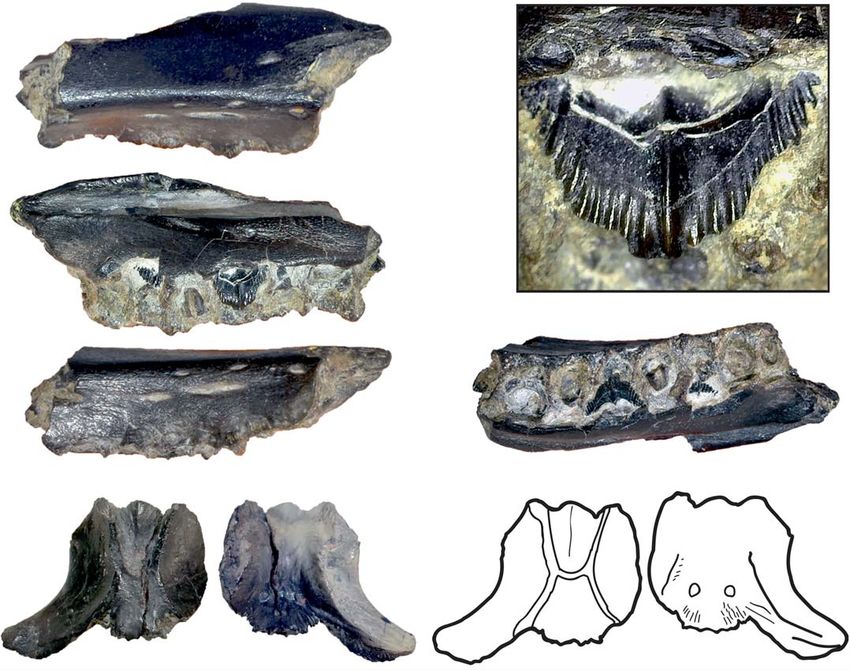
36                                                                      ACTA PALAEONTOLOGICA POLONICA 65 (1), 2020

municipality, Thulborn (1973) erected the taxon Alocodon       Geological settings
kuehnei based on isolated teeth. Most of the Late Jurassic
ornithischians of Portugal come from the outcrops of the       Specimens described here were recovered along the coastline
Lourinhã Formation (Antunes and Mateus 2003) includ-           in the Lourinhã municipality, western-central Portugal, from
ing Trimucrodon cuneatus and cf. Hypsilophodon from            a north-south transect that goes from north Vale Pombas to
the Porto Dinheiro locality (Thulborn 1973). Due to the        Santa Rita municipality (Fig. 1). All specimens come from
fragmented state of the specimens and the paucity of sub-      the famous and highly fossiliferous Upper Jurassic Lourinhã
sequent discoveries, a clear assessment of this material is    Formation (Kimmeridgian–Tithonian; Hill 1989). This litho-
problematic (Norman et al. 2004). Two undisputed iguano-       stratigraphic unit comprises a succession of sandstone and
dontian species were recovered from Lourinhã Formation:        mudstone beds, representing braided fluvial systems, allu-
the camptosaurid Draconyx loureiroi (Mateus and Antunes        vial fans and upper deltas, with the occasional presence of
2001) and the dryosaurid Eousdryosaurus nanohallucis           shallow marine limestone that represent short transgressive
(Escaso et al. 2014). The holotypes consist of fragmen-        events. The subdivision of the Lourinhã Formation into sub-
tary post-cranial elements. D. loureiroi has been treated as   units has been subjected to various revisions (Hill 1989;
a wildcard in several phylogenetic analyses dealing with       Manuppella 1998; Manuppella et al. 1999; Taylor et al. 2014).
basal iguanodontians relationships (McDonald 2012; Boyd        In the present work, we follow the subdivision proposed by
                                                               Mateus et al. (2017). From bottom to top, the beds outcrop-
2015), while E. nanohallucis is represented by an immature
                                                               ping belong to the following members: Porto Novo/Praia de
individual (Escaso et al. 2014). The scarcity of ornithopod
                                                               Amoreira, Praia Azul, Santa Rita.
and, in general, neornithischian remains, strongly contrasts       Porto Novo/Praia da Amoreira Member is characterized
with the abundance of other herbivorous dinosaurs, such as     by a percentage of sands that range 34–44%, and are formed
sauropods and thyreophorans (Antunes and Mateus 2003).         of fluvial channel deposits and calcrete-bearing floodplain
Up until now it has not been possible to assess if this gap    mud with occasional fine sand lenses. The depositional en-
may be due to differential preservation linked to ecologi-     vironment is interpreted as either distal mudflat of an al-
cal segregation as seen for ornithischians and saurischians    luvial fan, or of a braided river system. It is dated to the
in the Morrison Formation (Foster 2013) or if it reflects      uppermost Kimmeridgian (Fig. 1) (Hill 1989; Taylor et al.
inter-specific competition between different herbivorous       2014; Mateus et al. 2017).
taxa. Beside skeletal and dental ornithopod fossils, various       Praia Azul Member is characterized by a lower per-
tracks and track-sites have been reported from the Lourinhã    centage of sand with respect to the underlying Porto Novo/
Formation (Mateus and Milàn 2009) including a giant sized      Praia da Amoreira Member, ranging 12–25%. Three ex-
Iguanodontipus-like footprint which is 70 cm long, sug-        tensive carbonated shell layers, representing three marine
gesting the presence of a large sized iguanodontian possibly   transgression events, can be correlated along the whole ba-
larger than any previously known Late Jurassic species         sin, being important marker beds (Hill 1989). This mem-
(Mateus and Milàn 2008), but also smaller sized camp-          ber is dated to the Kimmeridgian–Tithonian interval, being
tosaurid tracks (as figured by Antunes and Mateus 2003:        the boundary between these two stages corresponding to
fig. 11) and Dinehichnus-like ones, probably made by dryo-     the second carbonated layer (Hill 1989; Taylor et al. 2014;
saurids or other small bipedal neornithischians (Mateus        Mateus et al. 2017).
and Milàn 2009).                                                   Santa Rita Member is constituted mainly of mudstone
                                                               with numerous pedogenic carbonated concretions, interca-
    Here, we extend the current knowledge of the Late
                                                               lated by levels of cross-bedded sandstone. The sandstone
Jurassic ornithopod fauna of Portugal, reporting and de-
                                                               elements include large scale point bars, flat tabular lenses,
scribing previously unpublished fossils housed at the Museu
                                                               crevasse splay, and levees bodies. This is the youngest mem-
da Lourinhã.                                                   ber, being dated to Tithonian (Mateus et al. 2017).
Institutional abbreviations.—CM, Carnegie Museum of
Natural History, Pittsburgh, PA, USA; IRSNB, Institut Royal
des Sciences Naturelles de Belgique, Brussels, Belgium;        Material and methods
NHMUK, Natural History Museum, London, UK; OUM,
Oxford University Museum, Oxford, UK; ML, Museu da             Here we report and describe material currently housed at
Lourinhã, Portugal; SHN, Sociedade de Història Natural,        the ML. The specimens were collected in annual field cam-
Torres Vedras, Portugal; YPM, Yale Peabody Museum, New         paigns of the museum during the last two to three decades
Haven, CT, USA; UMNH, Utah Museum of Natural History,          or donated to the institution by amateurs. Most of the latter
Salt Lake City, UT, USA.                                       lack detailed geological setting and sometimes even geo-
                                                               graphical location other than just the beach from where they
Other abbreviations.—CI, consistency index; GM, geome-         were collected. A detailed analysis of the accompanying
tric mean; PCA, Principal Component Analysis; RI, reten-       notations, remains of the matrix sediment the material, and
tion index; RMA, Reduced Major Axis; TL,tree length.           studying the geological mapping of the referred localities
ROTATORI ET AL.—LATE JURASSIC ORNITHOPOD DINOSAURS FROM PORTUGAL                                                                                                                                                  37

        A                                                                       C         Stratigraphy                                                                      Lithology

                                                                                                                Santa Rita Member
                                                                                                 Tithonian
         B

                                                                  Cenozoic
                                                                  Mesozoic
                                                                  Paleozoic

                                                                                                               Praia Azul Member

                                                                                                                                                                                        Ornithopoda
                                                                                                152
        B           Vale Pombas                                                                 Ma

                                                                                     Jurassic

                                                                                                                                                       Lourinha Formation
                   Vale Frades
                        Peralta
              Porto das Barcas
                                               Lourinha
                   Zimbral

                                                                                                               Porto Novo + Praia da Amoreira Member
              Porto Dinheiro
                                               Torres Vedras
                      Valmitao

                                                                                                Kimmeridgian
                                             Lisbon

                                                                                                                                                                                                      sandstone

                                                                                                                                                                                                      limestone

                                                                                                                                                                                                      mudstone

Fig. 1. General map of Iberian Peninsula (A), and a close-up of the Lourinhã coastline (B), describing the general sedimentology of the Lourinhã Formation
and the stratigraphic distribution of the material here discussed (C). The localities indicated represent the geographical provenance of the specimens. The
type localities of Draconyx loureiroi and Eousdryosaurus nanohallucis marked in bold. Credit for the Iberian Peninsula map: Eduardo Puértolas-Pascual.

has allowed us to refer most of the material to Praia Azul                     Eousdryosaurus nanohallucis (SHN 177) was added to the
Member, although at the present time is not possible to de-                    dataset. A heuristic search of 1000 replicates holding 10
termine if it is latest Kimmeridgian or earliest Tithonian in                  trees per replicate was performed for each of the multiple
age. ML 2206 comes from the Santa Rita Member and it can                       analyses conducted. The analyses were carried out in TnT,
be considered to be Early Tithonian in age. ML 563 has no                      version 1.5. (Goloboff and Catalano 2016). The complete
precise information regarding its provenance. Nomenclature                     data matrix used in the phylogenetic analysis is in SOM 1,
follows Galton (1981, 1983), Weishampel (1984) and Norman                      and the complete analyses consensus trees are in SOM 2
(1980, 1986). Further photographic comparison was car-                         (Supplementary Online Material available at http://app.pan.
ried out using photographic material including Dryosaurus:                     pl/SOM/app65-Rotatori_etal_SOM.pdf). In order to rein-
CM 3392, CM 11340, CM 21786, NHM 723, NHM 724,                                 force our results, the ML 768, ML 818, ML 2055, and ML
NHM 725, NMNHUK 812.                                                           563 were scored in the dataset of Boyd (2015), with the
    In order to assess their phylogenetic relationships, ML                    modifications of Madzia et al. (2018) and Bell et al. (2018).
768, ML 2055, ML 563, and ML 818 were included in the                          Three additional analyses were performed, using the same
matrix of Dieudonné et al. (2016). Despite the topology of                     tree search strategy as in the previous case. The two isolated
the consensus, Dieudonné et al. (2016) differs from the latest                 dentaries (ML 818, ML 768) were run separately (CI 0.344,
studies on the relations of some important clades (Herne                       RI 0.643, TL 914, CI 0.344, RI 0.644, TL 914, respectively),
et al. 2019; Rozadilla et al. 2019), it is still the best phylog-              while limb bones elements (ML 563, ML 2055) were run
eny to explore the relations of basal ornithopods, includ-                     together (CI 0.343, RI 0.642, TL 916). In order to improve the
ing dryosaurids. All character numbers and scores used                         resolution of the results, nine wild-card taxa were individu-
in the description refers to this dataset. The holotype of                     ated using the TnT function pcrprune. We safely removed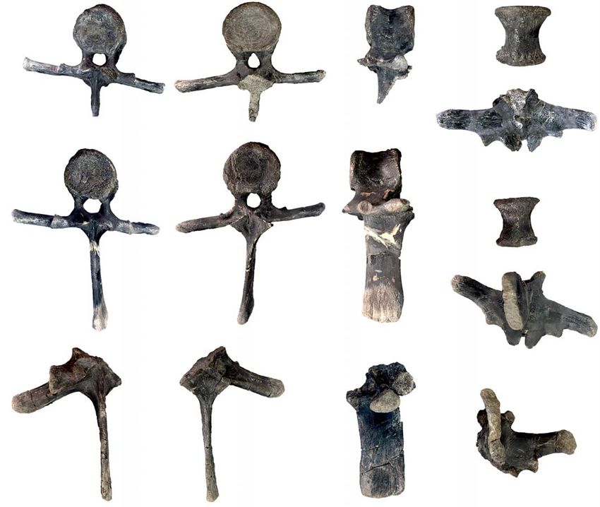
38                                                                                      ACTA PALAEONTOLOGICA POLONICA 65 (1), 2020

Table 1. Selected measurements (in mm) of the specimens here described.
                                               Width (proximal epiphysis)               Width (distal epiphysis)
                            Total length
                                             mediolateral   anteroposterior         mediolateral     anteroposterior
             ML 2055            250              46                43                   55                  47
Femora
             ML 563             188             26.7              27.3                 35.2                41.4
             ML 2055            220              40                19                   12                  30
Tibiae
             ML 505             235              29                44                   37                  17

                                                                                        Height           Thickness
                                                Total length       Width
                                                                                  max            min   (mediolateral)
                             ML 818                 150              –            62             49         23
Dentaries
                             ML 768                  30              –             7              2          8
Parietal                    ML 1851                  18              22                  –                   –
Coracoid                    ML 2206                 130             108                  –                   –
                        ML 2321 (complete)            –              51                  22                  –
Neural arches
                         ML 2321 (partial)            –              35                  19                  –

                                            Neural spine       Anterior surface    Posterior surface                 Length
                                           height   width      height width        height     width    anteroposterior transverse process
                        ML 864              140       65          –        –          –          –           75                85
Isolated dorsal
                        ML 452              150       50         60       65         65         75           60                90
vertebrae
                   ML 452 (incomplete)       –         –         65       73         70         70           65                90

                              Total length       Width (min)        Width (distal epiphysis)
Scapula           ML 2042         440                95                       120

a posteriori the following taxa: Burianosaurus augustai,                   Hübner (2018), including only the variables directly measur-
Gasparinisaura cincosaltensis, Laellynasaura amicagraph-                   able on the specimens for the femora and tibia (see supple-
ica, Micropachycephalosaurus hogtuyanensis, Morrosaurus                    mentary material in Hübner 2018 for further explanation).
antarticus, Qantassasaurus intrepidus, Stenopelyx valden-                  This methodological choice was embodied to minimize
sis, and Weewarrasaurus pobeni. The results of these new                   the impact of missing data on the analysis. Since allome-
analyses are shown in SOM 3 and SOM 4. Since the obtained                  tric variation is described by a power function, the data
results do not provide any particular deeper insight respect to            was log-transformed prior PCA to explore morphometric
the ones obtained with the matrix of Dieudonné et al. (2016),              linear relationships of the variables measured (Hammer
just the latter ones are discussed in detail here.                         and Harper 2008). PCA and RMA were calculated with
    The dryosaurids Dysalotosaurus and Dryosaurus are                      PAST v.3 (Hammer et al. 2001) In particular, the dataset is
represented in the fossil record generally by immature spec-               composed by linear measurements of post-cranial elements.
imens (Horner et al. 2009; Hübner 2012, 2018), and that has                The variates, the values of principal components and the
been proposed to be a distinctive trait of their life history              loading scores are shown in SOM 5–9. Descriptive mea-
(Hübner 2012). It is still not clear if this trait is common to            surements for all the specimens are given in the Table 1.
all dryosaurids, or just to Dysalatosaurus and Dryosaurus.
Recently Hübner (2018) estimated ontogenetic allometric
variation in the post-cranial skeleton in a population of the
dryosaurid Dysalotosaurus lettowvorbecki, based on a lin-                  Systematic palaeontology
ear morphometric approach. In order to test if the limb
bones elements ML 2055, ML 563, and ML 505 fitted the                      Dinosauria Owen, 1842
model proposed by Hübner (2018), Multivariate Analysis                     Ornithischia Seeley, 1887
was performed in two steps: PCA and subsequently RMA                       Ornithopoda Marsh, 1881
regression. PCA was embodied to explore the variation be-
tween ML specimens and the reference population accord-                    Iguanodontia Sereno, 1986
ing to the method described by Hammer and Harper (2008);                   Dryosauridae Milner and Norman, 1984
while RMA was used to extrapolate a growth trajectory for                  Dryosauridae indet.
Dysalotosaurus individuals plus ML specimens, plotting
                                                                           Figs. 2–4.
the log of GM of each specimen. Because the growth of
the bone is described by a power function, GM is a reliable                Material.—ML 1851, an almost complete isolated parietal
proxy of absolute size (Klingenberg 1996). The morphomet-                  from Praia da Peralta; ML 2321, two partial associated
ric dataset is constituted by a subset of the one presented by             neural arches from Praia de Porto Dinheiro; ML 768, a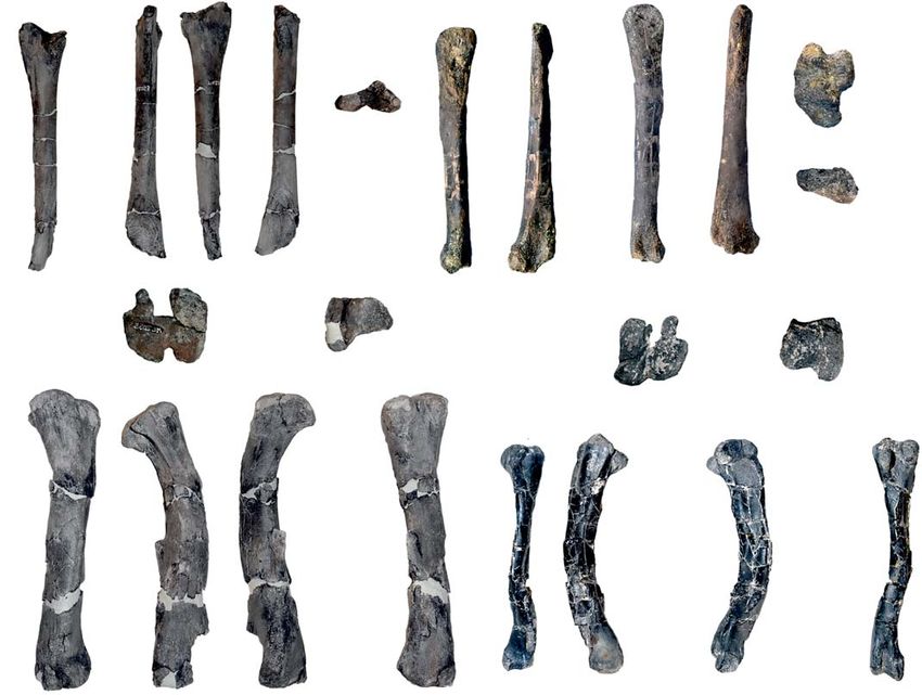
ROTATORI ET AL.—LATE JURASSIC ORNITHOPOD DINOSAURS FROM PORTUGAL 39
A1 A2 A3 A4
depression
lateral
process
10 mm
posterior notch nuchal crest
B1 B2
B3
B5 primary ridge
B5
splenial contact
Meckelian sulcus
B4 10 mm
1 mm
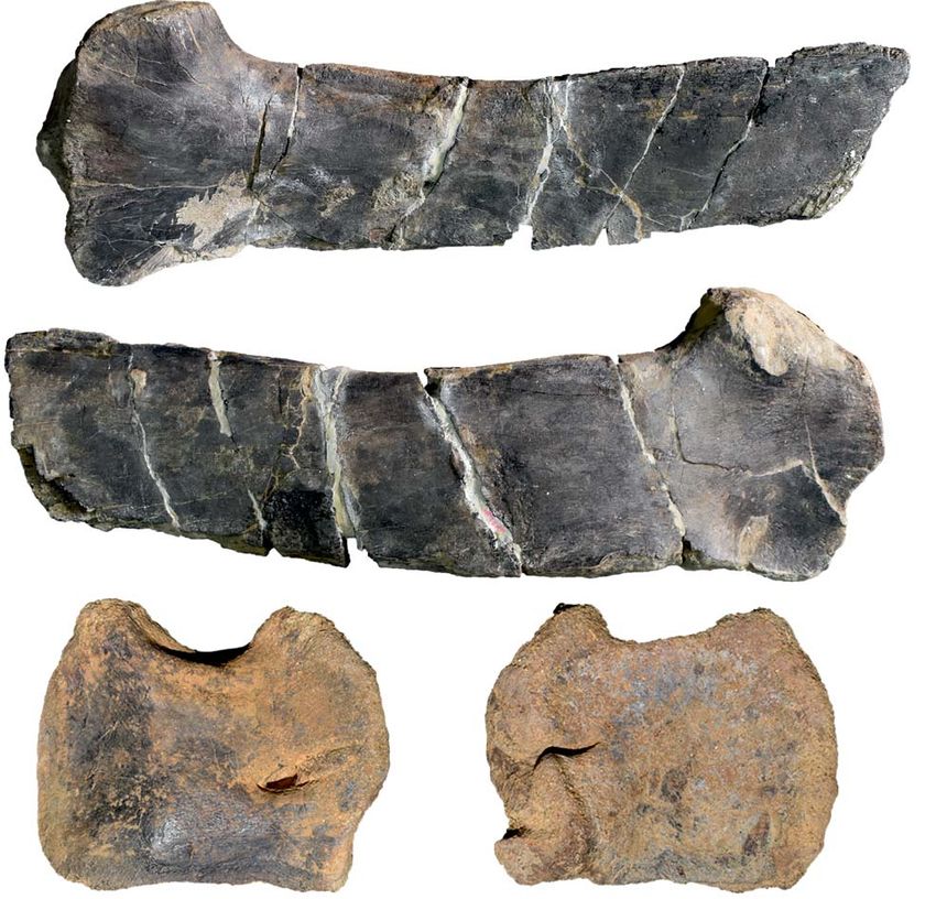
Fig. 2. Cranial material of Dryosauridae indet. from the Lourinhã municipality, Portugal, Lourinhã Formation, Kimmeridgian–Tithonian. A. ML 1851,
parietal in dorsal (A1, A3) and ventral (A2, A4) views. B. ML 768, dentary in lateral (B1), dashed frame indicates area with foramina, dorsal (B2), medial
(B3) and ventral (B4) views, detail of dentary tooth (B5).
partial tooth-bearing isolated dentary, ML 2055, associated The ventral surface is concave, flaring in a deep pit ante-
femur and tibia from Praia do Zimbral; ML 563, isolated riorly until the bone margin. Towards the midline pit, two
femur from Lourinhã coastline; ML 505, isolated tibia from small depressions are preserved.
Praia de Vale Pombas. All from Kimmeridgian–Tithonian, Dentary (Fig. 2B): ML 768 is a fragment of a right
Lourinhã, Portugal. dentary bone (length 30 mm, height 7 mm, latero-medial
Description.—Cranial skeleton: Parietal (Fig. 2A). An thickness 8 mm), fractured on both caudal and rostral ends,
isolated almost complete parietal (ML 1851), missing the first reported by Mateus (2006) as aff. Dryosaurus sp. It
right lateral half, is 18 mm in length and 22 mm in width. preserves seven tooth positions and one isolated tooth, two
It appears to be sub-triangular in shape, wider anteriorly erupting teeth and six roots of already worn down teeth.
than posteriorly. The preserved contact with the frontals The alveoli display an intra-alveolar border (character 136:
is W-shaped, presenting an interdigitated suture as seen 0), but the close proximity of the preserved crowns indicates
in Dysalotosaurus (Janensch, 1955). The preserved lateral the absence of intracrown spaces (character 154: 1). In dorsal
process projects antero-ventrally, having a smooth concave view, ML 768 is sinuous in shape and the medio-lateral sec-
surface. Posteriorly, the lateral processes converge medially tion is slightly concave-convex, towards the lateral surface.
to enclose a deep median notch between steep margins. The tooth row is convergent anteriorly and divergent poste-
Towards the midline of the notch, the proximal-most part of riorly (character 122: 1). The lateral surface is smooth, bear-
the nuchal crest is distinguishable although slightly eroded. ing seven visible vascular foramina on three different levels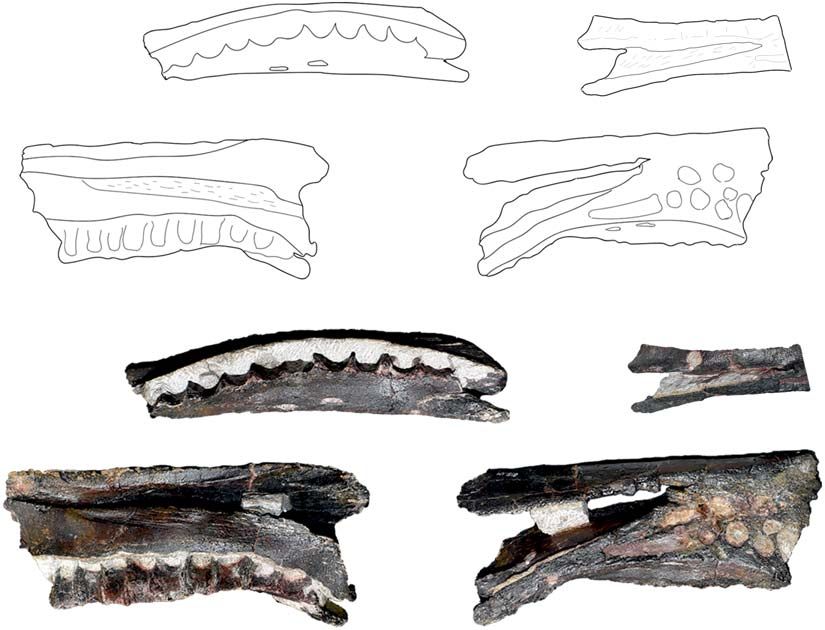
40                                                                          ACTA PALAEONTOLOGICA POLONICA 65 (1), 2020

(character 145: 0). On the medial surface, a deep Meckelian       Appendicular skeleton: Femora (Fig. 4A, B): The left femur
sulcus runs for the entire length of the tooth-row. The mar-      ML 563 is fragmented and heavily distorted. The proximal
gins of the sulcus are neat and straight. Caudally, on the me-    and distal ends are slightly eroded and fractured. Another,
dial surface, and dorsal to the Meckelian sulcus, the splenial    less distorted right femur (ML 2055) measures 250 mm
sutural contact is preserved and it exhibits a highly dense       in total length. In ML 563 the general outline of the fem-
capillary vascular system. The ventral surface does not pos-      oral shaft is strongly bowed anteriorly (character 248: 0).
sess a ventral flange (character 121: 0). The two preserved       The proximal epiphysis partially preserves the femoral
crowns are diamond-shaped (character 155: 1) and mesiodis-        head, which is separated posteriorly by a shallow depres-
tally expanded (character 135: 1) with a smoothly rounded         sion (inter-trochanteric fossa) from the greater trochanter
apex. The enamel is asymmetrically distributed (character         (character 249: 1). The surface of the greater trochanter is
140: 1), being present only on the lingual surface of the         flattened (character 252: 1). A broken blade-like surface,
crown. The crown margin possesses coarsely serrated hook-         which represents the base of the 4th trochanter, is located in
like non-mammilated denticles (character 139: 1). On the          the proximal half of the shaft (character 254: 0). Medially,
lingual side of the crown, an apicobasally extended primary       a collapsed surface overlaps the scar of the insertion of
ridge (character 158: 1) is slightly distally offset (character   Musculus caudofemoralis longus, which is restricted to the
156: 1), being surrounded by faint secondary ridges (charac-      medial surface of the shaft (character 256: 1). The distal
ters 141: 1, 159: 2). The ridges on the enamel are restricted     epiphysis includes both condyles, with the medial larger
to the lingual surface of the crown (character 160: 1). The       than the lateral. The two condyles are divided anteriorly by
denticles are generally not confluent with the crown ridges       a shallow, V-shaped extensor intercondylar groove (charac-
(character 142: 0), which are less than 10 in number (charac-     ters 257: 1, 258: 0) and posteriorly by a deep flexor grove. A
ter 157: 0). The worn down remains of dentary teeth (charac-      small lateral process overhangs on the flexor groove open-
ter 137: 1) do not show the presence of a cingulum between        ing (character 259: 1). The medial condyle is square shaped
the crown and the root (character 143: 1).                        and straight, while the lateral presents a slightly inclined an-
Axial skeleton: Neural arches (Fig. 3): Two small associated      terior edge, and a conspicuous finger-like posterior process
disarticulated neural arches (ML 2321) appear to be frac-         (character 260: 1). The medial condyle protrudes cranially
tured and subsequently restored. One is almost complete,          towards the lateral condyle (character 261: 1).
preserving both transverse processes (height 22 mm, width             As in ML 563, ML 2055 presents the femoral shaft is
51 mm) and missing part of the neural canal. The other el-        bowed anteriorly (character 248: 0), being thick and robust
ement preserves just the right transverse process (height 19      in general proportions. The section is sub-triangular in the
mm, width 35 mm). Both specimens show a low degree of             proximal and mid part of the shaft, becoming more rounded
distortion, due to taphonomic processes. The neural spine is      towards the distal epiphysis. The proximal epiphyisis pre-
low and blade like, as is common in vertebrae of other basal      serves part of the femoral head with a constriction between
iguanodontians from the cervical or anterior-dorsal series        it and the greater trochanter (character 249: 1). What is
(Norman 2004). Anteriorly, two slender and lobed prezyga-         preserved of the greater trochanter shows a slightly con-
pophyses are present, positioned at about 45° relative to the     vex surface (character 252: 0). On the medial surface of the
horizontal. There is a well-marked constriction between the       shaft, positioned towards the mid-shaft but slightly proximal
lobe located on the anterior-most end of the prezygapophy-        (character 254: 0), is a blade, like 4th trochanter (characters
ses, and the rest of the bony process. Posteriorly to the pre-    253: 1, 255: 0) that is directed medioventrally. The scar of the
zygapophyses, the prezygapophyseal lamina and the ante-           Musculus caudofemoralis longus extends more to the base of
rior centroparapophyseal lamina form a small notch. On the        the trochanter with respect to ML 563, but is still separated
transverse processes, the diapophyses are broken towards          from it (character 256: 1). Distally, the epiphysis has both
the distal edge, showing a sub-triangular transverse sec-         condyles preserved, divided anteriorly by a very prominent
tion. The parapophyses, located ventrally to the diapophyses      U-shaped extensor groove (character 257: 1, 258: 0). This
shaft, are constrained in the proximal half of the transverse     groove is proportionally deeper than in ML 563. Posteriorly,
process. Ventral to the parapophyses, on the lateral sides of     the two condyles are separated by a deeper, fully open, flexor
the neural arches, a shallow depression is present. The two       groove (character 259: 0). As in ML 563, the posterior finger-
postzygapophyses, lobed like the prezygapophyses, show            like process of the lateral condyle is strongly inset (character
the same degree of inclination with respect to the horizontal.    260: 1), whereas medial condyle does not protrude cranially
On the posterior side, wider and deeper notches, in compari-      to the lateral condyle (character 261: 0).
son to the notches present on the anterior side, are formed by        Tibiae (Fig. 4C, D): Two partially preserved right tibiae,
the postzygodiapophyseal lamina, posterior centrodiapoph-         present various degrees of erosion and fracturing. The spec-
yseal lamina and centropostzygapophyseal lamina. The              imen ML 505 is a heavily eroded but complete right tibia
same structure is seen in Valdosaurus (Barrett et al. 2011).      with some longitudinal fractures along the proximal and
Ventrally, the two lateral walls of the neural canal, present     distal epiphyses. It measures 235 mm in total length. The
a rugose surface, indicating an incomplete fused condition.       tibia ML 2055, associated with the ML 2055 femur, lacks
ROTATORI ET AL.—LATE JURASSIC ORNITHOPOD DINOSAURS FROM PORTUGAL                                                                                   41

   A1                                                    A2                                                      A3

                                                                                                           parapophysis
                                                                                                                          prezygapophyseal
A4                          postzygodiapophyseal                     A5                                     A6                 lamina
                            lamina

                                                                                                                                         pre-
                                                                                                                                     zygapophysis

                                           posterior centrodiapophyseal
                                           lamina                                                               anterior centroparapophyseal
                               centropostzygapophyseal                                                                      lamina
                               lamina
          B1                                          B2                                            B3

   constriction

                                                              20 mm
                                                               (A, B)

         B4                                                    B5                                         B6

     post-
 zygapophysis

                          pre-zygapophysis                                         pre-zygapophysis                            pre-zygapophysis
   C    constriction                                                D                                      E

                                      50 mm                                               50 mm                                      50 mm

Fig. 3. Axial skeleton elements of Dryosauridae indet. (A, B) from the Lourinhã municipality, Portugal, Lourinhã Formation, Kimmeridgian–Tithonian
compared with of Dryosaurus altus (C), Camptosaurus (“Uteodon”) aphanoecetes (D), and Mantellisaurus atherfieldensis (E). Dorsal vertebrae:
ML 2321a (A) and ML 2321b (B), in dorsal (A1, B1), anterior (A2, B2), lateral (A3, A6, B3, B6), posterior (A4, B4), and ventral (A5, B5) views. Dorsal
neural arches: YPM 1876 (C), CM 11337 (D), IRSNB 1551 (E), in dorsal view.

the proximal epiphysis, has various fractures all along the                  is divided from the fibular condyle by a rounded surface
shaft and was restored by epoxy resin, measuring 220 mm.                     (contrary to Valdosaurus, Barrett et al. 2011). The inner con-
   In the tibia ML 505 the proximal epiphysis exhibits a                     dyle points latero-posteriorly, being divided from the fibular
conspicuous cnemial crest, a well-developed fibular con-                     condyle by a narrow sulcus. The mid-shaft section is sub tri-
dyle, and a small inner condyle. The cnemial crest pos-                      angular in cross section, being flattened laterally and sharp
sesses a concave lateral edge and a convex medial edge. It                   medially, having a conspicuous crest departing from the
42 ACTA PALAEONTOLOGICA POLONICA 65 (1), 2020
distal-most part of the shaft (similar to the Eousdryosaurus is not well-discussed in literature, but this character is pres-
holotype). The overall morphology of the shaft has been ent in Dryosauridae (Janensch 1955; Galton 1981). On the
partially altered by the reconstruction process, therefore its contrary, ankylopollexians show stout and bulky prezyga-
taxonomical and systematical significance has to be consid- pophyses, see for instance Camptosaurus dispar, C. aph-
ered cautiously. The distal epiphysis does not preserve any anoecetes, Mantellisaurus atherfieldensis, and Iguanodon
visible features, only the size of the two sub-equal malleoli. bernissartensis (Gilmore 1909; Norman 1980, 1986, 2004;
In the tibia ML 2055 the proximal end is laterally com- Carpenter and Wilson 2008). Furthermore, the presence of
pressed and fractured. The section of the mid-shaft is round anterior and posterior notches constituted by the abovemen-
(character 263: 1) with a small crest on the lateral side. The tioned laminae, as seen in Valdosaurus canaliculatus, sup-
distal epiphysis preserves both malleoli and in anterior view ports the attribution to Dryosauridae (Barrett et al. 2011).
they exhibit a highly rugose surface. A deep sulcus, extends The unfused neural arch condition is common among dryo-
for most of the surface area of the lateral malleolus. The saurid individuals (Barrett et al. 2011; Barrett 2016; Hübner
medial malleolus possesses a less extensive notch, while the 2018) since even the largest individuals known so far did
lateral malleolus is more elongated posterolaterally (charac- not attain complete skeletal maturity (Horner et al. 2009;
ter 264: 1). In distal view, the two malleoli are at right angles Hübner 2012). This peculiar trait may explain why in this
to one another. case and others reported (Barrett et al. 2011; Barrett 2016),
isolated dorsal and cervical vertebral elements attributed to
Remarks.—Cranial skeleton: Parietal: Isolated parietals dryosaurids are often recovered disarticulated. On the con-
have not received much attention in literature, what hinders trary, vertebral elements of the caudal series fuse early in
detailed comparisons. ML 1851 differs from Othnielosaurus ontogeny (Hübner 2018) and therefore are usually recovered
rex, Hypsilophodon foxii, Camptosaurus dispar, and Dryo- articulated (Galton 2009; Barrett et al. 2011; Barrett 2016;
saurus altus in being sub triangular in general shape, hav- Hübner 2018).
ing less anteriorly expanded lateral processes, a narrower
Appendicular skeleton: Femora: The combination of an
sagittal crest, and proportionally longer posterior lateral
anteriorly bowed femoral shaft, the 4th trochanter proxi-
processes (Gilmore 1909; Galton 1974, 1981; Norman et
mally placed, and the medially relegated scar of Musculus
al. 2004). ML 1851 shows striking similarities with ma-
caudofemoralis longus are diagnostic for Dryosauridae
ture specimens of Dysalotosaurus lettowvorbecki (Janensch
(Butler et al. 2008; Barrett et al. 2011; Escaso et al. 2014;
1955; Galton 1981; Norman 2004), however, contrasting
Boyd 2015; Dieudonné et al. 2016). Nevertheless, ML 563
with the x-shaped morphology of the parietal present in im-
and ML 2055 differ from one another in some aspects. ML
mature specimens of the same species (Sobral et al. 2012).
563 is generally more gracile than ML 2055 in general pro-
As in adults of D. lettowvorbecki, ML 1851 possesses a
portions. This is herein interpreted as a difference in onto-
deeply arched lateral process which expands anteriorly, a
genetic stages between the two individuals and therefore,
W-shaped contact with the frontals, proportionately short
as it is discussed in the next paragraphs, ML 563 probably
posterior processes, and a wider sagittal crest (Hübner and
represents a more immature individual than ML 2055. The
Rauhut 2010; Sobral et al. 2012).
difference in size is reflected in the different shape and
Dentary: The two crowns present in ML 768 shows the
depth of the extensor groove, which is greatly influenced
characteristic diamond shape typical of iguanodontian dino-
by load. This is consistent from what is currently known in
saurs (Norman 2004; Galton 2006). The hook like denticles
other dryosaurid populations, which show great intra-spe-
are typical of dryomorphan taxa such as Dryosaurus altus,
cific variation of the extensor groove according to the size
Dysalotosaurus lettowvorbecki, Iguanodon bernissarten- (see, for instance, femora assigned to Valdosaurus canalic-
sis, Mantellisaurus atherfieldensis (Norman 2004; Galton ulatus, Dryosaurus altus, Dysalotosaurus lettowvorbecki,
2006). The number and position of primary and secondary Elrhazosaurus nigeriensis in Galton 1981, 2009; Barrett et
ridges is extremely variable among species and even within al. 2011; Hübner 2018). Another difference is the slightly
the same tooth row, depending on the tooth position. ML different position of the scar of Musculus caudofemoralis
768 differs from the coeval ankylopollexians in the lack longus. In ML 563 the position of the scar is well separated
of a marked and mesially offset secondary ridge (Galton from the base of the 4th trochanter while in ML 2055 it
2006). Instead, as in some specimens of Dryosaurus and extends further laterally to the base of the 4th trochanter,
Dysalotosaurus (Galton 1983, 2006; Carpenter and Galton although still separate. Although it is generally assumed that
2018) the single main ridge is positioned towards the mid- a “widely separated scar of Musculus of caudofemoralis lon-
line of the crown, slightly caudally offset. gus” is an unambiguous synapomorphy for Dryosauridae,
Axial skeleton: Neural arches: The two isolated and disar- in literature there are various examples of variation of this
ticulated neural arches can be ascribed to Iguanodontia on character even within the same population (Galton 1981).
the basis of stout transverse processes and zygapophyses Furthermore, Valdosaurus and Callovosaurus specimens
inclined 45° with respect to one another (Norman 2004). usually present the scar of the Musculus caudofemoralis
Among iguanodontians, the constriction between the lobe longus connected to the base of the 4th trochanter (Ruiz-
of the prezygapophyses and the rest of the bony processes Omeñaca et al. 2006; Barrett et al. 2011) similarly to MLROTATORI ET AL.—LATE JURASSIC ORNITHOPOD DINOSAURS FROM PORTUGAL 43
A1 A2 A3 A4 B1 B2 B3 B4
great
trochanter scar
of Musculus
scar caudofemoralis
of Musculus fourth
trochanter longus
caudofemoralis
longus
fourth
50 mm 50 mm
trochanter
lateral
condyle medial
extensor condyle
lateral groove extensor flexor
condyle groove groove
medial
extensor groove condyle B5 B6
lateral great
A5 A6 medial
condyle
condyle trochanter
lateral medial
femoral 20 mm
condyle condyle
head femoral head
flexor groove
20 mm
flexor groove great trochanter
cnemial crest fibular condyle
C1 C2 C3 C4 D1 D2 D3 D4
D5
40 mm lateral medial
50 mm cnemial
malleolus malleolus D6 crest
C5
fibular
medial 20 mm condyle
malleolus
inner condyle
lateral medial
lateral malleolus malleolus malleolus 20 mm
Fig. 4. Limb bones of Dryosauridae indet. from the Lourinhã municipality, Portugal, Lourinhã Formation, Kimmeridgian–Tithonian. A, B. Femur, ML
2055 (A), ML 563 (B), in anterior (A1, B1), lateral (A2, B2), medial (A3, B3), posterior (A4, B4), distal (A5, B5), and proximal (A6, B6) views. C, D. Tibia,
ML 2055 associated to femur ML 2055 (C), ML 505 (D), in anterior (C1, D1), lateral (C2, D2), posterior (C3, D3), medial (C4, D4), proximal (D5), and
distal (C5, D6) views.
2055; while Dryosaurus, Dysalotosaurus, Elrhazosaurus, possible attribution to Ankylopollexia is here considered
and Eousdryosaurus have it generally more widely sep- unlikely for ML 2055 since the medial condyle is compa-
arated (Galton 1981; Escaso et al. 2014) as in ML 563. rable in size to the lateral condyle, while in Camptosaurus
Besides the inter-specific variation, an incipient intra-spe- dispar the medial condyle is described as “sensibly more
cific variation is present within populations of Dryosaurus robust” by Gilmore (1909) and the same condition is present
and Dysalotosaurus (Galton 1981), where the insertion scar in the Portuguese ankylopollexian species Draconyx lou-
in some specimens is located more medially than others. reiroi. Furthermore, the flexor groove is widely open, while
As the extensor and flexor grooves, this character may also in most specimens ascribed to the genus Camptosaurus, an
be influenced by size and varies during ontogeny as sug- incipient lateral expansion overhangs on the flexor groove
gested by Hübner (2018). Therefore, the insertion scar of (Galton and Powell 1980; Carpenter and Wilson 2008;
the Musculus caudofemoralis longus may be more plastic Carpenter and Lamanna 2015).
than previously thought and possibly related to ontogenetic Tibiae: The tibia ML 505, despite presenting a high de-
development as well as with phylogeny. gree of erosion, has many recognizable characters, which
The more mature specimen represented by ML 2055 indicate dryosaurid affinities. In particular, ML 505 shares
resembles the overall morphology of an immature anky- with Dryosaurus, Dysalotosaurus, and Eousdryosaurus a
lopollexian as expected due to marked peramorphic condi- conspicuous fibular condyle placed in the midline of the
tions of basal ankylopollexians (i.e., camptosaurid) relative proximal epiphysis and the presence of a wide groove be-
to dryosaurids (see Horner et al. 2009). Nevertheless, this tween the posterior, inner, and fibular condyles (Janensch44 ACTA PALAEONTOLOGICA POLONICA 65 (1), 2020
1955; Galton 1981; Escaso et al. 2014). Ankylopollexians heavily eroded towards the anterior-most part. The lateral
exhibit a different condition of the proximal epiphysis of surface is heavily eroded, although preservation of the coro-
the tibia in having the fibular condyle directed posteriorly noid process is fairly complete, missing just the dorsal-most
(Norman 2004). end. The dorsal and ventral margins appear to be parallel to
It should be noted that ML 505 has a triangular diaphy- one another. The ventral surface is smooth, lacking a ventral
seal cross section, while in ML 2055 the cross section flange (character 121: 0). The tooth row preserves ten, pos-
is tear-drop. This result is consistent both with incipient sibly eleven, distinguishable close-packed alveoli (character
intra-specific variation as observed in Dryosaurus and 136: 1). The tooth row, which ends medial to the coronoid
Dysalotosaurus populations (Galton 1981). process, is encased in a parapet-like shelf that is smoothly
arched medially (character 122: 0). Two nutrient foramina
Ankylopollexia Sereno, 1986 are located on the marginal shelf of the tooth row (character
Ankylopollexia indet. 145: 0). Teeth are not preserved and the tooth-sockets do not
show any interdental plates.
Figs. 5–8.
On the medial surface, dorsally to the Meckelian sulcus, a
Material.—ML 2042, isolated, almost complete scapula longitudinally striated surface extends almost to the anterior
from Praia da Peralta; ML 452, two associated vertebrae, ML end of the specimen. Two other bones may have articulated
864, an isolated neural arch (not found in association with against this surface: the splenial and the pre-articular. There
ML 452) from Praia de Porto das Barcas; ML 818, isolated is no difference in rugosity to distinguish these two areas.
dentary from Praia Vale Frades; ML 2206, isolated cora- On the lateral surface the coronoid process projects pos-
coids from Lourinhã coastline. All from Kimmeridgian– tero-laterally (characters 124: 1, 125: 1) with respect to the
Tithonian of Lourinhã, Portugal. tooth-row and is inclined at about 30° to the horizontal. The
Description.—Cranial skeleton: Dentary (Fig. 5A): The contact with the surangular corresponds to the 4th alveolus
partial right dentary ML 818 is broken at both anterior and counted from the coronoid process cranially, so it reaches
posterior extremities, measuring 150 mm in total length. further anteriorly than coronoid process.
The maximum depth is reached in the posterior extremity Axial skeleton: Dorsal vertebrae and neural arch (Figs. 6,
being 62 mm tall, while the anterior extremity is 49 mm 7): ML 452 are two partially distorted and fractured associ-
tall. In general, the bone appears stout and compact, being ated vertebrae, while ML 864 is a broken and isolated neu-
A1 A2
50 mm
(A1–A3)
A3 A4
20 mm
surangular
coronoid process contact nutrient
alveoli foramina
A5 A6
splenial
alveoli
contact
50 mm
(A5–A7)
Meckelian sulcus surangular contact
nutrient foramina
A7 A8
20 mm
Fig. 5. Cranial material of Ankylopollexia indet. from the Lourinhã municipality, Portugal, Lourinhã Formation, Kimmeridgian–Tithonian. Dentary
ML 818, in medial (A1, A5), lateral (A2, A6), and dorsal (A3, A7) views, detail of the dentary/surangular contact (A4, A8).ROTATORI ET AL.—LATE JURASSIC ORNITHOPOD DINOSAURS FROM PORTUGAL 45
A1 A2 A3 A4 parapophysis
diapophysis
neural spine post-zygapophysis
100 mm posterior
diapophysis diapophysis
centrodiapophyseal
lamina
lamina post-zygapophysis
parapophysis pre-zygapophysis
posterior
parapophysis centrodiapophyseal lamina B4
centropostzygapophyseal
lamina parapophysis
B1 B2 B3 neural spine
neural spine
pre-zygapophysis diapophysis post-zygapophysis
post-zygapophysis parapophysis
parapophysis
pre-zygapophysis B5
neural canal diapophysis
diapophysis
centrum
pre-zygapophysis
100 mm
parapophysis
centrum
C4
neural spine
C1 C2 C3
parapophysis neural spine
parapophysis
diapophysis
neural canal C5
pre-zygapophysis
100 mm
centrum
centrum
Fig. 6. Dorsal vertebrae Ankylopollexia indet. from the Lourinhã municipality, Portugal, Lourinhã Formation, Kimmeridgian–Tithonian. A. Partial neural
arch, ML 864 in anterior (A1), posterior (A2), lateral (A3), and dorsal (A4) views. B, C. Dorsal vertebrae, ML 452a, complete (B) and ML 452b, incomplete
(C) specimens, in anterior (B1, C1), posterior (B2, C2), lateral (B3, C3), dorsal (B4, C4), and ventral (digitally modified) (B5, C5) views.
ral arch, which preserves both postzygapophyses and one 73 mm; posterior width 75 mm, 70 mm). The slight am-
prezygapophysis. The two vertebrae ML 452 were found phicoelous condition indicates a close proximity to the sa-
in association and therefore are interpreted to represent the crum. On the ventral surface, a slight constriction forms a
same individual, while ML 864 is a second individual re- smooth keel. The lateral surfaces of the centrum are longi-
covered from a different locality but comparable in size. tudinally convex, showing the presence of small vascular
Compared to the axial series of Camptosaurus dispar and foramina. The neural canal is fully open in both articulated
Mantellisaurus atherfieldensis, ML 864 is most likely lo- vertebrae, being sub-circular in shape. Dorsally, two clearly
cated at the 6–8th position of the dorsal series, while ML 452 distinguishable prezygapophysis are arranged in an angle of
are located after the 10th but before the 16th position of the 45° with respect to one another, as in other iguanodontians
dorsal series (Gilmore 1909; Norman 1986). The preserved (Norman 2004). Posterolateral to the prezygapophyses, the
centra are stout ranging 60–65 mm and consistently sub stout transverse processes originate, strongly inflecting dor-
cylindrical in shape as in Ankylopollexia (Norman 2004), sally in the partial neural arch. The associated vertebrae ML
slightly amphicoelous with the anterior facet slightly bigger 452 show a strong lateroventral inflection on the left lateral
than the posterior (anterior facet height 60 mm, 65 mm; side and a weaker dorsolateral inflection on the right side.
anterior width 65 mm, 73 mm; posterior height 60 mm, The similar orientation of the transverse processes caused46 ACTA PALAEONTOLOGICA POLONICA 65 (1), 2020
A1 B C D E F
dia par dia
A2
dia par par
dia par
par dia par
par dia
dia
I J K L
G H dia
dia
par
dia dia dia
par par par par
Fig. 7. Comparative dorsal vertebrae table of selected Ankylopollexians from the Late Jurassic and Early Cretaceous. A, B. Ankylopollexia indet.
Lourinhã municipality, Portugal, Lourinhã Formation, Kimmeridgian–Tithonian. A. ML 452 in lateral view (A1, A2). B. ML 864 in right lateral view.
C. “Uteodon” SHN.LPP 015 in left lateral view; Praia da corva, Torres Vedras Municipality, Portugal, Lourinhã Formation (Kimmeridgian–Tithonian) .
D. “Uteodon” aphanoecetes CM 11337 in left lateral view; East end of Carnegie Quarry at Dinosaur National Monument, Uintah County, Utah (USA),
Morrison Formation (Kimmeridgian–Tithonian). E. Camptosaurus dispar (unnumbered specimen) in left lateral view; Bone Cabin Quarry, Wyoming
(USA), Morrison Formation (Kimmeridgian–Tithonian). F. “Cumnoria” prestwichii OUM. J.3303 in lateral view; Oxford, UK, Kimmeridge Clay
Formation (Kimmeridgian–Tithonian). G. Hippodraco scutodens UMNH VP 20208 in left lateral view; Andrew’s Site , Grand County, Utah; Upper
Yellow Cat Memberof the Cedar Mountain Formation (upper Barremian–lowermost Aptian). H. Iguanacolossus fortis UMNH VP 20205 in right lateral
view; Don’s Ridge , Grand County, Utah, Lower Yellow Cat Member, Cedar Mountain Formation (?lower Barremian). I. Mantellisaurus atherfieldensis
IRSNB 1551 in left lateral view; Isle of Wight, Wessex Formation (Barremian). J. Barilium dawsoni NHMUK R798 in left lateral view; Shornden, East
Sussex, UK, Wadhurst Clay Formation (Valanginian). K. Hypselospinus fittoni NHMUK R604 in lateral view; Shornden Quarry, Hastings, UK, Wadhurst
Clay Formation (Valanginian). L. Iguanodon bernissartensis IRSNB “Individu S” in left lateral view; Bernissart, Belgium, Sainte Barbe Clays Formation
(Barremian). Abbreviations: dia, diapophysis; par, parapophysis. Scale bars 100 mm. Re-drawn from: C, Escaso 2014: fig. 6.5; D, Carpenter and Wilson
2008: fig. 11; E, Carpenter and Galton 2018: fig. 22D; F, Galton and Powell 1980: fig. 4; G, McDonald 2010b: fig. 27; H, McDonald 2010b: fig. 10;
I, Norman 1980: fig. 37; J, Norman 2011: fig. 4; K, Norman 2015: fig. 22; L, Norman 1980: fig. 31).
by the same diagenetic alteration further supports the asso- the parapophyses with respect to the diapophyses, being
ciation of these two specimens. Similar cases are reported on two well separated planes in ML 864 and almost on the
by Janensch (1955) describing bone beds of Dysalotosaurs. same one in ML 452 vertebrae, reflecting the general trend
Nevertheless, it is here noted that in Ornithopoda, the trans- within Iguanodontia (Norman 2004). The neural spines
verse processes generally are not directed ventrally, there- rise immediately posterior to the prezygapophyses, with
fore it is suggested the lateral left side has been affected by an anterior lamina encased by two lateral grooves, as in
taphonomical processes more than the right one. The right Camptosaurus, Mantellisaurus atherfieldensis, Iguanodon
transverse processes show an inclination which goes from bernissartensis, and other ankylopollexians (Gilmore 1909;
weakly anterodorsal to nearly sub-horizontal, in the case of Norman 1980, 1986, 2004). In the most-complete vertebra
the most incomplete vertebrae of the pair. Comparing with of the sample, the neural spine is highly elevated with re-
other taxa (Gilmore 1909; Norman 1980, 1986; Carpenter spect to the centrum as in other ankylopollexians, while
and Wilson 2008; Carpenter and Lamanna 2015; Carpenter dryosaurids have comparatively lower spines (Galton 1981).
and Galton 2018) we note that this degree of inclination The overall shape of the neural spine is rectangular, a char-
is consistent with undistorted specimens. Therefore, it is acteristic shared both by ML 452 and 864 individuals and
suggested that the right side has not been strongly affected different from the condition shown by Camptosaurus dispar
by taphonomy, the inclination being different on the right and C. aphanoecetes (Gilmore 1909; Carpenter and Wilson
lateral side a genuine character to discriminate the position 2008) but similar to that of Mantellisaurus atherfieldensis.
of the vertebrae along the vertebral column. The different Moderately deep grooves for the attachment of ossified ten-
inclination of the transverse processes indicates that ML dons are visible on the apical-most part of the neural spine.
864 is more cranially positioned than ML 452 vertebrae. Posteriorly, the postzygapophyses originate from the base of
This interpretation is corroborated also by the position of the neural spine.ROTATORI ET AL.—LATE JURASSIC ORNITHOPOD DINOSAURS FROM PORTUGAL 47
Appendicular skeleton: Coracoid (Fig. 8A): The left cora- (Galton 1983; Thomas 2015). Furthermore, ML 818 has a den-
coid ML 2206 is complete with the marginal sides slightly tary/surangular contact placed further anterior to the coronoid
eroded. It is sub-rectangular in shape, bowed medio-lat- process as in basal taxa: Hypsilophodon foxii, Dryosaurus
erally, and measures 130 mm in antero-posterior length altus, Dysalotosaurus lettowvorbecki, and Tenontosaurus sp.
and 108 mm in dorsoventral width. The dorsal margin is (Galton 1974, 1983; Thomas 2015). This condition is dif-
straight, while the ventral margin is deeply arched. The ferent from other akylopollexians such as Camptosaurus
scapular surface is slightly concave and relegated to the spp., Mantellisaurus atherfieldensis, Theiophytalia kerri,
dorsal-most part of the coracoid, while the glenoid surface Iguanodon bernissartensis, and Kukufeldia tilgatensis in
is wider and slightly convex. Ventrally, the glenoid deflects which the dentary/surangular contact is placed on the same
abruptly, forming with the bowed ventral margin a con- axis or immediately posterior to the coronoid process in a
spicuous labrum. The coracoid foramen is dorsally located nearly perpendicular fashion (Gilmore 1909; Norman 2004;
and totally enclosed on the lateral surface. On the medial Brill and Carpenter 2006; McDonald et al. 2010a).
surface, instead, it is open along the scapula-coracoid suture Axial skeleton: Dorsal vertebrae and neural arch: Ankylo-
as in Camptosaurus, Iguanodon, Mantellisaurus, and de- pollexians (and ornithopods in general) display a conser-
rived iguanodontians (Gilmore 1909; Norman 1980, 1986). vative axial skeleton during their evolutionary history,
With derived iguanodontians it also shares the width/length thus a taxonomic attribution of isolated material is prob-
ratio falling between 70–100% (Dieudonné et al. 2016). The lematic. The dorsal vertebrae ML 452 and the partial dor-
sternal process is wide and broad, a plesiomorphic condition sal neural arch ML 864 are noteworthy in terms of size,
within Ornithopoda (Weishampel et al. 2003). they are bigger than most of the material recovered from
Scapula (Fig. 8B): ML 2042 is an incomplete and eroded the Lourinhã Formation, although it is comparable in size
scapula measuring 440 mm, missing the distal-most part of with Camptosaurus sp. dorsal vertebrae from the Morrison
the blade and being partially eroded in the proximal end. Formation, in particular USNM 4282 (Gilmore 1909) and
The missing part is estimated to be not more than 10% of the specimen discussed in Carpenter and Galton (2018:
the total length of the whole scapular blade. The preserved fig. 19G). The holotype of Draconyx loureiroi is a ma-
dorsal and ventral margins appear to be parallel and pro- ture individual (Waskow and Mateus 2017), the caudal cen-
gressively converging towards the distal part of the blade tra preserved ranges 46–58 mm in length, being sensibly
as seen in Mantellisaurus atherfieldensis and Iguanodon smaller than the ones presented here (Mateus and Antunes
bernissartensis (Norman 1980, 1986), differently from the 2001). The general size proportions also closely resemble
condition exhibited by Camptosaurus dispar, C. aphanoece- the ones of Mantellisaurus atherfieldensis and Hippodraco
tes, and C. prestwichii (Gilmore 1909; Carpenter and Wil- scutodens (Norman 1986; McDonald et al. 2010b). Both
son 2008; McDonald 2011; Carpenter and Lamanna 2015), ML 452 and ML 864 share with Mantellisaurus atherfield-
which display a strong distal expansion towards the middle ensis, Hippodraco scutodens, Iguanodon bernissartensis,
of the blade. The scapular blade is dorsoventrally bowed as Barilium dawsoni, and Hypselospinus fittoni (Norman
in Mantellisaurus atherfieldensis and Iguanodon bernissar- 1980, 1986, 2011, 2015; McDonald et al. 2010b) the antero-
tensis (Norman 1980, 1986). The proximal part flares gently, posterior stout proportions, the rectangular shape of the
being concave on the lateral surface. The acromion process neural spine, and its ratio with respect to the centrum; while
is slightly rounded and forwardly directed, the underlying Camptosaurus dispar shows a paddle like structure towards
coracoid suture is straight and ventrally deflects into the gle- the dorsal-most part of the spine and generally slender pro-
noid. Contrarily to Camptosaurus dispar, C. aphanoecetes, portions (Fig. 7). Both ML 452 and ML 864 display a more
and C. prestwichii but similar to Mantellisaurus atherfield- developed neural spine with respect to the transverse pro-
ensis, ML 2042 does not possess high protuberances corres- cesses as in Mantellisaurus atherfieldensis, Iguanodon ber-
ponding to the acromion process and glenoid (Gilmore 1909; nissartensis, Barilium dawsoni, and Hypselospinus fittoni
Norman 1980, 1986; Carpenter and Wilson 2008; McDonald (Norman 1980, 1986, 2011, 2015); while in Camptosaurus
2011; Carpenter and Lamanna 2015). dispar specimens it is less developed (Gilmore 1909;
Remarks.—Cranial skeleton: Dentary: ML 818 shares with Norman 2004; Carpenter and Wilson 2008; Carpenter and
other ankylopollexians, such as Camptosaurus spp., Man- Galton 2018). Herein, ML 452 and ML 864 are conser-
tellisaurus atherfieldensis, Theiophytalia kerri, Iguanodon vatively assigned to Ankylopollexia, although the antero-
bernissartensis, and Kukufeldia tilgatensis, parallel margins posterior extension of the neural spine and its rectangular
of the dentary, closed packed alveoli, and a highly emargi- shape, its development respect to centrum and transverse
nated parapet-like structure which constrains the tooth row processes, suggests a possible affinity to Styracosterna.
(Gilmore 1909; Norman 1980, 1986; Brill and Car penter Appendicular skeleton: Coracoid: The isolated coracoid ML
2006; McDonald et al. 2010a). These characters suggest an 2206 possesses a short and broad sternal process, a typical
ankylopollexian affinity for ML 818. However, it differs from basal condition within Ornithischia. It shares with anky-
these taxa in having a more strongly inclined coronoid process, lopollexians, basal ornithopods (non-dryomorphans) and
a condition shared with dryosaurids and Tenontosaurus spp. neornithischians, a width/length ratio between 70–100%.48 ACTA PALAEONTOLOGICA POLONICA 65 (1), 2020
scapula facet
A1 A2
coracoid
foramen
50 mm
coracoid labrum glenoid facet acromion
process
B1
coracoid
suture
acromion process 50 mm
glenoid
B2
coracoid
suture
glenoid
Fig. 8. Ankylopollexian appendicular skeleton from the Lourinhã municipality, Portugal, Lourinhã Formation, Kimmeridgian–Tithonian. Coracoid
ML 2206 (A), scapula ML 2042 (B), in lateral (A1, B1) and medial (A2, B2) views.
It shares with dryomorphans, Anabisetia saldivai, and Mut- derived affinities. It is differentiated from Camptosaurus
taburrasurus langdoni an open coracoid foramen along the dispar, C. prestwichii, and dryosaurids in having the dor-
scapula-coracoid suture. The size of ML 2206 is greatly sal margin of the scapular blade convex and slender pro-
bigger than dryosaurids and other neornithischians (Galton portions of the proximal end with respect to the scapular
1974; Norman et al. 2004; Hübner 2018), while it is compa- blade. ML 2042 shares with Barilium dawsoni, Hippodraco
rable with ankylopollexians (Gilmore 1909; Norman 1980, scutodens, Hypselospinus fittoni, Iguanacolossus fortis,
1986; Car penter and Wilson 2008; Carpenter and Lamanna Mantellisaurus atherfieldensis, Iguanodon bernissartensis,
2015; Car penter and Galton 2018). The coracoid ML 2206 and more derived Hadrosauriformes the general bowed out-
does not present any distinguishable characters with respect line of the scapular blade (Norman 1980, 1986, 2011, 2015;
to the variability shown by other ankylopollexians present in Horner et al. 2004; McDonald et al. 2010a, b). The scapular
the Late Jurassic, such as Camptosaurus sp. (Dodson 1980; blade in C. prestwichii, M. atherfieldensis, Hypselospinus
Carpenter and Wilson 2008; Carpenter and Lamanna 2015). fittoni, and I. bernissartensis displays a concave proximal
Scapula: ML 2042 shows characters which are wide- end (Norman 1980, 1986, 2015; Carpenter and Wilson 2008;
spread among Ankylopollexia, although they hint to more McDonald 2011). The acromion process is rounded, differ-You can also read