SIR-ACTIN-LABELLED GRANULES IN FORAMINIFERA: PATTERNS, DYNAMICS, AND HYPOTHESES - BIOGEOSCIENCES
←
→
Page content transcription
If your browser does not render page correctly, please read the page content below
Biogeosciences, 17, 995–1011, 2020
https://doi.org/10.5194/bg-17-995-2020
© Author(s) 2020. This work is distributed under
the Creative Commons Attribution 4.0 License.

SiR-actin-labelled granules in foraminifera:
patterns, dynamics, and hypotheses
Jan Goleń1 , Jarosław Tyszka1 , Ulf Bickmeyer2 , and Jelle Bijma2
1 INGPAN – Institute of Geological Sciences, Polish Academy of Sciences, Research Centre in Kraków,
Biogeosystem Modelling Group, Senacka 1, 31-002 Kraków, Poland
2 AWI – Alfred-Wegener-Institut Helmholtz-Zentrum für Polar- und Meeresforschung, Am Handelshafen 12,

27570 Bremerhaven, Germany

Correspondence: Jan Goleń (ndgolen@cyf-kr.edu.pl)

Received: 10 May 2019 – Discussion started: 23 May 2019
Revised: 23 December 2019 – Accepted: 8 January 2020 – Published: 25 February 2020

Abstract. Recent advances in fluorescence imaging facilitate        is based on the assumption that actin granules are analogous
actualistic studies of organisms used for palaeoceanographic        to tubulin paracrystals responsible for efficient transport of
reconstructions. Observations of cytoskeleton organisation          tubulin. Actin patches transported in that manner are most
and dynamics in living foraminifera foster understanding of         likely involved in maintaining shape, rapid reorganisation,
morphogenetic and biomineralisation principles. This paper          and elasticity of pseudopodial structures, as well as in adhe-
describes the organisation of a foraminiferal actin cytoskele-      sion to the substrate. Finally, our comparative studies suggest
ton using in vivo staining based on fluorescent SiR-actin.          that a large proportion of SiR-actin-labelled granules prob-
Surprisingly, the most distinctive pattern of SiR-actin stain-      ably represent fibrillar vesicles and elliptical fuzzy-coated
ing in foraminifera is the prevalence of SiR-actin-labelled         vesicles often identified in transmission electron microscope
granules (ALGs) within pseudopodial structures. Fluores-            images.
cent signals obtained from granules dominate over dispersed
signals from the actin meshwork. SiR-actin-labelled gran-
ules are small (around 1 µm in diameter) actin-rich struc-
tures, demonstrating a wide range of motility behaviours,           1   Introduction
from almost stationarily oscillating around certain points to
exhibiting rapid motion. These labelled microstructures are         Since foraminifera were firstly recognised by science in
present both in Globothalamea (Amphistegina, Ammonia)               the beginning of the 19th century, thanks to the work of
and Tubothalamea (Quinqueloculina). They are found to be            d’Orbigny (Lipps et al., 2011), they have been the sub-
active in all kinds of pseudopodial ectoplasmic structures, in-     ject of extensive study. Most foraminifera species create
cluding granuloreticulopodia, globopodia, and lamellipodia,         shells (tests) that have great potential for preservation in the
as well as within the endoplasm. Several hypotheses are set         fossil record and are primarily important in Earth science
up to explain either specific or non-specific actin staining.       disciplines. Application of foraminiferal research include,
Two hypotheses regarding their function are proposed if spe-        among others, biostratigraphy, palaeoclimatology, palaeo-
cific actin labelling is taken into account: (1) granules are in-   environmental studies, and oil and gas exploration. As a con-
volved in endocytosis and intracellular transport of different      sequence, morphology, geochemical composition, and evo-
kinds of cargo, or (2) they transport prefabricated and/or re-      lution of their tests are much better understood than their bi-
cycled actin fibres to the sites where they are needed. These       ology. However, to properly understand fossils, it is essential
hypotheses are not mutually exclusive. The first hypothesis         to take into account the physiology of the living organisms.
is based on the presence of similar actin structures in fungi,      Recognition of this problem, together with advances in re-
fungi-like protists, and some plant cells. The later hypothesis     search methods, has led to an increasing number of studies
                                                                    concerning ultrastructure of foraminiferal cytoplasm and its

Published by Copernicus Publications on behalf of the European Geosciences Union.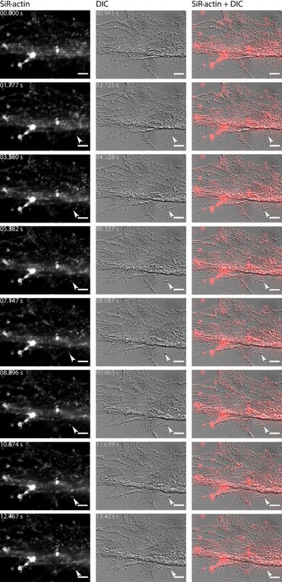
996                                                               J. Goleń et al.: SiR-actin-labelled granules in foraminifera

role in biomineralisation (e.g. Spero, 1988; de Nooijer et al.,    foraminifera. This active, bidirectional granular organisation
2009; Tyszka et al., 2019).                                        of actin was observed in all types of pseudopodial structures,
   Cytoplasm in foraminifera can be divided into two parts:        including reticulopodia, as well as globopodia and lamel-
ectoplasm (outside the test) and endoplasm (inside the test)       lipodia, during chamber formation of A. lessonii d’Orbigny.
(e.g. Boltovskoy and Wright, 1976). They differ not only           Motile granules followed relatively straight and often anas-
in location relative to the test but also in composition and       tomosing tracks (Tyszka et al., 2019, their Movies S1–S6).
appearance under the light microscope: endoplasm is much           However, the authors focused neither on this aspect of actin
thicker and is usually coloured even in the non-symbiotic          organisation nor on its dynamics. Structural and functional
species, and ectoplasm is less dense and transparent. In addi-     relationships between actin meshworks and their association
tion, many organelles, such as nuclei, ribosomes, and Golgi        with actin granularity have never been described or inter-
apparatus are reported to occur only in the endoplasm (Travis      preted (see Frontalini et al., 2019).
and Bowser, 1991). The most prominent ectoplasmic struc-              This paper is an attempt to fill the gap in our knowledge on
tures in foraminifera are pseudopods, which have a character-      actin organisation and dynamics in foraminifera. Therefore,
istic granular appearance, distinguishing foraminifera from        the main objectives of this study are as follows:
amoeba such as Gromia (Cavalier-Smith et al., 2018). This
                                                                       a. Live fluorescent labelling of actin within ectoplasmic
versatile network of branching pseudopods is involved in
                                                                          (pseudopodial) structures during various behavioural
motility (Kitazato, 1988), feeding, construction of the test,
                                                                          and/or physiological activities;
and responding to environmental stimuli (Goldstein, 1999).
As granuloreticulopodia are typically the outermost part of            b. Live fluorescent co-labelling of mitochondria to iden-
foraminiferal cell, these structures must fulfil a crucial role           tify a relative localisation and dynamics of granules rep-
in that process. The presence of granuloreticulopodia is the              resented by mitochondria and SiR-actin-labelled struc-
most fundamental morphological feature of foraminifera and                tures;
must have appeared very early in the evolutionary history of
this group (Pawlowski et al., 2003). Foraminifera probably             c. Identification and detailed description of the actin cy-
owe much of their evolutionary success to this versatile struc-           toskeleton organisation in foraminifera, with particular
ture.                                                                     focus on its granularity and dynamics by means of live
   Despite numerous studies concerning structure and func-                fluorescence imaging;
tion of granuloreticulopodia, many aspects of their organ-
                                                                       d. Assessment of unspecific labelling risk in order to eval-
isation and physiology are still unclear. The most striking
                                                                          uate reliability of staining results;
reticulopodial features are fine granules that exhibit various
behaviours. Granules move rapidly along threads of pseu-               e. Comparative analysis of published images of cytoplas-
dopods, and even along a single thread they exhibit move-                 mic foraminiferal ultrastructure observed in transmis-
ment in both directions (Jahn and Rinaldi, 1959; Kitazato,                sion electron microscope (TEM) images to identify
1988). There are numerous different categories of granules,               granular structures on TEM images that may correspond
including food particles (phagosomes), defecation vacuoles,               to SiR-actin-labelled granules;
mitochondria, dense bodies, clathrin-coated vesicles, and el-
liptical vesicles (Travis and Bowser, 1991). Granuloreticu-            f. Interpretation and discussion of working hypotheses
lopodia are not the only forms of exoplasmic (pseudopodial)               regarding the functionality of actin granularity and
structures present in foraminifera. Pseudopodial structures               its evolutionary consequences, taking into account the
are also represented by lamellipodia (see Travis et al., 1983;            physiological role of similar actin structures identified
Tyszka et al., 2019), globopodia, and frothy pseudopodia (see             and described so far in other organisms.
Tyszka et al., 2019). All of these pseudopodial structures are
highly functional and well expressed by their different mor-       2     Materials and methods
phologies and temporal organisation linked to life strategies
and behaviour.                                                     2.1    Foraminiferal culture
   Previous studies have shown that pseudopodial structures
in foraminifera depend on cytoskeleton organisation, which         Experiments were performed on three species of
includes microtubules (built from tubulin proteins) and actin      foraminifera: Amphistegina lessonii d’Orbigny, Ammo-
filaments (Travis et al., 1983; Koonce et al., 1986b; Tyszka       nia sp., and Quinqueloculina sp. They belong to both main
et al., 2019). The latest investigations of morphogenesis of       classes of multilocular foraminifera, i.e. the first two species
foraminiferal shells revealed that chamber formation and           belong to Globothalamea and the third species belongs to
biomineralisation are directly supported by actin meshworks        Tubothalamea. Specimens of A. lessonii were collected
and closely associated with microtubular networks (Tyszka          from the coral aquarium of Burgers’ Zoo in Arnhem (the
et al., 2019). The same study also reported granularity of         Netherlands). This aquarium contains a diverse assemblage
actin detected under fluorescent light of live actin-stained       of corals and other organisms from the Indo-Pacific, among

Biogeosciences, 17, 995–1011, 2020                                                         www.biogeosciences.net/17/995/2020/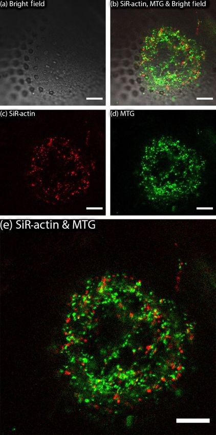
J. Goleń et al.: SiR-actin-labelled granules in foraminifera                                                                      997

them there are around fifty species of benthic foraminifera,         signal did not decrease below the detectable level. The sec-
including A. lessonii (Ernst et al., 2011). Samples of sedi-         ond method was employed for observations of reticulopodia.
ment with living foraminifera were transferred to the Alfred
Wegener Institute (AWI) in Bremerhaven (Germany) and                 2.2   Fluorescent probes and staining procedure
the Institute of Geological Sciences of the Polish Academy
of Sciences (ING PAN) in Kraków (Poland), where cultures             We focused on staining F-actin with SiR-actin but also used
were established in 10 L aquaria immediately after delivery.         MitoTracker™ Green to stain mitochondria and Celltrace™
Samples containing Quinqueloculina sp. were collected                calcein red–orange AM for staining cytoplasm. For experi-
in the oceanarium as a part of the Africarium in the Zoo             ments focusing on actin organisation during chamber forma-
Wrocław (Poland) and transported to the ING PAN in                   tion (Figs. 3–4 and S1–S2 in the Supplement and Movie S2;
Kraków, where they were cultured in 50 L aquaria. Cultures           for further information, please see the section “Video Sup-
of A. lessonii were kept in 12 : 12 light : dark cycles and          plements” at the end of the paper and the Supplement for an
natural seawater (salinity of 34). Samples of mud with Am-           explanatory text), we added a stock solution of probes pre-
monia sp. were collected from tidal flats in Dorum (Lower            pared according to the manufacturers’ instructions directly
Saxony, Germany), transported to ING PAN (Kraków), and               onto the imaging Petri dish containing living specimens of
stored in 0.25–0.5 L bottles with natural seawater (salinity of      A. lessonii up to a final concentration of 1 µM. For experi-
34) in a thermostatic cabinet (12 : 12 light : dark cycle; 8 ◦ C).   ments regarding reticulopodia (Figs. 1–2, 5–6, S3–S5, and
   We employed two different methods of sample preparation           Movie S1), the concentration of SiR-actin was 0.5 µM and
for observation during experiments. (1) Searching for asex-          the concentration of MitoTracker™ Green was 1 µM. SiR-
ual reproduction events, we monitored large individuals of           actin is a cell-permeable, fluorogenic probe-labelling F-actin,
A. lessonii climbing the glass walls of the aquaria. The juve-       thus it is suitable for live-staining (Lukinavičius et al., 2014).
nile individuals (2–5 d old) were picked up using a fine paint-         After 15–20 min the signal was sufficient to perform ob-
brush and transferred into a sterile imaging Petri dish (ibidi®      servations. Calcein red–orange AM is a cell-permeable dye
polymer coverslip bottom) containing 2 mL of clean culture           that stains the cytoplasm of living cells and is often used to
medium (up to 10 individuals per dish). After one dark phase         indicate the viability of cells (Frontalini et al., 2019). Calcein
of the light : dark cycle, when individuals attached to the cov-     AM is hydrolysed in the cytosol and fluoresce in the pres-
erslip bottom, they were examined under a stereomicroscope           ence of calcium ions. It differs in chemical structure and flu-
looking for pseudopodial activity and chamber formation.             orescence from the calcein AM used for staining cytoplasm
This method was applied to run chamber formation experi-             by Ohno et al. (2016). In our experiments the main purpose
ments (for more details, see the Supplement to Tyszka et al.,        of using Calcein red–orange AM was to indicate the limits
2019).                                                               of the cytoplasm and highlight 3-D structure of pseudopodia
   (2) In the second method, adult individuals of A. lessonii,       (e.g. globopodium). Live cell structures are stained with this
Ammonia sp., and Quinqueloculina sp. were picked from the            dye even if they are surrounded by calcium-free artificial sea-
culture aquaria or bottles and cleaned with fine paintbrushes        water, as calcium ions are always present within living cells.
under the stereomicroscope to remove algae and grains of             Table S1 (in the Supplement) summarises information about
sediment covering the specimens. Following this, they were           fluorescent probes used in the presented research.
transferred to glass-bottomed Petri dishes, which had been              Absorption and emission parameters of the SiR-actin
previously treated with hydrochloric acid over 16 h, contain-        probe overlap with the autofluorescence of chlorophyll from
ing 2 mL low-calcium artificial seawater, prepared as de-            endosymbionts, thus distinguishing between SiR-actin sig-
scribed in Bowser and Travis (2000). The reason for using            nal and autofluorescence emitted from the endoplasm of A.
low-calcium artificial seawater was to avoid the so-called           lessonii or other species hosting endosymbionts is difficult.
beading response (Bowser and Travis, 2000). This response            To minimise the problems caused by autofluorescence, we
causes transition of pseudopodia into droplets and may be            focused primarily on pseudopods where endosymbiotic algae
provoked by different chemical and physical factors (Travis          are not present. Chlorophyll and other substances present in
et al., 1983). After acclimation to the low-calcium seawater,        food particles captured by pseudopods may also give some
when reticulopodia were extended and adhered to the glass            fluorescent signal. To ensure that such food particles are ab-
bottom, specimens were stained and observed. Toyofuku et             sent within observed pseudopods, all specimens were starved
al. (2008) reported that incubation of foraminifera in low-          for 24 h prior to staining and observation.
calcium seawater for 3 d significantly decreases the level of
intracellular calcium. As a result, the presence of intracel-        2.3   Fluorescence and transmitted light microscopy
lular calcium cannot be deducted with fluorescence probes.
However, we always started our experiments immediately af-           Images were obtained with a Leica SP5 inverted confocal
ter reticulopodia were extended (usually within 0.5 h after the      microscope at the AWI and with a Zeiss Axio Observer Z1.
transfer to low-calcium seawater), and within this period the        equipped with ApoTome.2 at the ING PAN in Kraków. Apo-
                                                                     Tome.2 is a device that enables the removal of scattered light

www.biogeosciences.net/17/995/2020/                                                            Biogeosciences, 17, 995–1011, 2020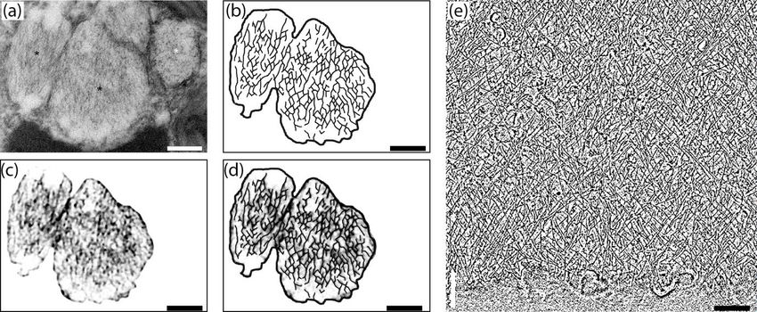
998                                                              J. Goleń et al.: SiR-actin-labelled granules in foraminifera

in fluorescence imaging. It takes between 3 and 15 images         value was set to up to 100. Blob is a technical term used in
with different positions of a grid placed in the light path       the tracking of any objects during time-lapse measurements.
between the fluorescent lamp and the sample. On the basis         These values were optimised by trial and error to minimise
of those images, the dedicated ApoTome software calculates        two types of errors: (1) lack of annotation of some objects
optical sections of the sample using a structured illumina-       that can be clearly identified on the images and (2) annota-
tion principle to enhance the signal to noise ratio of the im-    tion as spots areas with no apparent ALGs. It is not always
age (Weigel et al., 2009). In cases where living samples con-     easy to track which blobs in frame n + 1 match with those
taining moving structures it may result in the multiplication     in frame n. ALGs may temporally get so close to each other
of some rapidly moving objects. Because foraminiferal ec-         that it limits their separation in subsequent frames. To recog-
toplasmic structures are highly dynamic, we choose to set         nise this we allowed for merging and splitting the tracks. Af-
up ApoTome.2 to take only three pictures per frame and use        ter testing automatic and manual options for the second step
maximum light intensity to decrease exposure time. Despite        (creating links between spots), we decided to perform it man-
this, the most rapidly moving objects may appear in tripli-       ually, as automatic methods seem to give random results for
cate in some images. Hardware settings used for obtaining         the ALGs. When spots are annotated and links between spots
images are summarised in Table S2 (exposure time, binning         in the subsequent frames are created, velocities of spots are
mode, objectives, and Apotome mode in experiments con-            measured automatically.
ducted on a Zeiss Axio Observer Z1), Table S3 (hardware
settings in experiments conducted on a Leica SP5 inverted
confocal microscope), and Table S4 (filter sets used for dif-     3     Results
ferent fluorescent dyes for experiments performed on a Zeiss
                                                                  3.1    Identification of SiR-actin-labelled structures by
Axio Observer Z1). We optimised these settings using a trial
                                                                         fluorescence microscopy
and error method. To provide additional information over all
structures of observed individuals, we captured bright field      Fluorescent SiR-actin labelling has revealed three consid-
images for experiments with the Leica SP5 and Nomarski            erably different patterns of staining in Ammonia sp., i.e.
contrast imaging for the experiments with the Zeiss Axio          (1) weak but non-uniform staining following all membra-
Observer Z1. Nomarski contrast or differential interference       nous surfaces of pseudopodial structures, (2) linear or ring-
contrast (DIC) is a microscopy technique utilising the inter-     like structures showing intense fluorescence, and (3) small
ferometry principle for improving contrast in transparent ob-     but strongly labelled granular structures that often exhibit
jects (Lang, 1968).                                               very rapid dynamics (Fig. 1; Movie S1). The term SiR-actin-
                                                                  labelled granules (ALGs) is introduced here for these small
2.4   Measurement of velocity of the actin-labelled               oval objects. Their size has been estimated to be approxi-
      granules                                                    mately 1 µm. This is consistent with measurement of the size
                                                                  of objects corresponding to ALGs seen in Nomarski contrast
For measuring the velocities of granules in pseudopodia we        (DIC) images (Figs. 1, 2).
used time-lapse records of pseudopodia labelled with SiR-             ALGs are present within lamellipodia covering the
actin using the Zeiss Axio Observer Z1. A time-lapse movie        foraminiferal tests in A. lessonii (Figs. 3 and S1–S2 in the
consisting of 50 frames was recorded with 0.419 s time in-        Supplement) or any other structure they are attached to;
terval between timeframes (Movie S4). SiR-actin-labelled          finger-like rhizopodial structures, constructing outer protec-
granules (ALGs) can display very rapid movement, therefore        tive envelopes of chamber formation sites (Fig. 4); and retic-
tracking requires dense time-lapses. To minimise the time         ulopodia during feeding and locomotion (Fig. S3 in the Sup-
necessary for capturing a single frame, we used only one flu-     plement). They can be identified in endoplasmic structures
orescent channel (without ApoTome).                               within the chambers of non-symbiont-bearing species such
   The TrackMate plug-in (Tinevez et al., 2017), applied to       as Quinqueloculina sp., close to surfaces of internal walls of
the Fiji software (Schindelin et al., 2012), was used for cal-    the test (Fig. 5). At first glance ALGs seem to show fast and
culating the velocities of the ALGs. Calculating velocities       random movements but actually they can display different
required two main steps: (1) annotation of spots representing     “behaviours”.
ALGs in each of the frames of the time-lapse and (2) creat-           Structures labelled with SiR-actin can be matched with
ing links between particular spots in subsequent frames in the    pseudopodial structures and granular microstructures iden-
time-lapse (tracks of movement of spots). The software al-        tified in Nomarski contrast (DIC) or in a bright field im-
lows for choosing several options for both of the main steps.     ages. Figures 1–2 present a lamellipodial structure attached
This can be done either manually or automatically (with sev-      to the glass surface with a weak, dispersed fluorescent signal
eral different options in the latter case). We used the LoG       of SiR-actin staining the F-actin meshwork. Very fine bright
(Laplacian of Gaussian) detector for automatic annotation.        spots represent ALGs that match with granules observed with
As the approximate size of the ALGs is 1 µm, we used 1 µm         DIC optics.
as a “blob” size parameter in the LoG detector. The threshold

Biogeosciences, 17, 995–1011, 2020                                                      www.biogeosciences.net/17/995/2020/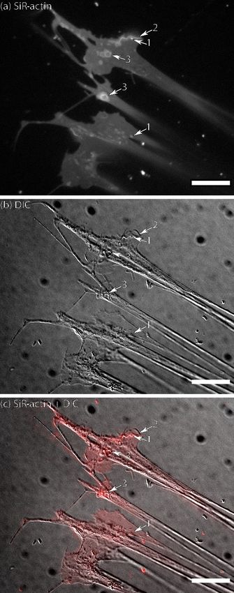
J. Goleń et al.: SiR-actin-labelled granules in foraminifera                                                                          999

                                                                        Figure 2. Pseudopodia of living Ammonia sp. attached to glass:
                                                                        (a) conventional fluorescence of SiR-actin-labelled structures,
                                                                        (b) DIC image of the same area, and (c) merged image of fluo-
Figure 1. Flattened lamellipodia of living Ammonia sp. attached to      rescence and DIC channels (since the reticulopodia were moving,
glass: (a) fluorescence of SiR-actin-labelled structures, (b) DIC im-   the DIC image is slightly shifted in relation to fluorescent one).
age of the same area, and (c) merged image of fluorescence and          Weak but non-uniform actin labelling following all membranes can
DIC channels (since the reticulopodia were moving, the DIC image        be seen in pseudopodia. The number indicate the following ob-
is slightly shifted in relation to fluorescent one). The numbers in-    jects: 1 – group of three SiR-actin-labelled granules (ALGs) trans-
dicate the following objects: 1 – actin-labelled granules (ALGs); 2     ported along one thread of pseudopodia; 2 – actin in the tip of thin
– linear SiR-actin-labelled structures; 3 – SiR-actin-labelled rings.   filopodium; 3 – larger SiR-actin-labelled areas showing smudgy flu-
Note the weak but non-uniform SiR-actin labelling following all         orescence weaker than in most ALGs; 4 – single ALG in bifurcation
membranous surfaces of pseudopodial structures. The linear struc-       of reticulopodia; 5 – a group of very bright densely packed ALGs
ture (2) was subsequently transformed into a ring structure (see        in the thick reticulopodium. Conventional fluorescence images were
Movie S1). Structures corresponding to ALGs, SiR-actin-labelled         obtained with a Zeiss Axio Observer Z1 (scale bar 10 µm).
rings, and linear structures can be seen in DIC image. Conventional
fluorescence images were obtained with a Zeiss Axio Observer Z1
(scale bar 20 µm).
www.biogeosciences.net/17/995/2020/                                                               Biogeosciences, 17, 995–1011, 2020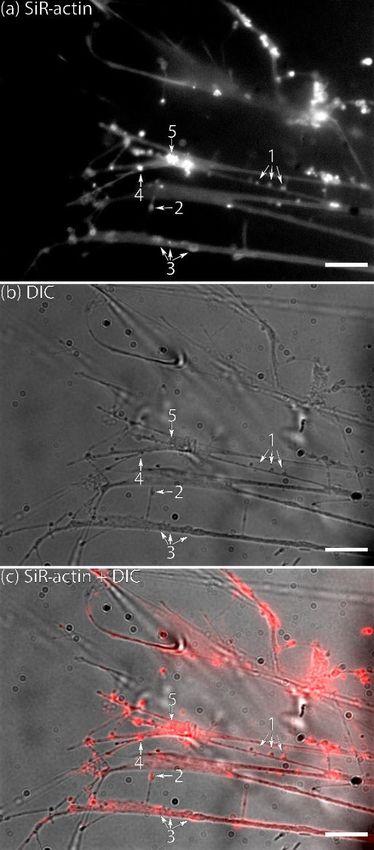
1000                                                                 J. Goleń et al.: SiR-actin-labelled granules in foraminifera

                                                                      the granules observed. Therefore, labelling of ectoplasmic
                                                                      granules is selective. In order to test ALGs’ relationships
                                                                      with selected, well-defined granules, mitochondria were cho-
                                                                      sen for double-labelling experiments. Mitochondria were the
                                                                      best candidates because they had frequently been recognised
                                                                      within the cytoplasm, including reticulopodia (e.g. Travis
                                                                      and Bowser, 1986; Hottinger, 2006; Nomaki et al., 2016;
                                                                      LeKieffre et al., 2018a). Mitochondria usually appear oval
                                                                      or kidney shaped in cross section, with a length in the range
                                                                      of 0.5 to 1 µm, although they are sometimes larger and take
                                                                      various, even tubular shapes (LeKieffre et al., 2018a).
                                                                         MitoTracker™ Green has been applied to living speci-
                                                                      mens of A. lessonii following the procedure described above
                                                                      (Sect. 2). This probe selectively accumulates in the mito-
                                                                      chondrial matrix by covalent binding to mitochondrial pro-
                                                                      teins (Presley et al., 2003). Results of replicated live ex-
                                                                      periments do not show co-localisation of ALGs and mito-
                                                                      chondria stained by MitoTracker™ Green (Figs. 3 and S3
                                                                      and Movie S2). Therefore, they indicate that mitochondria
                                                                      and SiR-actin-labelled granules are two non-overlapping cat-
                                                                      egories.

                                                                      3.3   Dynamics of SiR-actin-labelled granules

                                                                      The dynamics of the ALGs (velocity and overall pattern of
                                                                      movement) are described separately in granuloreticulopodia
                                                                      and in a globopodium during chamber formation. The dy-
                                                                      namics may vary for different locations in the cell. Not all
                                                                      of the ALGs have the same pattern of movement. At first
                                                                      glance their movement may appear chaotic, but closer analy-
                                                                      sis reveals some general patterns.
                                                                         For the sake of simplicity, particular threads of granu-
                                                                      loreticulopodia may be considered one-dimensional struc-
                                                                      tures that constrain the possible directions of movement: they
                                                                      can move along the thread of reticulopodia either inward
                                                                      or outward. Indeed bidirectional movement along a single
                                                                      thread is commonly observed in A. lessonii (Figs. S4–S5
                                                                      in the Supplement), however, in cases of thick pseudopo-
                                                                      dial threads there may by a spatial separation: in the core
                                                                      of pseudopodium, ALGs move towards the cell body, while
                                                                      in the cortex they travel in the opposite direction (Movie S3).
Figure 3. SiR-actin-labelled granules (ALGs) and mitochondria in      Usually one direction is dominant: when reticulopodia are
cross section of a newly forming chamber in Amphistegina lessonii
                                                                      formed, outward (centrifugal) transport is more common, but
during biomineralisation (pores are already visible in transmitted
light). ALGs and mitochondria do not show co-localisation. Images
                                                                      during retraction of reticulopodia inward (centripetal) move-
were obtained with Leica SP5 inverted confocal microscope (scale      ment is prevalent. During extension of a newly formed very
bar 10 µm). For the entire time-lapse, see Movie S2.                  fine thread of pseudopodium, there is usually a single ALG at
                                                                      the tip of this thread (Fig. 6). Sometimes clusters of granules
                                                                      moving together with the same speed along a pseudopodium
3.2    Testing the selectivity of labelling of granules:              may be identified. As the granuloreticulopodia themselves
       SiR-actin-labelled granules vs. mitochondria                   are very dynamic structures, it is not always possible to mea-
                                                                      sure displacement of ALGs due to the absence of a station-
Direct comparative analysis of fluorescence vs DIC images             ary reference frame. Another problem is that ALGs can be
of A. lessonii indicate that SiR-actin-labelled granules do not       so abundant in reticulopodia that they may be extremely dif-
overlap with all granules observed in DIC (Fig. S3 in the             ficult to track. To overcome this problem intervals between
Supplement). This means that SiR-actin does not stain all             subsequent frames in time-lapse movies were minimised. Us-

Biogeosciences, 17, 995–1011, 2020                                                          www.biogeosciences.net/17/995/2020/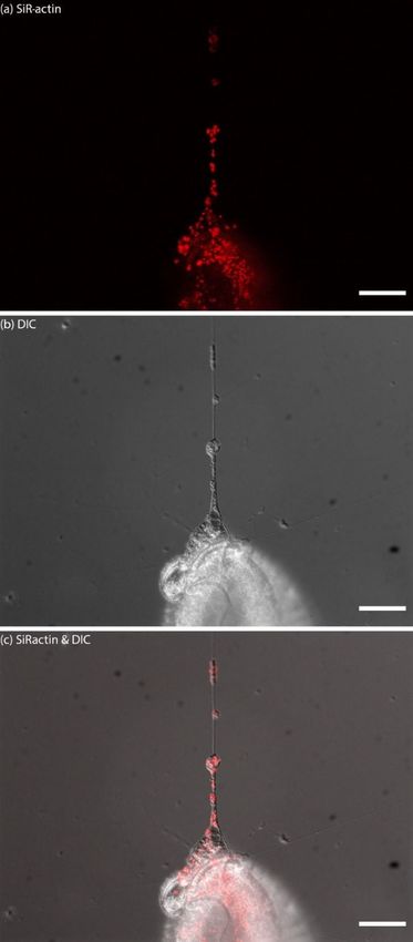
J. Goleń et al.: SiR-actin-labelled granules in foraminifera 1001
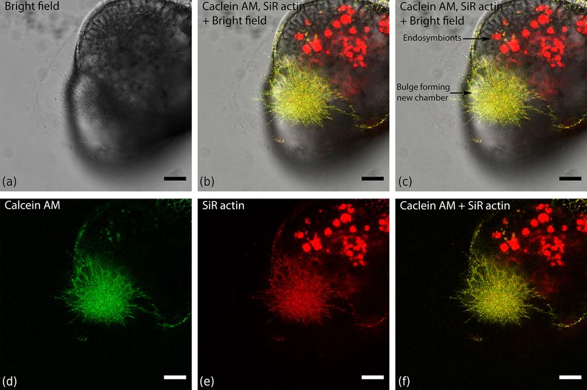
Figure 4. Organisation of actin within a finger-like structure preceding globopodium during chamber formation in Amphistegina lessonii,
compared with localisation of cytoplasm stained with calcein red–orange AM. Images was obtained with a Leica SP5 inverted confocal
microscope (scale bar 20 µm).
ing this strategy we recorded time-lapse movies (Movie S4) 4 Interpretation and discussion
showing a wide range of velocities of ALGs in reticulopodia
of A. lessonii up to 15.4 µm s−1 (Fig. S8 and Tables S5–S6 in 4.1 Assessment of unspecific fluorescent labelling risk
the Supplement).
Lamellipodia covering the test form two-dimensional All microscopy techniques are associated with a risk of cap-
sheets, resulting in a more complex pattern of displace- turing artefacts instead of imaging target structures. In the
ment of ALGs than the one observed in granuloreticulopo- case of fluorescence microscopy, the greatest danger is un-
dia. There are areas dominated by directional protoplasm specific labelling or autofluorescence. Comparison of stained
streaming and areas showing less organised behaviour. Ac- individuals of A. lessonii to an unstained control shows that
cordingly, actin granules can be divided into several cate- the SiR-actin fluorescent probe indeed stains endoplasmic
gories based on dominant movement patterns: (1) stationary structures in foraminifera (Fig. S6 in the Supplement). This
or almost stationary ALGs that oscillate within a very narrow may be caused when the concentration of the probe is too
space, (2) ALGs showing saltatory movements as described high or when the excitation intensities (or emission measure-
in Travis and Bowser (1991), and (3) ALGs exhibiting ex- ment sensitivity) are too high. Another problem might be that
tremely rapid movement that can be observed for up to a few the probe is not specific enough and binds to other chemical
seconds. Moreover, in some areas actin granules may move compounds in the cell, the structure of which mimics the tar-
along a single line but with different velocities and in differ- get structure. Most fluorescent probes were developed and
ent directions. They may pass some stationary granules with tested to study mammalian cells; therefore, the risk of unspe-
no significant interaction observed. cific fluorescent labelling should be addressed to avoid con-
fusion and over-interpretation of the results. Foraminifera are
placed in the actin phylogenetic tree with Bikonta (Flakowski
et al., 2005), thus the amino acid sequence of actin in
foraminifera is significantly distinct from the actin sequence
www.biogeosciences.net/17/995/2020/ Biogeosciences, 17, 995–1011, 2020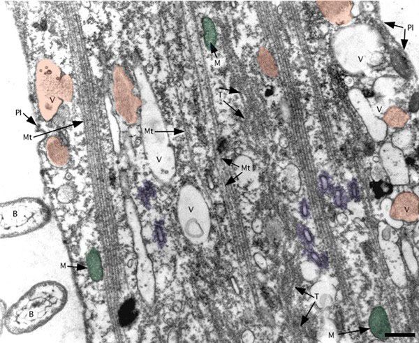
1002                                                                 J. Goleń et al.: SiR-actin-labelled granules in foraminifera

                                                                      in Metazoa or fungi (belonging to opisthokonts) that are sub-
                                                                      ject of most intensive research of actin physiology. Fluores-
                                                                      cent probes may therefore interact differently with actin in
                                                                      foraminifera. Moreover, foraminifera may contain other or-
                                                                      ganic compounds that mimic actin nanostructures and there-
                                                                      fore interact with fluorescence probes, such as SiR-actin. It
                                                                      should be noted, however, that our results are reproducible.
                                                                         As the granular pattern of SiR-actin staining is unusual
                                                                      compared to other eukaryotes, it requires an extensive dis-
                                                                      cussion of all possible scenarios (see Fig. S7). There are
                                                                      three possible scenarios in which ALGs may represent real
                                                                      F-actin-containing structures that are labelled by the SiR-
                                                                      actin probe (Fig. S6a–c in the Supplement) and three addi-
                                                                      tional possibilities that would reveal the observed patterns as
                                                                      artefacts (Fig. S6d–f in the Supplement). The first and most
                                                                      likely scenario (Fig. S6a in the Supplement) assumes that
                                                                      foraminifera possess granular structures filled with densely
                                                                      packed actin filaments that are specifically stained with SiR-
                                                                      actin. These structures possibly correspond to fibrillar vesi-
                                                                      cles known from TEM ultrastructure studies (see below in
                                                                      Sect. 4.5.1). According to the second scenario, labelled actin
                                                                      filaments surround some membranous vesicles (Fig. S7b
                                                                      in the Supplement). These vesicles are possibly involved
                                                                      in transport and endocytosis, and F-actin probably plays
                                                                      role in those processes. Alternatively, they may correspond
                                                                      to elliptical fuzzy-coated vesicles described by Koonce et
                                                                      al. (1986a) regulating motility of reticulopodia (see below
                                                                      Sect. 4.5.2 Elliptical fuzzy-coated vesicle). The third sce-
                                                                      nario assumes that actin filaments are located both inside and
                                                                      outside of some membrane-bound vesicles (Fig. S6c). Alter-
                                                                      natively, the observed staining pattern may be explained as
                                                                      an artefact if SiR-actin binds to another, unidentified, organic
                                                                      molecule that is different from (and thus not associated with)
                                                                      F-actin either inside (Fig. S6d in the Supplement) or outside
                                                                      (Fig. S6e in the Supplement) of membranous vesicles. Lastly,
                                                                      SiR-actin may induce assemblage of actin filaments in the ar-
                                                                      eas rich in G-actin (Fig. S6f in the Supplement), as suggested
                                                                      by Melak et al. (2017).
                                                                         The first argument supporting the reliability of SiR-actin
                                                                      live staining is the fact that attachment sites of pseudopo-
                                                                      dia to the substratum often demonstrate a strong fluores-
                                                                      cence signal (Fig. 1), as predicted from the essential role of
                                                                      actin for adhesion (Bowser et al., 1988). Secondly, as men-
                                                                      tioned above, granular actin structures are visible on images
                                                                      of fixed reticulopodia stained with phalloidin (see Koonce et
                                                                      al., 1986a, their Fig. 3c; Koonce et al., 1986b, their Fig. 1f). It
                                                                      cannot be excluded, however, that ALGs are associated with
                                                                      a defence strategy to remove and dispose of toxic compounds
Figure 5. SiR-actin-labelled granules (ALGs) in pseudopodia and
                                                                      introduced into the cell. If this were true, we would expect
endoplasm of Quinqueloculina sp. Panel (a) presents actin stained
with SiR-actin, panel (b) presents images obtained with DIC optics
                                                                      vesicles containing those probes to be transported outward.
(inverted LUT), and panel (c) presents merged fluorescent and DIC     As ALGs are often moving bidirectionally (both inwards and
channels. Conventional fluorescence images were obtained with a       outwards) (Figs. S4, S5 in the Supplement), this hypothesis
Zeiss Axio Observer Z1 (scale bar 50 µm).                             is not very convincing. In fact, ALGs’ inward movement is
                                                                      observed when a pseudopodial structure is being withdrawn
                                                                      and seems to indicate relocation of labelled actin into the en-

Biogeosciences, 17, 995–1011, 2020                                                           www.biogeosciences.net/17/995/2020/
J. Goleń et al.: SiR-actin-labelled granules in foraminifera 1003 Figure 6. Dynamics of SiR-actin-labelled granules (ALGs) in reticulopodia of Amphistegina lessonii. Eight frames of the time-lapse. Right column: actin stained with SiR actin; middle column: DIC; right column: overlay of fluorescent and DIC channels. Arrows indicate a granule in the tip of one very fine thread of forming pseudopodium. The numbers in top-right corner of each image of SiR actin and DIC channels indicate the time of acquisition. Conventional fluorescence images were obtained with a Zeiss Axio Observer Z1 (scale bar 10 µm). www.biogeosciences.net/17/995/2020/ Biogeosciences, 17, 995–1011, 2020
1004                                                                J. Goleń et al.: SiR-actin-labelled granules in foraminifera

doplasm. Such observations support the notion that live stain-       of different staining procedures. This is due to the fact that
ing using SiR-actin is specifically labelling actin and that the     every probe may have affinity to different epitopes of F-
inward movement of ALGs is a functional response during              actin, therefore, may not label all different F-actin-containing
withdrawal of pseudopodial structures.                               structures equally. The effectiveness of staining F-actin us-
   Another issue that needs to be considered is the risk of          ing different probes was compared by Lemieux et al. (2014).
interference of a probe with the physiology of actin itself,         They reported that different probes did not stain all subsets
as it may, for instance, cause an artificial polymerisation of       of F-actin equally. Apparently, the staining effectiveness of
F-actin (Melak et al., 2017). In that case, we would expect          F-actin depends on the location of actin filaments within the
negative interference of SiR-actin on morphogenesis and              cell. Even though this analysis does not include SiR-actin,
biomineralisation of new chambers. Nevertheless, such stain-         the same issue may apply to this probe. The interaction be-
ing artefacts have never been observed (Tyszka et al., 2019).        tween probes and F-actin may also lead to stabilisation or
Moreover, if SiR-actin causes polymerisation of F-actin, live        enhanced polymerisation of F-actin due to its structural sim-
actin staining should have a visible impact on organisation          ilarities to Jasplakinolide (Melak et al., 2017). In addition,
and motility of pseudopodia. In our observations we did not          cell fixation procedures may stabilise dynamic structures or
recognise any apparent long-term differences either in mor-          create some artefacts.
phology or in dynamics of reticulopodia after staining. Occa-           Previous studies of the dynamics of granules in
sionally, we observed a temporary retraction of pseudopods           foraminifera were conducted mostly on Allogromia and As-
immediately after adding the staining solution to the petri          tramina. The maximum speed of granules within reticulopo-
dish. However, after 10–15 min incubation with SiR-actin,            dia was reported to be approximately 25 µm s−1 , but most of
this effect was not visible any more, and reticulopodia were         them have velocities below 10 µm s−1 (Travis and Bowser,
spread out again, closely resembling the pre-staining struc-         1991). Velocities of ALGs fall within this range. The aver-
ture and dynamics. A. lessonii is less prone to retraction of        age speed of granules in foraminiferal pseudopodia reported
pseudopods and recovers faster than Ammonia sp. or Quin-             by Kitazato (1988) is 13 µm s−1 , which is roughly compara-
queloculina sp. after applying the staining solution.                ble to the maximum velocity recorded by us (15.5 µm s−1 ).

4.2    Previous studies of actin in foraminifera using               4.3   Main hypothesis regarding the functionality of
       fluorescent labelling                                               actin-labelled granules

The most commonly used method of fluorescent labelling               Actin-labelled granules described in this paper appear to be
of the actin cytoskeleton is phalloidin staining (Melak et           one of the main forms of actin cytoskeleton organisation in
al., 2017). Its utility is limited mostly to staining fixed          external cytoplasm (ectoplasm) of foraminifera. As they are
cells. Phalloidin staining was previously employed to study          ubiquitous in pseudopodia during feeding behaviour and in
the actin cytoskeleton in reticulopodia of a few species of          globopodia during chamber formation, they probably serve
foraminifera, i.e. mainly Reticulomyxa filosa (Koonce et al.,        an important physiological role. At present, it is difficult to
1986a, b) and Allogromia sp. (Bowser et al., 1988). Actin            determine their function, however, there are a few hypotheses
staining of R. filosa showed cable-like structures concen-           that could be proposed based on two sources of data.
trated in the cortex of the reticulopodia as a dominant pattern         As mentioned above, there are two possible explanations
of actin organisation in reticulopodia. Along those structures       of the role of ALGs: (1) either ALGs mediate transportation
there are visible granular actin structures in the figures of the    of various types of cargo or (2) they are involved in trans-
cited publications that are not discussed or mentioned by the        port of prefabricated or recycled actin fibres. The following
authors (Koonce et al., 1986a, their Fig. 3c; Koonce et al.,         paragraphs are dedicated to the discussion of these hypothe-
1986b, their Fig. 1f). In Allogromia sp. the actin cytoskele-        ses. Firstly, we will discuss the relation of actin granules in
ton has a different organisation depending on the location in        foraminifera to similar structures described in other organ-
the reticulopodium: in proximal parts of pseudopodia it is           isms. There are actin patches known from some fungi and
a thick linear fibre; in more distal regions flattened on the        fungi-like protists. Secondly, we compare actin granules to
glass it is visible only in a few locations, resembling the SiR-     different ultrastructures known mostly from TEM images of
actin-labelled granules in our study; and in the most periph-        foraminifera. We will focus on organelles or structures whose
eral areas actin staining is absent (Bowser et al., 1988, their      functions are questionable, e.g. fibrillar vesicles (LeKieffre
Figs. 1c, 2c, 3c). We suspect that the structure in the distal       et al., 2018a; Goldstein and Richardson, 2018) or ellipti-
regions flattened on the glass corresponds to the ALGs de-           cal fuzzy-coated vesicles, which are also known as motility-
scribed in our paper.                                                organising vesicles (Travis and Bowser, 1991).
   Although Fig. 1 demonstrates an SiR-actin-labelled linear
structure and Fig. 4d presents indistinct SiR-actin-labelled
fibres, clear cable-like structures are absent in our study in
comparison to previous publications which may be a result

Biogeosciences, 17, 995–1011, 2020                                                         www.biogeosciences.net/17/995/2020/
J. Goleń et al.: SiR-actin-labelled granules in foraminifera 1005
4.3.1 Comparison of actin structures in other Fibrillar vesicles (FVs) are the best candidates for the cor-
organisms responding structures that represent ALGs under TEM. They
are present in many different species of benthic foraminifera
Structures similar to SiR-actin-labelled granules described in that are relatively abundant in various parts of their cy-
foraminifera have been found in other organisms. They are toplasm (Angell, 1967; Hottinger, 2006; LeKieffre et al.,
present in water moulds, e.g. Saprolegnia ferax (Geitmann 2018a; Jauffrais et al., 2018; Koho et al., 2018). Their size
and Emons, 2000), Phytophthora infestans (Meijer et al., (up to ∼ 1 µm in diameter) and vesicular, globular shape
2014), as well as in yeast, Saccharomyces cerevisiae (Mose- (LeKieffre et al., 2018a; Goldstein and Richardson, 2018)
ley and Goode, 2006; Rodal et al., 2005; Waddle et al., 1996; both correspond to ALGs (Figs. 2–3, 5–6, S3, and S4–S5
Winter et al., 1997), where they are abundant in buds. They in the Supplement). Fibrillar vesicles appear to be separated
are referred to as cortical actin patches in budding yeast and from the cytosol by a lipid membrane (Figs. 7a, 8). Mem-
S. ferax (Geitmann and Emons, 2000) or actin plaques in P. branes enveloping the fibrillar vesicles may not cover the
infestans (Meijer et al., 2014). In these organisms they oc- entire vesicle. It may form characteristic open vase-shaped
cur alongside different actin structures such as actin cables structures (Goldstein and Richardson, 2018).
or rings. Although the chemical composition of FVs is uncertain we
Fluorescent images of Saprolegnia ferax (Geitmann and can assume from their ability to accumulate nitrogen (LeKi-
Emons, 2000) indicate that actin patches have a globular effre et al., 2018b) that they likely contain proteins. Internal
shape and diameters of approx. 0.5 µm. In yeast they ap- material within FVs appears to have a specific 3-D net-like
pear to have a similar size. Therefore, their size is compara- nanostructure. Most fibres are oriented along the long axis
ble to SiR-actin-labelled granules in foraminifera. The maxi- of the FVs, but they are not perfectly parallel. They form a
mum velocity of actin patches observed in yeast is 1.9 µm s−1 network of cross-linked and branching fibres, spreading in
(Waddle et al., 1996), thus it is significantly lower than two dominant directions and forming recurrent angles. This
the velocity of actin granules in foraminifera. Cortical actin organisation pattern resembles the actin meshwork observed
patches are most likely involved in endocytosis (Moseley and by cryoelectron tomography in Dictyostelium (Medalia et al.,
Goode, 2006) and cell growth (Geitmann and Emons, 2000). 2002) or in nano-tomography of lamellipodium in keratocyte
For instance in budding yeast actin patches are present during of zebrafish (Mueller et al., 2017). The similarity in the spa-
budding within the daughter cell. tial pattern of fibres inside FVs to the actin meshwork leads
In foraminifera, ALGs appear in large numbers in the to the conclusion that FVs contain a network of actin fila-
course of chamber formation, as well as within reticulopo- ments (Fig. 7). Similar but less organised structures of cross-
dia, which are known for their ability for rapid extension and linked fibres form an actin meshwork in the pseudopods of
retraction. Formation of a globopodium and reticulopodia in Allogromia (Bowser et al., 1988; Koury et al., 1985).
foraminifera and budding in yeasts require quick expansion It is not clear how FVs are formed. LeKieffre et al. (2018a)
of the cytoplasm and may share similar mechanisms facili- proposed that they are produced similar to the model of form-
tating those processes. Assembling actin filaments may gen- ing of Golgi vesicles published by Anderson and Lee (1991).
erate a physical force that can be used to provide the pressure This model assumes that they originate in the trans-surface
required for expansion of new protoplasm (Mogilner and Os- of Golgi apparatus, thus translation of the protein inside
ter, 2003). those vesicles must occur in the endoplasmic reticulum. This
seems to be inconsistent with our hypothesis that fibrillar ma-
4.3.2 Comparison of SiR-actin-labelled granules to terial consists of prefabricated actin filaments, as actin is a
organelles identified in TEM images of cytoplasmic protein, thus its translation takes place on ribo-
foraminifera somes in the cytosol and not in the endoplasmic reticulum
(ER). However, assuming that FVs are formed by enclos-
Transmission electron microscopy (TEM) is a principal
ing fibrillar material produced in the cytosol by the cisternae
method for investigating cell ultrastructures. However, TEM
of Golgi apparatus may resolve this issue. This assumption
images alone do not provide information about the chemical
agrees with findings by Goldstein and Richardson (2018) that
composition of certain structures. In contrast to TEM, flu-
the membrane may not cover the entire vesicle.
orescent labelling sometimes gives detailed insight into the
It is worthwhile to mention that Anderson and Bé (1976)
chemical composition of certain areas of the cell but in much
described, in planktic foraminifera, another subcellular struc-
lower resolution. Thus, combining the two approaches is es-
ture called the fibrillar system or the fibrillary bodies (ac-
sential to unravel the ultrastructure and chemical make-up
cording to Hemleben et al., 1989; Schiebel and Hemleben,
and thus provide clues about the function of cell components.
2017). Spero (1988) presented this system, which contained
Hence, for a better understanding of the role of actin granules
proteins involved in construction of a protective envelope
in foraminiferal cells, it is important to find the correspond-
during chamber formation in Orbulina universa. However,
ing structures on TEM images.
it is not clear whether these structures represent structurally
and functionally analogous organelles to FVs. Spero (1988;
www.biogeosciences.net/17/995/2020/ Biogeosciences, 17, 995–1011, 20201006 J. Goleń et al.: SiR-actin-labelled granules in foraminifera
Figure 7. Comparison of internal nanostructure of fibrillar vesicles (a–d) to actin meshwork (e) (scale bar 200 nm). (a) TEM image of a
fibrillar vesicle, reprinted from LeKieffre et al. (2018a), with permission from Elsevier (their Fig. 14). (b) Model of geometry of fibrillary
structures inside fibrillar vesicle based on panel (a). (c) The first step in drawing the model shown in (b). A fragment of panel (a) with the
background removed and processed in FIJI software in order to make the geometry more apparent. (d) Overlay of panel (c) with a sketch of
the internal structure of FB drawn in CorelDraw. (e) The structure of actin meshwork in lamellipodia based on a nano-tomograph reprinted
from Mueller et al. (2017), with permission from Elsevier (their Fig. 4b, modified).
their Figs. 4e, f, 5d) documented vesicles using TEM that
resemble FVs and are associated with the “primary organic
membrane” during chamber formation. In fact, “fibrillar” as
a descriptive term seems to describe different filamentous
structures at different spatial scales. Fibrillar vesicles show a
fibrillar internal ultrastructure, in contrast to the fibrillar sys-
tem that represents “massive fibrous deposits” constructed
from individual tubular structures called fibrillar units (see
Spero, 1988). Therefore, the fibrillar system is often tubular,
which contrasts with the granular (vesicular) appearance of
FVs and ALGs. Nevertheless, Hemleben et al. (1989) note
that fibrillar bodies originate in the cytoplasm inside the test
as small vacuoles filled with densely packed fibrous material
and that they typically enlarge and expand as they are trans-
ferred outside the test. However, the rhizopoda of Orbulina
universa may contain small vacuoles resembling FVs, e.g.
objects described as a vesicle containing an adhesive sub-
stance in Fig. 3.5(6) in Schiebel and Hemleben (2017). More
comparative studies are needed to reveal whether FVs in ben-
thic species are homologous to the fibrillar system in planktic
Figure 8. TEM image of ectoplasm of Assilina ammonoides
ones.
(Gronovius) (modified from Hottinger, 2006, their Fig. 67, based on
a Creative Commons Attribution 2.5 License) Areas marked in red
Finally, elliptical fuzzy-coated vesicles are additional ul-
indicate vesicles that we interpret as fibrillar vesicles. Areas marked trastructural cellular components that may correspond to
in violet indicate what we interpret as fuzzy-coated vesicles, also ALGs. These vesicles are structures unique to foraminifera.
known as motility-organising vesicles (MOVs). Green areas corre- They include elongated structures that are typically approx-
spond to mitochondria. B: bacteria; M: mitochondria; Mt: micro- imately 300 nm in length identified in TEM images of retic-
tubule; Pl: plasmalemma; T: tubulin paracrystals; V: vacuoles with ulopodia (Koury et al., 1985; Travis and Bowser, 1991). El-
or without fibrillar content (scale bar 500 nm). liptical fuzzy-coated vesicles consist of a membrane coated
with an unknown material that has a characteristic fibril-
lar appearance. They are reported to be involved in regula-
Biogeosciences, 17, 995–1011, 2020 www.biogeosciences.net/17/995/2020/J. Goleń et al.: SiR-actin-labelled granules in foraminifera 1007
tion of motility, thus the term motility-organising vesicles toplasm (Travis and Bowser, 1991), and as a consequence
was coined to describe those structures (Travis and Bowser, protein synthesis is restricted to the endoplasm. Therefore,
1991). The material coating these organelles has a character- foraminifera must have mechanisms to efficiently transport
istic fuzzy appearance that might resemble actin mesh. proteins needed for the formation of extensive pseudopodial
networks. This issue applies primarily to the transportation
4.4 Functional implications, evolutionary of the cytoskeletal proteins that are in high demand within
consequences, and future research prospects reticulopodia, due to their critical role in morphogenesis and
movement of this network. Simple diffusion of monomers of
SiR-actin-labelled granules (ALGs) are highly dynamic tubulin and assembly of microtubules on site may not be suf-
structures that are abundant in foraminiferal ectoplasm ficient (Bowser and Travis, 2002). Hence, it was proposed
(Figs. 1–3, 5–6). They are small organelles involved in the that foraminiferal cells use tubulin paracrystals as a storage
physiology of granuloreticulopodia and other types of pseu- of prefabricated MT (Travis and Bowser, 1991). Here, we
dopods, some of them directly involved in morphogene- suggest an analogous mechanism for efficient actin transport
sis of new chambers and biomineralisation of the wall (see in form of microfilaments. This mode of transport facilitates
Tyszka et al., 2019). As they are ubiquitous in the cells of a rapid formation, restructuring, and retraction of actin mesh-
many species of both globothalamean and tubothalamean work.
foraminifera (sensu Pawlowski et al., 2013), they have most Such functional mechanisms employed for optimisation of
likely evolved very early during evolution of foraminifera. It intracellular motility of building blocks, pseudopodial dy-
seems very likely that they correspond to fibrillar vesicles or namics and their overall morphogenesis may be one of the
fuzzy-coated vesicles observed in much higher resolution us- main evolutionary adaptations specific to foraminifera and
ing TEM (Fig. 8). More studies are needed to corroborate or possibly to related phylogenetic taxa included in the phy-
refute this hypothesis, particularly applying correlative light lum Retaria (see Cavalier-Smith et al., 2018). Similar gran-
and electron microscopy as a crucial method to solve this uloreticulopodial organisation of pseudopods is known from
puzzle. Radiolaria (Anderson, 1976, 2012). Radiolaria, also called
The second question that should to be addressed relates to Radiozoa, are very likely a sister group of foraminifera
the presence of analogue structures in other eukaryotic or- (Burki et al., 2010; Cavalier-Smith et al., 2018; Ruggiero et
ganisms. Indeed, in some fungi or fungi-like protists similar al., 2015). It is not clear if all types of granules in ectoplasm
actin structures have been identified in several previous stud- of Radiozoa and foraminifera are the same. It has been re-
ies (Geitmann and Emons, 2000; Meijer et al., 2014; Mose- ported that granules in radiolarian pseudopodia include mito-
ley and Goode, 2006; Rodal et al., 2005; Waddle et al., 1996; chondria, digestive and defecation vacuoles, and osmophilic
Winter et al., 1997). It is too early to state whether all these granules (Anderson, 2012).
structures serve the same physiological function and share Molecular phylogeny based on conservative actin gene se-
the same evolutionary origin. However, there are some facts quences suggests that actin in foraminifera evolved at higher
suggesting that this actually may be the case. Firstly, all of rates than in most other eukaryotes (Keeling, 2001). More-
them have a similar size and tend to be concentrated in a cor- over, duplication of gene-encoding actin occurred early in
tical layer of protoplasm just under the plasma membrane. the evolution of a lineage containing foraminifera, resulting
Moreover, all the cells containing them have the ability to in the presence of two paralogs of that gene in many species
rapidly expand the volume of protoplasm and actin networks (Flakowski et al., 2005). There is some evidence that this
and patches, which may be involved in generating the force duplication is shared by the group Acantharea belonging to
needed in this process. Investigation of the molecular basis Radiolaria (Burki et al., 2010). However, in at least some
of actin cytoskeleton regulation in broad phylogenetic con- foraminifera, actin genes have been duplicated many times,
text is required to address this issue. forming extraordinarily diverse gene families as is the case
Our working hypothesis is that ALGs most likely play a for Reticulomyxa filosa. It has been suggested that the diver-
crucial role in intracellular transport that may be two-fold: sification of actin genes was a key step in evolution of mech-
(1) they may be involved in transport of various cargo in- anisms of rapid transport between reticulopodia and the cell
ward (endocytosis) or outward (exocytosis), and/or (2) they body (Glöckner et al., 2014). This suggests that physiology,
may facilitate transfer of prefabricated actin filaments from dynamics, organisation and function of the actin cytoskele-
endoplasm to the external parts of the foraminiferal cell. If ton in foraminifera may differ significantly from most other
the second hypothesis is correct, ALGs are fundamental for organisms. More studies are essential for the understanding
extension and adhesion of reticulopodia, as well as the for- of the physiological functions of the actin cytoskeleton, in-
mation and shaping of the globopodium during chamber for- cluding the following examples:
mation.
Our hypothesis may solve the puzzle of efficient trans- 1. research regarding the expression of actin,
port of proteins within extensive pseudopodial networks. In
foraminifera, ribosomes are absent in the pseudopodial cy- 2. identification of actin-binding proteins in foraminifera,
www.biogeosciences.net/17/995/2020/ Biogeosciences, 17, 995–1011, 2020You can also read