Microphallus ochotensis sp. nov. (Digenea, Microphallidae) and relative merits of two-host microphallid life cycles - Isa Blasco Costa
←
→
Page content transcription
If your browser does not render page correctly, please read the page content below
Parasitology Research (2018) 117:1051–1068
https://doi.org/10.1007/s00436-018-5782-1

    ORIGINAL PAPER

Microphallus ochotensis sp. nov. (Digenea, Microphallidae) and relative
merits of two-host microphallid life cycles
Kirill V. Galaktionov 1,2       &   Isabel Blasco-Costa 3

Received: 21 July 2017 / Accepted: 23 January 2018 / Published online: 3 February 2018
# Springer-Verlag GmbH Germany, part of Springer Nature 2018

Abstract
A new digenean species, Microphallus ochotensis sp. nov., was described from the intestine of Pacific eiders (Somateria
mollissima v-nigrum) from the north of the Sea of Okhotsk. It differs from other microphallids in the structure of the metraterm,
which consists of two distinct parts: a sac with spicule-like structures and a short muscular duct opening into the genital atrium.
Mi. ochotensis forms a monophyletic clade together with other congeneric species in phylograms derived from the 28S and ITS2
rRNA gene. Its dixenous life cycle was elucidated with the use of the same molecular markers. Encysted metacercariae infective
for birds develop inside sporocysts in the first intermediate host, an intertidal mollusc Falsicingula kurilensis. The morphology of
metacercariae and adults was described with an emphasis on the structure of terminal genitalia. Considering that Falsicingula
occurs at the Pacific coast of North America and that the Pacific eider is capable of trans-continental flights, the distribution of Mi.
ochotensis might span the Pacific coast of Alaska and Canada. The range of its final hosts may presumably include other benthos-
feeding marine ducks as well as shorebirds. We suggest that a broad occurrence of two-host life cycles in microphallids is
associated with parasitism in birds migrating along sea coasts. The chances that migrating birds would stop at a site where both
first and second intermediate hosts occur are relatively low. The presence of a single molluscan host in the life cycle increases the
probability of transmission.

Keywords Digenea . Microphallidae . Trematoda . Marine parasites . Life cycle . Molecular phylogeny . Pacific distribution .
Marine ducks

Introduction                                                                   juvenilisation (a general simplification of morphology and phys-
                                                                               iology) and miniaturisation (Galaktionov and Dobrovolskij
The Microphallidae is a unique family of digenetic trematodes.                 2003). The miracidium hatches only after the egg gets into the
Their evolution has been closely linked with marine coastal                    gut of the molluscan host. Its germinal material contains one or
ecosystems and migratory birds (Belopol’skaya 1963; Deblock                    two germ cells and several non-differentiated cells. The mother
1971; Galaktionov 1993). The biology of these birds, in partic-                generation of parthenitae is represented by stolon-like germinal
ular the fact that many of them stop at coastal sites for a short              masses developing from the germinal material of the miracidium
time only, has shaped the adaptations of microphallids. All                    (Galaktionov and Dobrovolskij 1985; Galaktionov 1993). The
stages of their life cycles are characterised by a high degree of              germinal masses produce numerous daughter sporocysts.
                                                                               Cercariae developing in the sporocysts have a simple excretory
                                                                               formula, an underdeveloped ventral sucker and an undifferenti-
                                                                               ated germinal primordium but possess a specialised set of pro-
* Kirill V. Galaktionov
  kirill.galaktionov@gmail.com; kirill.galaktionov@zin.ru                      visional structures for searching and penetrating the second in-
                                                                               termediate host (tail with striated musculature, differentiated
1
                                                                               penetration glands and tegumental glands, the stylet) (reviewed
     Zoological Institute, Russian Academy of Sciences, St.
     Petersburg 199034, Russia
                                                                               in Galaktionov 1991a; Galaktionov and Dobrovolskij 2003;
2
                                                                               Galaktionov and Skírnisson 2007).
     Department of Invertebrate Zoology, St. Petersburg State University,
     St. Petersburg 199034, Russia
                                                                                  In the second intermediate host, mostly represented by
3
                                                                               crustaceans, the larvae undergo a complex and prolonged
     Natural History Museum of Geneva, Route de Malagnou 1,
     CH-1208 Geneva, Switzerland
                                                                               morphogenesis (Belopol’skaya 1963; Galaktionov and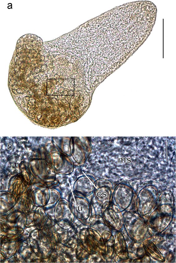
1052                                                                                                 Parasitol Res (2018) 117:1051–1068

Malkova 1993, 1994, 1995; Galaktionov et al. 1996;                  molecular methods (Blasco-Costa and Poulin 2017).
Galaktionov 1991a, 1993; Galaktionov and Dobrovolskij               Besides, the accumulation of molecular data on trematodes
2003). The metacercariae develops a complete, but yet non-          from the North Pacific and the North Atlantic offers tempting
functioning, reproductive system. Thus, the adult can start egg     possibilities for phylogeographic reconstructions. They will
production as early as few hours after the infection of the final   make it possible to elucidate the pathways of geographic ex-
host, dispersing eggs in the same biotope where the host has        pansion of the parasites to the north of the Palaearctic in the
been infected (Saville and Irwin 1991; Galaktionov 1993;            past and to forecast the impact of the ongoing climate changes
Field et al. 1998). The lifespan of adults in the host is short     on this process (Galaktionov 2017).
(5–10 days), which is due to their extreme juvenilisation and          This study continues the series of our studies of biodiver-
miniaturisation (reviewed in Belopol’skaya 1983;                    sity and life cycles of trematodes circulating in the coastal
Galaktionov 1993; Galaktionov and Dobrovolskij 2003).               ecosystems of the Sea of Okhotsk employing classical and
However, it is sufficient for the worms to complete their life      molecular-biological approaches (Galaktionov et al. 2012;
cycle at stopovers of migrating birds, minimising losses due to     Gonchar and Galaktionov 2017). Herein, we describe a new
their removal with the bird hosts outside the region of possible    microphallid species with a truncated two-host life cycle and
transmission (Belopol’skaya 1983; Galaktionov 1993).                an unusual structure of the terminal genitalia in adults.
   In general, three-host (trixenous) life cycles are typical of
trematodes. However, several phylogenetic lineages of
microphallids have independently evolved a truncation of trans-     Materials and methods
mission pathways resulting in two-host (dixenous) life cycles.
This means that cercariae do not leave the molluscan host. They     Material collection and treatment
transform (encysting or not encysting) into metacercariae infec-
tive for the final host directly in the mollusc (Belopol’skaya      The material was collected at the southern coast of the
1963; Deblock 1977, 1980; Galaktionov 1991b; Montoliu               Pyagina Peninsula, the Sea of Okhotsk (North Pacific) in
et al. 1992; Galaktionov and Skírnisson 2007).                      the Vnutrenn’aya Bay and the Shkiperov Bay in July–
   Microphallids are a dominant group of digeneans in the           August 2008 and 2012 (Table 1). Gastropod molluscs
coastal waters of Palaearctic seas, demonstrating the greatest      Falsicingula kurilensis (Pilsbry, 1905) were collected in
species diversity and the highest infection levels in final and     the intertidal and the upper subtidal zone. Pacific common
intermediate hosts (Galaktionov 1993). Nevertheless, while in       eiders Somateria mollissima v-nigrum Bonaparte, 1855 were
Atlantic seas their fauna and life cycles have been extensively     obtained by shooting in accordance with local regulations.
investigated (e.g. Reimer 1963; James 1968, 1969; Deblock           The molluscs were dissected under a stereomicroscope to
1980; Lauckner 1983; Galaktionov 1983, 1984, 1988, 1989;            identify those infected with sporocysts containing
Montoliu et al. 1992), similar studies in the North Pacific are     metacercariae of the undescribed species. Metacercariae
scarce. Until recently, the only such studies on Asian coasts       were excysted with the help of preparation needles. Some
have been those by Tsimbalyuk and colleagues dating back to         of them were studied in vivo, while others were fixed in
the 1960–1970s (Tsimbalyuk et al. 1968a, b, 1978; Kulikov           70% ethanol under a slight pressure of a coverslip. Eiders
et al. 1970; Pois et al. 1974). Ching (reviewed in Ching 1991)      were dissected in the field, and the microphallid individuals
carried out her research at approximately the same time at the      with noticeable spicule-like structure in the genital region
coast of the British Columbia. There are just a handful of more     (see BResults^) and individuals of Levinseniella sp. (identi-
recent studies on digeneans circulating in the coastal waters of    fied in accordance with Deblock (2008) by presence of sev-
the North Pacific (Miura et al. 2005; Galaktionov 2007;             eral accessory atrial sacs of type I associated with one sac of
Galaktionov et al. 2010, 2012). This is all the more regrettable,   type II) were selected from all the adult microphallids in their
as the biodiversity of digeneans in this region is high.            intestine under a stereomicroscope. These adults were fixed
Furthermore, the centres of origin of several taxa, including       in 70% ethanol following the same procedure as in the case of
the Microphallidae, are hypothesised to be associated with this     metacercariae. Samples of metacercariae and adults were
region (Belopol’skaya 1983). From there, they may have ex-          stored in 70 and 95% ethanol for further morphological and
panded into the North Atlantic after the opening of the Bering      molecular analysis correspondingly.
Strait in late Pliocene and the following mass flow of the             Carmine-stained whole mounts were used for morpholog-
Pacific marine fauna into the Atlantic (Belopol’skaya 1983;         ical studies and to make drawings. Measurements were made
Hoberg and Adams 2000; Galaktionov et al. 2012). In the             on 17 metacercariae and 22 adults. Additionally, one of the
light of this, studies on biodiversity and movements of             adults fixed in 70% ethanol was used to make a temporary
digeneans in the North Pacific remain a promising area of           preparation in a mixture of glycerine and lactic acid (10:3).
research with an enduring value, while the elucidation of life      This medium, which is used for clarification of nematodes,
cycles has become considerably easier with the advance of           also yields satisfactory results for the morphological analysis

Parasitol Res (2018) 117:1051–1068                                                                                         1053

Table 1 Taxa included in the
phylogenetic analyses and            Classification/species                      GenBank accession numbers
GenBank accession codes for
each sequence. Accession codes                                                   28S rDNA                    ITS2
in bold correspond to the newly
sequenced specimens                  Microphalloidea
                                       Lecithodendriidae
                                         Paralecithodendrium parvouterus         AY220617
                                          Pycnoporus heteroporus                 AF151918
                                     Microphallidae
                                      Longiductotrema tethepae                   KX712084                    KX712086
                                       Maritrema arenaria                        AY220629                    HM584171
                                       Maritrema brevisacciferum                 KT355818                    KT355824
                                       Maritrema corai                           KT880222
                                       Maritrema deblocki                        KJ144173
                                       Maritrema eroliae                         JF826247                    HQ650132
                                       Maritrema heardi                          AY220632
                                       Maritrema madrynense                                                  KF575167
                                       Maritrema neomi                           AF151927
                                       Maritrema novaezealandense                KJ144178                    KJ540203
                                       Maritrema oocysta                         AY220630                    HM584170
                                       Maritrema poulini                         KJ144175
                                       Maritrema prosthometra                    AY220631
                                       Maritrema subdolum                        AY151926                    HM584172
                                       Maritrema sp.                                                         KC222023–KC222024
                                       Maritrema sp. 1                                                       KC012521
                                       Maritrema sp. 2                                                       KC222022
                                       Microphallus abortivus                    AY220626                    HM5841731
                                       Microphallus basodactylophallus           AY220628
                                       Microphallus calidris                     HM584125                    HM5841831
                                       Microphallus fusiformis                   AY220633
                                       Microphallus kurilensis                   HM584140                    HM5841851
                                       Microphallus minutus                      KT355822                    KT355828
                                       Microphallus ochotensis sp. nov., adult   MG783586–MG783587           MG783581–MG783582
                                       Mi. ochotensis sp. nov., metacercaria     MG783588–MG783589           MG783583–MG783584
                                       Microphallus piriformes                   HM584122                    HM5841811
                                       Microphallus primas                       AY220627
                                       Microphallus pseudopygmaeus               HM584126                    HM5841981
                                       Microphallus pygmaeus                     HM584133                    HM5841901
                                       Microphallus similis                      HM584138                    HM5841781
                                       Microphallus triangulatus                 HM584139                    HM5841951
                                       Microphallus sp.                          HM584142                    HM5841751
                                       Probolocoryphe uca                                                    GQ377842
                                       Microphallidae gen. sp.                   KT355820                    KT355826
                                       Levinseniella sp.                         MG783585                    MG783580
                                     Pleurogenidae
                                       Parabascus duboisi                        AY220618
                                       Pleurogenes claviger                      AF151925
                                     Prosthogonimidae
                                       Prosthogonimus ovatus                     AF151928
                                     Plagiorchioidea
                                       Haematoloechidae
                                          Haematoloechus longiplexus             AF387801
1054 Parasitol Res (2018) 117:1051–1068
Table 1 (continued)
Classification/species GenBank accession numbers
28S rDNA ITS2
Plagiorchiidae
Plagiorchis vespertilionis AF151931
Telorchiidae
Telorchis assula AF151915
of trematodes. Three metacercariae fixed in 70% ethanol were concentrations) and 0.4 μM of each primer combination.
prefixed in Bouin fluid and analysed histologically. Then, they Thermocycling conditions used for amplification of the
were placed in paraffin wax after dehydration, according to rDNA regions followed Galaktionov et al. (2012). PCR
the standard technique and used to make longitudinal serial amplicons were purified prior to sequencing using exonucle-
sections 5 μm thick with the help of a microtome (Leica ase I and shrimp alkaline phosphatase enzymes (Werle et al.
RM2245). The sections were stained with Bömer 1994). Amplicons were cycle-sequenced from both strands
haematoxylin. The morphology of metacercariae and adults using PCR primers and an internal primer for the 28S frag-
was studied in vivo in the field using an Amplival microscope ment (L1200R: 5′-GCA TAG TTC ACC ATC TTT CGG-3′;
(Karl Zeiss, Jena). Microphotographs were made with the help Littlewood et al. 2000) at the commercial facility Macrogen
of an amateur Sony Cyber-shot camera DSC-W100 connected (Amsterdam, The Netherlands). Contiguous sequences were
to the eyepiece of the Amplival microscope. Stained total assembled and edited using Geneious® (v. 8.1 Biomatters
mounts of metacercariae and adults and sections of Ltd., Auckland, New Zealand) and submitted to GenBank
metacercariae were studied in the laboratory of the Zoological (see accession numbers in Table 1).
Institute under the Olympus CH40 compound microscope
equipped with an Olympus XC-30 digital camera. All measure-
ments presented in the paper are in micrometres, with the mean Molecular analyses
in parentheses. Drawings were made with the aid of a camera
lucida. Five newly generated sequences for the 28S rDNA and the
ITS2 fragments were aligned in two independent datasets to-
Molecular data generation gether with the published sequences of other microphallids
from GenBank (see accession numbers in Figs. 7 and 8 and
One adult specimen of Levinseniella sp. and two adult Table 1). The sequences were aligned using default parame-
microphallid specimens with noticeable spicule-like structures ters of MAFFT implemented in Guidance (Sela et al. 2015),
in the genital region from Pacific common eider, So. and the extremes of the alignment were trimmed to match the
mollissima v-nigrum, and two metacercariae specimens from shortest sequences. The 28S dataset (1281 bp long) included
individual molluscs, F. kurilensis, were characterised molecu- 13 representative sequences of Microphallus spp., 12 of
larly (Table 1). Genomic DNA was extracted from ethanol- Maritrema spp., one of Levinseniella and one of
fixed isolates in 200 μl of a 5% suspension of Chelex® in Longiductotrema retrieved from GenBank (Table 1).
deionised water and containing 0.1 mg/ml proteinase K, Additionally, an unidentified microphallid; five sequences of
followed by incubation at 56 °C for 5 h, boiling at 90 °C for species belonging to sister families of the Microphallidae, i.e.
8 min and centrifugation at 14,000g for 10 min. Partial frag- Lecithodendriidae, Pleurogenidae and Prosthogonimidae in
ments of the ribosomal RNA gene were amplified using the the Microphalloidea; and three sequences of species in the
following primers: the large ribosomal subunit (28S) Plagiorchioidea were retrieved from GenBank and included
[1800 bp; primers U178F: 5′-GCA CCC GCT GAA YTT as outgroups. The ITS2 dataset (394 bp long) included ten
AAG-3′ and L1642R: 5′-CCA GCG CCA TCC ATT TTC representative sequences of Microphallus spp.; 11 sequences
A-3′; Lockyer et al. 2003] and the internal transcribed spacer of Maritrema; one of Levinseniella, Probolocoryphe and
2 (ITS2) [500 bp; primers 3S: 5′-GTA CCG GTG GAT CAC Longiductotrema; and one of an unidentified microphallid.
GTG GCT AGT G-3′ and ITS2·2: 5′-CCT GGT TAG TTT The phylogenetic analyses were run on the two datasets indi-
CTT TTC CTC CGC-3′]. Polymerase chain reaction (PCR) vidually under the maximum likelihood (ML) and Bayesian
amplifications were performed in 20 μl reactions containing inference (BI) criteria, employing the nucleotide substitution
2 μl of extraction supernatant (~ 10–20 ng of template DNA), model GTR+Γ. ML analyses were conducted using the pro-
2× MyFi™ Mix (Bioline France, France; containing DNA gram RAxML v. 7.3 (Stamatakis 2006; Stamatakis et al.
Polymerase, dNTPs, MgCl 2 and enhancers at optimal 2008). All model parameters and bootstrap nodal supportParasitol Res (2018) 117:1051–1068 1055
values (1000 repetitions) were estimated using RAxML. BI paratypes (MHNG-PLAT-99644) and 5 metacercariae
trees were constructed using MrBayes v. 3.2 (Ronquist et al. vouchers (MHNG-PLAT-99645) deposited in the Muséum
2012), running two independent MCMC runs of four chains d’Histoire Naturelle de Genève (Geneva, Switzerland).
for 107 generations and sampling tree topologies every 103 Representative DNA sequences in GenBank: adults
generation. Burn-in periods were set automatically to 25% MG78 358 1–M G7 835 82; MG783 586 –MG78 358 7,
generations ensuring the remaining trees were obtained after metacercariae MG783583–MG783584; MG783588–
values for standard deviation of split frequencies were < 0.01. MG783589.
A consensus topology and nodal support estimated as poste- ZooBank registration: To comply with the regulations set
rior probability values (Huelsenbeck et al. 2001) were calcu- out in article 8.5 of the amended 2012 version of the
lated from the remaining trees. All MrBayes and RAxML International Code of Zoological Nomenclature, details of
analyses were performed on the computational resource the new species have been submitted to ZooBank. The Life
CIPRES (Miller et al. 2010). Genetic divergences amongst Science Identifier (LSID) for Mi. ochotensis nov. sp. is
taxa were calculated as uncorrected p-distances for each gene urn:lsid:zoobank.org:act:DC77B715-755D-4C4B-B621-
region using MEGA v. 6 (Tamura et al. 2011). EDC44BE712C2.
Etymology: The species was named after the site where it
was discovered, the Sea of Okhotsk.
Results
Molecular characterisation of adult microphallids with
spicule-like structures in the genital region and the Adult
metacercariae ex. F. kurilensis allowed us to link the two life
stages (see BMolecular results^ for details) of the new species. [Measurements based on whole mounts of 22 specimens
We provide a formal morphological description for both the (Figs. 1, 2 and 3).] Worms pyriform, with powerful postero-
adult and the metacercaria, below. lateral projections filled with glands (Fig. 1a). Body 330–
529 (369) long. Body width at mid-level of oesophagus
100–186 (129) and at level of testes 200–429 (267). Body
Description covered with spines, larger in anterior region. Oral sucker
24–36 × 29–43 (31 × 36), somewhat shorter than ventral
Family Microphallidae Ward, 1901 sucker 29–42 × 28–43 (35 × 35), the oral sucker width/
Supersubfamily Microphallidi Ward, 1901 ventral sucker width ratio is approx. 1:1. Prepharynx when
Subfamily Microphallinae Ward, 1901 visible very short, 4–11 (6). Pharynx oval, 14–25 (21) × 14–
Tribe Microphallini Ward, 1901 22 (19). Oesophagus length greatly variable, 72–194 (106),
Genus Microphallus Ward, 1901 mainly resulting from varying contraction condition of
Microphallus ochotensis sp. nov. fixed worms.
Type-final host: Pacific common eider Somateria Testes irregular to oval, 43–97 (62) × 25–54 (38). Seminal
mollissima v-nigrum Bonaparte, 1855 (Anatidae). vesicle and prostatic glands enclosed in a thin-walled mem-
Type-locality: Shelikhov Bay and bays of Pyagina brane-like structure. Seminal vesicle highly variable de-
Peninsula (north of the Sea of Okhotsk). pending on degree of filling with sperm (Fig. 2), 25–94
First intermediate host: Falsicingula kurilensis (Pilsbry, (54) × 29–72 (39). Prostatic part well developed, the ducts
1905) (Caenogastropoda, Falsicingulidae). of prostatic cells enveloping distal part of seminal vesicle and
Site in host: Final host: intestine; first intermediate host: ejaculatory duct and opening into ejaculatory duct at phallus
hepatopancreas and gonad. base. Ducts and pores of prostatic cells clearly visible in live
Prevalence: So. mollissima v-nigrum: in 4 of 6 dissected specimens serving as good diagnostic character during pre-
birds (male, female and 2 ducklings). liminary examination of material in the field. Phallus some-
F. kurilensis: Vnutrenn’aya Bay (entrance 3.3%, N = 30; what elongated along transverse axis, 17–25 (21) × 14–25
Chayachiy islet 2.2%, N = 186), Shkiperov Bay 4.6%, N = 87. (18). Genital atrium muscular, genital pore sinistral to ventral
Intensity in So. mollissima v-nigrum: varied from tens to sucker (Fig. 1b).
thousand. Ovary dextral to ventral sucker, adjacent to or slightly over-
Type-material: Holotype (slide # 3714-1), 21 paratypes lapping ventral sucker laterally, transversely elongate, approx-
(slides #3714-2–3714-5) and 17 metacercariae vouchers imately same size as testes, 36–101 (60) × 25–68 (38). Uterus
(slides # 3715-1–3715-4) deposited in the Collection of in hindbody, reaching to level of ventral sucker, often over-
helminthes, section Trematoda in Zoological Institute of the lapping testes ventrally. Metraterm complex, consisting of two
Russian Academy of Sciences (Saint Petersburg); four structurally different parts (Fig. 1b).1056 Parasitol Res (2018) 117:1051–1068
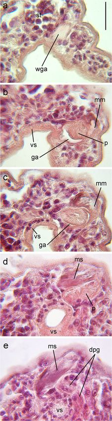
Fig. 2 Microphotograph of living adult of Microphallus ochotensis
(dorsal view), bar 100 μm. mls membrane-like structure, pg prostatic
gland cells, o ovary, s spicule-like structures, sv seminal vesicle, t testis
Metacercaria
[Measurements based on whole mounts of 17 specimens
(Figs. 4, 5 and 6).] Fully formed metacercariae enclosed in oval
cysts (Fig. 4a), 132–182 (160) × 119–172 (145). Larvae ex-
tracted from cysts tongue-shaped, with marked posterolateral
projections (Fig. 5a). Body length 157–257 (209); body width
Fig. 1 Adult of Microphallus ochotensis (ventral view). a General 86–129 (106) at mid-oesophagus level and 100–172 (140) at
morphology, bar 100 μm. b Terminal genitalia, bar 20 μm. dpg ducts testes level. Oval oral sucker 20–33 × 23–38 (28 × 31), some-
of prostatic gland cells, ga genital atrium, gp genital pore, mm muscular
part of metraterm, ms metratermal sac, p phallus, s spicule-like structures, what greater than round ventral sucker 20–28 × 20–28 (25 ×
u uterus 26), oral sucker width/ventral sucker width ratio is approx.
1.2:1. Prepharynx not present, pharynx oval 14–21 (18) × 10–
17 (14), oesophagus length 28–56 (42). Ducts of gland cells,
Distal muscular part of metraterm opening into sinistral arranged in 3 longitudinal rows on ventral and dorsal planes,
wall of genital atrium by broad aperture. Proximal part, opening at oral sucker margin and on forebody. Nucleus-
connecting with uterus (Fig. 3), forms large sac-like exten- containing cell bodies of gland cells concentrated in body re-
sion, which we refer to as metratermal sac, 31–76 (48) × 11– gions anterior to intestinal caeca. Agglomeration of gland cells
32 (21), dorsal and somewhat posterior to genital atrium. forms posterolateral body projections and opens at the tip of
Spicule-like structures arranged into two longitudinal bun- projection. Flame-cell formula 2[(2 + 2) + (2 + 2)] = 16.
dles visible in metratermal sac of fully formed adults (Fig. 1b Testes oval, 25–59 (38) × 15–38 (24), left testis often locat-
and 2). Pointed ends of spicules entering muscular part of ed posteriorly to the right one. Seminal vesicle rather small,
metraterm, sometimes extending into genital atrium. 15–28 (21) × 13–21 (17). Seminal vesicle and prostatic glands
Vitellarium represented by two compact groups of 3–6 folli- enclosed in thin-walled membrane-like structure. Prostatic
cles posterior to testes. Eggs small, 14–22 (19) × 7–14 (10). part well developed, prostatic cells agglomerated near distalParasitol Res (2018) 117:1051–1068 1057
clearly visible in live metacercariae (Fig. 4a, b). Phallus some-
what elongated along transverse axis, 14–21 (18) × 8–17 (13),
accommodated in muscular genital atrium.
Ovary subtriangular, 18–34 (26) × 13–21 (17). Metratermal
sac, 29–50 (38) × 11–28 (19), localised dorsally and somewhat
posteriorly of genital atrium (Figs. 5b and 6). Posterior shift of
left testis (see above) associated with position of metratermal
sac. Longitudinal cords of amorphous fibrous material (devel-
oping spicule-like structures) visible in metratermal sac (Fig. 6).
Cords tapering distally and entering muscular distal part of
metraterm opening into sinistral wall of genital atrium with
broad aperture (Fig. 4b and 6). Vitellarium forms two compact
groups of 3–5 follicles behind testes.
Hosts
Adults of Mi. ochotensis were found in most eiders examined
from the Sea of Okhotsk. The highest infection intensity
(more than 1000 individuals) was recorded in a duckling and
a female from the Shkiperov Bay, and single individuals of
Mi. ochotensis (identified based on molecular markers—see
above) were found in a male from the area of Cape Taygonos.
The prevalence of Mi. ochotensis sporocysts in molluscs
F. kurilensis examined in the Shkiperov Bay and the
Vnutrenn’aya Bay was low at 3–5%. Mature encysted
metacercariae were enclosed in thin-walled sporocysts. When
molluscs are dissected, the body wall of sporocysts bursts and
metacercariae pour out, creating a false impression that the
Fig. 3 Microphotographs of an adult of Microphallus ochotensis placed encysted larvae lie freely in the molluscan tissues. In some
on a temporary mount in a mixture of lactic acid and glycerine. a General sporocysts, earlier developmental stages were found alongside
view, boxed area—body part enlarged in b, bar 100 μm. b Body part at mature metacercariae. These developing larvae had a reduced
the site of connection between the metratermal sac and the uterus, bar
10 μm. cs connection site of the metratermal sac with the uterus; ms tail. No stylet nor penetration glands were seen in them.
metratermal sac, u uterus
part of seminal vesicle and around proximal part of ejaculatory Molecular results
duct. Ducts of prostatic cells enveloping distal part of ejacu-
latory duct opening into it at the phallus base (Fig. 5b). The two adults with spicule-like structures in the genital re-
Expanded distal parts of ducts of prostatic cells and pores gion and the two metacercariae isolates ex. F. kurilensis
Fig. 4 Microphotographs of
living metacercaria of
Microphallus ochotensis. a
Encysted metacercaria, bar
50 μm. b The body area of
metacercaria strongly pressed by
cover glass showing terminal
genitalia, bar 20 μm. ddpg ducts
of prostatic glands (distal parts),
ds developing spicule-like struc-
tures, ga genital atrium, mm
muscular part of metraterm, ms
metratermal sac, p phallus, pg
prostatic gland cells1058 Parasitol Res (2018) 117:1051–1068 Fig. 5 Metacercaria of Microphallus ochotensis (ventral view). a General morphology, bar 100 μm. b Terminal genitalia, bar 20 μm. ds developing spicule-like structures, dpg ducts of prostatic gland cells, ga genital atri- um, gp genital pore, mls membrane-like structure, mm muscular part of metraterm, ms metratermal sac, sv seminal vesicle, p phallus, pg prostatic gland cells, u uterus
Parasitol Res (2018) 117:1051–1068 1059
Fig. 6 A series of longitudinal sections (5 μm) (a–e) in the frontal plane rDNA fragment, whereas our specimens showed a genetic
(from the ventral side towards the dorsal) through the genital part of the divergence of 5.1–7.4% from other Microphallus spp. The
body of Microphallus ochotensis metacercaria, bar 20 μm. dpg ducts of
prostatic gland cells, ga genital atrium, mm muscular part of metraterm,
lowest genetic divergence was found between Microphallus
ms metratermal sac, p phallus, st sinistral testis; vs ventral sucker, wga pygmaeus (Levinsen, 1881) and Microphallus kurilensis
wall of genital atrium Galaktionov et al., 2010, and between Microphallus calidris
Belopol’skaja & Ryjikov, 1963 and Microphallus minutus
characterised molecularly showed identical sequences for Johnston, 1948, and the highest between Microphallus
each of the 28S and ITS2 rDNA regions sequenced (Figs. 7 abortivus Deblock, 1974 and Microphallus pseudopygmaeus
and 8), which allowed us to match the two life cycle stages of Galaktionov, 2009. The range of interspecific genetic diver-
the new species. Their sequences also matched with 100% gence amongst Microphallus spp. was similar to that amongst
identity to that of a previously sequenced adult from the same Maritrema spp. (0.6–9.2%). Average intergeneric divergence
host in Cape Taygonos, north Sea of Okhotsk, Russia amongst microphallids (Microphallus, Maritrema,
(GenBank accession codes HM584142, HM584175; Levinseniella and Longiductotrema) ranged from 7.0 to
Galaktionov et al. 2012). Interspecific genetic divergences 10.3% in the 28S rDNA region. The sequence of an uniden-
within Microphallus ranged from 0.4 to 8.6% in the 28S tified microphallid (KT355820) and Microphallus fusiformis
1/100
Plagiorchis vespertilionis AF151931
Telorchis assula AF151915
Haematoloechus longiplexus AF387801
1/100 Paralecithodendrium parvouterus AY220617
Pycnoporus heteroporus AF151918
1/92 Prosthogonimus ovatus AF151928
0.99/96 Parabascus duboisi AY220618
1/100 Pleurogenes claviger AF151925
L. tethepae KX712084 Longiductotrema
M.subdolum AY151926 M
1/100 1/100 M. arenaria AY220629 i
0.98/78
M. prosthometra AY220631 c
1/88 M. oocysta AY220630 r
0.98/70 M. neomi AF151927 o
1/83
1/96
M. deblocki KJ144173 Maritrema p
M. brevisacciferum KT355818 h
-/66
1/71
M. poulini KJ144175 a
M. corai KT880222
1/88
M. eroliae JF826247
M l
1/100
-/78 M. heardi AY220632 i l
M. novaezealandense KJ144178 c o
Levinseniella sp. Levinseniella r i
1/95 M. fusiformis AY220633 o d
Microphallidae gen. sp. KT355820 p e
0.99/77 M. basodactylophallus AY220628 h a
1/96 1/100 Microphallus sp. HM584142 ex. S. mollissima
M. ochotensis sp. nov. ex. S. mollissima & F. kurilensis
a
1/100 M. abortivus AY220626
l
1/93
M. primas AY220627 l
M. similis AY220625 i
0.99/92 Microphallus
M. minutus KT355822 d
1/98
M. piriformes HM584122 a
M. calidris HM584125 e
0.91/77 M. kurilensis HM584140
1/99
0.03 M. pygmaeus HM584133
substitutions/site 1/98 M. pseudopygmaeus HM584126
M. triangulatus HM584139
Fig. 7 Phylogenetic tree based on Bayesian inference and maximum (PP) followed by maximum likelihood bootstrap support (BS) percent-
likelihood analyses of the partial 28S rDNA dataset showing the ages (PP < 90 and BS < 60 not shown). GenBank accession codes for
relationships between the new species and other microphallids. Values sequences and host species names of the sequences of the new species
above the branches represent Bayesian inference posterior probabilities in grey1060 Parasitol Res (2018) 117:1051–1068
Longiductotrema tethepae KX712086
Maritrema sp. 1 KC012521 Gilardoni et al. 2011
Probolocoryphe uca GQ377842
0.98/97 Maritrema arenaria HM584171
Maritrema subdolum HM584172
1/- Maritrema brevisacciferum KT355824
0.99/81 -/64
Maritrema oocysta HM584170
0.99/92
Maritrema eroliae HQ650132
1/96 Maritrema sp. 2 KC222022 Gilardoni et al. 2011
0.96/81
0.99/93 Maritrema novaezealandense KJ540203
-/72
Maritrema sp. KC222023 Diaz & Cremonte, 2010
1/100
Maritrema madrynense KF575167
Maritrema sp. KC222024 Gilardoni et al. unpublished
Levinseniella sp.
Microphallidae gen. sp. KT355826
0.99/87 Microphallus similis HM5841781
Microphallus abortivus HM5841731
1/91 isolate 45_1 ex. Somateria mollissima
1/100
Microphallus sp. HM5841751 ex. Somateria mollissima
isolate Ex45_9 ex. Falsicingula kurilensis Microphallus
ochotensis sp. nov.
0.9/- isolate Ex45_4 ex. Somateria mollissima
isolate Ex45_10 ex. Falsicingula kurilensis
Microphallus pseudopygmaeus HM5841981
0.98/-
Microphallus minutus KT355828
Microphallus calidris HM5841831
0.98/63
Microphallus triangulatus HM5841951
Microphallus piriformes HM5841811
0.04
substitutions/site Microphallus pygmaeus HM5841901
1/96
Microphallus kurilensis HM5841851
Fig. 8 Phylogenetic tree based on Bayesian inference and maximum (PP < 90 and BS < 60 not shown). GenBank accession codes for se-
likelihood analyses of the ITS2 dataset showing the relationships quences and host species names of the sequences of the new species in
between the new species and other microphallid genera. Values above grey. Unidentified species names are followed by the name of the authors
the branches represent Bayesian inference posterior probabilities (PP) of the sequence after the GenBank code
followed by maximum likelihood bootstrap support (BS) percentages
Reimer, 1963 showed an average genetic divergence as high to be identical to Maritrema madrynense Deblock &
or higher than that observed between genera (on the average Rausch, 1968. Maritrema sp. 1 Gilardoni et al., 2011
10.5–12.7%). The genetic divergence between the unidenti- (KC012521) was the most divergent Maritrema species
fied microphallid and Mi. fusiformis was also high (10.5%). (20.6–23.8%). Average intergeneric genetic divergence be-
ITS2 genetic distances varied within the range 1.0–10.1% tween representative sequences of species of microphallid
between Microphallus spp. and 1.1–23.8% between genera ranged from 13.8 to 21.0% in the ITS2 region.
Maritrema spp. Sequences of Mi. ochotensis sp. nov. diverged Thus, Maritrema sp. 1 mentioned above showed a diver-
8.5–10.0% from all other Microphallus spp. Two unidentified gence similar to that expected between genera within the
sequences of Maritrema (KC222024 Gilardoni et al. unpub- Microphallidae.
lished and KC222023 Diaz & Cremonte 2010) were foundParasitol Res (2018) 117:1051–1068 1061
The BI and ML phylogenies based on the 28S rDNA region metraterm is well developed in all adults of Microphallus, no
were congruent and depicted the Microphallidae as monophy- such structure has ever been described before (Belopol’skaya
letic (Fig. 7), as well as the genera Maritrema and Microphallus 1952a, 1963; Deblock 1971, 2008). In particular, it is absent in
(the sequence of Mi. fusiformis should be disregarded from the adults of Mi. abortivus and Mi. primas (syn. Мicrophallus
Microphallus, see Kudlai et al. (2016) and the distance results canchei Biguet et al., 1958), the two species with which Mi.
above). However, support for the relationships amongst the ochotensis forms a clade in the 28S tree. It is also absent in Mi.
genera within the family was weak and resulted in the labile basodactylophallus, which is closely related to the aforemen-
placement of Longiductotrema as sister to Maritrema (BI tree) tioned species in the 28S phylogeny (Biguet et al. 1958;
or sister to the Microphallus + (Levinseniella sp. + Bridgman 1969; Deblock 1974). In other respects, the struc-
Microphallidae gen. sp. + Mi. fusiformis) clade (ML tree). Mi. ture of adults of Mi. ochotensis corresponds to that of other
ochotensis sp. nov. matched the sequence of Microphallus sp. Microphallus species. In Mi. primas, the metraterm is separat-
(HM584142; ex. So. mollissima) in the phylogenetic tree. The ed into two parts: a transitional (proximal) part lined with
new species fell within a strongly supported clade including papillae and a muscular terminal (distal) part connected to
Microphallus basodactylophallus (Bridgman, 1969), parasitic the genital atrium (Biguet et al. 1958; Deblock 1971).
in raccoons and rats, Microphallus primas (Jägerskiöld, 1908) However, these parts are developed much less strongly than
and Mi. abortivus, parasitic in birds. However, the relationships in Mi. ochotensis, and neither spicule-like structures nor lon-
amongst the species within this clade were unsupported. The gitudinal cords of fibrous material are present in the proximal
ITS2 phylogenies were also mostly congruent, except for the part.
placement of Probolocoryphe uca (Sarkisian, 1957) that either A powerful metraterm is characteristic of some species of
diverged earlier from Maritrema or was found within the Megalophallus (Cable et al. 1960; Prévot and Deblock 1970;
Maritrema clade as sister to Maritrema gratiosum Nicoll, Prévot 1974; Overstreet and Heard 1995). In some respects, it
1907 (syn. Maritrema arenaria Hadley & Castle, 1940) (Fig. is similar to the metraterm of Mi. ochotensis: it is also long,
8). The monophyly of Maritrema and Microphallus was broad and muscular and it also passes around the genital atri-
weakly supported. Maritrema sp. 1 of Gilardoni et al. (2011) um dorsally opening into the sinistral wall of the genital atri-
(KC012521) appeared as early divergent within the tree, not um. In contrast to Mi. ochotensis, Megalophallus spp. have no
closely related to other Maritrema spp., as noted above on the metratermal sac with spicule-like structures. However, in
basis of the high genetic divergence. The sequence of Megalophallus deblocki Kostadinova et al., 2006, the extend-
Levinseniella sp., a representative of the subfamily ed terminal portion of the metraterm forms two indistinct
Levinseniellinae, appeared as early divergent from the lobes covered with minute spines (Kostadinova et al. 2006).
Microphallidae gen. sp. and Microphallus clade. Mi. ochotensis Megalophallus spp. differ from all species of the genus
sp. nov. was depicted sister to Mi. abortivus within the Microphallus, including Mi. ochotensis, in the structure of
Microphallus clade. Thus, genetic data for both the 28S and the phallus, which is ornamented by several prominent
the ITS2 regions of the rRNA gene corroborated the morpho- structures.
logical distinctiveness of Mi. ochotensis sp. nov. and supported Amongst other microphallid genera, a structure superficial-
its distinct species status. ly similar to that in Mi. ochotensis is found in Spiculotrema
littorale Belopol’skaya, 1949 (subfamily Levinseniellinae
Stiles & Hassal, 1901, tribe Ascorhytini Deblock, 1971)
Remarks (Belopol’skaya 1949, 1952a; Deblock 2008). This large (by
the standards of microphallids) worm, 0.552–0.951 × 0.337–
Molecular data indicate clearly that Mi. ochotensis sp. nov. 0.410, parasitises shorebirds in Primorye (Russian Far East)
should be included in the genus Microphallus (Figs. 7 and (Belopol’skaya 1949, 1954; Tsimbalyuk et al. 1968b, 1978).
8). Its morphological description corresponds to the diagnostic Sp. littorale has an accessory sac connected to the genital
characters of the Microphallini and of the genus Microphallus atrium (type IV atrial sac according to Deblock (2008)). The
within this tribe (Deblock 1971, 2008). In particular, the adult sac contains a large sclerotised plate, pyriform in dorsoventral
and metacercaria described above possess a fleshly, muscular view and long spicule-like in profile (Deblock 2008).
phallus of Bmicrophalloid^ type, morphologically simple, According to Belopol’skaya (1949), this structure serves as
smaller than the ventral sucker; a simple genital atrium an organ of stimulation during copulation. The metraterm in
enveloping the phallus and the metraterm opening in the gen- Sp. littorale opens into the genital atrium independently. The
ital atrium is sinistral. The species described in this paper modification of the metraterm described in Mi. ochotensis is
differs from other known species of the genus Microphallus, probably also involved in copulation. Besides stimulation, it
first of all, in the structure of the metraterm, which consists of may serve for retention of the partner’s phallus in the
two distinct parts: a sac with spicule-like structures and a metraterm, which serves as vagina in trematodes.
muscular duct opening into the genital atrium. Although the1062 Parasitol Res (2018) 117:1051–1068
One character of the new species seems to be at variance Spiculotrema. However, the resemblance of the complex
with the classical diagnosis of the genus Microphallus and metraterm of Mi. ochotensis to type IV atrial sac is purely
Microphallini on the whole (Belopol’skaya 1963; Deblock superficial. These structures cannot correspond to each other
1971, 2008): the seminal vesicle and the prostatic part do for three reasons: (1) the connection of the metratermal sac to
not lie freely in the parenchyma but are enclosed in a fine the uterus, (2) its position in the worm’s body and (3) the
membranous envelope. This structure, which was also found position of the new species on the molecular tree. The con-
in other representatives of Microphallus, is a reduced male nection of the uterus with the metratermal sac is clearly seen in
genital pouch (see BDiscussion^ for details). live metacercariae and adult worms as well as in worms placed
In conclusion, we would like to note that, in contrast to into a mixture of glycerine and lactic acid (Fig. 3). In whole
many other microphallids, metacercariae and adults of Mi. mounts, the transition of the metratermal sac into the uterus
ochotensis are easy to identify. In live encysted metacercariae, proper is difficult to observe.
the distended distal parts of the ducts of the prostatic glands The large atrial sac in Sp. littorale is located sinistral to the
are clearly visible. They are arranged in a dense bundle and genital atrium. In Mi. ochotensis, the metratermal sac bearing
open into the prostatic chamber (Fig. 4a). The distended part longitudinal cords of amorphous fibrous material (in
of the metraterm (the metratermal sac) can be easily seen in metacercariae) or spicule-like structures (in fully formed
live metacercariae and adults (Fig. 4b) expanded under slight adults) lies dorsally of the genital atrium, either almost per-
pressure of a coverslip in a drop of water as well as in mounted pendicularly or at an acute angle to the longitudinal body axis.
specimens; in fully formed adults, the spicule-like structures Its distal part gradually becomes the muscular part, which
are also clearly visible (Fig. 2). These distinct diagnostic char- bends ventrally and opens into the sinistral wall of genital
acters allow a trouble-free identification of metacercariae and atrium. This position of the metraterm in the worm’s body is
adults even in the field, so that whole mounts in Canada bal- characteristic of Microphallini (see Deblock 2008).
sam are unnecessary. It should be noted that amongst Microphallini, consider-
able structural modifications of the metraterm are recorded
in Megalophallus spp. (see BRemarks^). However,
Discussion Kostadinova et al. (2006) in a detailed study of Me. deblocki
have shown that the metraterm in this species is short and
We described a new microphallid species, Mi. ochotensis, with tubular rather than enlarged and muscular, as in other species
a unique structure of the metraterm. No such metraterm struc- of the genus. At the same time, they found a large folded
ture has been described in this family before. In general, the structure considered as a modification of the genital atrium,
structure of the copulatory organ and the genital atrium in not of the metraterm. It is unclear whether this observation is
microphallids is highly variable, and their organisation forms also true of other species of Megalophallus, and further stud-
the basis of microphallid taxonomy (Belopol’skaya 1963; ies are necessary to elucidate this. Regarding the structures
Deblock 1971, 2008). However, the greatest modifications con- described as metratermal sac and the muscular part of the
cern the male copulatory organ and the genital atrium, which metraterm in Mi. ochotensis, their origin can only be
can develop diverticula and accessory sac(s). Variations of the ascertained in a detailed study of morphogenesis of female
metraterm are mostly represented by the degree of its differen- terminal genitalia and the genital atrium in the course of meta-
tiation and the development of the wall musculature. Yet cercarial development.
amongst the known species of Microphallus, the differentiation The genetic divergence of Mi. ochotensis from other
of the metraterm into two parts (the muscular part opening in Microphallus spp. falls within the range of interspecific genet-
the genital atrium and the transitional part neighbouring the ic divergences between species of this genus. In the 28S
uterus and lined with papillae) has been described in Mi. primas rDNA phylogenetic tree (Fig. 7), Mi. ochotensis forms a clade
(see BRemarks^). These parts are much more weakly differen- with morphologically typical species of Microphallus, but not
tiated in Mi. primas than in Mi. ochotensis, but the very pres- with Levinseniella sp., which belongs to the same subfamily
ence of the metraterm consisting of two parts indicates that a Levinseniellinae Stiles & Hassall, 1901 as Spiculotrema (see
tendency towards this kind of morphological and functional Deblock 2008). Though molecular data of other representa-
specialisation is present in this genus. tives of the Microphallidae are scarce, the phylogenetic posi-
Taking into account the differential ability of the genital tion of the newly described species clearly indicates that it
atrium and the metraterm to form structural modifications belongs to Microphallus. At the moment, it would be unrea-
(see above), it is logical to assume that the structures that we sonable to erect a new genus for the new species on the basis
described as parts of the metraterm in Mi. ochotensis (or, at of morphological characters (a complex structure of the
least, the muscular one) are also modifications of the genital metraterm). This would have made Microphallus
atrium. If so, they should be considered as type IV atrial sac, paraphyletic. We cannot rule out that with the accumulation
and then we will have to attribute the new species to the genus of genetic data for other genera of the tribe Microphallini mayParasitol Res (2018) 117:1051–1068 1063
promote taxonomic changes within this diverse group and a ochotensis parasitises not only eiders but also other marine
revision of the validity of some morphological characters con- benthos-feeding ducks and shorebirds such as the oystercatch-
sidered as diagnostic to date. er (Haematopus ostralegus), the red-necked stint (Calidris
The representative sequence of Mi. fusiformis (GenBank ruficollis), the red knot (Calidris canutus) and the great knot
accession number AY220633; Olson et al. 2003) appeared (Calidris tenuirostris). On autumn migration, these birds gath-
as highly divergent from other representatives of er in great numbers in the nearshore areas in the north of the
Microphallus spp. (divergence comparable to that found be- Sea of Okhotsk. During this time, shorebirds feed intensively
tween species belonging to different genera) in the current and on intertidal invertebrates including molluscs. For the great
previous molecular studies (Olson et al. 2003; Galaktionov knot, which has been studied in detail in this respect, molluscs,
et al. 2012; Kudlai et al. 2016). Given the molecular diver- including F. kurilensis, are the basis of the diet (Andreev
gence from other Microphallus and that Mi. fusiformis was not 2010). Incidentally, one of the largest accumulations of mi-
originally found in the mudsnail Hydrobia ulvae in the Belfast grating great knots and red knots is formed in the area of
Lough (see Field and Irwin 1999), the identification of the Vnutrenn’aya Bay, where we found F. kurilensis infected with
specimen used for generating this sequence (AY220633) is Mi. ochotensis.
likely to be erroneous. Mi. ochotensis has not, however, been recorded in
We shall discuss another morphological detail of Mi. Falsicingula spp. on the south coast of Sakhalin and the
ochotensis, which conflicts with the diagnoses of South Kuril Islands (Paramushir, Iturup and Kunashir)
Microphallus and Microphallidi in general, according to (Kulikov et al. 1970; Tsimbalyuk et al. 1978; our data). The
which the seminal vesicle and the prostatic part are embedded common eider is absent in these areas but migratory corridors
in the parenchyma (Belopol’skaya 1963; Deblock 1971, of shorebirds pass across them (Andreev 2005). Mi.
2008). This detail is a fine membranous structure enveloping ochotensis has not been recorded in shorebirds in Primorye
the seminal vesicle and the prostatic part. This structure has either (Belopol’skaya 1956; Deblock 1975). The area in the
been described before in other species of Microphallus as a Sea of Okhotsk where Mi. ochotensis was found coincides
reduced male genital pouch (Galaktionov 1983, 1984, 1991a; with the distribution of the Okhotsk population of the
Galaktionov et al. 2010). The study of morphogenesis of Pacific eider, which is limited by the north-western part of this
microphallid metacercariae (Mi. pygmaeus (Levinsen, 1881), basin (Babushkin Bay and Shelikhov Bay) (Krechmar and
Microphallus pirum (Lebour, 1907) and Maritrema subdolum Kondratyev 2006). The north-eastern border of the range lies
(Jägerskiöld, 1909) has shown that in all of them the primor- near the Taygonos Peninsula, where the male of Pacific eider
dium of the male genital pouch differentiates as a part of the infected with Mi. ochotensis was obtained in 2008. The
common genital primordium. However, in the studied species Okhotsk population of Pacific eider is rather small (6000–
of Microphallus, its development slows down considerably, 7000 individuals). It is isolated from the neighbour population
so that in fully developed metacercariae the male genital of eiders of the Bering Sea, not mixing with it even at winter-
pouch is a sac devoid of musculature and consists of two or ing sites. Eiders of the Okhotsk population spend the winter in
three layers of flattened cells (Galaktionov 1991a; ice-clear areas southwards of the Shelikhov Bay (Andreev
Galaktionov and Dobrovolskij 2003). The structure described 2005; Krechmar and Kondratyev 2006). No infection with
by Biguet et al. (1958) in Мi. primas (syn. Mi. canchei) and Mi. ochotensis was found in the F. kurilensis molluscs collect-
Мicrophallus debuni Biguet et al., 1958 as a fine but distinct ed in nearshore areas of the north-eastern Sea of Okhotsk
membrane separating the prostatic glands from the parenchy- (Srednyaya Bay, Seglan, Ol’skaya Bay), where the Pacific
ma was probably a reduced genital pouch. To note, a reduced common eider does not occur but shorebirds gather in large
genital pouch is easy to observe in live metacercariae and numbers during seasonal migrations (unpublished original da-
adults as well as in histological sections. However, it is very ta). All this may indicate that the range of final hosts of this
difficult to see on whole mounts, which seems to explain why species includes only the common eider and, possibly, some
this structure has been so rarely described in Microphallus other benthos-eating ducks.
species before. The distribution of this species seems to be limited to the
The use of molecular markers made it possible to elucidate coast of the northern Sea of Okhotsk, where the common
the life cycle of Mi. ochotensis. It involves only two hosts, the eider and molluscs F. kurilensis co-occur. Falsicingula
first intermediate and the final host. In the molluscan first aleutica, which is similar to this species, is common in the
intermediate host, F. kurilensis, the larvae develop inside coastal areas of the Aleutian Islands and in the Gulf of Alaska
daughter sporocysts until the stage of encysted invasive (Baxter 1987; Foster and Feder 2002), where, it would seem,
metacercariae. The final host is the Pacific common eider, the circulation of Mi. ochotensis is possible. Trans-
which becomes infected as it consumes infected molluscs. continental flights of sea ducks, including the Pacific eider,
Microphallids have a rather low specificity to the final host and shorebirds in the north of the Pacific Ocean are well-
(Belopol’skaya 1963). Therefore, it can be expected that Mi. documented (Petersen and Flint 2002; Webster et al. 2002;1064 Parasitol Res (2018) 117:1051–1068
Andreev 2005; Petersen et al. 2006; Alerstam et al. 2007). to one and the same clone. However, due to mitotic recombi-
This provides the background for an amphi-Pacific distribu- nation in the course of sporocysts’ reproduction, small
tion of Mi. ochotensis. intraclonal genetic variability is possible (Grevelding 1999;
As noted in the BIntroduction^, the elimination of the sec- Bayne and Grevelding 2003; Korsunenko et al. 2012;
ond intermediate host from the life cycle and the transition to Galaktionov et al. 2016).
dixenous life cycles, which is characteristic of Mi. ochotensis, When the final host eats the mollusc, adults of the same
are common amongst microphallids. Two-host life cycles oc- clone develop in it. Crossing between them is basically the
cur in various genera of these trematodes but are especially same as self-fertilisation. This can lead to the production of
common amongst species of Microphallus (Belopol’skaya offspring with a low genetic heterogeneity. Such a situation
1963; Deblock 1977; Galaktionov and Skírnisson 2007). was noted for Fascioloides magna Bassi, 1875, which has no
According to the classification of Galaktionov and second intermediate host in the life cycle, with a deficiency of
Skírnisson 2007, Mi. ochotensis falls into the second, the most heterozygous genotypes in local populations of flukes relative
species-rich category of dixenous life cycles of microphallids. to that expected for a randomly mating population (Mulvey
Morphogenesis of the larvae of the hermaphroditic generation et al. 1991). This begs the question: why are truncated life
(cercaria, metacercaria, adult) of these species is completed cycles so common amongst microphallids despite their evident
within daughter sporocysts, with the cercarial development disadvantages? The answer may be associated with the behav-
being suppressed. The tail is reduced to become a short, poor- ioural features of the final hosts of most microphallids, i.e. birds
ly differentiated appendage; the stylet and the penetration migrating along sea coasts. Belopol’skaya (1956, 1983) attrib-
glands are lost. The only cercarial provisional structures left uted microphallids to the group of Bmigratory^ parasites of
are tegumental glands, whose secretions are used for cyst con- birds. Transmission of many microphallid species is possible
struction (Galaktionov 1991a, b; Galaktionov and Skírnisson only at the sites where birds stop for a short time during their
2007). Mi. ochotensis is so far the only known species with seasonal migrations. As noted in the BIntroduction^,
such a life cycle in the North Pacific. All other such species microphallids are adapted to such hosts as they have a short
(14 described to date) are known from the Atlantic. The only maturation period (hours) and a short reproduction period of
exception is Atriophallophorus coxiellae Smith, 1974, whose adults in the final host (days). This increases the chances that
metacercariae encysted in sporocysts have been recorded from the birds would disperse eggs of these parasites in the same
the brackish-water mollusc Coxiella striata in Tasmania areas where they had been infected, that is, in the areas where
(Smith 1974). At the same time, microphallids of the completion of the life cycle is possible.
Bpygmaeus^ group (Mi. calidris, Mi. kurilensis, Mi. We should also remember that when a bird eats a single
pseudopygmaeus and Microphallus triangulatus), whose mollusc infected with a dixenous microphallid species, it gets
non-encysted metacercariae develop inside daughter sporo- from several hundred to several thousand metacercariae at
cysts in the molluscan host, are common at the coast of the once (up to 7600 in the case of Mi. pygmaeus from infected
Sea of Okhotsk. In their dixenous life cycles, metacercariae periwinkle Littorina saxatilis according to Belopol’skaya
develop inside daughter sporocysts without encysting (1952b)). This ensures high numbers of the infrapopulation
(Galaktionov et al. 2012). of adults in the final host (dozens to hundreds of thousand
Truncation of trematode life cycles entails several setbacks, individuals—Galaktionov (1996)), and, thus, a mass produc-
which have been analysed in detail by Poulin and Cribb (2002). tion of eggs. It is not very likely that migrating birds would
In the case of microphallids, which lose the second intermediate stop at the sites where suitable first and second intermediate
host, the most important setbacks are reduced possibilities of hosts occur. The situation is aggravated by the fact that
transmission and a possible decrease in genetic diversity in the microphallids have a narrow specificity not only to the first
final host. The former is due to the limitation of spatial disper- intermediate host, which is characteristic of all trematodes, but
sion determined by the activity of cercariae and the second also to the second intermediate host (Belopol’skaya 1963;
intermediate host and to the narrowing of the range of final Galaktionov 1993). This is probably due to the intensive meta-
hosts to the rather few mollusc-feeding vertebrates. The latter morphosis the larvae undergo in the second intermediate host
is due to the accumulation of metacercariae with different ge- (see BIntroduction^). One way to increase the probability of
notypes in the second intermediate host, which increases the transmission is to use abundant and broadly distributed crus-
genetic diversity of adults in the final host (Rauch et al. 2005). tacean species as second intermediate hosts, which is charac-
This, in turn, entails the production of offspring with a high teristic of microphallids. Another way is to eliminate the sec-
genetic variability and, potentially, a higher viability (Rauch ond intermediate host from the life cycle, i.e. to obtain a
et al. 2005; Keeney et al. 2007; Leung et al. 2009). On the dixenous cycle. This is an especially profitable variant for
contrary, in a mollusc infected with a single miracidium, all the species whose final hosts are benthos-feeding ducks, pri-
parthenitae, cercariae and metacercariae, if they develop in marily, the common eider, whose diet is rich in molluscs
the mother organism, have the same genotype, that is, belong (Madsen 1954; Cantin et al. 1974; Bustnes and ErikstadYou can also read