Rossellid glass sponges (Porifera, Hexactinellida) from New Zealand waters, with description of one new genus and six new species - ZooKeys
←
→
Page content transcription
If your browser does not render page correctly, please read the page content below
A peer-reviewed open-access journal
ZooKeys 1060: 33–84 (2021)
doi: 10.3897/zookeys.1060.63307                          RESEARCH ARTICLE
https://zookeys.pensoft.net                                                                                 Launched to accelerate biodiversity research

 Rossellid glass sponges (Porifera, Hexactinellida) from
     New Zealand waters, with description of one
              new genus and six new species

                    Henry M. Reiswig1,†, Martin Dohrmann2, Michelle Kelly3,
                       Sadie Mills4, Peter J. Schupp5,6, Gert Wörheide2,7,8

1 Biology Department, University of Victoria, Victoria, British Columbia, Canada 2 Department of Earth
and Environmental Sciences, Palaeontology and Geobiology, Ludwig-Maximilians-Universität München,
München, Germany 3 Coasts and Oceans National Centre, National Institute of Water and Atmospheric Re-
search, Auckland, New Zealand 4 NIWA Invertebrate Collection, National Institute of Water and Atmospheric
Research, Wellington, New Zealand 5 ICBM Terramare, University of Oldenburg, Wilhelmshaven, Germany
6 Helmholtz Institute for Functional Marine Biodiversity at the University of Oldenburg (HIFMB), Olden-
burg, Germany 7 SNSB – Bayerische Staatssammlung für Paläontologie und Geologie, München, Germany
8 GeoBio-Center, Ludwig-Maximilians-Universität, München, Germany

Corresponding author: Martin Dohrmann (m.dohrmann@lrz.uni-muenchen.de)

Academic editor: Pavel Stoev | Received 18 January 2021 | Accepted 4 August 2021 | Published 17 September 2021

                               http://zoobank.org/9CF1AD75-9AD3-4890-A7B3-59BEDA505C0D

Citation: Reiswig HM, Dohrmann M, Kelly M, Mills S, Schupp PJ, Wörheide G (2021) Rossellid glass sponges
(Porifera, Hexactinellida) from New Zealand waters, with description of one new genus and six new species. ZooKeys
1060: 33–84. https://doi.org/10.3897/zookeys.1060.63307

Abstract
New Zealand’s surrounding deep waters have become known as a diversity hotspot for glass sponges
(Porifera: Hexactinellida) in recent years, and description and collection efforts are continuing. Here
we report on eight rossellids (Hexasterophora: Lyssacinosida: Rossellidae) collected during the 2017 RV
Sonne cruise SO254 by ROV Kiel 6000 as part of Project PoribacNewZ of the University of Oldenburg,
Germany. The material includes six species new to science, two of which are assigned to a so far unde-
scribed genus; we further re-describe two previously known species. The known extant rossellid diversity
from the New Zealand region is thus almost doubled, from nine species in five genera to 17 species in
eight genera. The specimens described here are only a small fraction of hexactinellids collected on cruise
SO254. Unfortunately, the first author passed away while working on this collection, only being able to
complete the nine descriptions reported here. The paper concludes with an obituary to him, the world-
leading expert on glass sponge taxonomy who will be greatly missed.

† Deceased
Copyright Henry M. Reiswig et al. This is an open access article distributed under the terms of the Creative Commons Attribution License (CC
BY 4.0), which permits unrestricted use, distribution, and reproduction in any medium, provided the original author and source are credited.
34                   Henry M. Reiswig et al. / ZooKeys 1060: 33–84 (2021)

Keywords
Bathydorus, Caulophacus, Hexasterophora, Lyssacinosida, Nubes gen. nov., ROV Kiel 6000, RV Sonne,
Scyphidium

Introduction
The deep sea of the New Zealand region has only recently been recognised as a hotspot
of glass sponge diversity, with two major monographs treating the dictyonal and eu-
plectellid hexactinellids (Reiswig and Kelly 2011, 2018). However, the family Rossel-
lidae Schulze, 1885 (order Lyssacinosida Zittel, 1877), has not been extensively treated
thus far. In a literature review of the sponge fauna of New Zealand, Dawson (1993)
listed three species of rossellid glass sponges in New Zealand waters: Aulochone cylin-
drica Schulze, 1886 [now accepted as Crateromorpha (Aulochone) cylindrica], Rossella
ijimai Dendy, 1924, and Symplectella rowi Dendy, 1924 (see also Dohrmann 2016).
Symplectella rowi, and to a lesser extent, Rossella ijimai, are now fairly well known be-
cause they occur in relatively shallow water in many parts of the New Zealand Exclu-
sive Economic Zone, and they have distinctive morphologies that are easily identifiable
from images captured in situ by deep-water imaging systems.
     The endemic genus Symplectella Dendy, 1924 and only known species, S. rowi, was
first collected from the Terra Nova Expedition Station 96, 7 miles (11.5 km) east of
North Cape on the eastern tip of the North Island, at a depth of 70 fathoms (128 m).
It is now known to be relatively common around New Zealand, from the type locality
south along the East Coast to the Bay of Plenty, East Cape. In the South Island, the
distribution extends from southeast of Cook Strait and Kaikoura Coast out onto the
Chatham Rise and deep into the subantarctic New Zealand region. Symplectella rowi is
less common on the West Coast of both Islands, but this is not an unusual distribution
pattern for New Zealand Porifera and may reflect the lighter collection effort on that
coastline. The species is, however, quite common in Fiordland (Battershill et al. 2010),
where it is found in deep SCUBA-diving depths (up to ~ 30 m), along with other attrac-
tions such as endemic black and red corals, hydrozoan sea fans, and sea pens that also oc-
cur in shallow depths and are impressive tourist attractions. Recently, several important
regional populations have come to light in the North Island: off Rakitu Island, Great
Barrier Island, Hauraki Gulf (Lee et al. 2015; Kelly 2016); off Mimiwhangata, North-
land (Kerr and Doak, pers. comm.); and in the North Taranaki Bight (Jones et al. 2018).
     Rossella ijimai was collected from the same Terra Nova Expedition station as S.
rowi, ~ 12 km east of North Cape, at 128 m. It is now known from the continental
shelf around Northland on both the west and east coasts, and on the Chatham Rise.
Rossella ijimai and S. rowi often co-occur and so the important North Island regional
populations of the more abundant S. rowi include the odd specimen of R. ijimai. Of
special interest is the North Taranaki Bight population, discovered only in 2018, where
the two species co-occur in relatively high numbers. Recent NIWA and Department
of Conservation ROV surveys around Fiordland revealed the first, albeit unconfirmed
record of R. ijimai (Page and Handley, pers. comm.).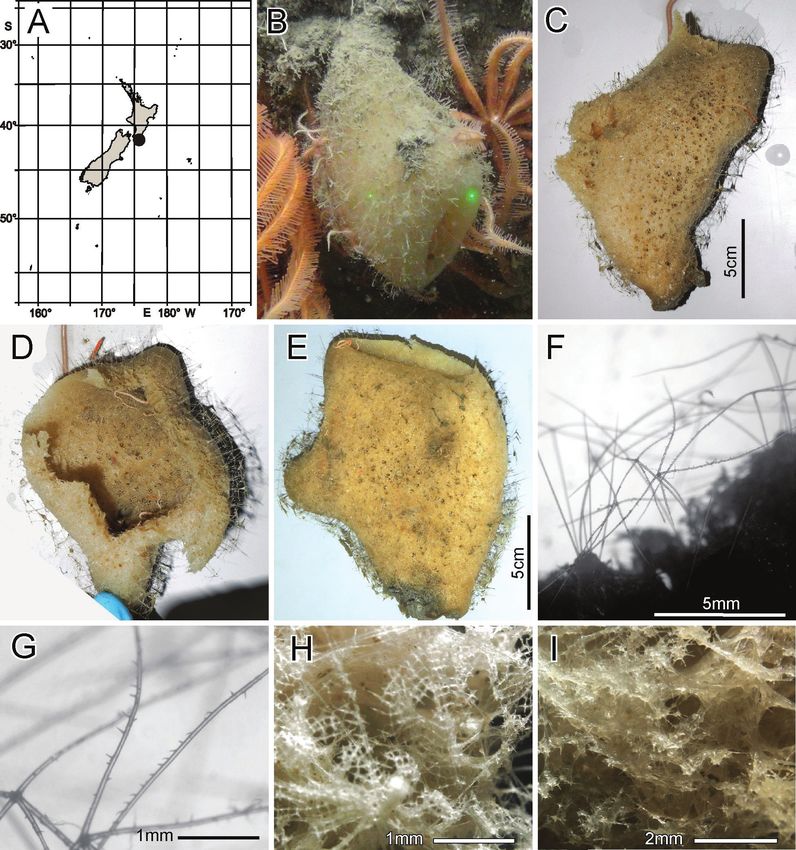
New Zealand Rossellidae                                35

     In the 2009 inventory of New Zealand biodiversity, Kelly et al. (2009) listed the fol-
lowing species, confirmed in a later draft manuscript on Rossellidae under preparation
by HMR and MK. These include Caulophacus hadalis Lévi, 1964, now accepted as Cau-
lophacus (Caulophacus) hadalis (not included in Dawson 1993), Crateromorpha cylindrica
(Schulze, 1886), now accepted as Crateromorpha (Aulochone) cylindrica, and Caulophacus
schulzei Wilson, 1904, now accepted as Caulophacus (C.) schulzei. Several species were
also included and confirmed for New Zealand in Kelly et al. (2009), from a draft 1980
manuscript under preparation by HMR: Crateromorpha (Aulochone) haliprum Tabach-
nick & Lévi, 2004; Crateromorpha (Caledochone) caledoniensis Tabachnick & Lévi in Ta-
bachnick (2002); Caulophacus (Caulodiscus) onychohexactinus Tabachnick & Lévi, 2004;
Sympagella clavipinula Tabachnick & Lévi, 2004. These species are beyond the scope of
this work and will be dealt with later. Kelly et al. (2015) confirmed the presence of Cau-
lophacus (C.) hadalis and Crateromorpha (A.) cylindrica in their Kermadec Islands review.
     In 2013, several well-preserved body fossils of a new species, Rossella cylindrica
Buckeridge & Kelly, 2013 (in Buckeridge et al. 2013), from the late Palaeocene-early
Eocene Red Bluff Tuff of Chatham Island, were confirmed by HMR. Both R. ant-
arctica Carter, 1872 and R. racovitzae Topsent, 1901 were stated to be present on the
Chatham Rise, but this remains unconfirmed. Various Rossellidae species were also
represented in the Oamaru Diatomite (Eocene) as hexactins, pentactins, and stau-
ractins, illustrated in Hinde and Holmes (1892), but microfossil spicules were not
recorded in the Tutuiri Greensand (Kelly and Buckeridge 2005), despite the relatively
high proportion of hexactinellid taxa in the fauna. Specimens compared to Rossella
racovitzae/R. antarctica on the Chatham Rise by Buckeridge et al. (2013) and Camp-
bell Plateau (Chin and Kelly, pers. comm.) form a small, squat, open-mouthed barrel
with a restricted base, and have a characteristic veil of hypodermal pentactins protrud-
ing beyond the surface of the sponge wall. This latter character is highly reminiscent
of the new species described herein, Nubes tubulata gen. nov., sp. nov. and Scyphidium
variospinosum sp. nov., the type localities of which are just north of Chatham Rise.
     The 2017 German RV Sonne (cruise SO254) expedition to New Zealand af-
forded an important collection of ~ 100 new hexactinellid specimens and corre-
sponding seafloor images, collected as part of Project PoribacNewZ of the Institute
for Chemistry and Biology of the Marine Environment (ICBM), Carl von Ossi-
etzky University of Oldenburg, using the GEOMAR Helmholtz Centre for Ocean
Research Kiel Remotely Operated Vehicle (ROV) Kiel 6000 (Schupp et al. 2017).
Preparation of a manuscript combining morphological descriptions and molecu-
lar systematics of these glass sponges was underway when we were devastated by
the untimely death of Henry Reiswig in July 2020. The objective of this work
is to provide the descriptions of specimens completed prior to Henry’s passing.
This work includes the establishment of a new endemic genus, Nubes gen. nov.,
with two new species; a new species of Bathydorus Schulze, 1886; redescription of
Scyphidium australiense Tabachnick, Janussen & Menschenina, 2008, and descrip-
tion of a new species of Scyphidium Schulze, 1900; redescription of Caulophacus
(Caulophacus) discohexaster Tabachnick & Lévi, 2004, and description of two new
species of Caulophacus (Caulophacus) Schulze, 1886.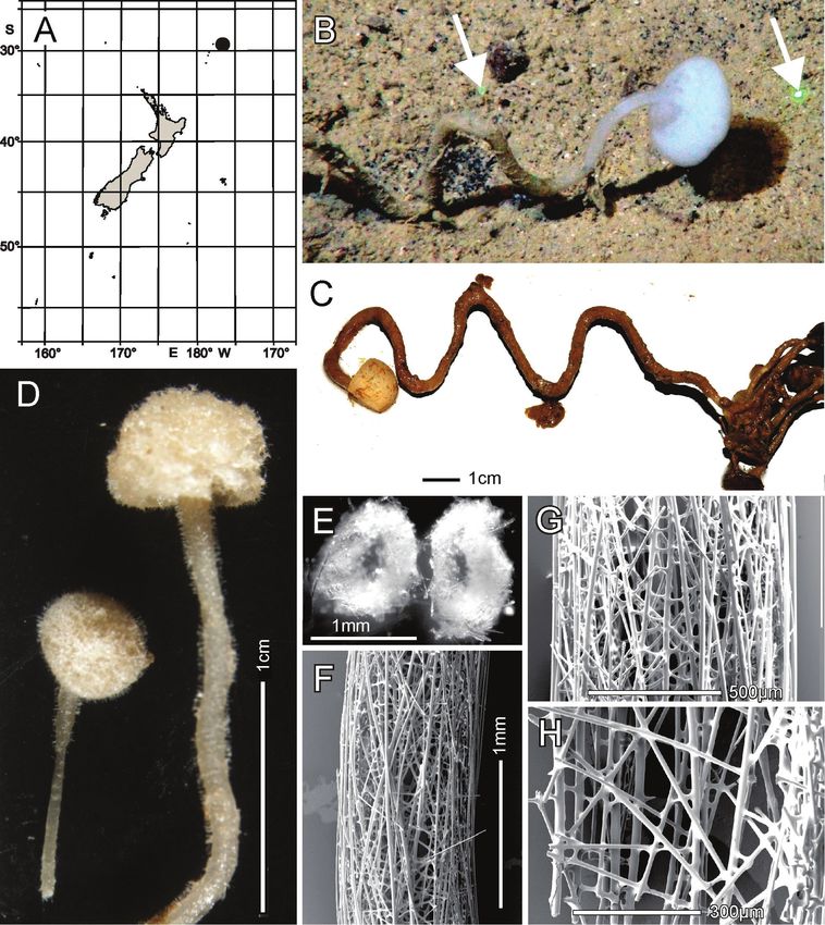
36                Henry M. Reiswig et al. / ZooKeys 1060: 33–84 (2021)

Materials and methods
Sample collection

Specimens, seafloor images, and videos were collected as part of Project Porib-
acNewZ of the Institute for Chemistry and Biology of the Marine Environment
(ICBM), Carl von Ossietzky University of Oldenburg, on the new German RV
Sonne (cruise SO254), using the GEOMAR Helmholtz Centre for Ocean Research
Kiel ROV Kiel 6000 (Schupp et al. 2017). With the exception of NIWA 126016,
which was collected in International Waters to the east of Norfolk Island and the
Three Kings Ridge, all other specimens were collected from the New Zealand Ex-
clusive Economic Zone (EEZ); collection sites and general distribution of the spe-
cies are shown in Fig. 1.

Sample preparation
Subsamples were taken on board, stored in appropriate preservatives for morpho-
logical and molecular work, and shipped to the Ludwig-Maximilians-Universität
(LMU) Munich. Specimens reported here, together with a much larger collection,
the remainder of which will be reported on elsewhere, were first subjected to a mo-
lecular phylogenetic study (results not shown) based on a mitochondrial 16S ribo-
somal DNA fragment (cf. Dohrmann et al. 2008) for initial assessment of their re-
lationships. This then allowed selection of interesting specimens for further study,
some of which we describe below. Preliminary morphological identifications of these
specimens were made by MD by analysing spicule content with light microscopy of
temporary bleach digestions of tissue pieces. Sample preparation for formal identi-
fication and description of taxa new to science (as well as re-descriptions of known
species where appropriate) were then performed by HMR using methods described
in Reiswig and Kelly (2011, 2018).

Registration of type and general material
Primary and secondary type materials of new species and additional material are de-
posited in the Invertebrate Collection (NIC) at the National Institute of Water and
Atmospheric Research (NIWA), Greta Point, Wellington, using the prefix NIWA – .
Registration numbers are cited in the text. Taxonomic authority is restricted to Re-
iswig, Dohrmann & Kelly.

Abbreviations
EEZ          Exclusive Economic Zone;
GEOMAR       Research Center for Marine Geosciences, Helmholtz Centre for Ocean
             Research Kiel, Germany;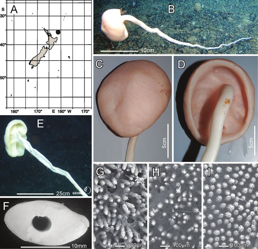
New Zealand Rossellidae                                      37

Figure 1. Study area showing the distribution of newly described rossellid sponges in the New Zealand
EEZ and in international waters.

ICBM     Institute for Chemistry and Biology of the Marine Environment, Carl
         von Ossietzky University of Oldenburg;
LM       light microscopy;
NIC–NIWA Invertebrate Collection, NIWA, Wellington, New Zealand;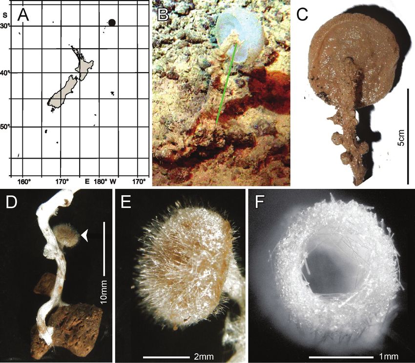
38                 Henry M. Reiswig et al. / ZooKeys 1060: 33–84 (2021)

NIWA          National Institute of Water and Atmospheric Research, Wellington,
              New Zealand;
SEM           scanning electron microscopy.

Systematics
PORIFERA Grant, 1836
HEXACTINELLIDA Schmidt, 1870
HEXASTEROPHORA Schulze, 1886
LYSSACINOSIDA Zittel, 1877
ROSSELLIDAE Schulze, 1885
ROSSELLINAE Schulze, 1885

Bathydorus Schulze, 1886

Bathydorus poculum Reiswig, Dohrmann & Kelly, sp. nov.

Nubes Reiswig, Dohrmann & Kelly, gen. nov.

Nubes tubulata Reiswig, Dohrmann & Kelly, sp. nov.
Nubes poculiformis Reiswig, Dohrmann & Kelly, sp. nov.

Scyphidium Schulze, 1900

Scyphidium australiense Tabachnick, Janussen & Menschenina, 2008
Scyphidium variospinosum Reiswig, Dohrmann & Kelly, sp. nov.

LANUGINELLINAE Gray, 1872

Caulophacus (Caulophacus) Schulze, 1886

Caulophacus (Caulophacus) discohexaster Tabachnick & Lévi, 2004
Caulophacus (Caulophacus) serpens Reiswig, Dohrmann & Kelly, sp. nov.
Caulophacus (Caulophacus) ramosus Reiswig, Dohrmann & Kelly, sp. nov.

Rossellidae Schulze, 1885

Diagnosis. The body is usually cup-like basiphytose or lophophytose; in the peduncu-
late forms the body can be mushroom-like. Prostalia lateralia, when present, are formed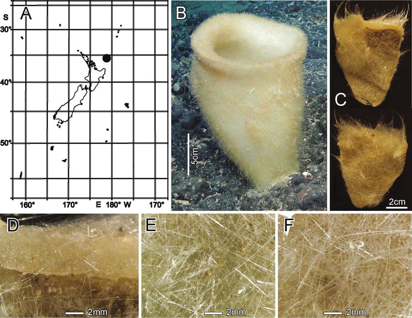
New Zealand Rossellidae                                39

with diactins or outwardly protruding hypodermal pentactins; prostalia basalia, when
present, are outwardly protruding hypodermal pentactins which are usually special-
ised (anchorate). Choanosomal skeleton consists of diactins, sometimes together with
less frequent hexactins. Hypodermal pentactins often present, usually they protrude
from the dermal surface serving as prostalia. Hypoatrial pentactins are rarely found
or absent in some taxa. Dermalia are combinations of various spicules usually pentac-
tins; stauractins and diactins, rarely hexactins. Atrialia are usually hexactins but other
holactinoidal spicules can be also found there. Microscleres are various: holactinoidal,
asterous and asters; they usually have discoidal or oxyoidal terminations but sometimes
floricoidal, onychoidal, or sigmoidal ones (after Tabachnick 2002).

Rossellinae Schulze, 1885

Diagnosis. As for family.
    Remarks. This subfamily is clearly not monophyletic (Dohrmann et al. 2017) and
retained here for historical reasons only.

Bathydorus Schulze, 1886

Diagnosis. Rossellinae with tubular, saccular, or plate-like gross morphology. Basi-
phytous or lophophytous, thin-walled. Dermalia are combinations of spicules from
hexactins to diactins. Regular pentactins make up a hypodermal layer. Choanosomal
skeleton composed of diactins, sometimes with hexactins. Atrialia are hexactins or
stauractins. Microscleres are combinations of oxyoidal hexasters, hemihexasters, and
hexactins; lacking pappocomes (from Kahn et al. 2013).
    Type species. Bathydorus fimbriatus Schulze, 1886

Bathydorus poculum Reiswig, Dohrmann & Kelly, sp. nov.
http://zoobank.org/1E8B4837-7A12-4A08-91B6-5A8E63CC79F2
Figs 2, 3; Table 1

Material examined. Holotype NIWA 126338, RV Sonne Stn SO254/85ROV19_
BIOBOX17, Southern Kermadec Ridge, 35.612°S, 178.852°E, 1150 m, 24 Feb 2017.
    Distribution. Known only from the type locality, the Southern Kermadec Ridge,
north of New Zealand (Fig. 2A).
    Habitat. Attached to hard substratum at 1149 m (Fig. 2B).
    Description. Morphology of the holotype is a thick-walled funnel attached to rock
substratum by a wide basal disc (Fig. 2B). Both dermal and atrial surfaces have a very
dense, bushy, cover of prostal diactins (Fig. 2C, E, F). The single terminal osculum is the
widest body part and the margin is abruptly sharpened; it has no marginalia (Fig. 2D).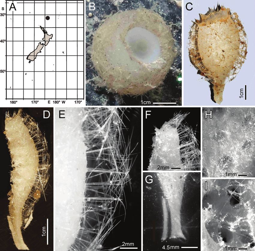
40                      Henry M. Reiswig et al. / ZooKeys 1060: 33–84 (2021)

Figure 2. Bathydorus poculum sp. nov., holotype NIWA 126338, distribution, skeleton, and morphology
A distribution in New Zealand waters B holotype in situ (scale bar approximate) C dermal (upper) and atri-
al (lower) sides of the preserved main part of the collected fragment D magnified area of the oscular margin,
showing the atrial surface curving out over the dermal surface E dermal surface with dense prostal diactins
F atrial surface with similarly dense prostal diactins. Image B captured by ROV Team GEOMAR, ROV
Kiel 6000 onboard RV Sonne (voyage SO254), courtesy of Project PoribacNewZ, GEOMAR, and ICBM.

Dimensions of the holotype are ~ 17.2 cm high and 12.8 cm wide; the measurements
are only approximate as only one of the two laser points could be certainly found on the
in-situ images. Wall thickness is 10.7 cm, excluding the 1–2.5 cm thick prostal cover
layers on each side. Texture is soft, compressible, and resilient, neither hard nor fragile.
Surfaces of both the inner and outer walls are hairy to the naked eye, and when inspected
at low magnification of a dissecting microscope, both are covered with a bushy layer of
prostal diactins. Colour in life is pale beige, and pale brown when preserved in ethanol.
     Skeleton. Choanosomal skeleton consists of a loose network of thin choanosomal
diactins amongst the thicker proximal ends of prostal diactins, and proximal rays of
hypodermal pentactins. No choanosomal hexactins are present. There is no evidence of
fusion between any spicules. Microscleres are scattered evenly throughout the choano-
some. Ectosomal skeleton of the dermal side consists of abundant prostal diactins pass-
ing through the distal tangential parts of hypodermal pentactins and dermalia, which
are mostly stauractins (62% of 126 assessed), pentactins (29%) and hexactins (10%).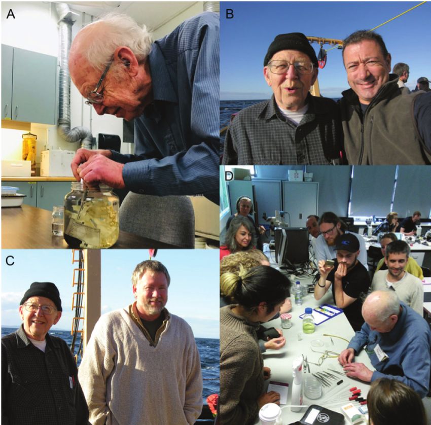
New Zealand Rossellidae                                     41

Figure 3. Bathydorus poculum sp. nov., holotype NIWA 126338 spicules A prostal diactin, whole and
enlarged end B hypodermal pentactin, whole and enlarged spicule centre, tangential and proximal ray
ends C choanosomal diactin, whole and enlarged end D stauractine dermalium, whole and enlarged ray
end E pinular hexactine atrialium, whole and enlarged ray end F oxyhexaster G enlarged whole primary
and secondary ray (left) and centre of spicule showing smooth primary and ornamentation of spines on
secondary rays (right) H hemioxyhexaster I hemioxystauraster.

The atrial ectosome lacks hypoatrial pentactins but has atrialia in the form of hexactins
(89% of 126 assessed), pentactins (8%), stauractins, and triactins (1.5% each). Micro-
scleres are present as in the choanosome.
     Spicules. Megascleres (Fig. 3; Table 1) are prostal diactins, hypodermal pentactins,
choanosomal diactins, dermalia mostly as stauractins, and atrialia mostly as hexactins.
Prostal diactins (Fig. 3A) are long bow-shaped spicules, smooth except for patches of
subterminal spines; the smooth tips are rounded or parabolic; the spicule centre is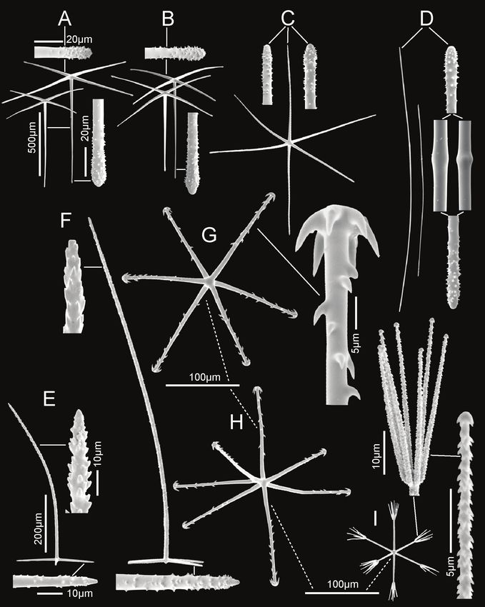
42                           Henry M. Reiswig et al. / ZooKeys 1060: 33–84 (2021)

Table 1. Spicule dimensions (µm) of Bathydorus poculum sp. nov., from holotype NIWA 126338.

                         Parameter                   mean          s.d.         range      no.
Dermal prostal diactin
     length (mm)                                      26.8         11.7       11.0–63.4    48
     width                                            65.6         14.1       16.1–92.6    63
Hypodermal pentactin
     tangential ray length                            476          110         218–995     60
     tangential ray width                             15.3         3.0         8.4–23.2    62
     proximal ray length (mm)                          1.6         0.6          0.7–4.2    62
     proximal ray width                               16.5         3.6         9.1–25.8    60
Choanosomal diactin
     length (mm)                                      16.8         11.1        1.4–31.5    8
     width                                            12.8          7.3        7.1–38.4    47
Dermalia, stauractin
     ray length                                       98.5         18.0       66.7–139.0   29
     ray width                                         5.0          0.8         3.4–7.7    32
Atrialia, pinular hexactin                                                                 50
     pinular ray length                              150.0         17.4      107.7–181.1   21
     pinular ray width                                 3.8          0.8         2.4–5.2    21
     tangential ray length                            92.8         10.6       73.1–110.5   21
     tangential ray width                              3.6          0.6         2.7–4.6    21
     proximal ray length                              79.4         13.2       62.6–108.0   16
     proximal ray width                                3.7          0.6         2.7–5.0    20
Atrialia, non-pinular hexactin
     ray length                                       89.2         9.5        73.6–106.2   21
     ray width                                         3.4         0.7          2.3–4.6    21
Oxy- and hemioxyhexaster
     diameter                                        109.0         21.8       66.2–164.3   30
     primary ray length                                4.6          0.9         2.9–7.3    30
     secondary ray length                             50.4         11.1        26.9–74.5   30
Oxyhexactin
     diameter                                        119.5         22.2       81.6–157.0   8
     ray width                                        1.5           0.3         1.2–1.9    8

not swollen. Hypodermal pentactins (Fig. 3B) are regular and crucial in form with
very long proximal rays, averaging 3.4 × tangential ray length, and fine spines evenly
scattered over the entire surface. All five rays have subterminal patches of larger spines
and smooth round tips. Choanosomal diactins (Fig. 3C) are straight, bent or more
commonly sinuous in shape. Most are broken so few intact spicules are measurable
for length. They are smooth except for subterminal inflated rough patches; the tip is
smooth and abruptly tapered to a point. The spicule centre is moderately swollen. Der-
malia (Fig. 3D) are mainly crucial stauractins completely covered with short, rounded
knobs or spines; rays are tapered to a round tip. Atrialia (Fig. 3E) are mostly hexactins
ca. half of which are pinular with one ray longer than the others. Like dermalia, these
are entirely covered with short, rounded knobs or spines but longer than those of the
dermalia; ray tips are rounded.
     Microscleres (Fig. 3; Table 1) are all oxyhexasters and their variants with hemiox-
yhexasters being the most common. Oxyhexasters (Fig. 3F, G) have short smooth pri-
mary rays and long straight secondary rays; the secondary rays are entirely ornamented
with reclined spines that increase in size from the ray tip to its proximal end. Secondary
New Zealand Rossellidae 43
rays on each primary ray vary from 2–5. Hemioxyhexasters (Fig. 3H) are similar to
oxyhexasters but at least one of the six primary rays bear only a single secondary ray.
Other rare variants include oxyhexactins, oxypentasters, and oxystaurasters (Fig. 3I).
Etymology. Named for the beaker-shaped morphology of this species (poculum,
beaker; Latin).
Remarks. This New Zealand specimen, NIWA 126338, is entirely consistent with
the diagnosis of Bathydorus and is assigned there. Each of the known species of the
genus differ from this specimen in the following characters: Bathydorus echinus Koltun,
1967 has prostal pentactins in addition to diactins, and dermalia as mainly pentactins;
B. fimbriatus Schulze, 1886 has prostalia including pentactins as marginalia only, and
no pinular atrialia; B. laevis laevis Schulze, 1886 has no prostalia lateralia and no pi-
nular atrialia; B. laevis pseudospinosus Tabachnick & Menshenina, 2013 has some large
choanosomal or prostal hexactins and smaller oxyhexasters to only 100 µm diameter;
B. laninger Kahn, Geller, Reiswig & Smith Jr., 2013 has a flat body form and no pros-
talia on the atrial (upper) surface; B. servatus Topsent, 1927 has no prostal diactins, and
dermalia as stauractins and diactins; B. spinosissimus Lendenfeld, 1915 has choanoso-
mal hexactins, and oxyhexasters with longer primary rays (4–12 µm); in the original
description of B. spinosus Schulze, 1886, there is no mention of hypodermal pentac-
tins; although Tabachnick and Menshenina (2013) include these, they fail to certify
that they are present in the holotype; this species also has wavy secondary rays on the
oxyhexasters; B. uncifer Schulze, 1899 has smooth dermal and atrial surfaces, and der-
malia as mainly pentactins and stauractins. These differences are sufficient to conclude
that the new form is a new species, here designated as Bathydorus poculum sp. nov.
Nubes Reiswig, Dohrmann & Kelly, gen. nov.
http://zoobank.org/032AA823-2695-4E82-888D-0051A86BC438
Diagnosis. Rossellinae with basiphytous, saccular, thick-walled body, unstalked or
with a short stalk. Hypodermalia are large, raised, paratropal or orthotropal pentactins
with strongly curved or straight tangential rays, smooth except for rough tips, form-
ing a cloud or veil around the thick-walled body. Prostal diactins are marginalia only.
Choanosomal spicules are diactins and sometimes large hexactins with curved rays,
smooth except for rough tips. Dermalia are mainly stauractins and pentactins. Atrialia
are mainly hexactins and sometimes pentactins. Microscleres are oxyhexasters, hemi-
oxyhexasters, and anisodiscohexasters.
Etymology. Named for the cloud of large hypodermal pentactins that veils the
body of these sponges (nubes, cloud; Latin).
Type species. Nubes tubulata sp. nov.
Remarks. This new genus diagnosis differs from those of most other anisodis-
cohexaster-bearing genera or subgenera in the following ways: from Anoxycalyx Kirk-
patrick, 1907 in not having anchorate hypodermalia, and having pleural hypodermalia
raised, having marginalia; in not including pappocomes and discohexasters other than44 Henry M. Reiswig et al. / ZooKeys 1060: 33–84 (2021)
anisodiscohexasters (strobiloidal discohexasters) as microscleres. It differs from that of
Crateromorpha (Crateromorpha) Gray in Carter, 1872 in body form, having marginal
diactins, and having main atrialia as hexactins. It differs from that of Rossella Carter,
1872 in having most atrialia as hexactins instead of stauractins, and no calycocomes.
However, it does not differ from the present diagnosis of Vazella Gray, 1870 (Tabach-
nick 2002) in any way, but below we offer a modified diagnosis of that genus to sepa-
rate the two groups.
Nubes tubulata Reiswig, Dohrmann & Kelly, sp. nov.
http://zoobank.org/352141EE-D1CC-4A5A-94F5-F52B731D5C73
Figs 4, 5; Table 2
Material examined. Holotype NIWA 126159, RV Sonne Stn SO254/36ROV10_
BIOBOX7, Seamount No. 986, off Hawkes Bay shelf, 39.990°S, 178.214°E, 782.8 m,
09 Feb 2017. Paratype NIWA 126160, RV Sonne Stn SO254/36ROV10_BIOBOX10,
Seamount No. 986, off Hawkes Bay shelf, 39.989°S, 178.214°E, 767 m, 09 Feb 2017.
Distribution. Known only from the type locality, Seamount 986 off Hawkes Bay
shelf, east of North Island, New Zealand (Fig. 4A).
Habitat. Attached to hard substratum; depth 767–783 m (Fig. 4B).
Description. Morphology of the holotype and paratype a thick-walled, tubular
sponge, attached to hard substratum by a narrow base (Fig. 4B). A round osculum of
moderate size is terminal and opens into a deep atrial cavity. The margin is sharp and
there are indications of sparse diactin marginalia in deck images, but we have been
unable to verify them in the material at hand. The dermal surface has a dense covering
of raised, prostal, hypodermal pentactins (Fig. 4C, D, I) projecting up to 1 cm from
the surface proper. There is indication in some of the deck images of long diactins
projecting sparsely up to 6 cm from the dermal surface, especially basally, but these
may be choanosomal diactins pulled out during collection; we have not found such
large diactins in the material we had for examination. Dimensions of the holotype are
~ 13.3 cm high, 7.0 cm wide, and 10.8 (9.2–13.3) (n = 9) mm in body wall thickness;
the osculum is 2.2 cm in diameter in situ. The paratype is 19.5 cm high, 13.4 cm wide,
and body wall is 7.4 (5.5–9.3) (n = 11) mm in thickness. The osculum is 4.2 cm in
diameter in situ. Texture is soft, compressible, and resilient, neither hard nor fragile.
Surface of the dermal side is covered by a thick layer of projecting hypodermal pen-
tactins (Fig. 4E). The dermal lattice is torn apart, and dermalia reside in preserved
specimens as attached flakes on the hypodermalia (Fig. 4G). The atrial layer retains the
atrial lattice covering smaller apertures (Fig. 4F, H); no large megascleres project into
the atrium. Colour in life is transparent white, preserved in ethanol is medium brown
(Fig. 4C).
Skeleton. Choanosomal skeleton consists of a loose, vacuolar network of thin choa-
nosomal diactins, large choanosomal hexactins, and the thicker proximal rays of the hy-
podermal pentactins. There is no evidence of fusion between any spicules. MicroscleresNew Zealand Rossellidae 45 Figure 4. Nubes tubulata gen. nov., sp. nov., holotype NIWA 126159, distribution, skeleton and morpholo- gy A Distribution in New Zealand waters B holotype in situ C holotype, deck image D holotype, deck image showing moderate-sized osculum and veil of hypodermal pentactins (deck images by PJS) E dermal surface with dense veil of prostal hypodermal pentactins F atrial surface without a hypodermal veil G closer view of dermal surface with disrupted lattice H closer view of atrial surface with intact lattice over exhalant apertures I section of body wall, dermal surface on left side. Image B captured by ROV Team GEOMAR, ROV Kiel 6000 onboard RV Sonne (voyage SO254), courtesy of Project PoribacNewZ, GEOMAR, and ICBM. are scattered evenly throughout the choanosome. Ectosomal skeleton of the dermal side consists of abundant prostal pentactins supporting a delicate lattice of hexactine, pen- tactine, and stauractine dermalia. The atrial ectosome lacks hypoatrial pentactins but has bands of diactins that support the atrial lattice of hexactins, providing greater sup- port than available to the dermal lattice. Microscleres are present as in the choanosome.
46                      Henry M. Reiswig et al. / ZooKeys 1060: 33–84 (2021)

Figure 5. Nubes tubulata gen. nov., sp. nov., holotype NIWA 126159, spicules A whole prostal hypoder-
mal pentactin and enlarged ray ends B whole curved and straight choanosomal diactins with two enlarged
ends; scales of whole spicules and parts in B and C as in A; C whole choanosomal hexactin with two en-
larged ray ends D two dermalia, a subhexactin, and a stauractin, with enlarged ray ends and centres E two
atrialia, a pinular subhexactin, and a regular hexactin with enlarged centrum of the pinular subhexactin
and a ray end; scales of whole spicules and parts as in D; F two oxyhexasters G enlarged terminal ray of an
oxyhexaster H anisodiscohexasters, from SEM preparation (above) and LM preparation (below).

    Spicules. Megascleres (Fig. 5; Table 2) are prostal hypodermal pentactins, choa-
nosomal diactins, choanosomal hexactins, dermalia, and atrialia. Prostal hypodermal
pentactins (Fig. 5A) are mostly large, raised paratropal forms (90% of 68 scored) with
long, very curved tangential rays, but some regular, crucial forms occur (10%) in small-
er forms especially near the margin. Tangential rays are 1.7 × the shorter, straighter
proximal rays. The spicules are smooth except for the rough sharp tips. Choanosomal
diactins (Fig. 5B) are straight or strongly curved, usually with undetectable central
swellings; they are smooth except for the rough, slightly inflated tips. Choanosomal
hexactins (Fig. 5C) are large forms with strongly curved or nearly straight, nearly equal
length rays, which are otherwise similar to those of the hypodermalia. Dermalia (Fig.
5D) are entirely spined and consist of stauractins (31% of 387 scored) and similar
forms with reduced fifth ray (subpentactins) or both fifth and sixth rays in one axis
(subhexactins) (64%) with a few (1–2%) as tauactins, diactins and paratetractins. It
New Zealand Rossellidae 47
Table 2. Spicule dimensions (µm) of Nubes tubulata gen. nov., sp. nov. from holotype 126159.
Parameter mean s.d. range no.
Prostal hypodermal pentactin, lateral body
tangential ray length (mm) 14.4 1.7 10.5–17.9 36
tangential ray width 42.5 3.3 36.8–50.4 35
proximal ray length (mm) 8.4 1.3 5.3–10.7 26
proximal ray width 46.6 5.0 36.8–59.4 28
Prostal hypodermal pentactin, margin
tangential ray length (mm) 2.0 1.4 0.6–7.4 32
tangential ray width 20.2 7.0 6.5–39.6 31
proximal ray length (mm) 2.8 1.7 0.8–6.0 25
proximal ray width 21.9 7.1 7.3–43.0 30
Choanosomal diactin
length (mm) 9.1 5.1 1.6–21.3 35
width 16.4 9.1 4.2–47.0 35
Choanosomal hexactin
ray length (mm) 5.9 1.9 2.5–10.9 46
ray width 39.0 8.9 21.0–60.7 45
Dermalia stauractin
ray length 132 17 91–174 36
ray width 5.7 0.8 4.5–7.3 20
Dermalia subpentactin/hexactin
ray length 142 17 107–180 36
ray width 5.4 0.8 4.2–7.1 20
Atrialia subhexactin short pinular
ray length 21 5 13–40 26
tangential ray length 176 25 130–230 28
proximal ray length 130 22 93–184 26
tangential ray width 5.8 0.9 4.1–7.8 26
Atrialia, non-pinular hexactin
ray length 171 17 139–220 27
ray width 5.8 1.1 3.8–8.0 26
Oxy- and hemioxyhexaster
diameter 130.5 14.1 90.2–165.7 32
primary ray length 5.3 1.1 3.7–8.9 32
secondary ray length 60.2 6.2 47.3–77.1 32
Anisodiscohexaster
diameter 70.9 7.3 47.5–81.7 35
primary ray length 5.5 0.9 4.0–7.5 35
longest secondary ray length 30.3 3.9 19.4–36.0 35
was not possible to differentiate the subpentactins and subhexactins either wet in dish-
es or mounted spicule microscope slides. Tips are either rounded or more often sharp.
Atrialia (Fig. 5E) are entirely spined and mostly subhexactins (71% of 125 scored)
with one ray reduced or hexactins (26%) with all rays of nearly equal length; a few
(1–2%) are stauractins and tauactins. Ray tips are sharp-pointed.
Microscleres (Fig. 5; Table 2) are oxyhexasters and their variants, with hemiox-
yhexasters being the most common, and anisodiscohexasters. Oxyhexasters and he-
mioxyhexasters (Fig. 5F, G) have very short smooth primary rays and long straight
secondary rays entirely ornamented with small, reclined spines. Secondary rays on each
primary ray vary from 1–4. Anisodiscohexasters (Fig. 5H) are spherical with stellate
discs with 4–6 marginal claws on the ends of terminal rays. Primary rays are smooth48 Henry M. Reiswig et al. / ZooKeys 1060: 33–84 (2021)
and end in strobiloid discs with a short central projecting knob. Each primary stro-
bilum supports 30–40 terminal rays with undulating, probably helically coiled, finely
rough shafts of unequal lengths. Terminal discs vary in diameter with shaft length, the
longer shafts carrying the larger discs, e.g., a series 1.7, 2.5, 3.1, 3.4, 3.6, 5.4, 6.9 µm
diameter for shafts 15.0, 20.5, 23.5, 27.3, 32.0. 33.4, 37.1 µm in length. These spic-
ules look very different in LM (lower image) and SEM (upper image) due to collapse
of the rays during drying for SEM and support of them by balsam in LM.
Etymology. Named for the tubular morphology of the sponge (tubulata, tu-
bular; Latin).
Remarks. The characters of these two New Zealand specimens are inconsistent with
the present diagnoses of all Rossellinae genera except Vitrollula Ijima, 1898. They differ,
however, from those of V. fertilis Ijima, 1898, the only species in the genus, in characters
not used as diagnostic. These are that V. fertilis has a smooth surface without raised hy-
podermalia, but the two new specimens have a bristly surface with raised hypodermalia,
and that the discohexasters of V. fertilis are isodiscohexasters while those of the new spe-
cies are anisodiscohexasters. In view of these differences, we opt not to include the new
species in Vitrollula nor to change the diagnosis of that genus at this time. We choose to
erect a new genus in Rossellinae with characters of this and the following second species
described below, and designate this species as Nubes tubulata gen. nov., sp. nov.
Nubes poculiformis Reiswig, Dohrmann & Kelly, sp. nov.
http://zoobank.org/2EBDD0FB-6EB9-498A-8749-595C64824C23
Figs 6, 7; Table 3
Material examined. Holotype NIWA 126016, RV Sonne Stn SO254/08ROV02_
BIOBOX10, Seamount No. 114 in International Waters to the east of Three Kings
Ridge and Norfolk Island, 31.301°S, 175.197°E, 1285 m, 31 Jan 2017.
Distribution. Known only from the type locality, Seamount No. 114, in Interna-
tional Waters to the east of Three Kings Ridge and Norfolk Island (Fig. 6A).
Habitat. Attached to hard substratum; depth 1285 m (Fig. 6B).
Description. Morphology of the holotype body is a thick-walled tubular sponge,
attached to hard substratum, by a moderately long, narrow stalk (Fig. 6B, C). A
moderately sized, round osculum is terminal and opens into a deep atrial cavity. The
margin is blunt, bordered by a band of diactine marginalia (Fig. 6D, F). The dermal
surface has a dense covering of raised, prostal, hypodermal pentactins (Fig. 6D, E),
projecting up to 7 mm from the surface proper. Some of the deck images indicate
long diactins projecting sparsely, up to 14 mm, from the dermal surface, but these
may be foreign in origin; we have not found such large diactins in the material avail-
able for examination. Dimensions of the holotype are ~ 6 cm in total length, includ-
ing the stalk of 1.8 cm length (Fig. 6G), and 3.5 cm in width; the maximum body
wall thickness is 13.9 mm. The osculum is 12.3 by 16.9 mm diameter in situ. Texture
is soft, compressible, and resilient, neither hard nor fragile. Surface of the dermal sideNew Zealand Rossellidae 49
Figure 6. Nubes poculiformis gen. nov., sp. nov., holotype NIWA 126016, distribution, skeleton and
morphology A distribution in New Zealand waters B holotype in situ C holotype, deck image (by PJS);
D longitudinal section of holotype, showing hypodermal pentactin veil E closer view of hypodermal pen-
tactin veil F edge of osculum with tuft of marginalia G close view of stalk subdivided for spicule prepara-
tion of smaller sample H dermal surface with intact lattice of dermalia over inhalant canals I atrial surface
with disrupted lattice of atrialia. Image B captured by ROV Team GEOMAR, ROV Kiel 6000 onboard
RV Sonne (voyage SO254), courtesy of Project PoribacNewZ, GEOMAR, and ICBM.
below the layer of projecting hypodermal pentactins is supported by an intact tight
lattice of dermalia (Fig. 6H). The atrial surface (Fig. 6I), in contrast, is torn apart by
removal from supporting fluids and the atrial lattice remains only as dismembered
patches attached to underlying diactins. Colour in life is pale brown as is the speci-
men preserved in ethanol.
Skeleton. Choanosomal skeleton consists of a loose, vacuolar network of thin choa-
nosomal diactins, large choanosomal hexactins and the thicker proximal rays of the hy-50 Henry M. Reiswig et al. / ZooKeys 1060: 33–84 (2021) Figure 7. Nubes poculiformis gen. nov., sp. nov., holotype NIWA 126016, spicules A three prostal hy- podermal pentactins, the lower one in plane of tangential rays, with enlarged ray ends B whole marginal diactins (ray ends unavailable) C two whole choanosomal hexactins with two enlarged ray ends and one centrum. Scale of whole spicules as in C; D four whole stalk diactins and enlarged end E dermalia, stau- ractin and pentactin with enlarged ray ends F atrialium and enlarged ray end G two oxyhexasters and enlarged terminal ray H whole anisodiscohexaster; an enlarged section showing disc diameter increasing in longer terminal rays; an enlarged side view of a terminal ray and end views of terminal ray discs. podermal pentactins. There is no evidence of fusion between any spicules. Microscleres are scattered evenly throughout the choanosome. Ectosomal skeleton of the dermal side consists of abundant prostal pentactins providing good support for the sturdy lattice of stauractine (60.0% of 315 assessed), pentactine (38.4%), and rare hexactine (1.64%) dermalia. The atrial ectosome lacks hypoatrial pentactins but has bands of diactins that provide poor support for the atrial lattice of mainly hexactins (86.4% of 118 assessed), pentactins (7.5%), and stauractins (5.1%). Microscleres are present as in the choanosome.
New Zealand Rossellidae                                         51

Table 3. Spicule dimensions (µm) of Nubes poculiformis gen. nov., sp. nov. from holotype 126016.

                        Parameter                mean          s.d.           range            no.
Prostal hypodermal pentactin
     tangential ray length (mm)                    3.9         0.8            1.9–5.2          73
     tangential ray width                         51.1         5.4           36.4–59.3         60
     proximal ray length (mm)                      6.5         1.1            3.3–8.0          69
     proximal ray width                           51.0         4.5           39.7–58.7         59
Marginal diactin
     length (mm)                                   4.5         0.7            2.8–6.0          58
     width (mm)                                   18.0         3.3           12.3–27.5         64
Choanosomal diactin
     length (mm)                                   2.5         1.4            0.6–4.9          52
     width (mm)                                   11.8         2.3           7.5–19.2          52
Stalk diactin
     length (mm)                                   7.4         2.1           2.5–11.5          25
     width (mm)                                   14.7         4.9           8.1–29.6          25
Dermalia stauractin ray
     length                                       200          24            130–243           51
     width                                        11.5         1.7           7.3–14.6          51
Dermalia pentactin tangential ray
     length                                       185          19            146–223           62
     width                                        11.3         1.5           6.3–15.5          63
Dermalia pentactin proximal ray
     length                                       155          20            119–190           21
     width                                        11.0         1.6           7.8–14.5          23
Atrialia hexactin
     ray length                                   227          25            176–283           50
     ray width                                    13.9         2.2           9.2–20.0          50
Oxyhexaster
     diameter                                     137          11             103–165          51
     primary ray length                            5.7         1.1             3.5–7.8         51
     secondary ray length                         62.6         4.9           46.7–72.9         51
Anisodiscohexaster
     diameter                                     148           34             87–201          54
     primary ray length                            9.0          1.4           6.0–12.6         54
     longest secondary ray length                 66.4         16.2          32.9–89.5         54

    Spicules. Megascleres (Fig. 7; Table 3) are prostal hypodermal pentactins, marginal
diactins, choanosomal diactins of the body, choanosomal diactins of the stalk, dermalia
and atrialia. Prostal hypodermal pentactins (Fig. 7A) are large, raised orthotropal forms
with long straight tangential rays. Tangential rays are ca. one half the length of the
longer straight proximal rays. The spicules are smooth except for the rough sharp or
round tips. Marginalia (Fig. 7B) are long, slightly curved diactins; no intact tips were
found in SEM surveys but an exhaustive survey with LM indicates tips taper to nearly
invisible thinness and are quite distinct from the thick roughened tips of choanosomal
diactins. Choanosomal diactins (Fig. 7C) are straight or slightly curved with undetect-
able central swellings; they are smooth except for the rough, slightly inflated tips. Stalk
diactins (Fig. 7D) are longer and thicker than the choanosomal diactins, but otherwise
similar. Dermalia (Fig. 7E) are mainly entirely rough stauractins and pentactins with
rounded ray tips. Atrialia (Fig. 7F) are entirely rough hexactins with equal length rays
and more acute ray tips.
52 Henry M. Reiswig et al. / ZooKeys 1060: 33–84 (2021)
Microscleres (Fig. 7; Table 3) are oxyhexasters, hemioxyhexasters, and anisodis-
cohexasters. Oxyhexasters (Fig. 7G) and hemioxyhexasters have very short, sparsely
spined or smooth, thick primary rays, ending in swollen hemispheres; 1–7, usually
3–4, rough, straight, terminal rays tapering to pointed tips emanate from the margins
and occasionally from the centre of the hemisphere. Short to very short spur-like
terminal rays are common. Anisodiscohexasters (Fig. 7H) have smooth primary rays
ending in ovoid strobila. Each strobilum supports ca. 20–30 rough, curved terminal
rays that end in discs with 4–7 marginal discs. The tuft of terminal rays from each
primary ray varies in length of rays, and with ray length the diameter of terminal
discs, in a pattern that is not yet clear, but the whole spicule resembles a radially sym-
metrical starburst.
Etymology. Named for the goblet shape of the sponge (poculiformis, goblet-
shaped; Latin).
Remarks. This species differs from Nubes tubulata sp. nov. in having a short stalk
and orthotropal hypodermal pentactins, but is otherwise similar enough to include it
in the genus Nubes as its second species, Nubes poculiformis sp. nov.
Vazella Gray, 1870
Diagnosis. Body is saccular, basiphytous. Choanosomal skeleton is composed of diac-
tins. Hypodermal pentactins are raised, thorned paratropal pentactins. Prostalia basalia
and marginalia are monaxons (diactins). Dermalia are stauractins and pentactins. Atri-
alia are mainly hexactins. Discoid microscleres are microisodiscohexasters and micro-
anisodiscohexasters; oxyoid microscleres are combinations of hexactins, hexasters, and
hemihexasters (modified from Tabachnick 2002).
Remarks. This modified diagnosis allows separation of the present genus, Nubes
gen. nov., from Vazella on the basis of lack of thorned hypodermalia and presence of
discoid microscleres that are not anisodiscohexasters in the former. Furthermore, mo-
lecular phylogenetic results do not support a close relationship of the two genera (MD,
unpubl. results).
Scyphidium Schulze, 1900
Diagnosis. Body is saccular, basiphytous, sometimes rhizophytous. Choanosomal skel-
eton is composed of diactins. Hypodermal spicules, if present, are pentactins. Prostalia,
if present, are hypodermal pentactins and/or diactins. Dermalia are stauractins and/or
pentactins in various combinations. Atrialia are mainly hexactins. Microscleres are dis-
cohexasters and oxyhexasters often with hemioxyhexasters and oxyhexactins; with two
or three types of discohexasters, none as calycocomes. Among the larger is a spherical
form with a restricted number of secondary rays (emended from Tabachnick 2002).New Zealand Rossellidae 53
Remarks. The genus diagnosis is emended of necessity, to accept S. australiense
Tabachnick, Janussen & Menschenina, 2008 and S. variospinosum sp. nov., de-
scribed below.
Type species. Scyphidium septentrionale Schulze, 1900.
Scyphidium australiense Tabachnick, Janussen & Menschenina, 2008
Figs 8, 9; Table 4
Note. From the ending of its name, Scyphidium is a neuter noun, and thus S. aus-
traliensis (as originally named by Tabachnick et al. 2008) should be S. australiense. This
is borne out by the names of conspecifics that are also adjectives (e.g., S. chilense, S.
septentrionale, S. tuberculatum) (J. Rosser, pers. comm.). We hereby make that change
and use the corrected name throughout this work.
Type and locality (not examined). Holotype – NIWA 155561, RV Sonne Stn
SO17/80 (NZOI Stn Z3951B), Chatham Rise, 43.553°S, 179.457°E, 409 m, 10 Apr
1981 [Originally cited in Tabachnick et al. (2008) as WAM (p14), RV Soela Stn SO
17–80, 43°33.10'–33.05'S, 179°27.25'–27.08'E, depth unknown].
Material examined. NIWA 126237, RV Sonne, Stn SO254/77ROV14_
BIOBOX02, Pegasus Canyon slope, off Christchurch shelf, 43.2927361°S,
173.6066742°E, 853 m, 20 Feb 2017.
Distribution. Chatham Rise and Pegasus Canyon slope, off Christchurch shelf
Christchurch shelf, New Zealand (Fig. 8A).
Habitat. Attached to hard substratum; depth 409–853 m.
Description. Body form is a heavy-looking, thick-walled, club-shaped, pendant
sponge with a narrow basal attachment, widening gradually to a hemispherical round-
ed terminal end (Fig. 8B, C) where a large osculum is centrally located. The osculum
opens into a deep atrial cavity (Fig. 8D). The margin is sharp-edged with indication
of sparse marginalia that do not differ from prostal diactins of the lower body. The
external surface of the upper body is fairly smooth, without prostalia, but the lower
half is conspicuously conulose with long prostal diactins projecting in small groups
from conules (Fig. 8E). We did not have access to the basal attachment so we cannot
comment on the basidictyonalia. Dimensions of the specimen are 27.6 cm in height,
11.7 cm in maximum width, 5.7–10.9 cm in diameters of the osculum, 10.0 mm
in maximum wall thickness, 8.3 mm in length of projecting part of prostal diactins.
Texture is firm but compressible and resilient, neither soft nor fragile. Surface of the
dermal side is covered by an intact lattice of dermalia (Fig. 8G) consisting mostly of
pentactins (98% of 302 assayed), and a few stauractins and diactins (1% each). The
upper body surface is fairly smooth, but the lower body is covered with conspicuous
conules up to 3.2 mm high, from which prostal diactins project in small groups of
one to four. One large pentactin was found but it was broken and assumed to be for-
eign. The atrial surface is covered by a felt-like layer of disarranged atrialia (Fig. 8H)54 Henry M. Reiswig et al. / ZooKeys 1060: 33–84 (2021) Figure 8. Scyphidium australiense Tabachnick, Janussen & Menschenina, 2008, NIWA 126237, distri- bution, skeleton and morphology A distribution in New Zealand waters, holotype as open circle, new specimen as filled circle B new specimen in situ (scale bar is approximate) C deck image (two sides, im- age by PJS) D osculum, deck image (by PJS) E preserved conulose outer surface of the lower body with prostal diactins F preserved wall section of the mid-body without conules G preserved dermal surface with intact pentactin lattice H preserved atrial surface with hexactins displaced from the atrial lattice. Image B captured by ROV Team GEOMAR, ROV Kiel 6000 onboard RV Sonne (voyage SO254), courtesy of Project PoribacNewZ, GEOMAR, and ICBM. composed of hexactins (57% of 168 assayed), pentactins (20%), paratetractins (8%), diactins (6%), stauractins (5%), and triactins (3%). Colour in life is very pale brown, preserved in ethanol is medium brown.
New Zealand Rossellidae                                          55

Figure 9. Scyphidium australiense Tabachnick, Janussen & Menschenina, 2008, NIWA 126237, spicules
A prostal diactin, whole and enlarged ends, one broken distal end and two intact proximal ends B choa-
nosomal diactins, whole long and short versions at different scales plus enlarged tips and central swellings
C dermalium: pentactin, whole and enlarged tips D atrialia, hexactin, whole and enlarged tip, pentactin,
whole with enlarged tips, and paratetractin, whole; scales are the same as those for dermalium E spheres
as small group of whole ones and one enlarged F discohexaster 1, whole and enlarged terminal ray end
G discohexaster 2, whole and enlarged part of one ray tuft H oxyhexaster, whole and enlarged terminal
ray end. Scales are the same for all whole microscleres and their enlarged parts.

    Skeleton. Choanosomal skeleton consists of a tight series of macroscopic parti-
tions of inhalant and exhalant channels running perpendicular to the body surfaces
(Fig. 8F). They consist of networks of choanosomal diactins and microscleres and in
the lower body the proximal ends of the prostal diactins. A few small patches of fused
choanosomal diactins occur but these are too rare to provide significant support
to the body. Ectosomal skeleton of the dermal side consists of the robust lattice of
56 Henry M. Reiswig et al. / ZooKeys 1060: 33–84 (2021)
Table 4. Spicule dimensions (µm) of Scyphidium australiense Tabachnick, Janussen & Menschenina,
2008 from holotype NIWA 126237.
Parameter mean s.d. range no.
Prostal diactin
length (mm) 10.9 3.9 5.7–18.3 31
width 83.9 27.7 37.8–172.3 46
Choanosomal diactin
length (mm) 2.0 1.3 0.4–4.4 38
width 13.1 3.6 6.1–21.7 50
Dermalia pentactin
tangential ray length 145 17 106–186 31
ray width 15.3 1.8 11.0–18.4 31
proximal ray length 119 19 57–165 31
ray width 14.4 1.8 12.0–18.2 31
Atrialia hexactin
ray length 206 80 88–359 40
ray width 14.3 3.4 7.7–24.5 40
Sphere
diameter 189 77 90–388 54
Discohexaster 1
diameter 69.8 10.2 50.0–91.2 32
primary ray length 4.8 0.7 3.4–6.8 32
secondary ray length 30.3 5.4 20.6–42.8 32
Discohexaster 2
diameter 50.2 10.0 33.4–79.4 68
primary ray length 4.8 0.9 2.7–7.0 68
secondary ray length 20.3 4.9 11.7–34.6 68
Oxyhexaster
diameter 86.2 10.6 63.5–111.3 59
primary ray length 5.6 1.2 3.2–9.0 59
secondary ray length 37.3 5.5 23.8–49.8 59
pentactine dermalia and in the lower body the projecting prostal diactins. The atrial
ectosomal skeleton consists of the felt-like lattice of atrialia and the supporting layer
of hypoatrial diactins.
Spicules. Megascleres (Fig. 9; Table 4) are prostal diactins, choanosomal diactins,
dermalia, and atrialia. Prostal diactins (Fig. 9A) are large, curved, and smooth spicules
with rounded proximal tips either smooth or bearing very low suggestions of obsolete
spines. They have neither an axial cross nor central swellings. Distal tips are invariably
broken off. Choanosomal diactins (Fig. 9B) come in three distinct forms. The larger
ones over 2 mm long are straight or slightly curved or sinuous and are smooth except
for the patches of spines at the rounded or abruptly pointed tips. Those between 1
and 2 mm long have sharp tips and longer spines on the tip patches. The shortest, less
than 1 mm long, are entirely spined with sharp tips and often with a central tyle or
four knobs. Dermalia (Fig. 9C) are thick stubby pentactins, entirely profusely spined
without a knob of a sixth ray. Atralia (Fig. 9D) are highly diverse; the most common
hexactins have thinner and less densely spined rays than the dermalia. Pentactin atrialia
are very similar to the dermal pentactins but have a knob in place of the sixth ray. Para-
tropal atrialia have rays similar to the hexactine atrialia. Spheres (Fig. 9E) are common
and here considered megascleres.New Zealand Rossellidae 57
Microscleres (Fig. 9; Table 4) are two types of discohexasters and one type of ox-
yhexaster and its variants, rare hemioxyhexasters and oxyhexactins. Discohexasters 1
(Fig. 9F) are spherical with very short smooth primary rays, each supporting 3.5 (2–5)
thick secondary rays ornamented with reclined spines. Terminal discs invariably have
six stout marginal teeth. Discohexasters 2 (Fig. 9G) are smaller spherical forms with
each smooth primary ray supporting 6.3 (5–8) thinner terminal rays; the terminal
discs also invariably have 6 marginal teeth. Oxyhexasters (Fig. 9H) are stout spheri-
cal forms with each short smooth primary ray supporting 3.2 (3–5) fully developed
secondary rays ornamented with dense reclined spines and ending in sharp tips. Each
oxyhexaster also has 2–12 poorly developed secondary rays only a few micrometres in
length. Only one hemioxyhexaster and three oxyhexactins, all of a similar size and ray
characters as the oxyhexaster, were discovered in microsclere surveys.
Remarks. The characters of this new specimen agree with those in the original
description of S. australiense by Tabachnick et al. (2008) except for the absence of
prostal diactins and sphere megascleres in the latter, and absence of the rare discohex-
actins in the former. Absence of prostal diactins in the holotype is likely attributable
to it being a distal fragment where we also found no prostalia in the new specimen.
Spheres appear to be spicules of erratic occurrence in hexactinellids and are unlikely
to be of phylogenetic significance. Absence of discohexactins in the new specimen is
not considered an important difference. Sizes and shapes of the common microscleres
are similar enough in both specimens to conclude that they are from specimens of the
same species. It is somewhat surprising that the authors of this species assigned it to
the genus Scyphidium without altering the generic diagnosis to encompass it; we have
done so here.
Prior to the discovery of a second specimen of S. australiense here, there was
considerable doubt as to the true type locality of the holotype described by Tabach-
nick et al. (2008). This work focused on hexactinellid sponges “sampled mainly off
the Australian West Coast”, and the holotype was named “after the type locality of
this species”, i.e., Australia. However, the latitude and longitude for RV Soela Stn
SO 17–80 (43°33.10'–33.05'S, 179°27.25'–27.08’E) placed the type locality as on
the north central Chatham Rise on the east coast of New Zealand. The Western
Australian Museum (WAM) has confirmed that the RV Soela carried out fieldwork
off western and northern Australia, and that the material covered in Tabachnick
et al. (2008) was sent to the MNHN to be worked on taxonomically. Unfortu-
nately, WAM has no details for “RV Soela Stn SO 17–80" (Jane Fromont, Western
Australian Museum, pers. comm.), but interestingly, the specimen reported here,
NIWA 126237, is also from Chatham Rise (Pegasus Canyon Slope, off Christch-
urch Shelf ), intensifying the mystery surrounding the type locality of this species.
Investigation of pre-2004 electronic records at NIC revealed that the specimen list-
ed from station “RV Soela Stn SO 17–80", given in Tabachnick et al. (2008), was
more likely to have been collected on the RV Sonne Cruise SO-17 on the Chatham
Rise phosphorite deposits east of New Zealand (Von Rad 1984), because the NZOI
Stn Z3951B from that cruise, a large grab with Porifera listed in the Remarks col-58 Henry M. Reiswig et al. / ZooKeys 1060: 33–84 (2021) Figure 10. Scyphidium variospinosum sp. nov.: A distribution in New Zealand waters, location of both holo- type NIWA 126279 and paratype NIWA 126274 on Wairarapa Slope B holotype NIWA 126279 in situ (green laser spots are 6.24 cm apart) C holotype, deck image, torn open on the left side. Note the distinct pentactin veil around body D holotype, superior end, deck image, where torn wall is obvious, and osculum is partly intact on the upper left side. Scale bar unavailable E paratype, NIWA 126274 (deck images by PJS) F close view of the prostal pentactins forming the veil of the holotype G Closer view of the thorns on the prostal pentactin tangential rays H dermal surface of preserved holotype with partly damaged lattice of dermalia I atrial surface of the preserved holotype with no lattice evident. Image B captured by ROV Team GEOMAR, ROV Kiel 6000 onboard RV Sonne (voyage SO254), courtesy of Project PoribacNewZ, GEOMAR, and ICBM. umn, has identical coordinates and similar station numbers. We are still unsure as to how the specimen reached Tabachnick’s attention at the MNHN, and indeed, the whereabouts of the holotype, but we know that errors were made in translation of the station data from the specimen labels to this publication, and it is possible that
You can also read