Cell identity specification in plants: lessons from flower development
←
→
Page content transcription
If your browser does not render page correctly, please read the page content below

Journal of Experimental Botany, Vol. 72, No. 12 pp. 4202–4217, 2021
doi:10.1093/jxb/erab110 Advance Access Publication 17 April 2021
This paper is available online free of all access charges (see https://academic.oup.com/jxb/pages/openaccess for further details)

FLOWERING NEWSLETTER REVIEW

Cell identity specification in plants: lessons from flower
development
Xiaocai Xu1, Cezary Smaczniak1, Jose M. Muino2 and Kerstin Kaufmann*,1,

                                                                                                                                                                   Downloaded from https://academic.oup.com/jxb/article/72/12/4202/6232700 by guest on 22 October 2021
1
    Plant Cell and Molecular Biology, Institute of Biology, Humboldt-Universität zu Berlin, Berlin, Germany
2
    Systems Biology of Gene Regulation, Humboldt-Universität zu Berlin, Institute of Biology, Berlin, Germany

* Correspondence: Kerstin.kaufmann@hu-berlin.de

Received 7 December 2020; Editorial decision 1 March 2021; Accepted 12 April 2021

Editor: Rainer Melzer, University College Dublin, Ireland

Abstract
Multicellular organisms display a fascinating complexity of cellular identities and patterns of diversification. The con-
cept of ‘cell type’ aims to describe and categorize this complexity. In this review, we discuss the traditional concept
of cell types and highlight the impact of single-cell technologies and spatial omics on the understanding of cellular
differentiation in plants. We summarize and compare position-based and lineage-based mechanisms of cell identity
specification using flower development as a model system. More than understanding ontogenetic origins of differen-
tiated cells, an important question in plant science is to understand their position- and developmental stage-specific
heterogeneity. Combinatorial action and crosstalk of external and internal signals is the key to cellular heterogeneity,
often converging on transcription factors that orchestrate gene expression programs.

Keywords: Cell lineage, cellular differentiation, flower development, phytohormone, plant cell type, positional regulation,
transcription factor.

Introduction                                                                      cellular diversity of cells within the major tissue and organ
                                                                                  types in plants.
How to define a cell type in plants                                                  In 1665, Robert Hooke first discovered the cells in a piece
The cell type concept                                                             of cork tissue, calling them ‘pores’ and later naming those
                                                                                  structures ‘cells’, implying both the form and function of the
A cell type is classically defined by its phenotype and function.                 cells (Mazzarello, 1999).The botanist Matthias Jakob Schleiden
More recently, molecular signatures such as gene expression                       (1804–1881) later suggested that every structural element of
profiles and epigenetic patterns have been introduced to assist                   plants is composed of cells or their products (Schleiden, 1838).
in defining and distinguishing cell types (Brady et al., 2007;                    With the improvement of microscopy and anatomy, cell types
Yadav et al., 2014; X. Zhang et al., 2018). Less than 20 major                    have historically been defined by morphology, localization, on-
cell types are classically assigned in vascular plants (Steeves and               togeny, and function (e.g. Carter et al., 1986). Concepts of plant
Sussex, 1989).Thus classical cell type concepts aim to generalize                 development have traditionally been strongly entangled with
cellular identity and may not entirely cover the complexity and                   the question of ontogenetic (and later evolutionary) origins of

© The Author(s) 2021. Published by Oxford University Press on behalf of the Society for Experimental Biology.
This is an Open Access article distributed under the terms of the Creative Commons Attribution License (http://creativecommons.org/licenses/by/4.0/), which per-
mits unrestricted reuse, distribution, and reproduction in any medium, provided the original work is properly cited.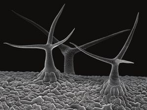
Plant cell identity | 4203

morphological structures. Classical concepts aim to trace the                   be illustrated by the cell types that together constitute the epi-
origins of ‘novel’ organs or structures from pre-existing struc-                dermis tissue (Fig. 1A). The plant epidermis forms the outer
tures, for example by exploring and conceptualizing ‘hidden’                    cell layer of the plant like the skin of the human body, and
evolutionary relationships and commonalities of shoots, and                     its primary functions are to protect inner tissues and to act as
vegetative and floral organs (see, for example, Arber, 1950).                   the communication and exchange surface with the environ-
With the origins of molecular genetics, evolutionary devel-                     ment (Glover, 2000). Epidermal pavement cells of Arabidopsis
opmental biology has revolutionized our understanding of the                    leaves are usually shaped like the interlocking pieces of a jigsaw
molecular mechanisms underlying the evolutionary origins                        puzzle. Pavement cells in sepals instead are boxy and differenti-
and diversification of plant organs and cell types. Gene duplica-               ated into giant and small cells, while the adaxial epidermal cells
tions followed by sub- and neofunctionalization, co-option of                   in petals are conical and uniform (Glover, 2000; Roeder et al.,
genes, and gene regulatory modules were found to contribute                     2010; Huang and Irish, 2015). Meristemoids within the abaxial
to the striking morphological complexity in higher plant spe-                   epidermis of developing leaves and in floral organs give rise to

                                                                                                                                                                Downloaded from https://academic.oup.com/jxb/article/72/12/4202/6232700 by guest on 22 October 2021
cies. However, in very practical terms, this creates challenges,                stomata.Trichomes develop typically at regular distances in the
for instance since many developmental control genes act in                      abaxial side of leaves and sepals; however, trichome anatomy is
more than one developmental process linked to evolutionary                      highly variable among organs of a plant. Cellular characteristics
history. For example, many genes controlling vegetative leaf                    reflect functional differences of epidermis cells among organs.
development are also expressed in floral organs, and their ac-                  For example, epidermal cells in petals enhance attractiveness to
tivities are modulated to give rise to specific structures of floral            pollinators. Specialized epidermal cells in carpels, the stigma,
organs. This often makes it hard to identify marker genes that                  receive and induce the germination of pollen grains (Glover
are very specifically expressed in only one cell type or differ-                and Martin, 1998; Balanzà et al., 2014). Together, the example
entiation stage. One might compare the problem of defining a                    of the epidermis shows that the cell type composition of tissues
‘cell type’ in plants with that of the ‘species concepts’ in plants             can be variable across organs in a plant, and that individual cell
(Christenhusz, 2020), in that it can be difficult to distinguish                types can have different phenotypes and function. Even more,
cell ‘types’ from ‘states’, and cells with the same ‘identity’ may              cellular morphology and cell type frequencies within tissues
appear very different from each other in terms of gene ex-                      can be plastic and affected by environmental factors (see, for
pression profiles. The challenge is thus to deduce cell ‘history’,              example, Wardlaw, 1952; Casson and Gray, 2008).
similar to deducing the natural history of species.                                Cellular differentiation in plants can be reversible under spe-
   The idea that a generalized cell type concept does not fully                 cific conditions. Plants are sessile organisms and have to cope
reflect the cellular diversity in form and function in plants can               with injuries caused by environmental stimuli or biotic attacks.

      A                                                                                       B

                                                                                                     GL3
                                                                                                             WD40
                                                                                                     GL1                                     GL1

            Stomata and pavement                    Trichome cells (leaf)
            cells (leaf)                                                                                          AG
                                                                                                                (In flower)          R3    WD40
                                                                                                       R3                            MYB
                                                                                                       MYB                           GL3

           Papillate cells          Giant cells and     Conical cells (petal)                       Trichome cell               Non-trichome cell
           (carpel)                 small cells (sepal)
Fig. 1. Diversity of epidermal cell types and control of trichome development. (A) Epidermal cell types, including stomatal guard cells and pavement cells
on a leaf, trichome cells on a leaf, papillate cells on a carpel (scale bar, 500 μm), giant cells and small cells on a sepal (scale bar, 100 μm), and conical
cells on a petal (scale bar, 15 μm). (B) Movement of R3 MYB functions as a positional signal for trichome patterning (after Grebe, 2012). AG serves as
an upstream lineage signal in flowers to suppress trichome formation. Solid lines represent direct regulation of target genes, and dashed lines represent
movement. SEM images in (A) are reprinted with permission from Riglet et al. (2020) (‘papillate cells’), Meyer et al. (2017) (‘giant cells and small cells’),
Yang et al. (2019) (‘conical cells’), Emmanuel Boutet (Plant biocurator for UniProtKB/Swiss-Prot) (‘stomata and pavement cells’), and Stefan Eberhard
(University of Georgia, USA; provided by Wellcome collection under a CC BY-NC 4.0 licence) (‘trichomes’).
4204 | Xu et al.
Somatic cells can be reprogrammed to regenerate new organs information or cell–cell interactions (Szymkowiak and Sussex,
or repair damaged tissues (Ikeuchi et al., 2019). Laser-assisted 1996). For instance, during leaf development, epidermal cells
elimination of cells in Arabidopsis root triggers the cells ad- regularly undergo anticlinal divisions to form the outermost
jacent to the injury to re-activate stem cell pathways, change layer. However, in chimeras, occasionally small sectors derived
the cell division orientation accordingly, and acquire the cell from periclinal divisions of epidermal cells have been observed.
fates of the missing cells to replace them (Marhava et al., 2019). These cells, although of epidermis lineage, adapt to their new
Other kinds of stresses, such as osmotic, heavy metal ion, and position and differentiate as internal mesophyll cells (Stewart
dehydration stress, can also induce plant cells to regenerate and Burk, 1970; Stewart and Dermen, 1975). This phenom-
(Ikeda-Iwai et al., 2003). The questions remain of which fac- enon has also been observed in the root meristem. If a cortical
tors and mechanisms determine stem cell identity in plants, initial cell is laser ablated, the adjacent pericycle cell switches
how different stem cell niches can be distinguished from one its fate and takes the position of the cortical cell to continue
another (beyond the activity of some master regulators), and forming corresponding cell files (van den Berg et al., 1995).
Downloaded from https://academic.oup.com/jxb/article/72/12/4202/6232700 by guest on 22 October 2021
what are the exact mechanisms underlying dedifferentiation Plant cells may alter their identity when positional signals are
versus terminal differentiation of specific cell types. changed.This is exemplified by observations on wound healing
Altogether, the concept of ‘cell type’ aims to simplify the and de-/re-differentiation. In the past ~30 years, the molecular
complex nature of cellular diversity in multicellular plants nature of many signaling mechanisms controlling cellular dif-
(and animals). Therefore, it is important to understand the ori- ferentiation and cell type specification in plants has been elu-
gins and consequences of heterogeneity within and among cidated. Combinations of genetic analyses and computational
cell types, and the mechanisms underlying their organ- and modeling have allowed us to gain insights into the regular na-
environment-specific modulation. ture of the positioning mechanisms.
Cell lineage versus positional signals
Signals controlling cell type specification
Understanding the principles that govern cell type specifica- in plants
tion in multicellular organisms is one of the major challenges
in developmental biology. The fundamental concept of ‘epi- Different mechanisms explaining the relationships between
genetic landscape’ introduced by Waddington in 1957 visu- relative position, spatial patterns, and cell fate in developing
alizes cell differentiation as a ball rolling down a valley in a organisms have been proposed. A major and often considered
landscape that is sculpted by regulatory genes and their com- mechanism is the formation of gradients of signaling molecules,
binatorial activities (Waddington, 1957). This path forms the such as morphogens, that result in the specification of distinct
developmental trajectory or ‘lineage’ of the cell defined by its cell fates in a concentration-dependent manner (Wolpert,
start position and by dynamic but predictable changes in gene 1996). Mobile signaling molecules in plants reported to con-
activities determined by the regulators. In the animal field, re- trol patterning include phytohormones, mobile transcription
construction of cell lineage history is often used to under- factors (TFs), non-coding RNAs, and small signaling peptides.
stand cellular differentiation programs (Kretzschmar and Watt, A second general model for explaining position-dependent
2012; Morris, 2019). A classical example for utilizing the lin- cell specification does not rely on a gradient, but on biochem-
eage concept in plants is stomata development, which is ini- ical signaling between neighboring cells in ‘boundary’ regions,
tiated from a lineage-specific stem cell via a series of defined essentially resulting in self-organization of the system (Sharpe,
asymmetric cell divisions controlled by consecutively acting 2019). The idea that mechanical signals play a role in cellular
regulatory factors and enforced by cell–cell signaling (Han and differentiation and patterning in plants has been recognized
Torii, 2016). for a long time (see, for example, Arber, 1950), but is also an
However, many experiments have shown that the devel- exciting focus of ongoing research.
opmental fate of a plant cell does not depend strictly on its
lineage, but on its exact position within the growing plant The phytohormone auxin in flower development
body (Stewart and Dermen, 1975; van den Berg et al., 1995;
Szymkowiak and Sussex, 1996; Berger et al., 1998; Kidner et al., Graded auxin accumulation has been shown to play important
2000; Costa and Shaw, 2006; Costa, 2016). In early studies, plant roles in developmental patterning, while a primary effect is on
scientists utilized mosaic or chimera experiments to trace the cell expansion (Leyser, 2018), thereby controlling processes such
lineage of a cell type by labeling cell clones with genetically as vascular development and specification of floral meristem
phenotypic traits, such as ploidy level or albinism, and tracking founder cells. Auxin is actively transported in a polar manner
the mitotic descendants of marked cells (Poethig, 1987, 1989; between cells via transport proteins, such as PIN-FORMED
Dawe and Freeling, 1991; Irish, 1991; Scheres, 2001). Chimera (PIN) efflux carriers. The pin1 mutant fails to produce flowers
studies have shown that plant cells do not follow strict lineages, and presents a pin-shaped inflorescence (Okada et al., 1991).
and their fates are not pre-determined but rely on positional Flower initiation can be rescued when indole acetic acid (IAA)Plant cell identity | 4205
is applied exogenously to the pin1 mutant (Reinhardt et al., size of the stem cell niche in the floral meristem has a direct
2000). Cellular specificity of auxin responses is linked to the effect on the number of floral organs that are produced by it.
complexity of auxin sensing and response pathways in the cell The clv3 mutant leads to overproliferation of the shoot ap-
(Leyser, 2018).After binding to receptors of the TRANSPORT ical meristem (SAM) cell population, while overexpression of
INHIBITOR RESPONSE1/AUXIN SIGNALING FBOX CLV3 eliminates the stem cell niche and results in termination
(TIR1/AFB) family, auxin controls gene expression in the nu- of shoot development (Fletcher et al., 1999; Kondo et al., 2006).
cleus via activation of AUXIN RESPONSE FACTOR (ARF) CLV3 is shown to be expressed in stem cells at the meristem
TFs by targeted degradation of AUXIN/INDOLE-3ACETIC apex. It diffuses toward inner layers of the meristem and organ-
ACID (AUX/IAA) repressor proteins (Weijers and Wagner, izing center, where it interacts with the leucine-rich receptor-
2016).The initiation of flower meristems is marked by the for- like kinase (LRR‐RLK) CLV1 and related proteins to restrict
mation of local maxima of auxin (Benková et al., 2003; Heisler the expression of a homeodomain TF protein WUSCHEL
et al., 2005). A series of auxin responses is started by the activa- (WUS), which is a positive regulator of the stem cell popula-
Downloaded from https://academic.oup.com/jxb/article/72/12/4202/6232700 by guest on 22 October 2021
tion of ARF5 (also known as MONOPTEROS), which pro- tion (Mayer et al., 1998; Fletcher et al., 1999; Schoof et al., 2000;
motes the expression of TFs mediating floral meristem identity, Kinoshita et al., 2010). Furthermore,WUS can also migrate to-
including LEAFY (LFY) (Yamaguchi et al., 2013, 2016). LFY ward the CLV3-expressing cell layers, where it activates CLV3
further promotes the expression of APETALA1 (AP1) (Parcy expression by directly binding to its genomic region (Yadav
et al., 1998; Wagner et al., 1999). LFY and AP1 play a cen- et al., 2011). This negative feedback loop between WUS and
tral role in flower meristem specification and they regulate a CLV3 is well established to maintain a proper cell population
large number of downstream genes for flower formation (Parcy in the SAM. Consequently, this pathway regulates meristem
et al., 1998; Kaufmann et al., 2010b;Winter et al., 2011). Besides size and by this controls the number of organs produced in a
a role in early activation of flower development, auxin has an flower: clv3 mutants typically produce more floral organs, while
instructive role in patterning of organs within the flower, as overexpression results in flowers without inner whorls of sta-
revealed by higher order yucca mutants that are defective in mens or carpels (Fletcher et al., 1999).
auxin biosynthesis (Zhao et al., 2001; Tobeña-Santamaria et al., miRNAs function by binding to complementary sites in
2002; Cheng et al., 2006). Besides this, other ARF TFs were mRNA molecules to trigger the degradation or translational
found to direct organ polarity as well as stamen differentiation inhibition of target genes. Small RNAs can diffuse across tis-
(Przemeck et al., 1996; Sessions et al., 1997; Nagpal et al., 2005; sues, resulting in concentration gradients, thereby potentially
Simonini et al., 2016; K. Zhang et al., 2018). To explain the mediating tissue patterning through dose-dependent activity
multiple functions of auxin, we need a better understanding of (D’Ario et al., 2017). miRNAs have been found to regulate the
the cell type-specific composition of the auxin response ma- activity of TFs and other regulatory proteins in flower develop-
chineries. This requires knowledge of quantitative abundance ment. A classical example is miR172, which accumulates in the
of specific auxin signaling factors across cellular differentiation SAM at the onset of flowering, preventing the transition of the
in the flower, direct ARF targets, along with their affinity and center of the SAM into a flower meristem by inhibiting AP2
specificity for protein–protein and protein–DNA interactions. expression (Aukerman and Sakai, 2003; Chen, 2004). miR172
At the same time, the chromatin landscape of a cell type may also accumulates in the center of the flower primordia to sup-
affect auxin response. Addressing these questions will help to press AP2, avoiding AP2 and AG co-expression and thereby
understand how transient peaks in auxin concentration can setting the boundary between petal and stamen whorls.
trigger cellular patterning responses, and to distinguish direct miR172 is itself down-regulated by AP2, establishing a nega-
effects from indirect downstream developmental decisions. tive feedback loop that is essential for the correct specification
of organ identity (Zhao et al., 2007; Wollmann et al., 2010).
Mobile regulatory molecules in flower development Several miRNAs are involved in the process of floral organ de-
velopment partially by crosstalking with plant hormones. For
Small peptides are usually4206 | Xu et al.
GL1, activating not only the expression of positive regulators cell, thus establishing a feedback loop (Uyttewaal et al., 2012).
for trichome cell fate determination but also the expression of Mechanical forces crosstalk with biochemical signals, for in-
R3-MYB TFs. R3-MYB proteins form a complex with GL3 stance in the generation of phyllotactic patterns. Auxin minima
in the neighboring cells, thus preventing the formation of the reside in organ boundaries, and these regions are characterized
GL3–GL1 complex (Pattanaik et al., 2014). Cell fate decision by the expression of a specific group of TFs, such as CUP-
during root hair development shares a similar mechanism, since SHAPED COTYLEDON1 (CUC1), CUC2, and CUC3,
movement of similar regulatory proteins between root hair and which limit cell growth and thus create a creased shape in
non-root hair cells reinforces their identity (Salazar-Henao the boundaries (Heisler et al., 2005; Rast and Simon, 2008).
et al., 2016) (Fig. 1B). During flower development, trichome Microtubule arrangement and polar auxin transport in the
formation in carpels is suppressed by the floral homeotic boundaries are regulated by mechanical forces (Heisler et al.,
AGAMOUS (AG) TF via several target pathways, including 2010; Landrein et al., 2015). Mechanical signals interplay not
direct repression of GL1 (Ó’Maoiléidigh et al., 2018). only with auxin, but also with miRNA regulation.The expres-
Downloaded from https://academic.oup.com/jxb/article/72/12/4202/6232700 by guest on 22 October 2021
Together, these examples show that cell to cell signaling sion of CUC genes in organ boundaries is regulated by both
impacts developmental patterning and cell identity specifica- miRNA and mechanical forces (Fal et al., 2016). Interestingly, it
tion. Developmental programs thus represent the sum of ‘en- was reported that the shape of nuclei correlates with cell shape
dogenous’ cellular status and different signaling cascades that and size in plants (Meier et al., 2016). Mechanotransduction
emerge from cell to cell signaling. can affect the shape of the nucleus via interaction with the
cytoplasmic microtubule cytoskeleton, and ‘nuclear stiffness’
Mechanical signals in floral organ differentiation may affect transcriptional regulation and gene activity (Finan
and Guilak, 2010; Goswami et al., 2020b; Irianto et al., 2013;
The role of mechanical forces in developmental patterning Lovett et al., 2013).
has long been acknowledged. For example, in The natural phil- In summary, mechanical forces provide positional signals
osophy of plant form (1950), Agnes Arber states ‘Judging merely by regulating organ growth rate, cell and organ shape, thereby
from inspection, it looks as if limitation of the space into which contributing to cellular differentiation and cell type frequen-
to expand, and the actual pressure which the developing parts cies within organs.
exert upon one another, must be the efficient cause [for dif-
ferent shapes of various members of the plant body]’.
Plant cells sense internal and external forces that can be per-
Insights into lineage-based mechanisms
ceived as growth signals that have important effects on and af-
fect the shape of cells and organs (Hervieux et al., 2017; Sapala The cell lineage concept suggests that a cell’s fate is deter-
et al., 2018). Mechanical signals defining epidermal cell morph- mined early by its progenitors. Cells pass on specific cell fate
ology are at least in part perceived by stress-dependent, katanin- decisions to their progeny across cell division (Stent, 1985).
mediated alignment of microtubules in the cytoplasm that in Although positional mechanisms play an important role in cell
turn guide cellulose-synthesizing complexes in the apoplast fate specification in plants, lineage-based mechanisms cannot
(Hamant et al., 2008; Jacques et al., 2013; Sampathkumar et al., be neglected. If cell fate is specified and maintained solely by
2014). Sepal development has been used as a model to study positional information, cells would have to re-establish their
the contribution of mechanical signals to local growth be- expression programs based on their new position at every div-
cause of its high variability in growth rate within tissues and ision (Costa, 2016). However, data from roots suggest that cells
its robustness in the final shape of the organ (Tauriello et al., that misexpress a cell identity marker gene frequently pass on
2015; Hervieux et al., 2017). Trichome precursor cells in sepals the ‘wrong’ identity to their progeny (Costa, 2016). Once cell
initially grow and expand much faster than surrounding epi- fate is specified by positional information, it is clonally main-
dermal cells, which potentially can distort the final shape of tained by lineage until they receive new positional input (Costa,
the sepal. However, neighboring cells of trichome precursors 2016). The critical role of TFs and epigenetic regulators also
organize their microtubule arrays according to the mechanical indicates the existence of a lineage-based component in plant
changes caused by trichome precursor cells, and thus grow at cell fate determination. Stomatal differentiation follows an evi-
a reduced rate to maintain sepal shape (Hervieux et al., 2017). dent cell lineage from meristemoid mother cells to mature
Mechanical forces are involved in the control of sepal size. guard cells (Yang and Sack, 1995; Larkin et al., 1997; Nadeau
Tangential tension at the tip of the sepal causes the arrest of and Sack, 2002). Several basic helix–loop–helix (bHLH) TFs—
growth at the tip by reorientation of the microtubule array SPEECHLESS (SPCH), MUTE, and FAMA—function at
(Hervieux et al., 2016). each stage to control the lineage. The overexpression of SPCH
Another example of mechanical signaling comes from the induces extra asymmetric divisions and the production of
SAM, where cells orient their cortical microtubules along excess stomata (MacAlister et al., 2007). Ectopic MUTE ex-
the lines of mechanical stress generated during tissue forma- pression in the petal that is normally devoid of stomata con-
tion, and this then affects the mechanical properties of the verted petal epidermal cells into stomata (Pillitteri et al., 2008).Plant cell identity | 4207
Induced expression of FAMA transforms cotyledon epidermal is mediated by direct promoter interactions (Kaufmann et al.,
cells into guard cells (Ohashi-Ito and Bergmann, 2006). 2010b; Winter et al., 2011). Recent work also explains how
In more general terms, key TFs have been identified that LFY binds DNA in a nucleosomal context and enhances chro-
trigger cell lineage differentiation, while the initial activation of matin accessibility at its target loci such as AP1 (Jin et al., 2021;
these TFs may be dictated by positional signaling at early stages Lai et al., 2021). The activation of LFY and AP1 is further-
in plants and animals (Scott and Carroll, 1987; St Johnston and more under positional control by ARF5 (Wagner et al., 1999;
Nüsslein-Volhard, 1992; Scheres, 2001). Yamaguchi et al., 2013, 2016).
Floral organ identity is specified by homeotic TFs of the
Pioneer transcription factors, organ identity, and cell MADS-box family that interact in a combinatorial manner to
type specification specify different types of floral organs (Causier et al., 2010).
Combined loss of function of floral homeotic proteins results
Over the past decades, genetic analyses have identified floral in the transformation of all floral organs into cauline leaf-like
Downloaded from https://academic.oup.com/jxb/article/72/12/4202/6232700 by guest on 22 October 2021
regulators and discovered detailed insights into how they organs (Bowman et al., 1991). Furthermore, the redundantly
interact and cooperate to control flower development. Plant acting SEPALLATA (SEP) TFs are essential for the specifica-
morphogenesis depends on the combinatorial interplay of tion of all floral organ types. Accordingly, loss of SEP func-
TFs to mediate distinct and dynamic spatiotemporal gene tion results in the conversion of the floral organs into leaf-like
expression, associated with feedback control (Zik and Irish, structures (Pelaz et al., 2000). SEP proteins act as mediators of
2003; Krizek and Fletcher, 2005; Alvarez-Buylla et al., 2010; higher order complex formation of other homeotic TF classes
Ó’Maoiléidigh et al., 2014; Thomson and Wellmer, 2019). (Theißen and Saedler, 2001; Immink et al., 2009). According
Floral organ specification essentially requires modification of to the floral quartet model, petals are specified by a complex
leaf developmental pathways, including changes in growth, consisting of AP1, APETALA3 (AP3), PISTILLATA (PI), and
cell type frequencies, cellular morphologies (e.g. trichomes in SEP proteins. In contrast, stamens are specified by a complex
sepals versus leaves; conical cells in the petal epidermis) (Fig. of AP3, PI, SEP, and AG proteins. Sepals are specified by com-
1A), and the establishment of flower-specific cell and tissue plexes formed by SEP and AP1 proteins, while carpels are
types that are not found in leaves (e.g. in reproductive organs). specified by a SEP/AG tetramer (Honma and Goto, 2001;
Cell type specification requires orchestrated changes in Theissen, 2001).
global gene expression programs. So-called ‘pioneer TFs’ con- Cooperative and combinatorial interactions are important
trol cell type programming and reprogramming by promoting for floral homeotic TFs and may facilitate their action as pi-
chromatin opening to make it accessible for other TFs (Zaret oneer factors (Kaufmann and Airoldi, 2018). Vegetative leaves
and Carroll, 2011; Iwafuchi-Doi and Zaret, 2014, 2016; Zaret of transgenic plants that constitutively express SEP3–AP3–PI
and Mango, 2016; Lai et al., 2018). Growing evidence suggests or AP1–AP3–PI are converted into petals, showing that these
that pioneer TFs contribute to the regulation of developmental TFs are not only required but also sufficient to specify floral
switches in plants (see, for example, Tao et al., 2017, 2019; Lai organ identity (Honma and Goto, 2001). SEM revealed that
et al., 2018). cells on both the abaxial and adaxial surface of the converted
Several key TFs are required for the leaf-to-flower transition. rosette leaves closely resembled cells on the surface of the
LFY specifies flower meristem identity. Combinatorial expres- petals (Pelaz et al., 2001). Cauline leaves of AP3–PI–SEP3–AG
sion of LFY and WUS induces the generation of floral organs ectopic expression lines were converted into staminoid organs,
on primary and lateral root tips (Gallois et al., 2004), and indu- and all floral organs are transformed into stamens or staminoid
cible expression of LFY in root explants is sufficient to trigger organs. Homeotically converted cauline leaves of these trans-
flower formation, bypassing elaboration of a shoot (Weigel genic plants consist of two distinct regions whose epidermal
et al., 1992;Wagner et al., 2004). Furthermore, expression of the cells exhibit a morphology similarity to that of anthers and
LFY co-regulator UNUSUAL FLORAL ORGANS (UFO) filaments, respectively (Honma and Goto, 2001). Combination
fused to a VP16 activation domain resulted in ectopic forma- of genome-wide ChIP-seq and Dnase I-seq time-series ex-
tion of flowers and inflorescences in vegetative leaves in the periments suggested that AP1 and SEP3 facilitate the opening
presence of a functional LFY gene (Risseeuw et al., 2013). of closed chromatin and promote gene activation in flower
Protein oligomerization via a SAM domain enabled LFY to development (Pajoro et al., 2014), indicating roles as pioneer
bind to closed chromatin regions (Sayou et al., 2016).The func- factors. Homeotic TFs are expressed throughout flower de-
tions of the floral meristem factors LFY and AP1 are closely velopment. The analysis of target networks of homeotic TFs
linked, and they regulate each other’s expression. For example, allows us to interrogate how homeotic TFs modulate organ
the expression level of LFY is reduced in ap1 cal double mu- growth and cellular morphology, and establish novel cell iden-
tants, and the onset of expression of AP1 is delayed in the lfy tities not found in vegetative leaves (Yan et al., 2016; Chen et al.,
mutant (Weigel and Nilsson, 1995). The ap1 mutant can sup- 2018). For example, the homeotic gene AG acts in concert
press the terminal flower phenotype of the constitutive expres- with the general organ polarity gene KANADI1 to suppress
sion of LFY (Weigel and Nilsson, 1995). This cross-regulation trichome initiation in the carpel epidermis (Ó’Maoiléidigh4208 | Xu et al.
et al., 2018). An example for a flower-specific gene activa- in the case of FLOWERING LOCUS T (FT) (Farrona et al.,
tion is SPOROCYTELESS, which is activated by AG in early 2011). In more general terms, the phenotypes of Polycomb
stages of floral organ development and plays an essential role mutants strongly suggest broad roles in the mediation of de-
in patterning processes related to sporogenesis (Schiefthaler velopmental phase transitions and the commitment to cellular
et al., 1999;Yang et al., 1999; Ito et al., 2004). The finding that differentiation (Mozgova et al., 2015).
floral organs were derived from leaf-like organs during evo- Epigenetic marks at specific genomic loci can be dynamic-
lution can also explain the fact that many developmental TFs ally regulated, for example by TFs that interact with epigenetic
with roles in cellular patterning appear to act in more than factors. Marks can be erased, re-written, or diluted by cell div-
one developmental process and display tissue- and organ spe- ision. For example, the B3 domain TFs LEAFY COTYLEDON
cific functions, since this could be explained by evolutionry 2 (LEC2) and FUSCA3 (FUS3) displace VAL1 and VAL2
co-option and diversification of ancestral regulatory programs. (two key components for Polycomb-mediated FLC silencing
In fact, this complicates analyses of cell identity in plants, since by vernalization) during early embryogenesis from the cold
Downloaded from https://academic.oup.com/jxb/article/72/12/4202/6232700 by guest on 22 October 2021
only a few genes are entirely characteristic to only one spe- memory cis-element of FLC to disrupt Polycomb silencing
cific cell type. For example, TFs controlling abaxial and ad- and thus prevent H3K27me3 maintenance at FLC during the
axial identity were recruited to control patterning in lateral rapid embryonic cell divisions (Tao et al., 2019). During flower
organs, the stem and roots, and their activity can be modulated initiation and morphogenesis, TFs such as LFY, MADS-box
in a floral organ-specific manner (e.g. Siegfried et al., 1999; proteins, and ARF have been shown to modulate chromatin
Yamaguchi et al., 2004). status by recruiting ATP-dependent nucleosome remodelers or
general transcriptional co-regulators (Smaczniak et al., 2012;
The role of epigenetics in plant cell lineage Wu et al., 2012, 2015). In sum, the current data suggest that
specification epigenetic programming plays a role in cell lineage commit-
ment in plants. The investigation of tissue- and stage-specific
While cell identity can be programmed by cell type-specific dynamics of epigenetic profiles can be expected to shed more
TFs, the robustness of the acquired transcriptional status de- light on the underlying molecular mechanisms.
pends on the chromatin environment (Hennig and Derkacheva,
2009; Costa and Dean, 2019). Epigenetic memory can be es- Synergistic action of position- and lineage-based cell
tablished during developmental progression because of stable fate control
and heritable epigenetic modifications (Huang et al., 2013;
Costa and Dean, 2019 and references therein). During cell div- The spatiotemporal expression pattern of floral homeotic
ision, the transcriptional status of genes can be recorded and TFs, and thereby the whorled organization of the flower, is
transmitted to daughter cells via epigenetic regulation (Iwasaki facilitated by multiple factors, including epigenetic factors,
and Paszkowski, 2014). To date, many epigenetic modifications positional signals, and regulatory feedback control (Alvarez-
have been found, such as DNA methylation, histone methyla- Buylla et al., 2010; Denay et al., 2017; Thomson and Wellmer,
tion, and histone acetylation. Chromatin modifications can be 2019). An example is provided by AG activity that is restricted
linked with gene activation or repression or a ‘poised’ state. For to the inner whorls of the floral meristem giving rise to sta-
example, H3K4me3 and H3K36me3 are associated with gene mens and carpels. Besides being a PcG (Polycomb Group)
activation, while H3K27me3 and H3K9me2 are commonly target, AG expression is prevented in the outer floral whorls
linked to transcriptional repression (Pikaard and Mittelsten via the activity of histone deacetylases (Tian and Chen, 2001;
Scheid, 2014). Chen and Tian, 2007). Moreover, miR172 acts as a positional
How epigenetic modifications store and transmit ‘memory’ signal to restrict AG action by regulating the spatiotemporal
to daughter cells has been intensely studied. Inheritance of his- activity of AP2, which is a known repressor of AG (Aukerman
tone marks by daughter cells requires the collaboration between and Sakai, 2003; Chen, 2004; Zhao et al., 2007; Wollmann
the DNA replication machinery, chromatin modifiers, and et al., 2010). Activation of AG in the inner whorls is mediated
chromatin modifications themselves (Stewart-Morgan et al., by the combined activity of several factors including WUS,
2020). Polycomb factors are known factors controlling epigen- LFY, and PERIANTHIA (Lenhard et al., 2001; Lohmann
etic memory across cell division that mediate trimethylation of et al., 2001; Maier et al., 2009). An autoregulatory feedback
histone 3 Lys27 (H3K27me3). During the DNA replication at loop, possibly involving the interaction with SEP factors, con-
mitosis, parental nucleosomes with H3K27 tri-methylation re- tributes to the stable AG activity (Gomez-Mena et al., 2005;
cruit polycomb repressive complex 2 (PRC2) which catalyzes Kaufmann et al., 2009). This and other examples show that
the trimethylation on daughter strand DNA (Jiang and Berger, spatiotemporal gene expression determining cell identity re-
2017).This mediates the stability of the repressed status at many quires combinatorial interplay of several factors (Fig. 2), and
developmental gene loci, across developmental stages, such as emphasizes the need for novel technological and computa-
in the case of FLOWERING LOCUS C (FLC) (reviewed in tional approaches to understand the underlying cis-regulatory
Costa and Dean, 2019), and in a tissue-specific manner, such as grammar.Plant cell identity | 4209
Downloaded from https://academic.oup.com/jxb/article/72/12/4202/6232700 by guest on 22 October 2021
Fig. 2. Cell type-specific gene expression integrates lineage, positional, and environmental gene expression. This model suggests an example
mechanism of combinatorial control by TFs that act together, resulting in cell type-specific activation of target genes. Lineage (TF1)- and position (TF2)-
specific factors can change the epigenetic status of the promoter region, for example by interacting with ATP-dependent nucleosome remodelers. In
some instances, cell identity or anatomy can be modulated by environmental factors, thereby, for example, changing the frequency of cell types or cell
shape in the epidermis. Abbreviations: LR, light receptor; SWI/SNF, SWItch/Sucrose Non-Fermentable (a type of ATP-dependent nucleosome remodeler);
PosRe, position-responsive element; CArG. CArG-box (MADS TF-binding motif, as an example for organ-specific regulation); LRE, light-responsive
element (an example for environmental regulation); basal, RNA polymerase II binding site.
Excellent examples for crosstalk of epigenetics and Scientists started to realize that stochasticity is needed to create
phytohormones have been described in controlling floral meri- small differences between identical cells, which are then amp-
stem specification. In general, some phytohormones, such as lified and stabilized by feedback loops to begin cell differenti-
auxin, gibberellic acid, and brassinosteroids, have been shown ation (Meyer and Roeder, 2014). Experimental confirmation
to affect epigenetic modifications (Yamamuro et al., 2016). of this theory is the study of the variable defects in the LEC2
In the absence of auxin, Aux/IAA proteins repress ARF5 ac- mutant embryo, where FUS3 expression appears in randomly
tivity in the SAM by interacting with ARF5 and recruiting positioned patches. The explanation might be that residual
the transcriptional co-repressor TOPLESS (TPL). TPL in turn ABSCISIC ACID INSENSITIVE3 (ABI3) expression fails to
interacts with histone deacetylase HDA19, thus removing his- induce the expression of FUS3 in some parts of the embryo
tone acetylation at ARF5 target loci, thereby preventing gene while it succeeds in triggering FUS3 expression in other parts
activation (Eberharter and Becker, 2002; Long et al., 2006; of the embryo, and the positive feedback loop can stabilize the
Szemenyei et al., 2008). Upon auxin sensing, Aux/IAA proteins expression of these two genes in these embryo parts (To et al.,
are rapidly degraded, leading to the dissociation of TPL and 2006).
HDA19, thereby freeing ARF5 to activate its targets (Wu et al., Stochasticity happens at both cellular and molecular levels.
2015; Lavy and Estelle, 2016). Furthermore, in the presence of For instance, during the growth of microtubule arrays, sto-
auxin, ARF5 recruits ATP-dependent SWI/SNF remodeling chastic disassembly of individual microtubules allows them
complexes to its targets, including LFY and FILAMENTOUS to go through various configurations and form optimal ones
FLOWER. This enhances chromatin accessibility at these loci (Holy and Leibler, 1994; Allard et al., 2010; Eren et al., 2010).
and activates transcription linked with increased H3K9ac (Wu Studies have shown that the growth rates of leaf epidermal
et al., 2015). cells in Arabidopsis differ by several fold from each other, and
change in time. This spatiotemporal variability is not related to
the size of either the cell or the nucleus (Elsner et al., 2012).
Stochastic gene expression has also been described in various
Stochasticity in cell fate determination
organisms, including plants (Elowitz et al., 2002; Paré et al.,
It is tempting to consider cell type specification as a fully deter- 2009; Dar et al., 2012; Ietswaart et al., 2017). Gene expression
mined process because of the highly reproducible tissue growth noise can be divided into two types: extrinsic noise which is
and organogenesis. However, the cellular and molecular be- due to fluctuations in the cellular or external environment that
haviors underlying cell type specification are often stochastic. affect the overall expression in a cell, and intrinsic noise which4210 | Xu et al.
is due to the inherent fluctuations of transcription and transla- An scRNA-seq experimental workflow usually begins
tion of a particular gene within a cell (Elowitz et al., 2002).The with the dissociation and isolation of single cells from a tissue.
use of a dual reporter system in plants helped distinguish be- However, in plants, the process of isolating single cells embedded
tween extrinsic and intrinsic noise in Arabidopsis, and revealed in a rigid cell wall matrix is technically challenging, and it is usu-
that fluctuation in gene expression is coupled in neighboring ally achieved by incubating plant tissues with cell wall-digesting
cells in young leaves (Araújo et al., 2017). The trichome distri- enzymes to release protoplasts. Protoplast response genes can
bution pattern also emerges stochastically, and the variability cause artifacts in the downstream data analysis (Tucker et al.,
in the trichome distribution pattern correlated with stochastic 2018; Denyer et al., 2019; Jean-Baptiste et al., 2019; Ryu et al.,
cell to cell variation in GL3 expression (Okamoto et al., 2020). 2019; Shulse et al., 2019), and the time and harshness of the di-
One of the most compelling examples of stochasticity is gestion that are required to digest the cell walls differ between
from cell type specification in the sepal epidermis. Sepals have tissues and organs. Thus, this method is not applicable to all tis-
both giant cells that are very long, usually stretching one-fifth sues, and longer digestion times may aggregate artifacts. An al-
Downloaded from https://academic.oup.com/jxb/article/72/12/4202/6232700 by guest on 22 October 2021
the length of the sepal, and small cells that are much smaller in ternative way to address this issue is to isolate nuclei, for example
size (Figure 1; Roeder et al., 2010, 2012). The correct propor- by tissue chopping or grinding and by cell membrane lysis (e.g.
tion of giant cells and small cells is required for the curvature Thibivilliers et al., 2020 Deal and Henikoff, 2010; Kaufmann
of the sepal; with an altered proportion of giant cells, sepals are et al., 2010a). It has been shown that the composition of the
unable to enclose and protect the developing floral organs in RNA pool from plant nuclei is representative of that from the
the inner whorls (Roeder et al., 2010, 2012). In the early whole cell (Deal and Henikoff, 2010). However, the isolation
stage of sepal development, the levels of the epidermis regu- procedure and the loss of mechanical connection with other
lator ARABIDOPSIS THALIANA MERISTEM LAYER1 cell components, particularly the cytoskeleton, may change the
(ATML1) fluctuate in sepal cells. When ATML1 reaches a shape of the nucleus and impact gene expression (Goswami
high level, specifically at the time of cell division, that cell will et al., 2020a). Additionally, dealing with the sparse RNA from
be determined to become a giant cell, whereas if the level of nuclei is challenging for both the experimental and the compu-
ATML1 is low at this point in time, the cell will keep dividing tational parts of the work. Further optimizing tissue dissociation
and remain small. Thus, the stochastic fluctuations in the con- methods, especially for recalcitrant plant tissues is fundamentally
centration of the TF ATML1 initiate the pattern of giant and critical to apply scRNA-seq to plant science.
small cells in the Arabidopsis sepal (Meyer et al., 2017). The What we learned from scRNA-seq is that no two cells are
examples presented above indicate that plants utilize stochastic transcriptionally the same (Choi and Kim, 2019). Nevertheless,
mechanisms to establish robust and reproducible morphology. clustering of cells with similar expression or epigenetic pro-
files is often used to annotate cell types. Subclusters can reflect
the variation of expression patterns among cells of the same
How does single-cell omics contribute to tissue type, and may represent cell types.Taking data from roots
understanding cell identity? as an example, in the stele cell cluster, there are protoxylem,
Single-cell omics technologies phloem-like, meristematic xylem, and pericycle cells (Shulse
et al., 2019). However, heterogeneity may also reflect differ-
Despite the limitations of the cell type concept, classifying ences in cellular states, or stochastic fluctuations in gene ac-
cells can help to understand how cells or tissues function and tivity (Trapnell, 2015; Wimmers et al., 2018).
interact, and to reveal specific mechanisms that govern pro- Cells of different ontogenetic origins may have similar func-
cesses that may influence a plant’s growth, development, and tions or ‘behaviors’ in terms of gene activity. In scRNA-seq
reproduction. Recent advances in profiling molecular fea- clustering, cells are grouped based on the similarity of their
tures at single-cell resolution provide novel insights into the transcriptome. For example, the lateral root cap (LRC) cells
understanding of cell types (see, for example, Özel et al., 2021). were found to cluster with the non-hair cells and columella
Benefiting from the development of single-cell omics tech- cells (Shulse et al., 2019), which indicates that although they
nologies, researchers can now study cellular heterogeneity are different types of cells that originate from different ini-
at the levels of the transcriptome, epigenome, or proteome. tial cells surrounding the quiescent center (QC), they share a
Single-cell RNA-seq (scRNA-seq) (Tang et al., 2009), ATAC- similar transcriptome that may provide them with the ability to
seq (Buenrostro et al., 2015), ChIP-seq (Rotem et al., 2015), protect the roots (Petricka et al., 2012). It has also been shown
DNA methylation (Hui et al., 2018; Lee and Smallwood, 2018), that meristematic cells cluster together independently of pre-
metabolomics (Minakshi et al., 2019), and proteomics (Marx, cise origin. The meristematic cell clusters are close to each
2019), have emerged and are used in animal and plant research, other and consist of meristematic cells of different identities,
but scRNA-seq is still the most commonly used technique. For such as cortex identity and trichoblast identity (Denyer et al.,
example, scRNA-seq permits analysis of the expression profiles 2019). The reason for this may be because these cells share
of thousands of individual cells at the same time and can reveal meristematic features such as a high division rate, although
the heterogeneity within a group of cells. they have different ultimate cell fates.Plant cell identity | 4211
The annotation of clusters in single-cell datasets other meristem based on reporter gene expression and in situ hy-
than roots—or in species other than Arabidopsis—is typically bridization provide a resource for this kind of computational
limited by the availability of tissue-specific reference datasets technologies (Refahi et al., 2021), and could be expanded to
and specific marker genes. Such pre-knowledge of tissue- comprehensively cover all tissue types in the developing flower.
specific data can strongly enhance our capacity to annotate cells Although these computational tools can regain the positional
in single-cell omics datasets. This is also the case for the data information of dissociated cells, the dissociation procedure it-
from developing flowers, where at least some stage-specific and self may cause plant cells to alter their identity, as already dis-
floral domain-specific datasets are available (e.g. Pajoro et al., cussed above. So, many efforts have been made to retain tissue
2014; Jiao and Meyerowitz, 2010). To follow the cellular differ- spacial context by using fluorescence in situ hybridization
entiation in depth, single-cell omics on fluorescence-activated (FISH)-based methods, such as multiplexed error-robust FISH
cell sorting (FACS)-selected populations of green fluorescent (MERFISH), spatially resolved transcript amplicon readout
protein (GFP)-labeled cells can be used to increase sensitivity, mapping (STARmap), and sequential fluorescence in situ hy-
Downloaded from https://academic.oup.com/jxb/article/72/12/4202/6232700 by guest on 22 October 2021
for example in the analysis of specific tissue types (e.g. epi- bridization (seqFISH+) (Shah et al., 2016; Wang et al., 2018;
dermis) or cells of a certain status of differentiation (e.g. floral Peng et al., 2019). SeqFISH+ can image mRNAs from up
stem cells). to 10 000 genes in single cells with high resolution, allowing
Different computational approaches try to order the tran- identification of cell types based on both transcriptional pro-
scriptome of the cells obtained by single-cell omics in some file and their spatial organization in situ (Peng et al., 2019).
type of differentiation trajectory. The earlier methods were Besides, seqFISH+ can also reveal subcellular mRNA local-
based on ordering cells in a pseudotime defined by similarity, ization in single cells. However, the drawback is that these
for example as implemented in Monocle or Palantir (Trapnell kinds of methods only allow targeted studies and lack unbiased
et al., 2014; Setty et al., 2019). In this way, cells with similar examination of the whole transcriptome. A recently published
transcriptomes were ordered together in a computationally technology, expansion sequencing (ExSeq), combined expan-
generated pseudotime. The main problem with these methods sion microscopy with long-read in situ RNA sequencing, re-
is the assumption that transcriptome similarity is related to a sulting in a nanoscale visualization of the position of transcripts
similar position in the differentiation pathway, because, as we in intact tissues (Alon et al., 2021). ExSeq does not need target
stated before, cells even from different origins can have similar genes, so it is unbiased compared with other in situ sequencing
transcriptomes. New approaches to infer lineage decisions are methods, as mentioned above (Alon et al., 2021).
based on estimating the dynamic ratios of spliced and unspliced Another experimental approach to retrieve cell positional
transcripts, for example as utilized in velo or scvelo (La Manno information is spatial RNA-seq. Researchers have generated
et al., 2018; Bergen et al., 2020). We can infer reaction rates of high-quality RNA-seq data with maintained two-dimensional
transcription, splicing, and degradation by modeling the abun- positional information by lysing histological sections on ar-
dance of spliced and unspliced transcripts, therefore providing rayed reverse transcription primers with unique positional
an estimation of the latent time behind these dynamics. barcodes (Ståhl et al., 2016). A similar method, Slide-seq, trans-
fers RNA from tissue sections onto DNA-barcoded drop-seq
Towards a virtual flower: understanding cell identity in beads arrayed on a surface with known positions, allowing
its positional context whole-genome sequencing of RNA with inferred locations
(Rodriques et al., 2019). However, these technologies can
Single-cell omics procedures are associated with the loss of only capture tissues in a thin section, and each bead is not
positional information of plant cells. However, as discussed in strictly capturing RNA from a single isolated cell. Combined
the previous sections, positional information is vital for mor- with scRNA-seq, these approaches may help to map or as-
phogenesis and cell identity in plants. By combining high- sign single-cell transcriptomics data back into a tissue context,
resolution imaging of marker gene activity with single-cell overcoming the need for targeted spatial expression analyses of
omics, the position of cells in their original tissue context can marker genes.
be predicted (Satija et al., 2015; Halpern et al., 2017; Cang and In sum, parallel imaging of the expression of multiple regu-
Nie, 2020). It is also possible to map scATAC-seq data to spa- latory genes or spatial omics approaches present promising av-
tial maps of gene activity (Bravo González-Blas et al., 2020). enues for mapping the expression and regulatory programs of
A computational framework called novoSpaRc was developed each individual cell in a developing flower, thus taking into
to this aim, which, in theory, can be used to de novo recon- account position and lineage. To trace plant cell lineages, it
struct single-cell spatial gene expression without prior spatial would be interesting to test the applicability of CRISPR/
information, although the use of prior spatial information en- Cas9 [clustered regularly interspaced palindromic repeats /
hances its performance (Nitzan et al., 2019). Attempts to map CRISPR-associated protein 9]-based lineage tracing in plants
expression of selected regulatory genes to a virtual 3D floral (Spanjaard et al., 2018).4212 | Xu et al.
The promises of single-cell omics noise with computational methods if these cell behaviors are
not of interest (Leng et al., 2015; Barron and Li, 2016).
Single-cell level transcriptomics can define cell types and iden-
tify marker genes (Trapnell, 2015). Although the clustering of
cells based on ‘similarity’ has limitations (explained in the next
section) in cell type identification, the techniques provide us Conclusion and outlook
with new insights into cellular heterogeneity: (i) single-cell The difficulty of defining cell type conceptionally reflects the
technology enables the discovery of novel cell types; (ii) detects complex and dynamic nature of cells in plants. Researchers
subtypes or cell states in a single cell type; and (iii) orders cells over the past decades have proposed genetic mechanisms and
in ‘time’ along a trajectory makes it possible to infer, or at least models to elucidate how plants build up their bodies with di-
predict, differentiation pathways (Shekhar and Menon, 2019; verse cell types. Both the position and ‘history’ of a plant cell
Torii et al., 2019; Rich-Griffin et al., 2020). are important for its identity. Positional signals such as auxin,
Downloaded from https://academic.oup.com/jxb/article/72/12/4202/6232700 by guest on 22 October 2021
Single-cell omics can also provide new ways to study the small peptides, miRNAs, and mobile TFs, as well as mechanical
function of positional signals in cell identity determination or forces, have been shown to contribute to cell type specifica-
tissue patterning. For example, a combination of single-cell tion and patterning in plants. TFs, including pioneer TFs asso-
omics and genetic perturbation would allow us to decipher cell ciated with epigenetic modifications that evoke or consolidate
type-specific auxin response pathways and dosage-dependent cellular ‘history’, reflect the role of cell lineage in plant cell
mechanisms in the flower. Another interesting application of fate determination. It is the synergistic action of position- and
scRNA-seq is to detect gradients of regulatory molecules lineage-based cell fate controlling factors, together with un-
within tissues. For example, scRNA-seq with roots has shown regulated factors (stochasticity) that eventually determines cell
that some genes, such as SCARECROW and UPBEAT, rep- identity.
resent concentration gradients along the clusters (Ryu et al., Single-cell technology brings new insight into the under-
2019). Underlying mechanisms, such as mobility or gradients standing of cell identity by its advantage of studying gene ex-
of regulation, will require combination of different experi- pression, chromatin status, and other cellular features at the
mental set-ups. single-cell level. Despite the room for improvement, it allows
Since every cell of a plant is exposed to an environment that us to dissect cellular heterogeneity by defining novel cell types
is inherently heterogeneic, single-cell omics may also help us or states that may have been neglected by classical studies.
understand cell type-specific environmental responses in the Single-cell technology can also order cells along a trajectory
future. and make it possible to infer the origins and consequences of
differentiation.
Current limitations of single-cell omics in exploring cell The combination of genetic analyses with single-cell tech-
identities nologies, reporter gene analyses, and spatial omics can be
expected to deepen our knowledge on mechanisms and con-
Although single-cell omics techniques are gaining more and sequences of cell identity specification and organ patterning in
more popularity, we have to be aware of their limitations. For plants in the future.
example, the power of scRNA-seq is limited because of its
inability to sensitively capture all transcripts, leaving false-
negative ‘zeros’ in gene expression (Dal Molin and Di Camillo,
2019). In addition, clustering methods are typically based on Acknowledgements
the assumption that cells with similar transcriptional features This work was funded by DFG project 316736798 (to KK).
are ontogenetically closely related. However, the actual rela-
tionships among the profiled cells are not known because ex-
pression only represents one layer of cellular regulation, and
Author contributions
transcriptomic similarity may not always reflect ontogenetic
origin. Integrated profiling with other molecular features and XX: drafting and correcting the manuscript; KK, JM, and CS: editing and
spatial reconstruction will overcome these limitations (see e.g. improving the manuscript, and contributing to specific sections.
Macaulay et al., 2015; Angermueller et al., 2016; Cao et al.,
2018).
One of the uses of scRNA-seq is to define subtypes or states References
within a cell type. However, fluctuations in gene expression Allard JF, Wasteneys GO, Cytrynbaum EN. 2010. Mechanisms of self-
can be caused by oscillatory cell behavior, linked to cell div- organization of cortical microtubules in plants revealed by computational
simulations. Molecular Biology of the Cell 21, 278–286.
ision, apoptosis, the circadian clock, and stochastic or bursty
Alon S, Goodwin DR, Sinha A, et al. 2021. Expansion sequencing: spa-
transcription (Stegle et al., 2015; Dal Molin and Di Camillo, tially precise in situ transcriptomics in intact biological systems. Science
2019). Nevertheless, it is possible to correct the expression 371, eaax2656.You can also read