TRANSABDOMINAL CHORIONIC VILLUS SAMPLING (CVS) FOR PRENATAL DIAGNOSIS OF GENETIC DISORDERS
←
→
Page content transcription
If your browser does not render page correctly, please read the page content below
ORIGINAL ARTICLE

TRANSABDOMINAL CHORIONIC VILLUS SAMPLING (CVS)
FOR PRENATAL DIAGNOSIS OF GENETIC DISORDERS
Suhaib Ahmed

   A BSTRACT
   Objective: To determine the safety and outcome of transabdominal Chorionic Villus Sampling (CVS) for prenatal diagnosis
   of genetic disorders.
   Design: Descriptive study.
   Place and Duration of Study: Department of Pathology, PNS Shifa, Karachi, from January 2003 to December 2004.
   Patients and Methods: A total of 143 couples with request for prenatal diagnosis of various genetic disorders were
   studied. Transabdominal CVS was done under local anesthesia and ultrasound guidance. A Co-axial Chorion Biopsy
   needle set with an outer guide and an inner aspiration needle was used. The needle was introduced into the placenta in
   its longitudinal direction. Once the needle was adequately placed, the chorionic villi were aspirated with a to and fro jiggling
   movement of the aspiration needle and a suction force was applied through a syringe. Results were recorded and
   analyzed for descriptive statistics.
   Results: A total of 144 CVSs were done in the outdoor on 143 couples including one with a twin pregnancy. The most
   common indication was β-thalassaemia (97%). Most procedures (76%) were done between 12 and 14 weeks (range 10-
   21 weeks). All placental positions including 52% anterior and 48% posterior were approachable through the trans-
   abdominal route. Most aspirations were easy, however, in 28% the aspiration was difficult due to a variety of factors. The
   overall success rate was 100%. In 85% of the cases sample yield was >25mg while in the remaining cases 10-25mg of
   sample was obtained that allowed a comfortable diagnosis. The procedure related abortion occurred in 1/144 (0.7%).
   Conclusion: Transabdominal CVS is a useful outdoor procedure for prenatal diagnosis. Placentae in almost any position
   can be approached without significant risk to the mother and the fetus.

   KEY WORDS:            Chorionic villus sampling. Prenatal diagnosis. Pakistan. Transabdominal.

                                                                     one with a twin pregnancy) requested prenatal diagnosis for
INTRODUCTION
Genetic disorders are a fairly common cause of morbidity and
mortality in Pakistan. Most such disorders are either not            various genetic disorders at the Department of Pathology,
treatable or the cost of treatment, if available, is out of the      PNS Shifa, Karachi. At the time of booking the couples were
reach of the population at large.1 Early prenatal diagnosis and      counseled about the genetic risks, the procedure and
selective termination of the affected pregnancies have               complications of fetal sampling, errors in diagnosis and the
become an important component of the management of                   termination of pregnancy and its religious implications. Before
genetic disorders. The sample can be obtained by Chorionic           the procedure, a written consent was obtained from all
Villus Sampling (CVS) through the transabdominal or the              couples.
trans-cervical route. The transabdominal route is considered
safer as well as convenient for the patient than the                 A preliminary ultrasound scan was done to determine the fetal
transcervical route.2                                                viability, gestational age, number and placental position. When
                                                                     the gestational age was 10 weeks or more, CVS was carried
A clinical service for the prenatal diagnosis of genetic             out immediately. Otherwise, the procedure was deferred till a
disorders was introduced in Pakistan in 1994.3 Since then a          date corresponding to about 12 weeks gestation.
large number of affected families have benefited from this
facility. 4,5 The objective of this study was to determine the       The ultrasound scanning (USG) was done either on Sonica C
safety and outcome of transabdominal CVS for the prenatal            scanner and Aloka SSD 900 scanner, using a 3.5MHz convex
diagnosis of common genetic disorders.                               probe. The size and position of placenta was ascertained and
                                                                     a suitable site for introducing the needle on the anterior
                                                                     abdominal wall was selected. The abdominal skin in a radius
PATIENTS AND METHODS                                                 of about 10 cm was cleaned with Pyodine. Approximately
Between January 2003 and December 2004, a total of 143               5-10 ml of 2% xylocain was injected with a 23 gauge spinal
                                                                     needle. The whole tract of the CVS needle from the skin to the
couples (including four couples who used the test twice and
                                                                     uterine serosa was infiltrated with the local anesthetic.
Department of Pathology, PNS Shifa, Karachi.                         A co-axial chorion biopsy needle set (Luer Lock) 18G x 165
                                                                     mm outer needle and 20G x 200 mm inner needle (Rocket,
Correspondence: Dr. Suhaib Ahmed, Department of Haematology, Armed
Forces Institute of Pathology, Rawalpindi, Pakistan.                 UK) was used (Figure 1). The inner needle can be passed
E-mail: suhaib955@hotmail.com                                        freely through the outer needle when the stillet of the latter is
Received January 28, 2005; accepted February 13, 2006.               removed leaving about 30mm of the inner needle to protrude

204                                                                                                   JCPSP 2006, Vol. 16 (3): 204-207
Transabdominal chorionic villus sampling
beyond the outer that the needle was in the placenta and not in the uterine wall.
needle. The needle sets Once the inner needle was in place, the plunger of the syringe
were re-used after was pulled back to about 25 ml mark to create a suction force.
sterilization and sealing The position was maintained by locking the plunger with the
in a sterile pack. four fingers of the right hand. The aspiration syringe and the
While standing on the inner needle in the locked position were jiggled to and fro
left side of the patient, about 10-12 times. This caused localized damage to the
the CVS outer needle placenta and with the simultaneous suction force the disrupted
was introduced from the villi were sucked into the needle. The aspiration needle was
puncture site of the removed and the outer needle was left as such. The sample
local anesthetic. was flushed into a sterile Patri dish containing normal saline.
Thereafter, the USG Adequate amount of grayish white placental villi confirmed a
probe was held in the successful aspiration. In case of a poor yield of the sample, a
left hand and the CVS second or rarely a third aspiration attempt was made through
needle was maneuvered the same outer needle left in place. Finally, the outer needle
with the right hand. An was also removed and the puncture mark was sealed with a
important step in the sterile elastic bandage.
procedure was to keep A post-aspiration USG scan was done to see the fetal well-
the needle tip visible at being, any haematoma formation, or placental separation. The
all times. After piercing patients were allowed home 30 minutes to one hour after the
the uterine wall, the procedure with an advice to take bed rest for 24 hours. Two
needle was pushed with tablets paracetamol were advised for pain relief, if required. No
a jerky movement to prophylactic antibiotics were used. Follow-up regarding any
enter the placenta in its complication was done after 1 week at the time of report
longitudinal plane. The Figure 1: Co-axial chorion biopsy needle set collection.
jerky forward push was (Luer Lock, Rocket, UK). From left to right are
the assembled needle set, 200mm 20G The ethical issues related to prenatal diagnosis, and a
helpful in avoiding any aspiration (inner) needle, 165mm 18G outer
placental separation at needle, stillet, and the 30 ml syringe. possible termination of pregnancy to follow, were discussed
the site of the needle with the couples in the light of a religious verdict (fatwa).6
entry. For an anterior placenta, the needle was kept in a Descriptive statistics were applied to the data using Sigmastat
horizontal direction while for a posterior placenta, the needle version 2.0.
was kept vertically placed. While approaching a posterior
placenta special care was required to avoid intestinal loops.
The point of entry into the uterine wall was particularly RESULTS
sensitive to pain and its adequate anesthesia was essential. A total of 144 CVSs were done on 139 couples (including one
with a twin pregnancy and four who requested the test twice
during the study period). The indications in 144 CVSs included
β-thalassaemia (140), Down syndrome (03) and Becker
muscular dystrophy (01).
The gestation at CVS ranged from 10-21 weeks. Most
procedures (76%) were done between 12 and 14 weeks. Only
6/144 (4%) were done after the 16th week. There were 75
(52%) anterior and 69 (48%) posterior placed placentae. In 7
patients, the placenta was posterior and very low-lying. In 3
such cases the uterus was also retroverted. All low- lying
placentae were approached either through the right or the left
iliac fossa. Most aspirations were easy, however, in 40/144
(28%), the aspiration was difficult. The factors associated with
a difficult aspiration were obesity, previous cesarean section
(mostly in anterior placentae), fibroids, retroverted uterus and
thin placentae. In 123/144 (85%) cases, the sample yield was
very good (>25 mg). However, in the remaining cases, the
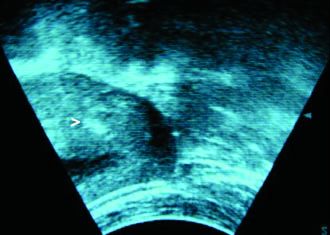
Figure 2: An ultrasound image showing the tip of the CVS needle (>) in a sample was adequate enough (10-25 mg) to allow a
posterior placenta. comfortable lab diagnosis. In the vast majority (90%),
aspiration was successful in the first attempt. In the remaining
The entry of needle into the placenta was marked by a loss of cases, a second or rarely a third attempt from the same outer
resistance. Once the needle was in the placenta (Figure 2), it needle, left in place, was required. There were only 3 patients
was sufficiently advanced to leave at least 2cm of the placental in whom the aspiration was unsuccessful and all 3 were called
tissue ahead of the needle tip. A 30 ml disposable syringe was for a repeat CVS one week later which was successful. The
attached to the CVS inner needle and it was rinsed with about overall success rate was 100%. The time for one aspiration,
1 ml sterile normal saline. The stillet was removed and the from introduction of outer needle to its removal, ranged from 5-
inner needle was introduced through the outer needle. Lack of 15 minutes (average 10 minutes).
resistance for the tip of the inner needle was another indication
JCPSP 2006, Vol. 16 (3): 204-207 205Suhaib Ahmed
Most patients felt pain and discomfort lasting upto 36 hours spinal needle to the co-axial chorion biopsy needles.2,13 The
after the procedure that was relieved by simple analgesics. latter have several advantages including greater length, better
Haematoma formation within the placental tissue at the site of visibility under ultrasound, special design to disrupt placental
aspiration was seen in 7/144 (4.9%). Three patients developed villi, and the option of multiple sampling attempts through the
spotting shortly after the procedure. Two recovered by rest for same outer needle left in place.14 Whichever needle is chosen,
3-6 hours in the hospital while the third patient aborted a larger syringe and needle size yields a larger quantity of
spontaneously six hours after the procedure. No case of post- chorionic villi.15
procedure infection was observed. On the whole the
CVS is a safe procedure in experienced hands. Mild and
procedure related abortion occurred in 1/144 (0.7%).
transient postprocedure pain due to uterine cramps, not more
than that felt after amniocentesis, is common. 16 Bleeding and
DISCUSSION spotting are uncommon and may result due to direct damage
Prenatal diagnosis through early fetal sampling has played a to the placental edge.2 Transfixation of the large intestine and
pivotal role in the prevention of genetic disorders.7 Ultrasound bacterial contamination is a rare but serious complication that
guidance adds to the safety for the fetus as well as the mother. is best avoided by taking utmost care in avoiding the intestinal
Nevertheless, an elaborate learning process to master the loops.2 Pregnancy loss is the most serious complication after
technique remains indispensable.8 The choice is between CVS. The overall rate of fetal loss is 0.5-1.0%.11 The results
amniocentesis and chorionic villus sampling. The main of this study also conform to the internationally accepted data.
disadvantage of amniocentesis is the increased risk of One reason for the low rate of miscarriage after an invasive
pregnancy loss and higher incidence of talipes, if done earlier procedure may be the low reactivity of the myometrium to
than 15 weeks. 9 transfixation during first trimester. 17
Chorionic villus sampling was introduced in the early 80s and Late obstetric complications could not be followed up in this
since then it has given a new dimension to prenatal study. But the incidence of complications like preterm delivery,
diagnosis.10 The procedure can be done as early as 9 weeks premature rupture of membranes, placental disorders,
of gestation. However, attempts to do it earlier than 9 weeks perinatal mortality, such as low birth weight, and congenital
have resulted in an increased risk of fetal limb reduction defects compare favourably well in the general population not
defects.11,12 In this study majority of the procedure were done exposed to CVS.18
between 12-14 weeks. At this stage, the placenta is of
adequate size that can be sampled without much difficulty. The Prenatal diagnosis is an essential recommendation for all
best time for CVS appears to be around 12-13 weeks. There couples with recessive genetic disorders like thalassaemia
is no upper time limit for doing the procedure. Due to with upto 25% recurrence risk. Prenatal diagnosis is also
anatomical reasons, it was easier to aspirate a placenta of an offered to women aged 35 years or above, or who are found
earlier stage than late. Another very important reason for doing by screening to be at a higher risk of having an infant with
the procedure early is that if it is to be followed by termination Down's syndrome or another chromosomal abnormality. A
of pregnancy then it should be done within a reasonable time- recent cost utility analysis of chorionic villus sampling and
frame defined by consensus. Most Islamic scholars in Pakistan amniocentesis versus no invasive testing, using data from
have a consensus on legal termination of a pregnancy before randomized trials, case registries, and a utility assessment of
120 days (17 weeks) of gestation if the fetus is found to have pregnant women, aged 16-47 years, have shown that prenatal
a serious abnormality. 6 The response of the Pakistani couples
diagnosis is cost-effective at any age or risk level.19 Chorionic
to prenatal diagnosis and termination of pregnancy has shown
villus sampling has the great advantage over mid-trimester
that over 90% of the couples are willing to accept the test and
amniocentesis of producing early results. Moreover, rapid
terminate the pregnancy before 17 weeks.5
analytic techniques have significantly reduced the waiting time
The CVS is done either transabdominally or through the between sampling and diagnosis, whereas progress in
transcervical route.2 In Pakistan, at least five different centers recombinant DNA technology and human gene mapping has
are involved in doing the procedure using the transcervical as led to an increase in the range of conditions it can detect. 20
well as transabdominal routes. A disadvantage of the trans-
cervical route is the possibility of transmitting infection from the
contaminated cervical canal. 4 The transcervical CVS is more CONCLUSION
technically demanding than the transabdominal CVS with Ultrasound guided transabdominal CVS is a useful outdoor
more failures to obtain sample and more multiple insertions.9 procedure for fetal sampling and prenatal diagnosis. It can play
Its main advantage is the ease with which the low-lying an important role in the prevention of genetic disorders that are
posterior placentae may be sampled.13 The trans-abdominal otherwise incurable. A placenta in almost any position can be
route has an obvious advantage of mechanical similarity to approached without much difficulty. The procedure is also safe
amniocentesis that makes CVS easier and familiar to for the mother as well as the fetus.
perform.4 In this study, practically all positions of placenta were
sampled through the transabdominal route without much ACKNOWLEDGEMENTS: The author wishes to acknowledge
difficulty that makes it the most feasible choice for use in Pakistan Council for Science and Technology for granting the
routine practice. The transcervical route may be used as a Research Productivity Allowance (RPA) that was the main
complementary procedure to improve the results in posterior financial support for carrying out the work and the subsequent
placentae.2,13 DNA analysis. The author also wishes to acknowledge the
technical advice and support by Prof Yasmeen Raashid of
The choice of needle for CVS may vary from a simple 18-20G
King Edward Medical College, Lahore.
206 JCPSP 2006, Vol. 16 (3): 204-207Transabdominal chorionic villus sampling
guidance of real time ultrasound. Br Med J 1983; 286: 1542-4.
REFERENCES
1. Alwan AA, Modell B. Community control of genetic and congenital 11. Chorionic villus sampling and amniocentesis: recommendations
disorders. EMRO technical publication. Series 24. Egypt: WHO for prenatal counseling. Centers for disease control and
Mediterranean Regional Office 1997. prevention. MMWR Recomm Rep 1995; 44: 1-12.
2. Brambati B, Lanzani A, Oldrini A. Transabdominal chorionic villus 12. Golden CM, Ryan LM, Holmes LB. Chorionic villus sampling: a
sampling. Clinical experience of 1159 cases. Prenat Diagn 1988; distinctive teratogenic effect on fingers? Birth Defects Res A Clin
8: 609-17. Mol Teratol 2003; 67: 557-62.
3. Ahmed S, Saleem M, Rashid Y. The first prenatal diagnosis of 13. Silver RK, MacGregor SN, Sholl JS, Elesh RH, Beaird JA, Waldee
thalassaemia in Pakistan: a case report. Pak J Pathol 1994; 5: 69-
JK. Initiating a chorionic villus sampling program. Relying on
71.
placental location as the primary determinant of the sampling
4. Raashid Y, Ahmed S, Saleem M, Tahir M, Waheed I, Jafri H. route. J Reprod Med 1990; 35: 964-8.
Transabdominal chorionic villus sampling for prenatal diagnosis of
genetic disorders. Mother Child 1995; 33: 63-6. 14. Maxwell D, Lilford R, Czepulkowski B, Heaton D, Coleman D.
5. Ahmed S, Saleem M, Petrou M, Sultana N, Raashid Y, Waqar A, Transabdominal chorionic villus sampling. Lancet 1986; 1: 123-6.
et al. Prenatal diagnosis of β-thalassaemia in Pakistan: 15. Cochrane L, Ainscough M, Alfirevic Z. The influence of needle and
experience in a Muslim country. Prenat Diagn 2000; 20: 378-83. syringe size on chorionic villus sampling of term placentae: a
6. Petrou M. Genetic counselling. In: Galanello R, Eleftheriou A, randomised trial. Prenat Diagn 2003; 23: 1049-51.
Traeger-Synodinos J, Old J, Petrou M, Angastiniotis M, (edi).
Prevention of thalassaemias and other haemoglobin disorders. 16. de Crespigny L, Robinson HP, Ngu A. Pain with amniocentesis
Nicosia: Thalassaemia International Federation, 2003. and transabdominal CVS. Aust N Z J Obstet Gynaecol 1990; 30:
308-9.
7. Ball RH. Invasive fetal testing. Curr Opin Obstet Gynecol 2004;
16: 159-62. 17. Huszar G. Physiology of the myometrium. In: Creasy RK, Resnik
8. Levy R, Arfi JS, Daffos F. Fetal sampling techniques. Gynecol R, (edi). Maternal fetal medicine. Philadelphia: WB Saunder,
Obstet Fertil 2003; 31: 550-5. 1984.
9. Alfirevic Z, Sundberg K, Brigham S. Amniocentesis and chorionic 18. Brambati B, Oldrini A, Ferrazzi E, Lanzani A. Chorionic villus
villus sampling for prenatal diagnosis. Cochrane Database Syst sampling: an analysis of the obstetric experience of 1000 cases.
Rev 2003; 3: CD003252. Prenat Diagn 1987; 7: 157-69.
10. Ward RH, Modell B, Petrou M, Karagozlu F, Douratsos E. Method
19. Harris RA, Washington AE, Nease RF Jr, Kuppermann M. Cost
of sampling chorionic villi in first trimester of pregnancy under
utility of prenatal diagnosis and the risk-based threshold. Lancet
lllll O lllll
JCPSP 2006, Vol. 16 (3): 204-207 2072004; 363: 276-82.
20. Brambati B. Chorionic villus sampling. Curr Opin Obstet Gynecol
1995; 7: 109-16.
JCPSP 2005, Vol. 15 (02): 208You can also read