Loss of sympathetic innervation to islets of Langerhans in canine diabetes and pancreatitis is not associated with insulitis - Nature
←
→
Page content transcription
If your browser does not render page correctly, please read the page content below
www.nature.com/scientificreports
OPEN Loss of sympathetic innervation
to islets of Langerhans in canine
diabetes and pancreatitis
is not associated with insulitis
Chen Gilor1,2*, Jully Pires1, Rachel Greathouse3, Rebecca Horn3, Mark O. Huising4,
Stanley L. Marks1, Brian Murphy3 & Amir Kol3*
Canine diabetes mellitus (DM) affects 0.6% of the canine population and yet, its etiology is poorly
understood. Most affected dogs are diagnosed as adults and are insulin-dependent. We compared pan-
leukocyte and sympathetic innervation markers in pancreatic islets of adult dogs with spontaneous
DM (sDM), spontaneous pancreatitis (sPanc), both (sDMPanc), toxin-induced DM (iDM) and controls.
We found evidence of decreased islet sympathetic innervation but no significant infiltration of islets
with leukocytes in all disease groups. We show that loss of sympathetic innervation is ongoing in
canine DM and does not necessarily precede it. We further found selective loss of islet-associated beta
cells in dogs with sDM and sDMPanc, suggesting that collateral damage from inflammation in the
exocrine pancreas is not a likely cause of DM in these dogs. The cause of this selective loss of beta cells
needs to be further elucidated but overall, our findings are not supportive of an autoimmune process
as a cause of sDM in adult dogs. The loss of sympathetic innervation in sPanc in dogs that do not
suffer from DM links the disease in the exocrine pancreas to a pathological process in the endocrine
pancreas, suggesting pancreatitis might be a potential precursor to DM.
Diabetes mellitus (DM) is a common disease in dogs affecting about 0.6% of dogs in the USA with increasing
prevalence1. Like Type 1 diabetes mellitus (T1DM) human patients, most diabetic dogs are insulin-dependent
and ketosis-prone, and insulin resistance does not play a significant role in the disease p rocess2. In both species,
DM can be associated with inflammation in the exocrine pancreas (pancreatitis) although the cause and effect
relationship is uncertain3–7. As in human T1DM, canine DM is associated with significant selective loss of beta
cells in pancreatic islets8. Because of these similarities, canine DM has often been labeled as T1DM, although
evidence for the hallmark of T1DM, autoimmune beta cell destruction, has been identified in only a small frac-
tion of canine cases2,7,9. Therefore, we set out to further characterize the pathogenesis of canine DM and examine
similarities, or lack thereof, to T1DM in humans. Specifically, we aimed to (1) assess the frequency of insulitis
(inflammation within the islets of Langerhans) based on current c riteria10 (2) assess changes in sympathetic
innervation of islets11 (3) compare these findings between dogs diagnosed with spontaneous DM (sDM) lack-
ing histological evidence of pancreatitis, dogs diagnosed concurrently with spontaneous DM and pancreatitis
(sDMPanc), dogs with pancreatitis and no DM, and purpose-bred research dogs with alloxan-streptozotocin-
induced DM (iDM).
In humans with T1DM and in rodent models, pancreatic pathology is characterized not only by a predomi-
nately lymphocytic insulitis, but also by early, sustained, islet-selective loss of sympathetic innervation11. The
pathogenesis of this neuropathy is unclear but it appears to be a result of the combination of beta cell loss together
with the inflammatory milieu of the autoimmune process within the islet. This neuropathy is not observed
in Type 2 DM or when beta cells are destroyed by a toxin in rodent models11–13. It has not been determined
whether the loss of sympathetic innervation in T1DM occurs secondary to other inflammatory diseases in which
1
Department of Veterinary Medicine and Epidemiology, College of Veterinary Medicine, University of California,
One Shields Ave., Davis, CA 95616, USA. 2Department of Small Animal Clinical Sciences, College of Veterinary
Medicine, University of Florida, 2015 SW 16th Ave, Gainesville, FL 32610, USA. 3Department of Pathology,
Microbiology and Immunology, College of Veterinary Medicine, University of California, One Shields Ave., Davis,
CA 95616, USA. 4Department of Neurobiology, Physiology and Behavior, College of Biological Sciences, University
of California, One Shields Ave., Davis, CA 95616, USA. *email: cgilor@ufl.edu; akol@ucdavis.edu
Scientific Reports | (2020) 10:19187 | https://doi.org/10.1038/s41598-020-76091-5 1
Vol.:(0123456789)
www.nature.com/scientificreports/
Group Age (years) Sex Body weight (kg)
4.6 ± 2.6a FS = 1, FI = 0 27.4 ± 18.8
Controls (N = 5)
4 (0.8–7.0) MC = 4, MI = 0 23.0 (2.4–51.7)
10.2 ± 2.9b FS = 4, FI = 1 26.0 ± 15.8
sDM (N = 11)
10 (5–14) MC = 5, MI = 1 28.2 (4.4–52.0)
7.9 ± 3.8 FS = 6, FI = 0 20.2 ± 8.1
sPanc (N = 10)
7.0 (0.8–14) MC = 4, MI = 0 20.2 (10.4–30.0)
9.7 ± 3.0 FS = 6, FI = 1 21.5 ± 18.0
sDMPanc (N = 10)
10.0 (4.0–14) MC = 2, MI = 1 12.9 (5.0–60.0)
3.0 ± 0 FS = 0, FI = 0 9.9 ± 1.5
iDM (N = 7)
MC = 7, MI = 0 10.5 (7.5–11.2)
Table 1. Study population demographics. Data are presented as Mean ± SD; Median (range). Different
superscripts indicate significantly different values (p ≤ 0.05). DM diabetes mellitus, sDM spontaneous DM,
sDMPanc spontaneous DM and concurrent pancreatitis, sPanc spontaneous pancreatitis, iDM toxin-induced
DM, FS female spayed, FI female intact, MC male castrated, MI male intact.
lymphocytes do not necessarily predominate (such as pancreatitis). Loss of sympathetic innervation to islets
might serve as a marker for prior autoimmune destruction of insulin-producing beta cells.
In humans with T1DM, the number of affected islets and the number of infiltrating immune cells in each
affected islet varies but is usually low, especially in patients diagnosed in middle age10. There are no established
criteria for diagnosis of insulitis in canine DM, and until recently, none existed for T1DM. In a recent consensus
guideline, the threshold for diagnosis of insulitis in people with T1DM has been established as a minimum of
three affected islets and a minimum of 15 CD45+ (leukocyte common antigen) cells per islet (either intra-islet,
or at the periphery of the islet)10. Importantly, these standards were established based on a comparison to a non-
diabetic control group because it is recognized that background inflammation (as well as presence of resident
lymphocytes) in the exocrine pancreas is common in humans. To our knowledge, these standards have not been
applied to histopathological samples from canine DM to date. To that end, we aimed to compare the number and
distribution of CD45+ leukocytes in pancreata from dogs with sDM and sDMPanc to dogs that have no evidence
of either DM or pancreatitis and dogs with pancreatitis but no DM.
In dogs and human patients, DM is often diagnosed concurrently with pancreatitis . The association between
DM and pancreatitis in dogs and in people is complex and establishing cause and effect is d ifficult4,9. There is
evidence that at least in a subset of affected dogs, pancreatitis precedes and increases the risk of DM. Some
histopathological evidence of chronic pancreatitis is found in most dogs that die from diseases unrelated to the
pancreas and that do not present with clinical signs associated with pancreatitis or DM. In one study, 90% of
dogs that died of a variety of causes had histopathological evidence of pancreatitis (acute and chronic)14. Even
if pancreatitis does not cause DM directly, its high prevalence confounds the interpretation of histopathological
findings of inflammation in the endocrine pancreas and should be considered at least as “background” when
assessing the degree or presence of insulitis.
We hypothesized that in diabetic dogs, leukocytes will infiltrate islets of Langerhans at a higher frequency
compared to non-diabetic dogs. We also hypothesized that sympathetic innervation of islets will be decreased
in diabetic dogs, but not when DM is associated with pancreatitis or in cases of toxin-induced DM. Finally, we
hypothesized that in pancreatitis, DM is the result of indiscriminate collateral damage to islets. Therefore, in dogs
with DM and concurrent pancreatitis, beta cell loss would be associated with loss of other endocrine cell types,
leading to a greater reduction in endocrine cell mass in these dogs compared to dogs suffering from DM alone.
Here we report for the first time on sympathetic denervation of pancreatic islets in adult dogs, not only with
DM, but also in dogs with pancreatitis and no DM. We also show that sympathetic denervation is ongoing in
canine DM and as previously reported, we found no evidence for insulitis in canine DM. Together, these findings
are not supportive of an autoimmune process as a cause of DM in adult dogs. We further found selective loss of
islet-associated beta cells in dogs with spontaneous DM, with or without pancreatitis, suggesting that collateral
damage from inflammation in the exocrine pancreas is not the cause of DM in these dogs. The cause of this
selective loss of beta cells needs to be further elucidated.
Results
Age, sex and body weight are presented in Table 1. Body condition scores were unavailable for most dogs and
therefore not reported. Except for the iDM group which included only purpose-bred Beagles, there was no
dominant breed in any group, with a total of 18 breeds represented and 14 mix-breed dogs in the four groups
(Table S3).
To investigate the role of inflammation in the pathogenesis of canine DM and its potential association with
loss of sympathetic innervation, we stained pancreata with TH, a rate-limiting enzyme of catecholamine bio-
synthesis and a marker of sympathenic innervation, and the pan-leukocyte cell surface marker CD45. Islet
area was visualized by staining CgA. An early and sustained islet-selective loss of sympathetic innervation was
shown previously in human patients with T1DM and models of autoimmune insulitis11. In all disease groups,
TH surface area per islet was decreased compared to controls (Fig. 1A). The decrease in TH expression per islet
Scientific Reports | (2020) 10:19187 | https://doi.org/10.1038/s41598-020-76091-5 2
Vol:.(1234567890)www.nature.com/scientificreports/
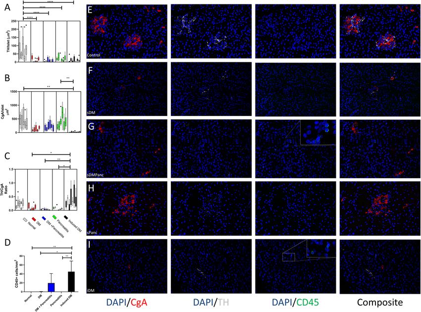
Figure 1. Loss of sympathetic innervation in dogs with pancreatic disease. DM diabetes mellitus, sDM
spontaneous DM, sDMPanc spontaneous DM and concurrent pancreatitis, sPanc spontaneous pancreatitis,
iDM toxin-induced DM. (A) Sympathetic innervation (indicated by TH area per islet), is markedly reduced
in all disease groups compared with control dogs. (B) Islet size (indicated by CgA area per islet), is markedly
decreased in iDM dogs vs. controls and sPanc groups. (C) When TH loss is controled for endocrine cell loss,
iDM dogs have increased TH-to-CgA ratio compared with all other disease groups. (D) In iDM dogs, more
leukocytes (CD45+ cells) infiltrate the exocrine pancreas compared with all other groups, other than sDMPanc.
Reprasentative immunofluorescence confocal images of CgA (red), TH (grey), CD45 (green) and nuclei (blue)
in (E) control dogs, (F) sDM, (G) sDMPanc, (H) sPanc and (I) iDM. P-values represented by * (P ≤ 0.5), **
(P ≤ 0.01), *** (P ≤ 0.001) and **** (P ≤ 0.0001).
was most extreme in the sDMPanc (90%, p < 0.0001), sDM (86%, p < 0.0001) and iDM (85%, p < 0.0001) but was
also significant in the sPANC group (75%, p < 0.0001, Fig. 1A). While the mean size of islets, as determined by
CgA positive area, was higher in control and sPanc dogs compared with sDM and sDMPanc dogs (433.2, 407.3,
117.9 and 215.7 µm2, respectively), changes were not statistically significant (Fig. 1B). The only group that had
a significant reduction in islet size compared with controls (p < 0.01) and Panc (p < 0.01) was iDM (433.2, 407.3
and 19.98 µm2, respectively). After adjusting for loss of islet area (TH/CgA ratio), sDM, sDMPanc and sPANC
groups were not different from the control group (Fig. 1C). TH/CgA ratio was significantly increased in the iDM
group, suggesting that the decrease in TH expression per islet in iDM is likely secondary to marked endocrine
cell loss and not due to loss of islet sympathetic innervation as in the other disease groups.
There were no intra-islet nor peri-islet CD45+ cells consistent with insulitis in controls, sDM, sDMPanc, sPanc
or iDM. In the vicinity of the islets within the exocrine pancreas, there were rare CD45+ cells scattered in dogs
with sDMPanc (0–40 cell/mm2) and in iDM (0–68 cells/mm2; Fig. 1D). This degree of immune cell infiltration
falls well below the lowest category of inflammation consistent with p ancreatitis14. The morphology of these cells
was most consistent with lymphocytes. Representative images depicting CgA, TH and CD45 staining patterns
in the various groups are included (Fig. 1E–I).
We hypothesized that in dogs with pancreatitis, exocrine pancreatic inflammation induces indiscriminate
collateral damage to islets. In order to test our hypothesis, we determined numbers of alpha cells and beta cells
per islet via multicolor confocal microscopy in pancreas tissues from controls, sDM, sDMPanc, and sPanc. We
hypothesized that if DM in the sDMPanc group is driven by indiscriminate collateral damage to islets, alpha
cells and beta cells should be equally decreased and total islet area would be smaller compared to sDM without
Scientific Reports | (2020) 10:19187 | https://doi.org/10.1038/s41598-020-76091-5 3
Vol.:(0123456789)www.nature.com/scientificreports/
Figure 2. Selective β-cell loss in dogs with spontaneous diabetes mellitus (DM) and dogs with pancreatitis
and concurent DM. sDM Spontaneous DM, sDMPanc spontaneous DM and concurrent pancreatitis, sPanc
spontaneous pancreatitis. (A) Pancreatic β-cells are significantly reduced in numbers/islet in pancreata of dogs
with spontaneous DM and dogs with pancreatitis and DM compared with normal control dogs and dogs with
pancreatitis and no DM. (B) There is no significant difference in pancreatic α-cell numbers/islet between all
groups. Reprasentative immunofluorescence confocal images of insulin (green), glucagon (red) and DNA (blue)
representing β-cells, α-cells and nuclei, respectively, in pancreate from (C) normal control dogs compared to (D)
dogs with DM, (E) DM and (F) pancreatitis and pancreatitis only. p-values represented by *** (P ≤ 0.001) and
**** (P ≤ 0.0001).
pancreatitis. We found that the number of beta cells per islet in sDM and sDMPanc dogs did not differ and
both groups had decreased beta cells compared with control dogs (p < 0.0001) and compared with sPanc dogs
(p < 0.001, Fig. 2A). The number of alpha cells per islet was not different in the sDM, sDMPanc and sPanc groups
(Fig. 2B), suggesting that there is a selective beta cell loss in both the sDM and sDMPanc groups. Representative
images of alpha and beta cell distribution within pancreatic islets in control dogs, sDM, sDMPanc and sPANC
are depicted (Fig. 2C–F). Further supporting the hypothesis that there is similarly selective beta cell loss in
sDM and sDMPanc, was the finding of a similar decrease in islet area (as represented by CgA area) in sDM and
sDMPanc (Fig. 1A). Representative images of CgA area in control dogs, sDM, sDMPanc and sPANC are depicted
(Fig. 1E–I). In human T1DM, a characteristic histological feature is the presence of islets which are entirely
devoid of insulin across the pancreas. In contrast, this was not a common finding in dogs of this study. Iselts
that are entirly devoid of insulin were observed in only 3 of the 11 dogs in the sDM group (31%, 35% and 85% of
islets in each) while in three other dogs in this group, no islets were identified at all but clusters of 1–3 beta cells
were scattered throughout the pancreas (Figure S1). Of the eight dogs in the sDMPanc group, three had islets
completely devoid of insulin (6%, 7%, 16% of islets) and in a 4th dog of this group no islets were identified but
clusters of 1–3 beta cells were scattered throughout the pancreas.
Median [range] DM duration was significantly shorter in sDMPanc (5 [1–120] days) compared to sDM (638
[3–1095] days, p = 0.0016, Fig. 3A). When sDM and sDMPanc were combined, beta cell numbers were negatively
correlated with DM duration (rho = − 0.48, p = 0.036, Fig. 3B) and there was also a trend towards overall loss of
islet area (represented by CgA expression, rho = − 0.54, p = 0.09, Fig. 3C) but there was no correlation between
disease duration and alpha cell loss (p = 0.5, data not shown). With the progressive loss of beta cells and total
islet area, there was also a strong negative correlation between TH area per islet and DM duration (rho = − 0.70,
p = 0.02, Fig. 3D). These findings suggest that loss of sympathetic innervation is ongoing and does not precede
beta cell loss as is the case when autoimmunity is the cause of D M11–13.
Discussion
The etiology of canine DM is poorly understood, however, we report for the first time in this study on the sym-
pathetic denervation of the islets of Langerhans as a key characteristic of this disease that helps narrow down
possible etiologies. Overall, our histological assessment of pancreas pathology in dogs with DM does not reveal
clear evidence of autoimmunity as the underlying cause of canine DM and suggests that the cause of selective
beta cell loss should be further investigated. Canine DM is often proposed as a valuable, but overlooked, animal
Scientific Reports | (2020) 10:19187 | https://doi.org/10.1038/s41598-020-76091-5 4
Vol:.(1234567890)www.nature.com/scientificreports/
Figure 3. (A) Duration of diabetes mellitus (DM) in spontaneous DM without pancreatitis (sDM, open
triangles) vs. sDM with concurrent pancreatitis (sDMPanc, closed circles). Horizontal solid line represents
median. When all diabetic dogs were considered as one group (sDM, and SDMPanc), duration of DM is
negatively corrleatd (spearman’s rho) with (B) beta cell numbers, (C) islet area (as expressed by Chromogranin
A [CgA] area) and (D) sympathetic innervation as expressed by tyrosine hydroxylase (TH) area/islet. Dashed
lines (with corresponding r 2) represent best fit for one phase decay regression. Dotted lines represent median of
healthy controls.
model of human T1D, and our work further highlights the need to deepen our understanding of the pathogenesis
odel15.
of this m
Sympathetic denervation of islets was reported in three animal models of autoimmune DM as well as in
T1DM in people, but not in type 2 DM11–13, leading to the suggestion that this phenomenon is tightly linked
with autoimmune insulitis. This phenomenon has been dubbed eSIN (early Sympathetic Islet Neuropathy)16
because in contrast to the classic Diabetic Autonomic Neuroparthy (DAN), it precedes the loss of beta cells.
We showed that islet sympathetic denervation is a feature of canine DM, but also occurs in canine pancreatitis
without DM, suggesting that at least in dogs, it is inflammation in general, not necessarily autoimmunity, that
leads to islet neuropathy. Furthermore, in contrast to eSIN, we found evidence that in dogs, islet neuroparthy
is ongoing, correlated to ongoing beta cells loss, and does not precede beta cell loss as in eSIN. These findings
are important not only because they do not lend support to the autoimmune etiology hypothesis in canine DM
but also because of their clinical implication. Islet neuropathy is associated with decreased glucagon responses
Scientific Reports | (2020) 10:19187 | https://doi.org/10.1038/s41598-020-76091-5 5
Vol.:(0123456789)www.nature.com/scientificreports/
in diabetics and inability to respond appropriately to insulin over-dosing which increases the risk and severity
of life-threatening h ypoglycemia16. Our finding that loss of sympathetic innervation is associated with beta cell
numbers in canine DM suggests that the risk of hypoglycemia increases with disease duration in dogs. This
hypothesis should be further tested, but if true, it would imply that glycemic targets should be modified in dogs
based on disease duration, a strategy that is not currently employed in dogs.
Importantly, we demonstrated throughout our study that the pathological changes in islets of dogs with sDM
do not differ from those of dogs with sDMPanc, increasing the likelihood that the etiology of DM in both groups
is the same, and that pancreatitis is a complication in a subset of sDM dogs. We found a selective loss of beta cells
in dogs with spontaneous DM, with and without pancreatitis, as was previously shown in d ogs8. This was evident
by sparing of alpha cells as well as by an overall similar reduction in islet size in sDM and SDMPanc. These data
suggest that collateral damage from inflammation in the exocrine pancreas is not a major contributor to beta cell
loss in these dogs, especially considering the absence of CD45 positive cells in or near islets.
Shields et al., looked for evidence of autoimmune insulitis in canine DM by staining for CD3+ l ymphocytes8.
However, insulitis in people is sometimes dominated by B lymphocytes (CD20+)17. By using CD45 as a pan-
leukocyte marker and applying the current low-threshold standard for diagnosis of insulitis10, we were more
likely than previously to detect insulitis. To minimize the risk of false positive staining associated with this greater
sensitivity and to avoid misinterpreting “background noise” of inflammation or resident lymphocytes as insu-
litis, we used samples from dogs with pancreatitis and dogs with no pancreatitis as a control group to diabetic
dogs. Strikingly, no pancreatic tissue and no single islet in our study met the low-threshold standard criteria for
diagnosis of insulitis. In fact, we found no inflammatory cells at all within or adjacent to islets in control dogs,
dogs with DM, and even in dogs with DM and pancreatitis. It has been hypothesized that the lack of evidence for
autoimmunity in canine DM is related to the fact that the autoimmune process subsides when beta cells are no
longer present in the islet8. This hypothesis might explain the apparent absence of insulitis in histopathological
samples from diabetic dogs that died long after the diagnosis of the disease, but it has a few deficiencies: (1) we
showed that after DM is clinically established in dogs, loss of beta cells continues and therefore, the cause of that
ongoing loss should still be present. (2) Insulitis is also not observed in samples from dogs with early diagnosis
of DM and when beta cells are still present in islets8. (3) Although early studies demonstrated the presence of
antibodies to insulin or to beta cells in some diabetic dogs, more recent studies failed to replicate these findings
and demonstrated no humoral response to either insulin or beta cells prior to insulin t herapy2,9,18. It has been
proposed previously that canine DM is pathogenically similar to Latent Autoimmune Diabetes of Adults (LADA),
explaining its typical diagnosis in dogs at middle age or older and in the absence of insulitis19. However, even in
LADA, at least 29% of human patients have evidence of insulitis, even when including older studies that have not
used the current low-threshold s tandard10,20. As mentioned above, and even if ignoring the CD45 data subset,
the pathology of islets in dogs with sDM and sDMPanc is similar. Combining these two groups, if canine DM
was similar to LADA, we would have expected at least some of the cases in our study to demonstrate insulitis.
Our data therefore do not support autoimmunity, acute or chronic, as a cause of canine DM.
In people, autoimmune DM is more commonly diagnosed in juveniles. Reports on DM in juvenile dogs
(< 1 year old) are rare, and while they uniformly describe insulin-dependent dogs, they describe various histo-
pathological abnormalities of the pancreas, with no clear etiology2,8. Only rarely, these reports support autoim-
munity as a cause of juvenile DM8. Overall, less than 2.2% of all dogs diagnosed with DM are diagnosed before
they are 1 year old, and still less than 3% of all dogs diagnosed with DM are diagnosed before they are 3 years
old1. We therefore chose to exclude juvenile dogs from the DM groups in our study, because it is likely that their
disease process does not represent the disease process in the majority of the population.
Gestational diabetes has been reported in dogs and is thought to be associated with progesterone-induced
GH production in the mammary glands, leading to insulin r esistance21. However, the prevalence of DM in intact
females is not higher than the general canine population and being intact vs. spayed does not increase the risk
of DM in a female dog1, suggesting that the underlying cause of DM in intact females is not different than in the
rest of the population. Rather, it is likely that insulin resistance caused by progesterone-induced GH production
exposes the underlying pancreatic lesion during diestrous. We therefore did not exclude intact females from
our study.
One potential limitation of studies of patient-sourced tissues (from people or pets), especially retropesctive
ones, is the inability to quantify the extent to which entire pancreata is affected by pancreatitis. Pancreatitis can
be focal or multi-focal and therefore a single section of the organ might not represent the extent of organ tissue
injury. As such, correlating histopathological lesions with function and clinical outcome may be discordant.
Histopathological evidence of pancreatitis is found in most dogs that die from diseases unrelated to the pan-
creas and that do not present with clinical signs associated with pancreatitis. In one study, 63 of 70 (90%) dogs
that died of a variety of causes had histopathological evidence of pancreatitis (acute and chronic) although only
17 had biochemical evidence of pancreatitis (increased serum pancreatic lipase) and only 11 had clinical signs
consistent with pancreatitis14. In an earlier study, 73 pancreata were collected consecutively from dogs that died
from a variety of causes and 47 of them (64.4%) had histopathological evidence of pancreatitis22. Therefore, the
clinical significance of the histopathological diagnosis of pancreatitis remains unknown. In both of these stud-
ies, diagnosis of pancreatitis was based on systematic serial sectioning of the entire pancreas (every 2 cm) and
standard scoring of each section. Dogs were then diagnosed with pancreatitis if lesions were observed in any of
the sections, regardless of distribution throughout the entire organ. We used tissues from one of these s tudies14
and selected only cases that had no evidence of pancreatitis on any of the section, therefore confirming the
appropriatenss of categorizing these cases as controls for our study. This systematic approach, however, is not
routinely implemented in clinical cases (human and veterinary). Therefore, in most cases available to us, only
a single location of the pancreas was sectioned based on ad-hoc judement of the attending pathologist, and the
presence or absence of pancreatitis was based on these single tissue section.
Scientific Reports | (2020) 10:19187 | https://doi.org/10.1038/s41598-020-76091-5 6
Vol:.(1234567890)www.nature.com/scientificreports/
In conclusion, we found no evidence to support the hypothesis that selective beta cell loss in canine DM is
caused by autoimmunity or by collateral damage from pancreatitis. The decrease in sympathetic innervation
to islets in non-diabetic dogs suffering from pancreatitis suggests that pancreatitis might be a precursor to islet
pathology by yet unknown mechanisms that should be further explored.
Material and methods
Animals. All animal studies perfomed at UC Davis in this study were approved by the Institutional Ani-
mal Care and Use Committee (IACUC protocol #21100) and followed IACUC guidelines. All pancreatic tissue
samples in this study were collected from archived tissues at the UC Davis William R. Pritchard Veterinary
Medical Teaching Hospital (VMTH) Pathology laboratory, except for samples from dogs with induced DM (see
below). Study cases were identified by searching the electronic medical records at UCD VMTH for diagnoses of
DM and pancreatitis in dogs from 1988 to 2016. The selected cases were recorded in an Excel spreadsheet and
were divided into five groups: Group 1 (Control, N = 7) was of non-diabetic dogs that in a previous study were
confirmed to have no clinical and histopathologic evidence of pancreatic disease14. Specifically, in each dog, for
each limb of the pancreas, serial transverse sections were made every 2 cm throughout the pancreas, and each
individual section was examined and scored for acute and chronic inflammation on H&E using a semiquanti-
tative histopathologic grading scheme by a board certified veterinary pathologist as previously described14. In
brief, nine histopathologic parameters were evaluated including suppurative inflammation, acinar cell necrosis,
hemorrhage, interstitial edema, peripancreatic fat necrosis, mononuclear inflammation, fibrosis, atrophy, and
cellular degeneration. Each transverse section was individually scored for all nine parameters on a scale from
0 to 3. A score of 0 indicated none of the sectional surface area was affected by that particular histopathologic
change. Scores of 1, 2 and 3 had < 10%, 10–40% and > 40% of the examined surface area affected by the specific
lesion parameter, respectively. Additional histopathologic changes not captured by this scoring system were also
recorded for each pancreas (e.g. nodular hyperplasia and neoplasia). Dogs were included in the control group
only if their cumulative score was 0. Group 2 represented dogs that were diagnosed with spontaneous DM
without pancreatitis (sDM, N = 11); group 3 dogs were diagnosed with spontaneous DM and spontaneous pan-
creatitis (sDMPanc group, N = 10); group 4 dogs were diagnosed with spontaneous pancreatitis but not with DM
(sPanc, N = 10). Inclusion into groups 3 and 4 required a standard histological diagnosis (including the nine his-
tologic parameters specified above) by a board certified veterinary pathologist. Group 5 dogs were purpose-bred
Beagle dogs with alloxan-streptozotocin induced DM (iDM, N = 7). For each case, the following clinical data was
extracted from the medical record: age, sex, breed, body weight, histopathologic diagnosis and duration of DM.
Cases were excluded if they did not have a necropsy report, were clinically diagnosed with pancreatitis but
the diagnosis was not confirmed histopathologically, had an autolyzed postmortem body condition, or did not
have any archived pancreatic tissue. Dogs under the age of 4 years were also excluded from the spontaneous DM
(sDM) and sDM + spontaneous pancreatitis (sDMPanc) groups.
Induction of DM was achieved as previously described 23 at Sinclair Research LLC, as part of a different
study which was approved by institutional ethical committee. Dogs were induced 8 months before tissue. All
procedures performed at UC Davis on these dogs were approved by the Institutional Animal Care and Use
Committee (IACUC protocol #21100). Futher details on the husbendary of iDM dogs are available in “Sup-
plementary Methods”.
Tissue processing, image acquisition and image analysis. All pancreatic tissues were fixed in 10%
neutral buffered formalin and embedded in paraffin blocks. Four µm sections were cut from each pancreatic
tissue block. Tissue sections were prepared routinely for multiplex immunofluorescence staining. Primary anti-
bodies targeting insulin, glucagon, chromogranin A (CgA), tyrosine hydroxylase (TH) and CD45 were used
(Tables S1 and S2). All immunohistochemistry and immunofluorescence studies had several layers of negative
and positive controls to ensure appropriate interpretation of our findings. A Leica TCS SP8 STED 3X confocal
laser scanning microscope was used to take 63× Z-stack images of islets and a 20× stitched image of the canine
pancreatic section. For each image, ImageJ (NIH, Bethesda, MA) was used to measure the following when appli-
cable: Total Image Surface Area, Number of Islets, CgA Area of Islet(s), TH Area of Islet(s), total CD45+ cells,
and %TH relative to %CgA. Futher details on tissue processing, image acquisition and image analysis are avail-
able in “Supplamentary Methods”.
Statistical analysis. Statistical analysis was performed using a commercially available computer software
(GraphPad Prism, version 8.0; GraphPad Software Inc, La Jolla, CA). Visual inspection of the data and the
Shapiro–Wilk test were used to assess normal distribution. Demographic data are presented as mean ± SD and
median (range). For distribution of beta cells, alpha cells, TH area, CgA area, and CD45+ cells, nested tables were
created and a non-parametric 1-way ANOVA was applied to compare between disease groups after removal of
outlier values (ROUT method, Q = 0.5%). Adjusted p values are presented for post-hoc multiple comparisons.
Duration of disease was not normally distributed and was compared between sDM and SDMPanc using the
Mann–Whitney test. For the association between duration of disease and beta cells, TH area, and islet area,
a non-linear correlation (Spearman’s rho) was calculated, and a regression model of one-phase decay was fit.
Significance was set at p ≤ 0.05.
Data availability
The datasets generated during and/or analysed during the current study are available from the corresponding
author on reasonable request.
Scientific Reports | (2020) 10:19187 | https://doi.org/10.1038/s41598-020-76091-5 7
Vol.:(0123456789)www.nature.com/scientificreports/
Received: 8 June 2020; Accepted: 21 October 2020
References
1. Guptill, L., Glickman, L. & Glickman, N. Time trends and risk factors for diabetes mellitus in dogs: Analysis of veterinary medical
data base records (1970–1999). Vet. J. 165, 240–247. https://doi.org/10.1016/s1090-0233(02)00242-3 (2003).
2. Gilor, C., Niessen, S. J., Furrow, E. & DiBartola, S. P. What’s in a name? Classification of diabetes mellitus in veterinary medicine
and why it matters. J. Vet. Intern. Med. 30(4), 927–940. https://doi.org/10.1111/jvim.14357 (2016).
3. Wang, Y. et al. Concurrent diabetic ketoacidosis in hypertriglyceridemia-induced pancreatitis: How does it affect the clinical course
and severity scores?. Pancreas 46, 1336–1340. https://doi.org/10.1097/mpa.0000000000000937 (2017).
4. Davison, L. J. Diabetes mellitus and pancreatitis—Cause or effect?. J. Small Anim. Pract. 56, 50–59. https://doi.org/10.1111/
jsap.12295(2015).
5. Hess, R. S., Saunders, H. M., Van Winkle, T. J., Shofer, F. S. & Washabau, R. J. Clinical, clinicopathologic, radiographic, and ultra-
sonographic abnormalities in dogs with fatal acute pancreatitis: 70 cases (1986–1995). J. Am. Vet. Med. Assoc. 213, 665–670 (1998).
6. Hess, R. S., Saunders, H. M., Van Winkle, T. J. & Ward, C. R. Concurrent disorders in dogs with diabetes mellitus: 221 cases
(1993–1998). J. Am. Vet. Med. Assoc. 217, 1166–1173. https://doi.org/10.2460/javma.2000.217.1166 (2000).
7. Hume, D. Z., Drobatz, K. J. & Hess, R. S. Outcome of dogs with diabetic ketoacidosis: 127 dogs (1993–2003). J. Vet. Intern. Med.
20, 547–555. https://doi.org/10.1892/0891-6640(2006)20[547:oodwdk]2.0.co;2 (2006).
8. Shields, E. J. et al. Extreme beta-cell deficiency in pancreata of dogs with canine diabetes. PLoS ONE 10, e0129809. https://doi.
org/10.1371/journal.pone.0129809 (2015).
9. Ahlgren, K. M. et al. Lack of evidence for a role of islet autoimmunity in the aetiology of canine diabetes mellitus. PLoS ONE 9,
e105473. https://doi.org/10.1371/journal.pone.0105473 (2014).
10. Campbell-Thompson, M. L. et al. The diagnosis of insulitis in human type 1 diabetes. Diabetologia 56, 2541–2543. https://doi.
org/10.1007/s00125-013-3043-5 (2013).
11. Mundinger, T. O. et al. Human type 1 diabetes is characterized by an early, marked, sustained, and islet-selective loss of sympathetic
nerves. Diabetes 65, 2322–2330. https://doi.org/10.2337/db16-0284 (2016).
12. Mei, Q., Mundinger, T. O., Lernmark, A. & Taborsky, G. J. Jr. Early, selective, and marked loss of sympathetic nerves from the islets
of BioBreeder diabetic rats. Diabetes 51, 2997–3002. https://doi.org/10.2337/diabetes.51.10.2997 (2002).
13. Taborsky, G. J. Jr. et al. Loss of islet sympathetic nerves and impairment of glucagon secretion in the NOD mouse: Relationship to
invasive insulitis. Diabetologia 52, 2602–2611. https://doi.org/10.1007/s00125-009-1494-5 (2009).
14. Trivedi, S. et al. Sensitivity and specificity of canine pancreas-specific lipase (cPL) and other markers for pancreatitis in 70
dogs with and without histopathologic evidence of pancreatitis. J. Vet. Intern. Med. 25, 1241–1247. https://doi.org/10.111
1/j.1939-1676.2011.00793.x (2011).
15. O’Kell, A. L. et al. Comparative pathogenesis of autoimmune diabetes in humans, NOD mice, and canines: Has a valuable animal
model of type 1 diabetes been overlooked?. Diabetes 66, 1443–1452. https://doi.org/10.2337/db16-1551 (2017).
16. Mundinger, T. O. & Taborsky, G. J. Jr. Early sympathetic islet neuropathy in autoimmune diabetes: Lessons learned and opportuni-
ties for investigation. Diabetologia 59, 2058–2067. https://doi.org/10.1007/s00125-016-4026-0 (2016).
17. Willcox, A., Richardson, S. J., Bone, A. J., Foulis, A. K. & Morgan, N. G. Analysis of islet inflammation in human type 1 diabetes.
Clin. Exp. Immunol. 155, 173–181. https://doi.org/10.1111/j.1365-2249.2008.03860.x (2009).
18. Kim, J. H. et al. Anti-insulin immune responses are detectable in dogs with spontaneous diabetes. PLoS ONE 11, e0152397. https
://doi.org/10.1371/journal.pone.0152397 (2016).
19. Catchpole, B., Ristic, J. M., Fleeman, L. M. & Davison, L. J. Canine diabetes mellitus: Can old dogs teach us new tricks?. Diabetologia
48, 1948–1956. https://doi.org/10.1007/s00125-005-1921-1 (2005).
20. In’t Veld, P. Insulitis in human type 1 diabetes: The quest for an elusive lesion. Islets 3, 131–138. https: //doi.org/10.4161/isl.3.4.15728
(2011).
21. Eigenmann, J. E. et al. Progesterone-controlled growth hormone overproduction and naturally occurring canine diabetes and
acromegaly. Acta Endocrinol. (Copenh.) 104, 167–176. https://doi.org/10.1530/acta.0.1040167 (1983).
22. Newman, S. et al. Localization of pancreatic inflammation and necrosis in dogs. J. Vet. Intern. Med. 18, 488–493 (2004).
23. Anderson, H. R., Stitt, A. W., Gardiner, T. A., Lloyd, S. J. & Archer, D. B. Induction of alloxan/streptozotocin diabetes in dogs: A
revised experimental technique. Lab. Anim. 27, 281–285. https://doi.org/10.1258/002367793780745426 (1993).
Acknowledgements
Parts of this study were presented as a poster in the Student Training in Advanced Research (STAR) Program
‘STAR in Science Day’, UC Davis, August 2018.
Author contributions
C.G.: conceptualization; data curation, formal analysis; methodology; investigation; supervision; writing—
original draft; writing—review and editing. J.P.: conceptualization; data curation; methodology; investigation;
supervision; writing—review and editing. RG: data curation, writing—review and editing. R.H.: data curation,
formal analysis, writing—review and editing. M.O.H., S.L.M., B.M.: conceptualization; writing—review and
editing. A.K.: data curation, formal analysis; methodology; investigation; supervision; writing—original draft;
writing—review and editing.
Funding
University of California, Davis, Center for Companion Animal Health (CCAH) #2016-57F and Students Training
in Advanced Research (STAR) program: NIH T35 0d010956-19.
Competing interests
The authors declare no competing interests.
Additional information
Supplementary information is available for this paper at https://doi.org/10.1038/s41598-020-76091-5.
Correspondence and requests for materials should be addressed to C.G. or A.K.
Reprints and permissions information is available at www.nature.com/reprints.
Scientific Reports | (2020) 10:19187 | https://doi.org/10.1038/s41598-020-76091-5 8
Vol:.(1234567890)www.nature.com/scientificreports/
Publisher’s note Springer Nature remains neutral with regard to jurisdictional claims in published maps and
institutional affiliations.
Open Access This article is licensed under a Creative Commons Attribution 4.0 International
License, which permits use, sharing, adaptation, distribution and reproduction in any medium or
format, as long as you give appropriate credit to the original author(s) and the source, provide a link to the
Creative Commons licence, and indicate if changes were made. The images or other third party material in this
article are included in the article’s Creative Commons licence, unless indicated otherwise in a credit line to the
material. If material is not included in the article’s Creative Commons licence and your intended use is not
permitted by statutory regulation or exceeds the permitted use, you will need to obtain permission directly from
the copyright holder. To view a copy of this licence, visit http://creativecommons.org/licenses/by/4.0/.
© The Author(s) 2020
Scientific Reports | (2020) 10:19187 | https://doi.org/10.1038/s41598-020-76091-5 9
Vol.:(0123456789)You can also read