HISTOLOGICAL EVALUATION OF PERIRADICULAR TISSUE INFLAMMATORY REACTIONS AND CALCIFIED TISSUE FORMATIONS AFTER IMPLANTATION OF EXPERIMENTAL CALCIUM ...
←
→
Page content transcription
If your browser does not render page correctly, please read the page content below
Acta Veterinaria-Beograd 2021, 71 (1), 85-97
                                                                          UDK: 615.463:616.314.16
                                                                     DOI: 10.2478/acve-2021-0006
Research article
HISTOLOGICAL EVALUATION OF PERIRADICULAR
TISSUE INFLAMMATORY REACTIONS AND CALCIFIED
TISSUE FORMATIONS AFTER IMPLANTATION
OF EXPERIMENTAL CALCIUM SILICATE AND
HYDROXYAPATITE BASED NANOSTRUCTURAL
CEMENTS INTO ROOT CANALS OF RABBITS TEETH

PETROVIĆ Violeta1*, OPAČIĆ-GALIĆ Vanja1, JOKANOVIĆ Vukoman2,
SOPTA Jelena3, PROKIĆ Bogomir Bolka4, ŽIVKOVIĆ Slavoljub1

1
 University of Belgrade, School of Dental Medicine, Department for Restorative Dentistry and
Endodontics, Belgrade, Serbia; 2University of Belgrade, Vinča Institute of Nuclear Sciences, Belgrade,
Serbia; 3University of Belgrade, Medical Faculty, Institute of Pathology, Serbia; 4University of Belgrade,
Faculty of Veterinary Medicine, Department for Surgery, Orthopedic and Ophthalmology, Serbia

(Received 30 November 2019, Accepted 23 December 2020)

        The aim of the study was to evaluate inflammatory tissue reactions and the formation
        of calcified tissue after implantation of experimental nanostructured calcium silicate
        cement (CS) and hydroxyapatite with calcium silicate cement (HA-CS) into root canals
        of rabbits’ teeth. The study was conducted on four rabbits of the genus Oryctolagus
        cuniculus. After instrumentation and irrigation, the root canals of the central incisors
        were dried and filled with CS, HA-CS and control material (MTA Angelus). The animals
        were sacrificed after 28 days. After histological preparation and hematoxylin-eosin
        staining, tissue samples were evaluated for the intensity and extension of inflammatory
        tissue reaction; continuity, morphology and thickness of the newly formed calcified
        tissue; and presence of giant cells, materials particles and microorganisms. Kruskal Wallis
        and Dunn’s post hoc test were used for data analysis (α=0.05). There were no significant
        differences in the intensity of inflammatory reactions between CS, HA-CS and MTA
        control. HA-CS showed significantly better results than MTA and CS with respect to
        continuity of the newly formed calcified tissue (P=0.003 and P=0.010, respectively).
        Significant differences in thickness of the calcified tissue existed between CS and MTA
        (P=0.004) and between HA-CS and MTA (P=0.012). Application of CS and HA-CS
        resulted in minimal inflammatory tissue response, similar to the MTA control. CS and
        HA-CS were more efficient than MTA in supporting hard tissue formation. The best
        organized newly formed calcified tissue was seen after HA-CS application.
        Keywords: biocompatibility; calcium silicate; hard tissue formation; hydroxyapatite.

*Corresponding author: e-mail: petrovic.violeta.bg@gmail.com

                                                                                                        85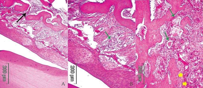
Acta Veterinaria-Beograd 2021, 71 (1), 85-97

                                      INTRODUCTION

Calcium silicate cements are superior to most endodontic materials, taking into
account their biocompatibility, bioactivity and sealing properties. These cements
promote the formation of dentin, cementum, bone and regeneration of the periodontal
ligamentum. They are considered to be the materials of choice for numerous clinical
indications such as pulp capping, root end closure in apical surgery, treatment of root
perforations, apexifications and pulpotomies [1,2]. Good biological properties of
calcium silicate cements are related to calcium hydroxide, which is released during the
material setting and stimulates the proliferation and differentiation of different cells
response for tissue repair [3-5]. The first generation of commercial calcium silicate
cements known as MTAs cements, has a long initial setting time (3h), as a result of
their chemical composition and hygroscopic nature [6] and contain a trace of heavy
metals as a result of the manufacturing by purifying and modification of Portland
cement [7]. Also, these cements usually contain bismuth oxide as radiopacifier, which
interferes with the hydration processes of cement [8] and may react with dentin
collagen resulting in tooth staining [9]. Newer generations of MTAs cements have a
shorter initial setting time, as a result of changes in chemical composition and contain
non toxic radiopacifiers such as zirconium oxide [10].
Hydroxyapatite, despite biocompatibility and bioconductivity did not find its place
in endodontic therapy, mainly because of inappropriate mechanical properties.
Combinations of hydroxyapatite with calcium silicate were initially promising, since
these composite cements showed improved mechanical properties and bioactivity,
compared to pure calcium phosphate cements [11,12].
Technological progress has led to the synthesis of nanostructured materials. Distinct
activity of nano particles enhances hydration of nanostructured calcium silicate
cements, improving their hardening and setting as well as physical and chemical
properties [13,14]. Still, there is concern about the biological behavior of these
materials since nano particles are typically deposited in the mitochondria, causing
structural cell damage. According to the available literature data, a commercial
nanostructured calcium silicate cement with added hydroxyapatite (BioAggregate,
Innovative bioceramix, Vancouver, BC, Canada) shows a similar toxicity in cell cultures
[15], but lower systemic toxicity [16] compared to commercial microstructural MTAs,
which was related to differences in manufacturing and consequently differences in
heavy metals content.
Recently, two new nanostructured cements have been developed in an attempt to
synthetize materials with good biological properties, short setting time and without
heavy metals and bismuth oxide. The first cement (CS) is based on dicalcium and
tricalcium silicate and the other (HA-CS) is a mixture of hydroxyapatite with CS, in
2:1 ratio. According to Jokanović et al. [17], CS was synthetized using hydrothermal
sol-gel methodology and self-propagating combustion waves. Hydroxyapatite was
synthetized by a hydrothermal method. Both materials contain barium sulphate for

86
Petrović et al.: New biomaterials biocompatibility and biofunctionality

radiopacity. Enhanced nanotechnology used in its synthesis resulted in the short setting
time of CS and HA-CS (10 minutes and 15 minutes, respectively). Also, addition of
rheological modifiers prolonged working time and enhanced handling properties of
these materials. Initial in vitro testing of CS and HA-CS has shown the absence of toxic
effects on human cells in terms of genotoxicity [18]. Implantation of these cements
in subcutaneous rat tissue or as pulp capping materials in rabbits’ teeth resulted in
minimal inflammatory response, confirming their biocompatibility [19, 20].
The aim of this study was to evaluate periradicular tissue inflammatory reactions and
the formation of calcified tissue after implantation of CS and HA-CS into root canals
of rabbits’ teeth.

                           MATERIALS AND METHODS

Experimental animals
Experiments were conducted at the Faculty of Veterinary Medicine, University of
Belgrade. The research protocol was approved by the Ethical Committee of the School
of Dental Medicine, University of Belgrade, Serbia (Protocol No.36/21/2013). Four
rabbits of the genus Oryctolagus cuniculus, aged 12 months and average weight of 4 kg
were included in the study. The study was performed in accordance with ISO 10993-2
(Animal welfare requirements) and ISO 7405 [21,22].

Experimental procedure
The animals were kept in standard, individual cages, given ad libitum access to standard
rabbit feed and water and daily monitored during the experiment. Before the surgical
procedure, the animals were introduced into general anesthesia by Xylazine (2%
Xylazine, Check Republic) 35 mg/kg body weight and Ketamidor (100% Ketamidor
100 mg/ml, Richter Pharma AG, Austria) 5 mg/kg body weight. The surgical procedure
was performed in aseptic conditions. The working field was disinfected with 5% iodine
tincture and class I cavities were prepared in the upper and lower central incisors
using round diamond burs. Access to the cavity was prepared and coronal pulp tissue
removed using sterile, round, carbide burs. After extirpation of the radicular pulp, root
canals were instrumented with K files #40 (VDW Gmbh, Germany), and irrigated
with 5 ml of saline between each instrument. A new set of endodontic instruments
was used for each animal. Then, the canals were dried with paper points and filled
with freshly mixed materials. Experimental, nanostructured cements, CS and HA-CS
were mixed with distilled water in 2:1 [17]. Control material, Mineral trioxide aggregate
(White MTA, Angelus ® Solu ões odontológicas, Londrina , Brazil) was mixed in a 3:1
powder to water ratio, according to manufacturers’s instructions. The control material
was implanted in the right maxillary incisors of all four animals. CS was implanted in
the left maxillary incisors and both mandibular incisors of the two animals, and HA-
CS was implanted in the left maxillary incisors and both mandibular incisors of the

                                                                                                        87
Acta Veterinaria-Beograd 2021, 71 (1), 85-97
remaining two animals. Into the root canals, materials were applied with a lentulo spiral
and compacted by a hand compactor. Class I cavities were sealed with resin modified
cement (GC FUJI VIII, GC Corporation, Tokyo, Japan). Postoperatively, the animals
received subcutaneously an analgetic (Butorfanol,10mg/ml, Richter Pharma AG
Austria), 0.1 mg/kg body weight, every 8 h for the next three days and an antibiotic
(Baytril®, 25mg/ml, KVP Pharma und Veterinär Produkte GmbH), 10 mg/kg body
weight, daily for the next five days.
The animals were sacrificed after 28 days, by intravenous injection of 10 ml
Pentobarbital solution (Pentobarbital sodium salt 100 mg ml-1, Sigma-Aldrich Chemie
GmbH, Steiheim, Germany).
Histological procedure and histological analysis
After the removal of soft tissues and separation of the upper and lower jaw, the treated
teeth were cut with a diamond disk and fixed in 10% formalin. After decalcification
(8% HCl from 37% (v/v) concentrate and 10% HCOOH from 89% (v/v) concentrate
in the PBS during 24h at 37◦C), the tissue was fixed in a semi-enclosed benchtop
tissue processor (Leica TP1020, Leica Biosystems, Wetzlar, Germany) and embedded
in paraffin blocks. Serial tissue sections (eight per sample) 5μm thick, were cut from
the paraffin blocks and stained with haematoxylin eosin (HE) according to standard
procedure. The slides were analyzed by optical microscopy (Olympus 5 microscope)
using morphometric software package „Cell-B“ (Olympus), at magnification 40x, 100x
and 200x, by an experienced pathologist blinded to the types of the tested materials. The
histological parameters were analyzed qualitatively (extension of inflammation, general
state of the tissue, continuity and morphology of calcified tissue), semi-quantitatively
(presence of giant cells, material particles and microorganisms) and quantitatively
(inflammation intensity, thickness of calcified tissue). Histomorphometric analysis
was carried according to the cellularity and thickness of calcified tissue. Parameters
were scored using a 1 - 4 scoring system according to modified criteria of Accorinte
et al.[23].
Statistical analysis
The data were analyzed statistically using non-parametric Kruskal-Wallis test and
Dunn’s post hoc test for inter-group comparison (α = 0.05). Non-parametric testing
was chosen to compare categorical variables. Statistical analysis was done in Minitab
16 software package (Minitab Inc., State College, PA, USA).
RESULTS
The results of the histological examination are presented in Table 1. and Figures 1-3.
88Petrović et al.: New biomaterials biocompatibility and biofunctionality
Table 1. Histological analysis for each material according to the scores
CS HA-CS MTA
Score Score Score Intragroup
range Med range Med range Med p-value
Inflammatory reaction
Intensity 1-4 2 1-2 1 1-3 2 0.004*
Extension 2-3 2.5* 1-2 1* 2 2
General state of the tissue 1-3 2 1 1 1-2 2
Calcified tissue
Continuity 2-3 2.5* 1-2 1*# 2-3 3# 0.010* 0.003#
Morfology 1-3 2 1-2 2 2-3 2
Thickness 1-2 1* 1-2 1.5# 3 3*# 0.004* 0.012#
Other findings
Giant cells 1-2 2 1 1 1-2 2
Material particles 2-4 3.5* 1-2 2* 2 2 0.003*
Microorganisms 1 1 1 1 1 1
CS-calcium silicates cement; HA-CS-hydroxyapatite with CS; MTA-Mineral trioxide aggregate.
Med-Median. Within a row, cells with the same superscript are significantly different and matched with
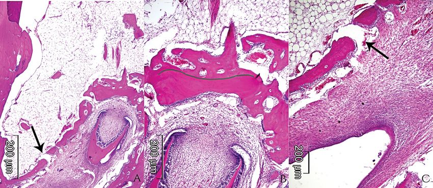
the intragroup P-value with same superscript (pActa Veterinaria-Beograd 2021, 71 (1), 85-97 After implantation of material CS, half of the samples showed no inflammatory reaction (score 1), i.e only few inflammatory cells next to the implanted material were detected (Figure 1 A-C). A moderate inflammatory reaction was found in two samples (score 3). A severe inflammatory reaction (score 4) was observed in one sample, with extension of the inflammatory cells deeper into the tissue (score 3) and abscess formation (score 3). Material particles were detected in all samples, albeit in a different number (score 2 - 4). Half of the samples were giant cells - free (score 1) and in the other half giant cells were detected in a small number (score 2). Microorganisms were not detected in any of the samples. After implantation of material HA- CS, the tissue was unchanged (score 1) in most of the samples (Figure 2 A-C). A mild inflammatory reaction (score 2) was observed in two samples with inflammatory cells next to the implanted material (score 2). A small number of material particles (score 2) was detected in most of the samples. No giant cells or microorganisms was found (score 1). After implantation of MTA, an inflammatory reaction of a different intensity (score 2 - 3) was noticed with inflammatory cells localized next to the implanted material (score 2) (Figure 3 A-C). A small number of material particles (score 2) was detected in all samples. Similarly, a small number of giant cells was found in most of the samples (score 2). No microorganisms were found (score 1). Statistical analysis did not show significant differences in the intensity of inflammatory reactions between the tested materials. There were statistically significant differences between CS and HA-CS with respect to the extension of inflammation (p = 0.004) and the number of material particles (p = 0.003). There were not significant differences among materials tested with respect to giant cells or microorganisms. Figure 2. HA-CS. A: Continuous calcified tissue with lamellar structure (HE, 40x). B and C: Details from the previous photomicrography. Calcified tissue with regular mineralization, lamellar structure and slight to moderate cellularity. Viabile osteocyte is presented in newly formed bone (black arows). Particles of the material (green arrows) can be seen as well as chronic proliferative inflammation of a moderate intensity with signs of angiogenesis and acute hyperaemia around them. (HE,200x). 90
Petrović et al.: New biomaterials biocompatibility and biofunctionality

In the samples with CS, newly formed calcified tissue was mostly of an irregular
morphology (score 2), deposited at a thickness exceeding 250 μm (score 1-2) in most
of the samples, but discontinuous with foci of fibrovascular tissue (score 2-3) (Figure
1 A-C).
In most samples with HA-CS, the implanted material was completely separated from
the adjacent tissue by newly formed, regularly structured, calcified continuous tissue
(score 1). The thickness of newly formed tissue varied between 150 and -250 μm (score
1 - 2). Mesenchymal cells with osteoblastic differentiation were observed on the
outskirts of the newly calcified tissue (Figure 2 A-C).

Figure 3. MTA. A: Partially discontinuous newly formed calcified tissue permeated with
foci of fibrovascular proliferation (black arrow) (HE,40x). B and C: Details from previous
photomicrography. Regular structure of calcified tissue with increased cellularity and many
osteocytes in lacunar spaces. Presence of material particles. Scattered particles of material
(green arrows) surrounded with multinuclear foreign body giant cells can be seen (yellow
arrows) (HE, 200x).

In all of the samples with MTA, newly calcified tissue was deposited in small quantities,
up to 150 μm thick (score 3). The newly formed calcified tissue was irregularly structured,
discontinuous with foci of fibrovascular proliferation (score 2-3) (Figure 3 A-C).
HA-CS exhibited significantly better results than MTA and CS with respect to the
continuity of the newly formed calcified tissue (p = 0.03 and p = 0.010, respectively).
There were significant differences in thickness of the calcified tissue between CS and
MTA (p = 0.004) and between HA-CS and MTA (p=0.012).

                                      DISCUSSION
In vivo tests enable the evaluation of complex interactions between materials and
host tissue. Therefore, such tests, beside biocompatibility, enable the assessment of
materials biofunctionality. With respect to ethical principles, actual studies in animal
models are conducted on laboratory animals, mainly rodents [24, 25]. In this study,

                                                                                                         91
Acta Veterinaria-Beograd 2021, 71 (1), 85-97 materials effects were evaluated after their implantation in the root canals of rabbit teeth. Application of CS and HA-CS in this study resulted in periradicular tissue inflammatory reactions similar to control material (MTA) with respect to intensity. In most of the samples, inflammatory reactions were scored as mild to moderate suggesting good tolerance of the host tissue to the applied materials. These findings are consistent with results of other authors who evaluated biocompatibility of the materials with similar chemical composition [23, 26]. Inflammatory reactions after application of calcium silicate cements are considered to be the result of calcium hydroxide release during material setting. Alkaline pH causes local tissue necrosis with the development of local inflammatory reactions [24]. It is known that calcium silicate cement induces the expression of proinflammatory cytokines (IL-6 and IL-8) also as a result of high pH [27]. Although tissue necrosis is in general, considered to be an initiator of mineralization processes [28], some studies suggested that repair processes could commence even without necrosis or acute inflammation [29]. The amount of released calcium hydroxide from calcium silicate cements decreases over time [30], along with material setting, creating favorable conditions for the start of the repair processes. Although no statistically significant differences between CS, HA-CS and MTA were found with respect to inflammatory response, tissue conditions in the samples with HA-CS were rated the best. This finding may be due to the composition of this material. HA-CS consists mostly of hydroxyapatite with a lower pH value than CS and MTA, although still alkaline [19]. It was confirmed that lower pH values allow alkaline phosphatase activity, but cause a smaller zone of superficial necrosis compared to highly alkaline materials such as calcium silicate cements [31]. A small number of giant cells in the samples with CS and MTA, or their absence in the samples with HA-CS implicated low activity of tissue hystocytes, and good tissue tolerance to the implanted materials. Still, giant cells would presumably be detected in deeper tissues sections. Newly calcified tissue was observed in all samples of tested materials. This finding confirms that tested materials have, besides biocompatibility, an inductive potential. The present results are consistent with previous studies which reported the formation of mineralized tissue after application of materials with a similar chemical composition in different clinical indications [26]. All of the tested materials belong to the group of bioactive materials which are characterized by the release of biologically active ions. As mentioned, the major soluble fraction of these cements is calcium hydroxide, released during material setting. Since calcium silicates are slow setting materials, they release calcium hydroxide over several weeks [30]. In contact with tissue and tissue fluids, calcium hydroxide dissolve to calcium and hydroxyl ions. Continuous release of calcium ions from the material is considered to be crucial for the induction of calcified tissue formation. In addition to its role in chemotaxis, calcium regulates cell proliferation, differentiation and mineralization [28]. 92
Petrović et al.: New biomaterials biocompatibility and biofunctionality

It was confirmed that calcium releasing materials induce proliferation of periodontal
fibroblasts, growth and differentiation of pulp cells, osteoblasts, osteoblast-like cells
and cementoblasts [3-5].
The processes of tissue mineralization are also associated with the release of hydroxyl
ions. The pH increase results in an increase of alkaline phosphatase (ALP), the
expression of growth factors and the formation of calcified nodules. Also, hydroxyl
ions neutralize mediators of inflammation and possess antimicrobial activity [28].
The tested materials are composed of Si ions which are known to have a role in
material bioactivity [11] and proliferation and differentiation of osteoblast-like cells.
High concentrations of Si ions (> 30 ppm) can inhibit the growth of osteoclasts
and resorption processes, but can also increase the level of ALP participating in the
mineralization of the newly calcified tissue [32].
The thickness and continuity of the newly calcified tissue observed in this study was
different and material-dependent. The application of both nanostructured-materials
resulted in a thicker layer of calcified tissue compared to MTA. Materials synthetized
by the sol-gel method, as CS and HA-CS in the present study have improved
bioactivity compared to the same materials obtained by other methods [33]. It is also
known that the topography of the material surface, which is related to their chemical
composition and structure, affects cell activity, especially their adhesion and viability
[34]. It is possible that the nanostructure of CS and HA-CS, which is similar to the
nanocrystalline structure of the bone, could be associated with the obtained results.
Additionally, newly formed calcified tissue associated with HA-CS was continuous
and without foci of vascularized fibroblast proliferation, which was not the case with
MTA and CS. Unlike CS and MTA, HA-CS contains phosphate ions which could be
associated with the aforementioned histological finding. More efficient calcified tissues
were previously reported after application of calcium silicate cements containing
phosphate ions, compared to pure calcium silicate cement [26]. The authors attributed
these findings to a greater amount of phosphate ions available for the hydroxyapatite
formation. Similarly, Zhang et al. [35] reported pronounced mineralization and
odontoblast differentiation of human pulp cells induced by calcium silicate-based
materials with added hydroxyapatite (Bioaggregate and iROOT BP Plus) compared
to pure calcium silicate cement (MTA). The present and cited findings support the
hypothesis that hydroxyapatite-containing materials with high phosphate ion content
have a higher potential for tissue mineralization than MTA.
Microorganisms were not detected in any sample of the tested materials. The microbial
presence is usually correlated with inappropriate crown restorations and subsequent
microleakage [36]. Good sealing properties of GIC used in this study might be the
reason for the obtained result. Still, it must be pointed out that microorganisms are
difficult to detect with this type of histochemical staining and that they could be
removed during tissue preparation for histological analysis.

                                                                                                        93
Acta Veterinaria-Beograd 2021, 71 (1), 85-97
CONCLUSION
Application of CS and HA-CS resulted in a minimal inflammatory tissue response
similar to control MTA. CS and HA-CS were more efficient than MTA in inducing
hard tissue formation after implantation in the root canals of rabbit teeth. The best
organized newly formed calcified tissue was seen after HA-CS application. The present
results serve as a solid foundation for further CS and HA-CS testing.
Acknowledgements
This study was financially supported by the Ministry of Education and Science of the
Republic of Serbia; Projects No 172026 and No 172007.
Authors’ contributions
PV carried out the study, wrote, prepared and formated the manuscript for
publication. OGV participated in experimental procedures and analysis of the results.
JV synthesized the experimental material. SJ carried out the histological examination.
PBB participated in experimental procedures. ŽS gives the idea of experiment, a lead
of role in planning the experiment and revised the manuscript.
Declaration of conflicting interests
The author(s) declared no potential conflicts of interest with respect to the research,
authorship, and/or publication of this article.
REFERENCES
1. Torabinejad M, Parirokh M, Dummer PMH: Mineral trioxide aggregate and other bioactive
endodontic cements: an updated overview-part I: vital pulp therapy. Int Endod J 2018,
51:177-205.
2. Torabinejad M, Parirokh M, Dummer PMH: Mineral trioxide aggregate and other
bioactive endodontic cements: an updated overview-part II: other clinical applications and
complications. Int Endod J 2018, 51:284-317.
3. Takita T, Hayashi M, Takeichi O et al: Effect of Mineral trioxide aggregate on proliferation
of cultured human dental pulp cells. Int Endod J 2006, 39:415-422.
4. Silva LAB, Pieroni KAMG, Nelson-Filho P et al: Furcation perforation.periradicular
tissue response to Biodentine as a repair material by histopathologic and indirect
immunofluorescence analyses. J Endod 2017, 43:1137-1142.
5. Hakki SS, Bozkurt BS, Ozcopur B, Gandolfi MG, Prati C, Belli S: The response of
cementoblasts to calcium phosphate resin-based and calcium silicate-based commercial
sealers. Int Endod J 2013, 46:242-252.
6. Okiji T, Yoshiba K: Reparative dentinogenesis induced by mineral trioxide aggregate: a
review from the biological and physicochemical points of view. Int J Dent 2009; 2009.
94Petrović et al.: New biomaterials biocompatibility and biofunctionality
7. Kum KY, Kim EC, Yoo YJ. Et al: Trace metal contents of three tricalcium silicate materials:
MTA Angelus, Micro Mega MTA and Bioaggregate. Int Endod J 2014, 47:704-710.
8. Camilleri J: Hydratation characteristics of calcium silicate cements with alternative
radiopacifiers used as root-end filling materials. J Endod 2010, 36:502-508.
9. Ramos JC, Palma PJ, Nascimento R et al: 1-year in vitro evaluation of tooth discoloration
induced by 2 calcium silicate-based cements. J Endod 2016, 42:1403-1407.
10. Camilleri J, Sorrentino F, Damidot D. Investigation of the hydration and bioactivity of
radiopacified tricalcium silicate cement, Biodentine and MTA Angelus. Dent Mater 2013,
29:580-593.
11. Huan Z, Chang J: Calcium-phosphate-silicate composite bone cement: self-setting
properties and in vitro bioactivity. J Mater Sci: Mater Med 2009, 20:833-841.
12. Zhao Q, Qian J, Zhou H, Yuan Y, Mao Y, Liu C: In vitro osteoblast-like and endothelial
cells response to calcium silicate/calcium phosphate cement. Biomed Mater 2010, 5:1-8.
13. Asgary S, Eghbal MJ, Parirokh M, Ghoddusi J: Comparason of Mineral trioxide aggregate’s
composition with Portland cements and a new endodontic cement. J Endod 2009, 35:243-
250.
14. Saghiri MA, Godoy FG, Gutmann JL, Lotfi M, Asatourian A, Sheibani N, Elyasi M:
The effect of pH on solubility of nano-modifiedd endodontic cements. J Conserv Dent
2014,17:13-17.
15. De Deus G, Canabarro A, Alves G, linhares A, Senne MI, Granjeiro JM: Optimal
cytocompatibility of a bioceramic nanoparticulate cement in primary human mesenchymal
cells. J Endod 2009, 35:1387-1390.
16. Khalil WA, Eid NF: Biocompatibility of BioAggregate and Mineral trioxide aggregate on
the liver and kidney. Int Endod J 2013, 46:730-737.
17. Jokanović V, Čolović B, Jokanović B, Živković S: Superplastic, quick‐bonding endodontic
mixtures and their hydration. Int Jour Appl Cer Tech 2015;12(S2).
18. Opačić-Galić V, Petrović V, Živković S. et al: New nanostructural biomaterials based on
active calcium silicate systems and hydroxyapatite:characterization and genotoxicity in
human peripheral blood lymphocytes. Int Endod J 2013, 46:506-516.
19. Petrović V, Opačić Galić V, Jokanović V, Jovanović M, Basta Jovanović G, Živković S:
Biocompatibility of a new nanomaterial based on calcium silicate implanted in subcutaneous
connective tissue of rats. Acta Vet-Beograd 2012, 62:697-708.
20. Opačić-Galić V, Petrović V, Jokanović V, Živković S: Histological evaluation of tissue
reactions to newly synthetized calcium silicate-and-hydroxyapatite-based bioactive
materials-In vivo study. Srp Arh Celok Lek 2017, 145:370-377.
21. International Organization for Standardization. ISO 10993: 2009 ed.
22. International Standard Organization. ISO 7405 Dentistry-Preclinical Evaluation of
Biocompatibility of Medical Device Used in Dentistry-Test method for Dental Material.
Geneva, Switzerland;1997.
23. Accorinte Mde L, Holland R, Reis A, Bortoluzzi MC, Murata SS, Dezan E, Souza V,
Alessandro LD: Evaluation of mineral trioxide aggregate and calcium hydroxide cement as
pulp-capping agents in human teeth. J Endod 2008, 34:1-6.
24. Tran XV, Gorin C, Willig C, et al: Effect of a calcium-silicate-based restorative cement on
pulp repair. J Dent Res 2012, 91:1166-1171.
95Acta Veterinaria-Beograd 2021, 71 (1), 85-97
25. Maslamani M, Almusawi A; Joseph B, Gabato S, Andersson L: An experimental model
for studies on delayed tooth replantation and ankylosis in rabbits. Dent Traumatol 2016,
32:443-449.
26. Zarrabi MH, Javidi M, Jafarian AH, Joushan B: Histologic assessment of human pulp
response to capping with mineral trioxide aggregate and a novel endodontic cement. J
Endod 2010, 36:1778-1781.
27. Chen CC, Shie MY, Ding SJ: Human dental pulp cell response to new calcium silicate-based
endodontic materials. Int Endod J 2011, 44:836-842.
28. Sangwan P, Sangwan A, Duha J, Rohilla A: Tertiary dentinogenesis with calcium hydroxide:
A review of proposed mechanisms. Int Endod J 2013, 46:3-19.
29. Leprince JG, Zeitlin BD, Tolar M, Peters OA: Interaction between immune system and
mesenchymal stem cell in dental pulp and periapical tissue. Int Endod J 2012, 45:689-701.
30. Gandolfi MG, Siboni F, Primus CM, Prati C: Ion release, porosity, solubility and bioactivity
of MTA Plus tricalcium silicate. J Endod 2014, 40:1632-1637.
31. Silva EJNL, Rosa TP, Herrera DR, Jacinto RC, Gomes BPFA, Zaia AA: Evaluation of
cytotoxicity and physicochemical properties of calcium silicate-based endodontic sealer
MTA Fillapex. J Endod 2013, 39:274-277.
32. Pietak AM, Reid JW, Stott MJ, Sayer M: Silicon substitution in the calcium phosphate
bioceramics. Biomater 2007, 28:4023-4032.
33. Li P, de Groot K: Better bioactive ceramics through sol-gel process. J Sol-Gel Sci Tech
1994, 2:797-801.
34. Anselme K: Osteoblast adhesion on biomaterials. Biomat 2000, 21:667-681.
35. Zhang S, Yang X, Fan M: Bioaggregate and iRoot BP Plus optimize the proliferation and
mineralization ability of human dental pulp cells. Int Endod J 2013, 46:923-929.
36. Gillen BM, Looney SW, Gu L, et al: Impact of the quality of coronal restoration versus the
quality of root canal fillings on success of root canal treatment: a systematic review and
meta-analysis. J Endod 2011, 37:895-902.
HISTOLOŠKA ANALIZA ZAPALJENSKIH REAKCIJA
U PERIRADIKULARNOM TKIVU I FORMIRANJA
KALCIFIKOVANOG TKIVA POSLE IMPLANTACIJE
EKSPERIMENTALNIH NANOSTRUKTURNIH
CEMENATA NA BAZI KALCIJUM SILIKATA I
HIDROKSIAPATITA U KANALE KORENA ZUBA KUNIĆA
PETROVIĆ Violeta, OPAČIĆ-GALIĆ Vanja, JOKANOVIĆ Vukoman,
SOPTA Jelena, PROKIĆ Bogomir Bolka, ŽIVKOVIĆ Slavoljub
Cilj rada je bio da se ispitaju zapaljenske reakcije u periradikularnom tkivu i formiranje
kalcifikovanog tkiva posle implantacije eksperimentalnih, nanostrukturnih cemenata
na bazi kalcijum silikata (CS) i mešavine hidroksiapatita i kalcijum silikata (HA-CS) u
kanale korena zuba kunića.
96Petrović et al.: New biomaterials biocompatibility and biofunctionality
Kanali korena centralnih sekutića su posle instrumentacije, ispiranja i sušenja napu-
njeni materijalima CS, HA-CS i kontrolnim materijalom, mineral trioksid agregatom
(MTA). Životinje su žrtvovane posle 28 dana. Posle histološke pripreme, uzorci tkiva
su analizirani u pogledu inteziteta i raširenosti zapaljenske reakcije; kontinuiteta, mor-
fologije i debljine novoformiranog kalcifikovanog tkiva; prisustva džinovskih ćelija, če-
stica materijala i mikroorganizama. Dobijeni rezultati su statistički obrađeni (α = 0,05)
Nisu uočene statistički značajne razlike u intezitetu zapaljenske reakcije između CS,
HA-CS i MTA. U pogledu kontinuiteta novostvorenog kalcifikovanog tkiva HA-CS je
pokazao bolje rezultate u odnosu na MTA i CS (p=0,003 i p=0,010). Značajne razlike
utvrđene su u pogledu debljine kalcifikovanog tkiva između CS i MTA (p=0,004), kao
i HA-CS i MTA (p=0,012).
Aplikacija materijala CS i HA-CS je rezultirala minimalnom zapaljenskom reakcijom
tkiva, slično kontrolnom materijalu (MTA). CS i HA-CS su bili efikasniji u pogledu
stimulacije formiranja kalcifikovanog tkiva u odnosu na MTA. Najbolje organizovano
novoformirano tkivo uočeno je posle aplikacije materijala HA-CS.
97You can also read