Journal Pre-proof - IUPHAR
←
→
Page content transcription
If your browser does not render page correctly, please read the page content below
Journal Pre-proof

Lianhuaqingwen exerts anti-viral and anti-inflammatory activity against
novel coronavirus (SARS-CoV-2)

Li Runfeng, Hou Yunlong, Huang Jicheng, Pan Weiqi, Ma Qinhai,
Shi Yongxia, Li Chufang, Zhao Jin, Jia Zhenhua, Jiang Haiming,
Zheng Kui, Huang Shuxiang, Dai Jun, Li Xiaobo, Hou Xiaotao,
Wang Lin, Zhong Nanshan, Yang Zifeng

PII:                    S1043-6618(20)30743-X
DOI:                    https://doi.org/10.1016/j.phrs.2020.104761
Reference:              YPHRS 104761

To appear in:           Pharmacological Research

Received Date:          29 February 2020
Revised Date:           14 March 2020
Accepted Date:          17 March 2020

Please cite this article as: Runfeng L, Yunlong H, Jicheng H, Weiqi P, Qinhai M, Yongxia S,
Chufang L, Jin Z, Zhenhua J, Haiming J, Kui Z, Shuxiang H, Jun D, Xiaobo L, Xiaotao H, Lin
W, Nanshan Z, Zifeng Y, Lianhuaqingwen exerts anti-viral and anti-inflammatory activity
against novel coronavirus (SARS-CoV-2), Pharmacological Research (2020),
doi: https://doi.org/10.1016/j.phrs.2020.104761
This is a PDF file of an article that has undergone enhancements after acceptance, such as
the addition of a cover page and metadata, and formatting for readability, but it is not yet the
definitive version of record. This version will undergo additional copyediting, typesetting and
review before it is published in its final form, but we are providing this version to give early
visibility of the article. Please note that, during the production process, errors may be
discovered which could affect the content, and all legal disclaimers that apply to the journal
pertain.

© 2020 Published by Elsevier.
Lianhuaqingwen exerts anti-viral and anti-inflammatory activity against novel
                           coronavirus (SARS-CoV-2)

 Li Runfeng a, #, Hou Yunlong e,#, Huang Jichengd, #, Pan Weiqia,#, Ma Qinhaia, Shi
  Yongxiad, Li Chufanga,#, Zhao Jina, Jia Zhenhuae , Jiang Haiminga , Zheng Kuid,
Huang Shuxiangd,Dai Jund, Li Xiaobod, Hou Xiaotaoc, Wang Linc, Zhong Nanshana,
                                 Yang Zifeng a,b,c*
a
  State Key Laboratory of Respiratory Disease, National Clinical Research Center for
Respiratory Disease, Guangzhou Institute of Respiratory Health,the First Affiliated
Hospital of Guangzhou Medical University, Guangzhou, Guangdong, postcode,
P.R.China
b
  State Key Laboratory of Quality Research in Chinese Medicine, Macau Institute for
Applied Research in Medicine and Health, Macau University of Science and

                                                             of
Technology, Taipa, Macau SAR, China
c
  KingMed Virology Diagnostic & Translational Center
d
  Technology Centre, Guangzhou Customs
e

                                                           ro
  Hebei University of Chinese Medicine, College of Integrated Chinese and Western
Medicine.
#
  Li Runfeng, Hou Yunlong, Huang Jicheng, and Pan Weiqi have equal contributions
to this study.

*
 To whom correspondence should be addressed.
                                                 -p
State Key Laboratory of Respiratory Disease, National Clinical Research Center for
                                        re
Respiratory Disease,Guangzhou Institute of Respiratory Health,the First Affiliated
Hospital of Guangzhou Medical University,Guangzhou, Guangdong,
postcode,P.R.China
Tel.: +86 83205181
                           lP

E-mail: jeffyah@163.com (Zifeng Yang).

Abstract
                 na

Purpose: Lianhuaqingwen (LH) as traditional Chinese medicine (TCM) formula has
been used to treat influenza and exerted broad-spectrum antiviral effects on a series of
influenza viruses and immune regulatory effects[1]. The goal of this study is to
        ur

demonstrate the antiviral activity of LH against the novel SARS-CoV-2 virus and its
potential effect in regulating host immune response.
Methods: The antiviral activity of LH against SARS-CoV-2 was assessed in Vero E6
Jo

cells using CPE and plaque reduction assay. The effect of LH on virion morphology
was visualized under transmission electron microscope. Pro-inflammatory cytokine
expression levels upon SARS-CoV-2 infection in Huh-7 cells were measured by
real-time quantitative PCR assays.
Results: LH significantly inhibited SARS-CoV-2 replication in Vero E6 cells and
markedly reduced pro-inflammatory cytokines (TNF-α, IL-6, CCL-2/MCP-1 and
CXCL-10/IP-10) production at the mRNA levels. Furthermore, LH treatment resulted
in abnormal particle morphology of virion in cells.
Conclusions: LH significantly inhibits the SARS-COV-2 replication, affects virus
morphology and exerts anti-inflammatory activity in vitro. These findings indicate that
LH protects against the virus attack, making its use a novel strategy for controlling the
COVID-19 disease.
Keywords: Lianhuaqingwen, coronavirus, SARS-CoV-2,anti-inflammatory
Introduction
Coronaviruses are a group of enveloped viruses named for their coronary appearance
with positive single-stranded RNA genomes[2]. In addition to six known strains of
coronaviruses that are infectious to humans, a novel coronavirus (SARS-CoV-2) was
detected recently in Wuhan, China[3, 4]. Like the other two highly pathogenic
coronaviruses SARS-CoV and MERS-CoV, SARS-CoV-2 also caused severe
of
respiratory illness and even death. Moreover, the population's susceptibility to these
highly pathogenic coronaviruses has contributed to large outbreaks and evolved
into the public health events, highlighting the necessity to prepare for future
ro
reemergence or the novel emerging viruses[5].
Similar to SARS-CoV and MERS-CoV, SARS-CoV-2 is initiated by zoonotic
transmission likely from bats and spreads rapidly among humans[6]. The basic
-p
reproduction number (R0) of person-to-person spread is about at 2.6, which means that
the SARS-CoV-2 infected cases grow at an exponential rate. As of February 07, 2020,
57,620 cases of the SARS-CoV-2 have been reported in China, including 26,359
suspected cases, and a sustained increase is predictable. The initial patient cluster with
re
confirmed SARS-CoV-2 infection was reported Wuhan pneumonia with unknown
aetiology, which bore some resemblance to SARS-CoV and MERS-CoV infections
and was associated with ICU admission and high mortality. Moreover, High
lP
concentrations of cytokines were recorded in plasma of patients requiring ICU
admission, such as GCSF, IP10, MCP1, MIP1A, and TNFα, suggesting that the
cytokine storm was associated with disease severity[7]. A retrospective clinical study
indicated the risk of fatality among hospitalized cases at 4.3% in single-center case
na
series of 138 hospitalized patients[8], and the infection fatality risk could be below 1%
or even below 0.1% in a large number of undetected relatively mild infections[9].
However, It is challenging to judge the severity and predict the consequences with the
information available so far. Since no specific antiviral treatment for COVID-19 is
ur
currently available, supportive cares, including symptomatic controls and prevention
of complications remain the most critical therapeutic regimens, especially in
preventing acute respiratory distress syndrome[10]. Although the control of
Jo
SARS-CoV-2 still presents multiple challenges in the short term, more potent antiviral
drugs are urgent to be developed[4].
At present, some drugs are effective in eliminating SARS-CoV-2 and improving
symptoms. The most promising antiviral drug for SARS-CoV-2 is remdesivir that is
currently under clinical development for the treatment of Ebola virus infection[11].
However, the efficacy and safety of remdesivir for SARS-CoV-2 pneumonia patients
need to be assessed by further clinical trials. In addition, in the prevention and
treatment of COVID-19, Tranditonal Chinese medicines have received broad adoption,
especially in treating cases of mild symptoms[12]. Lianhuaqingwen (LH), a Chinese
patent medicine composed of 13 herbs, has played a positive role in the treatment ofSARS-CoV-2. A retrospective analysis of clinical records was conducted in the
SARS-CoV-2 infected patients at Wuhan Ninth Hospital and CR & WISCO General
Hospital. LH combination could significantly relieve cardinal symptoms and reduce
the course of the COVID-19[13], making it successively included in the Guideline for
the Diagnosis and Treatment of Novel Coronavirus (2019-nCoV) Pneumonia (On
Trials, the Fourth/Fifth/Sixth/Seventh Edition) issued by National Health Commission
of the People’s Republic of China and also recommended by 20 provincial health
commissions including Hubei, Beijing, and Shanghai as well as National
Administration of Traditional Chinese Medicine for the treatment of COVID-19.
Moreover, LH exerted broad-spectrum effects on a series of influenza viruses by
inhibiting viral propagation and regulating immune function and achieved similar
therapeutic effectiveness with Oseltamivir in reducing the course of H1N1 virus
infection[1, 14, 15]. Notably, the anti-influenza activity of LH in infected mice might
depend on the regulation of cytokines, particularly in cytokine storm associated
cytokines, such as IP-10, MCP-1, MIP1A, and TNF-α[1]. In the present study, we
of
evaluated the antiviral and anti-inflammatory efficiency of LH against a clinical isolate
of SARS-CoV-2 from Guangzhou in vitro.
ro
Materials and Methods
Cell lines and virus
The African green monkey kidney epithelial (Vero E6) cells and the human
hepatocellular carcinoma (Huh-7) cells were cultured in Dulbecco’s Modified Eagle’s
-p
medium (DMEM, Gibco, USA) supplemented with 10% fetal bovine serum (FBS) at
37°C. A clinical isolated SARS-CoV-2 virus (Genebank accession no. MT123290.1)
was propagated in Vero E6 cells, and viral titer was determined by 50% tissue culture
re
infective dose (TCID50) according to the cytopathic effect by use of Reed-Muench
method [17]. All the infection experiments were performed in a biosafety level-3
(BLS-3) laboratory.
lP
Reagent preparation
LH capsule (Lot No.A2001108) was obtained from Yiling Pharmaceutical Co. Ltd.
(Shijiazhuang, China). UPLC fingerprints of LH consist of 32 common peaks. 9 of 32
na
common peaks are identified. The similarities in 10 batches of LH Capsules samples
were all above 0.96 (Supplementary figure 1). The black powder of raw material of
LH was first dissolved in dimethyl sulfoxide (DMSO) to 240 mg/mL. After shaking
for 30 min at room temperature, the LH solution was diluted with serum-free DMEM
ur
to 24 mg/mL as a stock solution and stored at -20°C before using. Remdesivir was
kindly provided by Prof. Jiancun Zhang from Guangzhou Institutes of Biomedicine
and Health, Chinese Academy of Sciences and was dissolved in DMSO to 100 mM
Jo
and stored at -20 °C before using. DMEM with 2% FBS was used as the dilution
buffer in the follow-up experiments.
Cytotoxicity assay
The cytotoxic effects of the LH on Vero E6 and Huh-7 cells were evaluated by Methyl
Thiazolyl Tetrazolium (MTT) assay. Briefly, monolayers of Vero E6 cells and Huh-7
cells in 96-well plates were rinsed with phosphate-buffered saline (PBS) followed by
incubation with indicated concentrations of LH. After 72 h, the cells were stained with
MTT solution at 0.5 mg/mL for 4 h. The supernatants were then removed, and the
formed formazan crystals were dissolved in 200 μL DMSO. The absorbance wasmeasured at 490 nm using Multiskan Spectrum reader (Thermo Fisher, USA). The 50%
cytotoxic concentration (CC50) was calculated by the GraphPad Prism 7.0 software.
Cytopathic effect (CPE) inhibition assay
The Vero E6 cell monolayers were grown in 96-well plates and inoculated with 100
TCID50 of coronavirus strains at 37 ̊C for 2 h. The inoculum was removed, and the
cells were subsequently incubated with indicated concentrations of LH or the positive
control remdesivir. Following the 72 h of incubation, the infected cells shown 100%
CPE under the microscope. The percentage of CPE in LH-treated cells were recorded.
The 50% inhibition concentration (IC50) of the virus-induced CPE by LH was
calculated by the Reed-Muench method[17].
Plaque reduction assay
The Vero E6 cell monolayers in 6-well plates were infected with 50 plaque-forming
units (PFU) of SARS-CoV-2 for 2 h at 37 °C. After incubation, the cell monolayers
of
were covered with agar overlay (final concentration: 0.6% agar, 2% FBS, indicated
concentrations of LH or remdesivir). The plates were then incubated for 48 h at 37 °C
with 5% CO2. Subsequently, the agar overlays were removed, and the cell monolayer
ro
was fixed with 10% formalin, stained with 1% crystal violet, and then the plaques
were counted and photographed.
-p
RNA isolation and reverse transcriptase-quantitative PCR analysis (RT-qPCR)
The Huh-7 cell monolayers in 12-well plate were rinsed with PBS and then exposed to
coronavirus at a multiplicity of infection (MOI) of 1 for 2 h at 37°C. The inoculum
was removed and replaced with the indicated concentrations of LH or mock-treated
re
with DMEM supplemented with 2% FBS for subsequent 48 h incubation at 37 °C with
5% CO2. The cells were then harvested for RNA isolation and qPCR as described
previously [16]. The primer and probe sequences used for analysis are listed in
lP
Supplementary Table 1. The relative mRNA expression was calculated using the 2-△△Ct
method with GAPDH as an internal reference gene.
Electron microscope
na
Monolayers of Vero E6 cells in 6-well plates were incubated with SARS-CoV-2 at
a MOI of 0.001 for 2h at 37°C. The virus inoculum was then removed and replaced
with DMEM medium supplemented with 2% FBS containing LH (600 μg/mL) or
remdesivir (5 μM). At 48 h p.i., the cells were fixed, dehydrated and embedded as
ur
described previously [18]. Ultrathin sections (70 nm) of embedded cells were prepared,
deposited onto Formvar-coated copper grids (200 mesh), stained with uranyl acetate
and lead citrate, and then observed under JEM-1400 PLUS transmission electron
Jo
microscopy (Japan Electron Optics Laboratory Co., Ltd., JEM-1400 PLUS).
Statistical Analyses
Statistical analysis was performed using GraphPad Prism 7.0 software. The differences
in mRNA expression levels of cytokines were compared using a one-way analysis of
variance (ANOVA). Values of pThe cell viability after LH or remdesivir treatment was determined by MTT assay in
both Vero E6 and Huh-7 cells. LH showed unapparent cytotoxicity for both cell lines
at concentrations up to 600 μg/mL (Fig. 1A, 1C). The positive control remdesivir
showed no cytotoxicity to cells at a concentration of 50µM (Fig. 1B, 1D).
of
ro
Fig. 1. Cytotoxic effect of the LH and remdesivir on Vero E6 cells (A, B) and Huh-7
cells (C, D). Data are presented as mean ± SD. The experiments were performed in
triplicate. -p
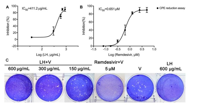
To investigate the antiviral effect of LH against SARS-CoV-2 virus, the Vero E6 cells
were infected with 100 TCID50 of virus and incubated with LH at various
re
concentrations for 72h. As shown in Fig. 2A, LH inhibited the replication of
SARS-CoV-2 virus with an IC50 value of 411.2 µg/mL by CPE assay (Fig.2A).
Meanwhile, treatment with LH following infection also had a dose-dependent
lP
inhibitory effect on plaque formation of the SARS-CoV-2 virus (Fig.2C). We selected
remdesivir as the positive control in our study and the results showed that remdesivir
potently inhibited virus-induced CPE with an IC50 of 0.651 µM and a total plaque
na
ur
Jo
formation inhibition at 5 μM (Fig.2B, 2C).
Fig. 2. Antiviral activity of LH and remdesivir against SARS-CoV-2 in Vero E6 cells.
(A, B) The inhibitory curve for LH and remdesivir. (C) Plaque reduction assay of LHagainst the SARS-CoV-2 virus. Data are representative of three independent
experiments.
To further confirm the efficacy of LH in inhibiting SARS-CoV-2 virus replication in
cells, we detected the viral particles in ultrathin sections of infected cells under
electron microscopy. At 48h p.i., viral particles were found in cytoplasm, intracellular
vesicles, endoplasmic reticulum, and cell membrane and presented spherical
crown-like appearance, which was typical coronavirus morphology (Fig. 3B, G). LH
(600 μg/mL) and positive control remdesivir (5 μM) treatment resulted in a reduction
of the number of virions compared with mock-treated infected cells (Fig. 3G, H, J). It
was interesting to note that some virions in the surface of LH-treated cells presented
spindle sharp which was in contrast to the typical spherical particles in the
mock-treated cells (Fig. 3I).
Fig. 3 Virions in the ultra sections of infected Vero E6 cells under electron microscope.
of
(A, F) uninfected cells, (B, G) mock-treated SARS-CoV-2 virus infected cells, (C, D,
H, I) infected cells with LH treatment, (E, J) infected cells with remdesivir treatment.
White arrows indicated the spindle sharp of viral particles in infected cells with LH
ro
treatment.
-p
re
lP
na
Inhibition of SARS-CoV-2-induced cytokine and chemokine expression by LH in
vitro
To determine the effect of LH on the expression of cytokines and chemokines induced
ur
by SAR2-CoV-2, the mRNA expression levels of TNF-α, IL-6, CCL-2/MCP-1, and
CXCL-10/IP-10 were detected and compared between the LH-treated and
mock-treated Huh-7 cells. The results showed that the elevated expressions of these
Jo
four cytokines were significantly inhibited by LH treatment in aconcentration-dependent manner (Fig. 4).
Fig. 4 Effects of LH treatment on the mRNA expression levels of inflammatory
mediators in SARS-CoV-2-infected Huh-7 cells. A: TNF-α, B: IL-6, C:
CCL-2/MCP-1, D: CXCL-10/IP-10. Data are presented as the mean±SD obtained
from three separate experiments. *p< 0.05; **p< 0.01; ***p< 0.001, compared with
mock-treated cells.
Discussion
Starting from December 2019, a pandemic of respiratory illness caused by a novel
coronavirus named SARS-CoV-2 is sweeping the mainland of China. This virus has
spread to several foreign countries, threatening to trigger a global outbreak. Several
antiviral agents can be envisaged to control or prevent viral infections by antiviral
assay in vitro[14, 17]. However, the efficacy and safety of novel candidates need
of
validations in vivo, even for those clinically approved medicines, which means that it
will take months to years for clinical practices. At present, symptomatic and
supportive treatments remain key to clinical practices. Thus, Traditional Chinese
ro
Medicines (TCM) carried both the antiviral effect and the symptomatic relief might
bring more clinical benefits[12]. As a classical TCM prescription for respiratory
diseases, LH is the only approved medicine in the treatment of SARS and influenza.
-p
After the outbreak of SARS-CoV-2, LH as a representative TCM prescription was
recommended again in the latest Guideline for the Diagnosis and Treatment of Novel
Coronavirus (2019-nCoV) Pneumonia issued by National Health Commission of the
re
People’s Republic of China The purpose of this study was to demonstrate whether the
therapeutic effects of LH on the COVID-19 targeting virus replication and
immunological regulation as it did on the infection caused by influenza viruses.
lP
Our previous study showed that LH exhibited in vitro anti-influenza activity with IC50
ranging from 200-2000 μg/mL[1]. Here we demonstrated that LH also has a
comparable antiviral potency against the SARS-CoV-2 virus with an IC50 value of
411.2 μg/mL (Fig. 2). Transmission electron microscopy (TEM) has been a potent tool
na
to observe virus entry, virus particle assembly, viral ultrastructure, and budding from
the plasma membrane[17]. To understand the antiviral details of LH, EM pictures were
taken from each group. Abundant virus particles assembled at the surface of
ur
membrane, cytoplasm, and plasma vesicles in the SARS-CoV-2 infected cells,
decreased in the treatment of LH at 600ug/mL. Notably, slight deformation of virus
particles was seen in the LH treatment, which required us to make further studies.
Jo
Highly pathogenic coronaviruses such as SARS-CoV and MERS-CoV cause fatal
pneumonia, which is mainly associated with rapid virus replication, massive
inflammatory cell infiltration and elevated proinflammatory cytokine/chemokine
responses. Although the pathophysiology of fatal pneumonia caused by highly
pathogenic coronaviruses has not been completely understood, accumulating evidence
suggests that the cytokine storm plays a crucial role in causing fatal pneumonia[18].
Excessive amounts of proinflammatory cytokines were reported (eg, IL-1β, IL-6,
IL-12, IFN-γ, IP-10, and MCP-1) in the serum of SARS patients[18], similar in the
serum of MERS patients[19]. Chaolin Huang et al. confirmed the occurrence of thecytokine storm in the COVID-19 patients in ICU rather than those in non-ICU
patients[7]. Based on the excessive cytokines responses, Suxin Wan et al. claimed that
IL-6 and IL-10 levels could be used as one of the bases for predicting the outcome and
prognosis of the COVID-2019[20]. In this study, host cells infected with HCoV-229E
and SARS-COV-2 increased the cytokine release such as TNF-α, IL-6, CCL-2/MCP-1,
and CXCL-10/IP-10, which was suppressed by LH in a dose-dependent manner. The
change of cytokine profiles suggested that LH might have a potential effect on the
inhibition of cytokine storm induced by SARS-COV-2, which also needed to be
validated in vivo.
Conclusion
Since the launch of LH, it has been widely used as a broad spectrum of antiviral agent
in the clinical practice, especially for various respiratory virus infections. Previous
studies have shown that LH a broad spectrum of effects on a series of influenza viruses
of
by interfering with both viral and host reactions. Although LH significantly relieved
the clinical symptoms of the COVID-19, the underlying mechanism of antiviral effects
ro
on coronavirus, especially in the SARS-COV-2, was still elusive. In this study, we
demonstrated that LH exerted its anti-coronavirus activity by inhibiting virus
replication and reducing the cytokine release from host cells, which supported the
-p
clinical application of LH in combination with existing therapies to treat
COVID-2019.
re
Acknowledgement
The study was funded by Beijing Municipal Science and Technology Commission
lP
NCP Emergency Project; Hebei Provincial Department of Science and Technology
NCP prevention and control emergency scientific research project ( Grant no.
20277708D);The Science research project of the Guangdong Province (Grant no.
2020B111110001);Daxing District Science and technology development projects
na
(Grant no. KT202008013).
References
1. Ding, Y., et al., The Chinese prescription lianhuaqingwen capsule exerts
ur
anti-influenza activity through the inhibition of viral propagation and impacts
immune function. BMC Complement Altern Med, 2017. 17(1): p. 130.
Jo
2. Fung, T.S. and D.X. Liu, Human Coronavirus: Host-Pathogen Interaction. Annu
Rev Microbiol, 2019. 73: p. 529-557.
3. Du Toit, A., Outbreak of a novel coronavirus. Nat Rev Microbiol, 2020.
4. Carlos, W.G., et al., Novel Wuhan (2019-nCoV) Coronavirus. Am J Respir Crit
Care Med, 2020.
5. Nkengasong, J., China's response to a novel coronavirus stands in stark
contrast to the 2002 SARS outbreak response. Nat Med, 2020.
6. Zhou, P., et al., A pneumonia outbreak associated with a new coronavirus of
probable bat origin. Nature, 2020.7. Huang, C., et al., Clinical features of patients infected with 2019 novel
coronavirus in Wuhan, China. Lancet, 2020.
8. Wang, D., et al., Clinical Characteristics of 138 Hospitalized Patients With 2019
Novel Coronavirus-Infected Pneumonia in Wuhan, China. JAMA, 2020.
9. Wu, P., et al., Real-time tentative assessment of the epidemiological
characteristics of novel coronavirus infections in Wuhan, China, as at 22
January 2020. Euro Surveill, 2020. 25(3).
10. Zumla, A., et al., Coronaviruses - drug discovery and therapeutic options. Nat
Rev Drug Discov, 2016. 15(5): p. 327-47.
11. Mulangu, S., et al., A Randomized, Controlled Trial of Ebola Virus Disease
Therapeutics. N Engl J Med, 2019. 381(24): p. 2293-2303.
12. Ren, J.-l., A.-H. Zhang, and X.-J. Wang, Traditional Chinese Medicine for
COVID-19 Treatment. Pharmacological Research, 2020: p. 104743.
13. Yao, K, et al., Retrospective Clinical Analysis on Treatment of Novel
of
Coronavirus-infected Pneumonia with Traditional Chinese Medicine Lianhua
Qingwen. Chinese Journal of Experimental Traditional Medical Formulae: p.
ro
1-7.
14. Lu, H., Drug treatment options for the 2019-new coronavirus (2019-nCoV).
Biosci Trends, 2020.
15. -p
Duan, Z.P., et al., Natural herbal medicine Lianhuaqingwen capsule
anti-influenza A (H1N1) trial: a randomized, double blind, positive controlled
clinical trial. Chin Med J (Engl), 2011. 124(18): p. 2925-33.
re
16. Li, Z., et al., Radix isatidis Polysaccharides Inhibit Influenza a Virus and
Influenza A Virus-Induced Inflammation via Suppression of Host TLR3
Signaling In Vitro. Molecules, 2017. 22(1).
lP
17. Wang, M., et al., Remdesivir and chloroquine effectively inhibit the recently
emerged novel coronavirus (2019-nCoV) in vitro. Cell Res, 2020.
18. Leong, H.N., et al., Clinical and laboratory findings of SARS in Singapore. Ann
Acad Med Singapore, 2006. 35(5): p. 332-9.
na
19. Assiri, A., et al., Epidemiological, demographic, and clinical characteristics of
47 cases of Middle East respiratory syndrome coronavirus disease from Saudi
Arabia: a descriptive study. Lancet Infect Dis, 2013. 13(9): p. 752-61.
20. Wan, S., et al., Characteristics of lymphocyte subsets and cytokines in
ur
peripheral blood of 123 hospitalized patients with 2019 novel coronavirus
pneumonia (NCP). medRxiv, 2020: p. 2020.02.10.20021832.
JoYou can also read