Glucocorticoids protect HEI-OC1 cells from tunicamycin-induced cell damage via inhibiting endoplasmic reticulum stress
←
→
Page content transcription
If your browser does not render page correctly, please read the page content below
Open Life Sciences 2021; 16: 695–702
Research Article
Zhibiao Liu#, Bing Fei#, Lisheng Xie, Jin Liu, Xiaorui Chen, Wenyan Zhu, Lingyun Lv, Wei Ma,
Ziwen Gao, Jie Hou, Wandong She*
Glucocorticoids protect HEI-OC1 cells from
tunicamycin-induced cell damage via inhibiting
endoplasmic reticulum stress
https://doi.org/10.1515/biol-2021-0057 Abstract
received July 10, 2020; accepted March 24, 2021 Background ‒ To analyze mechanisms of action of gluco-
corticoid treatment for endoplasmic reticulum stress
(ERS) in sensorineural hearing loss (SNHL), we aimed
to evaluate the expression and activation status of the
protein kinase RNA-like ER kinase (PERK)–C/EBP homo-
# These authors contributed equally to this work. logous protein (CHOP) pathway, which is the major
pathway in the ERS.
Methods ‒ In the present study, we established an in vitro
* Corresponding author: Wandong She, Department of
Otolaryngology-Head and Neck Surgery, Nanjing Drum Tower ERS model using tunicamycin-treated hair-cell-like HEI-
Hospital Clinical College of Nanjing Medical University, 321 OC1 cells. The effect of dexamethasone on proliferation
Zhongshan Road, Nanjing 210008, China; Department of inhibition, apoptosis, and ATF4–CHOP pathway in HEI-
Otolaryngology-Head and Neck Surgery, Nanjing Drum Tower OC1 cells was examined by CCK-8 assay, flow cytometry,
Hospital Clinical College of Traditional Chinese and Western
western blotting, and reverse transcription PCR, respectively.
Medicine, Nanjing University of Chinese Medicine, Nanjing 210000,
China; Department of Otolaryngology-Head and Neck Surgery, Results ‒ In HEI-OC1 cells, dexamethasone was shown
Nanjing Drum Tower Hospital, The Affiliated Hospital of Nanjing to significantly reduce the tunicamycin-induced expres-
University Medical School, Jiangsu Provincial Key Medical sion of ATF4 and CHOP in the context of sustained viability
Discipline, Nanjing, China, e-mail: shewandong@163.com and proliferation, a therapeutic effect that was reversible
Zhibiao Liu, Wenyan Zhu, Lingyun Lv: Department of
by co-treatment with a glucocorticoid antagonist.
Otolaryngology-Head and Neck Surgery, Nanjing Drum Tower
Conclusion ‒ Dexamethasone can protect hair-cell-like
Hospital Clinical College of Nanjing Medical University, 321
Zhongshan Road, Nanjing 210008, China; Department of HEI-OC1 cells from ERS damage, which may be one of the
Otorhinolaryngology-Head and Neck Surgery, The Affiliated Huaian mechanisms of action for GCs in SNHL treatment.
No. 1 People’s Hospital of Nanjing Medical University, Huaian,
Nanjing, China Keywords: endoplasmic reticulum stress, PERK–CHOP
Bing Fei: Department of Otorhinolaryngology-Head and Neck pathway, dexamethasone, mifepristone
Surgery, Affiliated Huai’an Hospital of Xuzhou Medical University,
62 South Huaihai Road, Huai’ an 223002, China
Lisheng Xie, Wei Ma, Ziwen Gao: Department of Otolaryngology-
Head and Neck Surgery, Nanjing Drum Tower Hospital Clinical 1 Introduction
College of Nanjing Medical University, 321 Zhongshan Road, Nanjing
210008, China Endoplasmic reticulum (ER) is an important organelle to
Jin Liu, Xiaorui Chen: Department of Otolaryngology-Head and Neck
maintain normal cellular homeostasis. When eukaryotic
Surgery, Nanjing Drum Tower Hospital Clinical College of Traditional
Chinese and Western Medicine, Nanjing University of Chinese cells are exposed to pathophysiological stressors, a large
Medicine, Nanjing 210000, China number of misfolded proteins accumulate in the ER and
Jie Hou: Department of Otolaryngology-Head and Neck Surgery, activate endoplasmic reticulum stress (ERS) [1]. ERS is
Nanjing Drum Tower Hospital Clinical College of Nanjing Medical related to many human diseases [1,2]. During the early
University, 321 Zhongshan Road, Nanjing 210008, China;
stages of ERS, cells can adapt to altered environmental
Department of Otolaryngology-Head and Neck Surgery, Nanjing
Drum Tower Hospital, The Affiliated Hospital of Nanjing University
conditions by reducing unfolded or misfolded protein.
Medical School, Jiangsu Provincial Key Medical Discipline, Nanjing, However, if stress conditions persist, cells undergo apop-
China tosis [3]. Protein kinase RNA-like ER kinase (PERK) is a
Open Access. © 2021 Zhibiao Liu et al., published by De Gruyter. This work is licensed under the Creative Commons Attribution 4.0
International License.
696 Zhibiao Liu et al.
predominant ERS-induced apoptotic signaling pathway concentration of mifepristone-mediated antagonism of
and it is activated by phosphorylation, thereby phosphor- the therapeutic effects elicited by dexamethasone. The
ylating eukaryotic initiation factor 2α (eIF2α). p-eIF2α can inhibition rate of cell proliferation was detected using
promote the expression of activating transcription factor the CCK-8 Cell Proliferation Detection Kit (Tianjin Bayang
4 (ATF4) and C/EBP homologous protein (CHOP) [4,5]. Huake Biotechnology Co., Ltd, China.). The optimized
After CHOP expression increases considerably, CHOP conditions were then used to conduct comparative ana-
accumulates in the nucleus and ultimately results in lyses between cultures, using appropriate controls con-
apoptosis [5]. In several animal models of sensorineural taining no drugs or with individual drug treatments.
hearing loss (SNHL), ERS was believed to be associated
with inner ear injuries [6–9].
Glucocorticoids (GCs) regulate many complex sig-
naling pathways [10–12]. It has been reported that, under 2.2 Flow cytometry (FACS)
ERS conditions, there is crosstalk between CHOP and
GR signaling, which is associated with a glucocorticoid Flow cytometric analysis has been done using Annexin
receptor (GR)-CHOP heterocomplex formation [13]. V/Propidium Iodide (PI) Apoptosis Detection Kit (Beyotime,
Therefore, we hypothesized that GCs might protect Shanghai, China) according to the manufacturer’s instruc-
inner ear cells from ERS damage. In the present study, tions. HEI-OC1 cells (3 × 10⁵) were collected and stained
we examined the effects of GCs on the expression of pro- with 5 μL Annexin V-APC and 5 μL PI in the dark at room
teins associated with the PERK–CHOP pathway in HEI- temperature for 10 min. Data were then acquired on a BD
OC1 cells to validate a putative role of ERS in SNHL and to Accuri™ C6 Plus flow cytometer (BD, Franklin Lakes, NJ,
determine whether GCs can reduce ERS. USA) and analyzed by Flow Jo V10 software (Tree Star
Software, San Carlos, CA, USA).
2 Materials and methods 2.3 qPCR and mRNA extraction
Total RNA was extracted from HEI-OC1 Cells using TRIzol
2.1 Cell culture and drug administration
reagent. cDNA was then obtained by reverse-transcrip-
tion. Real-time PCR was performed with the Applied
HEI-OC1 cells were obtained from the Chinese academy
Biosystems QuantStudio 6 Flex Real-Time PCR System
of medical science. The cells were maintained in DMEM
(Applied Biosystems, Singapore). The M-MLV was applied
medium (Life technologies) supplemented with 10% Fetal
to synthesize cDNA through reverse-transcription. For cDNA
Bovine Serum (FBS Life technologies) and 100 U/mL
synthesis, samples were incubated at 43°C for 30 min, 97°C
penicillin along with 200 mg/mL streptomycin. 1 × 10⁴
for 5 min, and 5°C for 5 min. The thermal cycle conditions for
HEI-OC1 cells were seeded in 96-well microplates and
real-time PCR included an initial denaturation at 95°C for
cultured for 24 h. Cultures were then assigned to three
30 s, followed by 40 cycles of 5 s denaturation at 95°C and
groups. In the first group, cells were cultured with var-
ious concentrations of tunicamycin (TM) (0.1, 0.5, 1, 5,
or 10 µg/mL) in DMEM culture medium for 12, 24, 36, or Table 1: Sequences for real-time PCR primers
48 h to determine the optimal concentration and culture
Gene Sequences (5′ → 3′) Amplification
time for tunicamycin-mediated inhibition. In the second
efficiency
group, cells were pretreated with various concentrations
of dexamethasone (DEX) (0.2, 2, 20, or 200 nmol/mL) for PERK F: GTACTGACTCCAATGCCAGCCTA 1.00
12 h and then treated with the optimal concentration of R: CATCTGGGTGCTGAATGGGTA
eIF2α F: ATGGTTATGAAGGCATTGATGCTG 1.00
tunicamycin in DMEM culture medium to determine the
R: TGTCATCACATACCTGGGTGGAG
optimal concentration of dexamethasone for reducing ATF4 F: CTATGGATGATGGCTTGGCCA 1.01
tunicamycin-mediated inhibition. In the third group, cells R:CCAACGTGGTCAAGAGCTCAT
were pretreated with different concentrations of mifepris- CHOP F: AGTGCATCTTCATACACCACCACA 1.02
tone (MIF) (0.2, 2, 20, and 200 nmol/mL) and the optimal R: CAGATCCTCATACCAGGCTTCCA
β-Actin F: AGAGGGAAATCGTGCGTGAC 1.03
concentration of dexamethasone for 12 h followed by
R: CAATAGTGATGACCTGGCCGT
culturing with tunicamycin to determine the optimal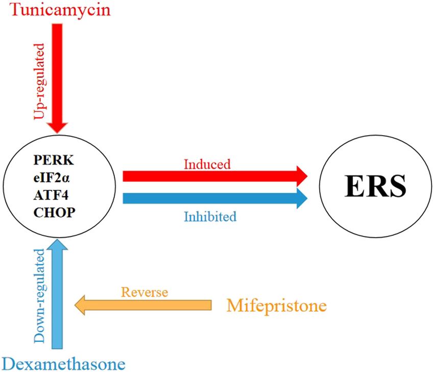
Glucocorticoids protect HEI-OC1 cells from cell damage 697
30 s extension at 60°C. Relative fold changes were deter- membrane was blocked with 5% BSA for 1 h at room
mined by 2ΔΔCt method [14]. Primer sequences of PERK, temperature and then incubated with the primary anti-
eIF2α, ATF4, CHOP, and β-actin that were used in real- bodies (PERK, or eIF2α, or p-eIF2α, Cell Signaling Tech,
time PCR are listed in Table 1. USA; p-PERK, ImmunoWay, USA; ATF4, Abcam, UK;
CHOP, or BAX, or Bcl-2, Proteintech, USA. 1:1,000/each
antibody) at 4°C overnight. After washing with TBST, the
membranes were incubated with appropriate secondary
2.4 Western blotting antibodies (anti-rabbit IgG, 1:10,000, Fcmacs, China) for
2 h at room temperature. ECL substrate was used to visua-
Total protein was extracted from HEI-OC1 Cells by using lize the bands, and the blots were developed by Tanon 5200
RIPA buffer with protease and phosphatase inhibitors. Multi fully automatic fluorescence/chemiluminescence
Protein concentration was determined by BCA assay. image analysis system (Tanon Science & Technology Co,
Thirty micrograms of protein were resolved by SDS- Ltd, Shanghai, China). Protein bands were analyzed for
PAGE and then transferred onto a PVDF membrane. The densitometry using NIH Image J software.
Figure 1: The effects of tunicamycin, dexamethasone, and mifepristone on the proliferation of HEI-OC1 cells. (a) Dose-response of tunica-
mycin on the proliferation of HEI-OC1 cells. While significant inhibition of HEI-OC1 cell proliferation was observed at all test doses of
tunicamycin and at all time points (all p < 0.05), the strongest inhibition of tunicamycin on the proliferation of HEI-OC1 cells was observed at
36 h postexposure at a concentration of 5 µg/mL (p < 0.05). (b) Dose-response profile of dexamethasone pretreatment on the proliferation
of HEI-OC1 cells. HEI-OC1 cells were pretreated with various concentrations of dexamethasone (0–200 nmol/mL) for 12 h before culturing
with 5 µg/mL of tunicamycin. Dexamethasone alleviated the inhibition of tunicamycin on HEI-OC1 cell proliferation (all p < 0.05). The
optimal dose of dexamethasone was 20 nmol/mL (p < 0.05). (c) Dose-response profile of mifepristone-mediated antagonism of dexa-
methasone protection of HEI-OC1 cells from tunicamycin-induced proliferation inhibition. Cells were pretreated with various concentrations
of mifepristone (0–200 nmol/mL) and 20 nmol/mL of dexamethasone for 12 h and then cultured with 5 µg/mL tunicamycin for 36 h.
Mifepristone reversed the protective effect of dexamethasone (all p < 0.05). The optimal concentration of mifepristone was 20 nmol/mL
(p < 0.05). All results are expressed as X̄ ± SD, * indicates p < 0.05.698 Zhibiao Liu et al.
2.5 Statistical analysis (0.2, 2, 20, or 200 nmol/mL) of glucocorticoid dexa-
methasone (DXM); after 12 h, 5 µg/mL of TM was added
All data were expressed as mean ± standard deviation. into the medium and cells were cultured for additional
SPSS20.0 software (IBM Corp. Armonk, NY, USA) was 36 h. The optimal concentration of DXM was found to
used for statistical analyses. The independent samples be 20 nmol/mL (Figure 1b). All doses of DXM reduced
t-test was used to compare the values of means between the TM-induced inhibition of HEI-OC1 cell proliferation
groups. A value of p < 0.05 was considered a statistically (all p < 0.05).
significant difference. All experiments have been done in To understand the glucocorticoid receptor (GR)
three independent replicas. role in this therapeutic response, HEI-OC1 cells were
pretreated with the optimal therapeutic dose of DXM
(20 nmol/mL) in the presence of various concentrations
(0.2, 2, 20, or 200 nmol/mL) of the GR antagonist, mife-
pristone (MIF). After 12 h, 5 µg/mL of TM was added into
3 Result
the medium, and the cells were cultured for an additional
36 h. All test doses of MIF reduced the protective effect of
3.1 Dexamethasone reversed mifepristone dexamethasone (20 nmol/mL) against TM-induced inhi-
and tunicamycin’s ERS and apoptotic bition of HEI-OC1 cell proliferation (p < 0.05, 1C).
effect in HEI-OC1 cells Investigating DXM potential protection against the
damage induced by ERS, HEI-OC1 cells were incubated
HEI-OC1 cells were treated with various concentrations with 5 µg/mL of TM alone or following pretreatment with
(0.1, 0.5, 1, 5, 10 µg/mL) of tunicamycin (TM) for 12, 24, DXM (20 nmol/mL), MIF (20 nmol/mL), or DXM + MIF.
36, and 48 h. TM inhibited the proliferation of HEI-OC1 cells Flow cytometry was used to detect apoptosis. Compared
in a dose- and time-dependent manner. TM (5 µg/mL – 36 h) to the normal control group, no increased apoptosis was
significantly inhibit the proliferation of HEI-OC1 cell in both observed in cells treated with DXM or MIF alone (p > 0.05).
time and dose-dependent manners (p < 0.05) (Figure 1a). HEI- Increased apoptosis was observed in the TM, TM + DXM,
OC1 cells were then pretreated with various concentrations TM + MIF, and TM + DXM + MIF groups compared to the
Figure 2: Dexamethasone protects HEI-OC1 cells from tunicamycin-induced apoptosis. Examples of flow cytometry analysis of apoptosis in
the control group (a), the DXM group (b), the MIF group (c), the TM group (d), the TM + DXM group (e), the TM + MIF group (f), and the TM +
DXM + MIF group (g). Apoptosis rates were statistically analyzed (h). Compared to the control group, no increased apoptosis was observed
in the DXM and MIF groups (all p > 0.05). Significantly more apoptosis was observed in the TM, TM + MIF, TM + DXM, and TM + DXM + MIF
groups compared to the control group (all p < 0.05). However, dexamethasone treatment (TM + DXM) significantly protected HEI-OC1 cells
from tunicamycin-induced apoptosis, and mifepristone (TM + DXM + MIF) reversed this protective effect (all p < 0.05). Mifepristone
pretreatment did not alter the apoptosis rate in the TM + MIF group compared to the TM alone group (p > 0.05). All results are expressed
as X̄ ± SD, * indicates p < 0.05.Glucocorticoids protect HEI-OC1 cells from cell damage 699
control group (p < 0.05, Figure 2d–g). However, apop- expression of ATF4 and CHOP proteins was significantly
tosis was significantly decreased in the TM + DXM group increased with increasing TM concentrations and culture
compared to the TM group (p < 0.05, Figure 2d and e), and times (Figure 3a and b). A dose-response profile for
more apoptotic cells were counted in the TM + DXM + MIF ATF4 and CHOP expression was observed across the
group compared to the TM + DXM group (p < 0.05, Figure 0.5–5 µg/mL concentration range of TM (p < 0.05,
2e and g). These results indicate that TM-induced ERS Figure 3c and d). This TM-induced ATF4 and CHOP
promoted apoptosis in HEI-OC1 cells and that DXM- expression were significantly increased with extended
mediated protection of HEI-OC1 cells from this patho- cultured intervals (p < 0.05, Figure 3e and f).
logical response could be reversed by MIF.
3.3 Effects of dexamethasone and
3.2 Tunicamycin upregulated the expression mifepristone on the upregulation of
of ATF-4 and CHOP proteins in HEI-OC1 ATF-4 and CHOP induced by tunicamycin
cells in HEI-OC1 cells
To study the effects of TM-induced ERS at the molecular The ERS-related expression of PERK, eIF2α, ATF4, and
level, HEI-OC1 cells were treated with increasing concen- CHOP in HEI-OC1 cells was then examined by western
trations of TM (0–10 µg/mL) between 0 and 48 h. The blot and Qrt-PCR in the context of therapeutic
Figure 3: Effects of tunicamycin on the expression of ATF4 and CHOP in HEI-OC1 cells. (a) Example of western blots of ATF4 and CHOP protein
expression in HEI-OC1 cells treated with various concentrations of tunicamycin (0–10 µg/mL) for 36 h. The expression of ATF4 and CHOP
proteins in HEI-OC1 cells gradually increased with increasing concentrations of tunicamycin. (b) Example of western blots of ATF4 and CHOP
protein expression in HEI-OC1 cells cultured with 5 µg/mL of tunicamycin for various culture times (0–48 h). The expression of ATF4 and
CHOP proteins in HEI-OC1 cells gradually increased in the presence of TM with longer culture times. The western blots were quantitatively
analyzed (c–f). A dose-response on ATF4 and CHOP expression was observed across the concentration range of 0.5–5 µg/mL of tunicamycin
(all p < 0.05, c and d). At a fixed dose of 5 µg/mL tunicamycin, increased ATF4 protein was expressed when the cells cultured longer (all p <
0.05, e). Similar time-dependent effects on protein expression were also observed for CHOP when the cells were cultured for 24–36 h in the
presence of tunicamycin (all p < 0.05, f). All results are expressed as X̄ ± SD, * indicates p < 0.05.700 Zhibiao Liu et al.
Figure 4: Effects of dexamethasone and mifepristone on the upregulation of ATF-4 and CHOP induced by tunicamycin in HEI-OC1 cells. (a and b)
Example of western blots of ATF4, CHOP, PERK, eIF2α, BAX, and Bcl-2 expression after drug treatment. Upregulation of ATF4, CHOP, BAX, PERK,
and eIF2α expression and downregulation of Bcl-2 were observed in the TM, TM + MIF, and TM + DXM + MIF groups. The upregulation of ATF4,
CHOP, BAX, PERK, and eIF2α and the downregulation of Bcl-2 were blocked by dexamethasone in the TM + DXM group. (c) Similar results were
also observed in the expression pattern of ATF4 and CHOP mRNA levels in each test group. The expression of PERK and eIF2ɑ mRNA was not
changed after drug treatment (all p > 0.05). All results are expressed as X̄ ± SD, * indicates p < 0.05.
pretreatment with DXM. TM treatment significantly upre- ERS is considered a common cause of various sensor-
gulated the protein expression of BAX, p-PERK, p-eIF2α, ineural deafness. Sensorineural hearing loss is reported
ATF4, and CHOP (Figure 4a and b), downregulated the to be associated with ERS in animal studies. Whether GCs
protein expression of Bcl-2, and upregulated mRNA can protect inner ear cells by inhibiting ER stress remains
expression of ATF4 and CHOP (p < 0.05) (Figure 4c); unclear [20–22]. We speculate that ERS may be promi-
pretreatment with DXM reversed TM’s effect (all p < nent in the inner ear cells of patients suffering from
0.05, Figure 4a and b). DXM-mediated inhibition of ERS SNHL. However, the relationship between GCs and ERS
was reversed by co-treatment with MIF, demonstrating spe- is immensely complicated, and the effects of GCs whether
cificity for the antagonistic response at the molecular inhibiting or promoting ERS can differ upon different
levels. These results suggest that DXM protects HEI-OC1 cells [23,24]. Question remains, if the inner ear cells are
cells from ERS-induced apoptosis by inhibiting BAX, p-PERK, damaged in SSNHL patients, will GCs promote or inhibit
p-eIF2α, ATF4, and CHOP expression and increasing the ERS? To answer this question, we used tunicamycin (the
Bcl-2 expression. most common drug used to induce ERS) to treat hair-cell-
like HEI-OC1 cells as an in vitro system for modeling inner
ear ERS damage. Additionally, we pretreated HEI-OC1
cells with GCs to investigate whether GCs have protective
4 Discussion effects against ERS damage. We found that dexamethasone
can effectively protect HEI-OC1 cells from ERS damage.
GCs have vast effects on the metabolic, immunological, Also, these effects could be inhibited by mifepristone, a
and homeostatic functions. In the inner ear, it directly well-studied GC antagonist.
targets the glucocorticoid receptor (GR) [15]. After GCs In order to explore the relationship between ERS and
were delivered in the inner ear, thousands of inner ear apoptosis, we further determined the protein expression
genes were affected and this number increased signifi- of BAX and Bcl-2 in HEI-OC1 cells. We found that TM
cantly [16]. GCs have been widely used in the protection significantly upregulated the protein expression of BAX
of inner ear injury. For example, GCs could significantly and downregulated the protein expression of Bcl-2 in
improve the auditory brainstem response threshold after HEI-OC1. Previous studies have found that ERS can induce
acoustic overexposure [17]. Also, recently many hospitals apoptosis in H9c2 cell and MLTC-1 cells, which was similar
consider GCs being applied perioperatively in patients to our results. To determine the role of GCs in ERS-induced
undergoing cochlear implantation as a promising treat- apoptosis, we examined the protein expression of ERS
ment regimen [18]. On the other hand, high doses of marker genes. Interestingly, GCs not only inhibited the
corticosterone can impair auditory nerve processing [19]. expression of p-PERK, p-eIF2α, ATF4, and CHOP, butGlucocorticoids protect HEI-OC1 cells from cell damage 701
also reversed the expression of apoptosis-related proteins. with SNHL. This study provided a theoretical basis for
These results indicated that GCs may reduce apoptosis by clinical treatment of SNHL.
alleviating ERS.
After binding to GRs, GCs enter the nucleus and con- Acknowledgment: The authors would like to thank Drs.
trol the activity of large gene networks associated with a Xiaoping Du and Matthew B. West (Hough Ear Institute,
variety of developmental and metabolic processes [25]. OK, USA) for their critical reviews and thoughtful feed-
GCs may inhibit ERS and protect cells through multiple back during the preparation of our manuscript.
pathways. For instance, GCs can inhibit ERS by pro-
moting the secretion of correctly folded proteins and Funding information: This work was supported by the
degradation of misfolded proteins; GRs may undergo National Natural Science Funds of China (81271074); the
re-localization and phosphorylation by ERS inducers, Six Talent Peaks Project of Jiangsu Province (WSW-075);
thereby decreasing ERS; GCs can alleviate ERS response the Medical Science and Technology Development key
by inducing leucine zippers, and the interactions of GR- Foundation of the Department of Health of Nanjing
bound GCs with CHOP can reverse tunicamycin-induced City (ZKX17019); and the Jiangsu Provincial Key Medical
cell death [13,24,26]. To determine the molecular mechan- Discipline (ZDXKB2016015), P.R. China.
isms of GC-mediated mitigation of ERS damage in inner
ear cells, dexamethasone and mifepristone were used to Author contributions: W.S., Substantial contribution to
pretreat HEI-OC1 cells in this study. The Figure 5 was drew the design of the manuscript, revising it critically for
to explain the mechanisms of action of glucocorticoid important intellectual content. Z.L.: Preparing the main
treatment ERS in SNHL. paper. Z.L., B.F.: Substantial contribution to literature
We found that dexamethasone could suppress tuni- search, data analysis, and interpretation. L.X., J.L., X.C.,
camycin-induced increases in ATF4 and CHOP expres- W.Z., L.L.,W.M., Z.G., J.H.: Substantial contribution to lit-
sion in HEI-OC1 cells. Attenuation of ERS in inner ear erature search. Z.L.: Drafting the manuscript and revising
cells may, therefore, represent an important mechanism it critically for important intellectual content. All authors
of action for GCs to elicit their therapeutic effects in read and approved the final manuscript. All listed authors
patients with SNHL. have approved the manuscript before submission, including
In conclusion, our results suggest that GCs can the names and order of authors.
inhibit ERS-related ATF4 and CHOP expression and
confer protective effects against ERS damage and poten- Conflict of interest: The authors state no conflict of
tial apoptosis in inner ear cells; and also that GCs may interest.
alleviate SNHL by inhibiting ERS, which may be one of
the mechanisms of action for GC treatment in patients Data availability statement: The datasets generated during
and/or analyzed during the current study are available
from the corresponding author on reasonable request.
References
[1] Liu MQ, Chen Z, Chen LX. Endoplasmic reticulum stress: a
novel mechanism and therapeutic target for cardiovascular
diseases. Acta Pharmacol Sin. 2016;37(4):425–43.
[2] Louessard M, Bardou I, Lemarchand E, Thiebaut AM, Parcq J,
Leprince J, et al. Activation of cell surface GRP78 decreases
endoplasmic reticulum stress and neuronal death. Cell Death
Differ. 2017;24(9):1518–29.
[3] Hetz C, Chevet E, Harding HP. Targeting the unfolded protein
response in disease. Nat Rev Drug Discov. 2013;12(9):703–19.
[4] Nougarède A, Tesnière C, Ylanko J, Rimokh R, Gillet G,
Andrews DW. Improved IRE1 and PERK pathway sensors for
multiplex endoplasmic reticulum stress assay reveal stress
response to nuclear dyes used for image segmentation. Assay
Figure 5: Mechanisms of action of glucocorticoid treatment ERS Drug Dev Technol. 2018;16(6):350–60.
in SNHL.702 Zhibiao Liu et al.
[5] Kim J, Song H, Heo HR, Kim JW, Kim HR, Hong Y, et al. [16] Trune DR, Shives KD, Hausman F, Kempton JB, MacArthur CJ,
Cadmium-induced ER stress and inflammation are mediated Choi D. Intratympanically delivered steroids impact thousands
through C/EBP-DDIT3 signaling in human bronchial epithelial more inner ear genes than systemic delivery. Ann Otol Rhinol
cells. Exp Mol Med. 2017;49(9):e372. Laryngol. 2019;128(6_suppl):134S–8S.
[6] Kalinec GM, Thein P, Parsa A, Yorgason J, Luxford W, Urrutia R, [17] Tabuchi K, Murashita H, Sakai S, Hoshino T, Uemaetomari I,
et al. Acetaminophen and NAPQI are toxic to auditory cells via Hara A. Therapeutic time window of methylprednisolone in
oxidative and endoplasmic reticulum stress-dependent path- acoustic injury. Otol Neurotol. 2006;27(8):1176–9.
ways. Hear Res. 2014;313:26–37. [18] Honeder C, Zhu C, Gausterer JC, Schöpper H, Ahmadi N,
[7] Zong S, Liu T, Wan F, Chen P, Luo P, Xiao H. Endoplasmic Saidov N, et al. Sustained-release triamcinolone acetonide
reticulum stress is involved in cochlear cell apoptosis in a hydrogels reduce hearing threshold shifts in a model for
cisplatin-induced ototoxicity rat model. Audiol Neurootol. cochlear implantation with hearing preservation. Audiol
2017;22(3):160–8. Neurootol. 2019;24(5):237–44.
[8] Hu J, Li B, Apisa L, Yu H, Entenman S, Xu M, et al. ER stress [19] Singer W, Kasini K, Manthey M, Eckert P, Armbruster P,
inhibitor attenuates hearing loss and hair cell death in Vogt MA, et al. The glucocorticoid antagonist mifepristone
Cdh23erl/erl mutant mice. Cell Death Dis. 2016;7(11):e2485. attenuates sound-induced long-term deficits in auditory nerve
[9] Xue Q, Li C, Chen J, Guo H, Li D, Wu X. The protective effect of response and central auditory processing in female rats.
the endoplasmic reticulum stress-related factors BiP/GRP78 FASEB J. 2018;32(6):3005–19.
and CHOP/Gadd153 on noise-induced hearing loss in guinea [20] Ermutlu G, Süslü N, Yılmaz T, Saraç S. Sudden hearing loss: an
pigs. Noise Health. 2016;18(84):247–55. effectivity comparison of intratympanic and systemic steroid
[10] Sevilla LM, Pérez P. Roles of the glucocorticoid and minera- treatments. Eur Arch Otorhinolaryngol. 2017;274(10):3585–91.
locorticoid receptors in skin pathophysiology. Int J Mol Sci. [21] Chin CJ, Dorman K. Sudden sensorineural hearing loss. CMAJ.
2018;19:7. 2017;189(11):E437–8.
[11] Alam MM, Okazaki K, Nguyen L, Ota N, Kitamura H, [22] Lai D, Zhao F, Jalal N, Zheng Y. Intratympanic glucocortico-
Murakami S, et al. Glucocorticoid receptor signaling represses steroid therapy for idiopathic sudden hearing loss: meta-
the antioxidant response by inhibiting histone acetylation analysis of randomized controlled trials. Med (Baltim).
mediated by the transcriptional activator NRF2. J Biol Chem. 2017;96(50):e8955.
2017;292(18):7519–30. [23] Smith M, Wilkinson S. ER homeostasis and autophagy. Essays
[12] Whirledge S, DeFranco DB. Glucocorticoid signaling in health Biochem. 2017;61(6):625–35.
and disease: insights from tissue-specific GR knockout mice. [24] André F, Corazao-Rozas P, Idziorek T, Quesnel B, Kluza J,
Endocrinology. 2018;159(1):46–64. Marchetti P. GILZ overexpression attenuates endoplasmic
[13] Mihailidou C, Panagiotou C, Kiaris H, Kassi E, Moutsatsou P. reticulum stress-mediated cell death via the activation of
Crosstalk between C/EBP homologous protein (CHOP) and mitochondrial oxidative phosphorylation. Biochem Biophys
glucocorticoid receptor in lung cancer. Mol Cell Endocrinol. Res Commun. 2016;478(2):513–20.
2016;436:211–23. [25] Bain DL, Yang Q, Connaghan KD, Robblee JP, Miura MT,
[14] Livak KJ, Schmittgen TD. Analysis of relative gene expression Degala GD, et al. Glucocorticoid receptor-DNA interactions:
data using real-time quantitative PCR and the 2(-Delta Delta C binding energetics are the primary determinant of sequence-
(T)) method. Methods. 2001;25(4):402–8. specific transcriptional activity. J Mol Biol. 2012;422(1):18–32.
[15] Kumagami H, Terakado M, Takahashi H. Distribution of gluco- [26] Hu DD, Mai JN, He LY, Li PQ, Chen WX, Yan JJ, et al.
corticoid receptors and 11β-hydroxysteroid dehydrogenase Glucocorticoids prevent enterovirus 71 capsid protein VP1
isoforms in the human inner ear. Otol Neurotol. induced calreticulin surface exposure by alleviating neuronal
2013;34(1):151–7. ER stress. Neurotox Res. 2017;31(2):204–17.You can also read