Original Article Small cell carcinoma of the ovary, hypercalcemic type (SCCOHT): a challenge for clinicopathological diagnosis
←
→
Page content transcription
If your browser does not render page correctly, please read the page content below
Int J Clin Exp Pathol 2019;12(6):2166-2172
www.ijcep.com /ISSN:1936-2625/IJCEP0094237

Original Article
Small cell carcinoma of the ovary, hypercalcemic type
(SCCOHT): a challenge for clinicopathological diagnosis
Ran Li1*, Ting Zhou1*, Shaohua Chen1*, Nan Li1, Zhaogen Cai1, Yunzhi Ling2, Zhenzhong Feng1*

Departments of 1Pathology, 2Anesthesiology, The First Affiliated Hospital of Bengbu Medical College, Bengbu
Medical College, Bengbu, Anhui, P. R. China. *Equal contributors.
Received March 22, 2019; Accepted April 25, 2019; Epub June 1, 2019; Published June 15, 2019

Abstract: Small cell carcinoma of the ovary, hypercalcemic type (SCCOHT) is an extremely aggressive ovarian tu-
mor, with a poor prognosis and high mortality for young women. This paper aims to inform clinical physicians of
new clinical improvements and further understanding of SCCOHT. Two cases diagnosed with SCCOHT from our
medical database were reconfirmed and immunohistochemically stained with vimentin, CK, EMA, S-100, ER, PR,
and SMARCA4. Diffuse small, round cells with scant cytoplasms, small nucleoli, hyperchromatic nuclei, and active
nuclear divisions were detected in the microscopy. The immunohistochemical markers indicated minor positive
but notably were SMARCA4 negative, which led to the final diagnosis. SCCOHT is a rare and lethal ovarian tumor in
young women. The loss of SMARCA4 or the presence of SMARCA2 is a specific marker for the disease. Susceptibility
to CDK4/6 inhibitors associated with downregulation of SMARCA4 targeted cyclin D1 may be a probable therapeuti-
cal mechanism for the disease.

Keywords: SCCOHT, SMARCA4, ATRT, PD1, CDK4/6

Introduction                                                 [7]. Also, more than 70% of SCCOHT cases
                                                             present with a recurrence within 6 months,
Small cell carcinoma of the ovary, hypercalce-               according to Young’s report [5]. For its rarity,
mic type (SCCOHT) is an aggressive ovarian                   only about 350 cases have been described
malignancy. It’s important to distinguish it from            worldwide, and half of these were reported in
other mimics because of its dismal prognosis,                Young’s retrospective research [5]. The origin of
highly likely recurrence, and early onset age [1].           SCCOHT is still obscure. Neither an epithelial
It was first reported by Robert Scully in 1979,              origin nor a germ cell neoplasm etiology has
who named it for its morphology and hypercal-                been confirmed, and the World Health Or-
cemic features [2]. Generally, in microscopy,                ganization (WHO) still classifies it as a “miscel-
the tumor cells are mainly small, round cells                laneous tumor [3]”.
that diffuse into a sheet, but it also presents
with rhabdoid morphological large cells in some              We report two clinical cases of unilateral
rare cases [3, 4]. And in a digital analysis, about          SCCOHT that presented in our hospital. And we
two-thirds of patients may show elevated serum               also review a possible rationale, a current study
calcium, which can return to a normal level                  of the immunochemotherapy, as well as some
after tumor resection [5]. Epidemiologically,                treatment options for the disease. The paper
young females are mainly affected, with a medi-              aims to inform clinical physicians of new cli-
an age of 23 years, and many patients are                    nical improvements and further knowledge of
admitted for a unilateral accessory mass with                SCCOHT.
an average diameter of 15.3 cm [1, 2, 6].
Meanwhile, the disease-free survival rate is                 Patients and methods
approximately one-third for stage IA disease,
with exponentially poorer prognoses for about                Collected from the First Affiliated Hospital of
10% of patients in the more advanced stages                  Bengbu Medical College, the only two cases in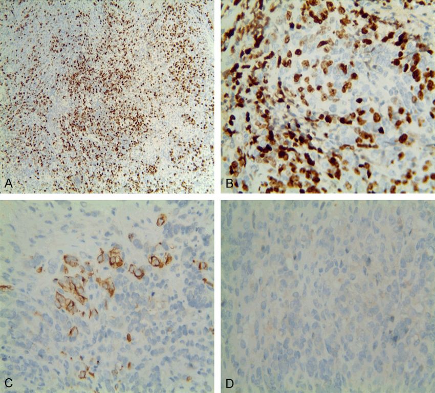
Advanced knowledge of SCCOHT

Table 1. The sources of the antibodies used
in the immunohistochemical analysis
Source             Antibody
CK                 Monoclonal, clone AE1/AE3
CA125              Monoclonal, clone TA347
EMA                Monoclonal, clone E29
PLAP               Monoclonal, clone 8A9
CD30               Monoclonal, clone Ber-H2
S-100              Monoclonal, clone 4C4.9
Inhibin-a          Monoclonal, clone R1
Ki-67              Monoclonal, clone MIB-1
SMARCA4            Rabbit Polyclonal Antibody
WT1                Monoclonal, clone WT49
Desmin             Monoclonal, clone D33
Vimentin           Monoclonal, clone V9                  Figure 1. Imaging figures (case 1). Ultrasound of the
ER                 Monoclonal, clone SP1                 pelvis demonstrating a 10 cm × 7.1 cm mixed echo
PR                 Monoclonal, clone P2                  figure in the right ovary.
MyoD               Monoclonal, clone 5.8A
All antibodies were obtained from Maixin Biotech, Inc.   the ethical guidelines of the Declaration of
(Fuzhou, China) and were ready to use.
                                                         Helsinki and was approved by the Ethics
                                                         Committee of the First Affiliated Hospital of
Table 2. The results of the immunohisto-                 Bengbu Medical College.
chemical features
                                                         Results
Immunohistochemical markers              Result (+/-)
CK                                           -/+         Clinical features
CA125                                          -
EMA                                            -         Both of the patients were admitted to our hos-
PLAP                                           -         pital with persistent abdominal pain lasting for
CD30                                           -
                                                         one year or more. A small pelvic mass and
                                                         abdominal pain during menstruation were not
S-100                                          -
                                                         apparent until the condition became aggravat-
Inhibin-a                                      -
                                                         ed. In case 1, a soft mass of about 10 cm diam-
Ki-67                                   +, about70%      eter was examined in the right groin area, and
SMARCA4                                        -         ultrasound verified it had a mixed echo figure of
WT1                                            -         10 cm × 7.1 cm in the right accessory (Figure
Desmin                                         -         1). No obvious abnormality was found in the
Vimentin                                      +          preoperative examinations other than the
ER                                             -         serum CA125 of about 57.5 IU/ML. Most nota-
PR                                             -         bly, the blood calcium was 2.41 mmol/L, a nor-
MyoD                                           -         mal level which prevented us from making a
                                                         morphological diagnosis. In case 2, there was a
                                                         14 cm × 18 cm lump in the left ovary, and the
our database were aged 26 and 44 years when              preoperative examination showed the CA125
diagnosed with SCCOHT. Surgical specimens                at about 233 U/ML, while the serum calcium
were taken regularly and HE-stained sections             was about 3.53 mmol/L.
were examined by our experienced patholo-
gists. Immunohistochemical staining was per-             A clinical operation of the total uterus, a bilat-
formed using the EnVision two-step method.               eral oophorectomy, a greater omentum resec-
The antibody used and its results are shown in           tion, and a pelvic lymph node dissection were
Tables 1 and 2 respectively. The clinical records        performed. A TP (paclitaxel + carboplatin) regi-
were obtained from the patients’ medical                 men was given to the patients monthly as post-
records. The study was conducted according to            operative adjuvant chemotherapy.

2167                                                        Int J Clin Exp Pathol 2019;12(6):2166-2172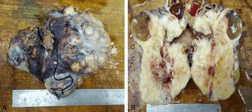
Advanced knowledge of SCCOHT
with the foci of hemorrhage,
cystic degeneration, and ne-
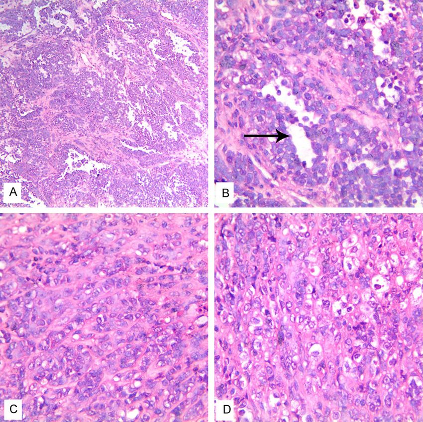
crosis (Figure 2B). In the HE
stain photomicrograph of the
right ovary, the neoplastic cells
were mainly diffusing sheets of
small round cells, punctured
by follicular-like architecture
(Figure 3A, 3B). The follicles of
various sizes were mainly
empty, and few contained any
eosinophilia fluid (Figure 3B).
Figure 2. A: Gross image of the external surface of the left ovary (Case 2).
B: Representative cut surfaces of the left ovary (Case 2). The tumor mass The tumor cells predominately
is lobulated, with cystic degeneration, hemorrhage, necrosis, and mucus. had scant cytoplasms, small
nucleoli, hyperchromatic nu-
clei, and active nuclear division
(Figure 3C, 3D). The nucleoli
could be observed but were
rarely prominent. The mitotic
figures were about 18 per
every 10 high-power field. Fina-
lly, all the lymph nodes select-
ed from the omentum and pel-
vic cavity were negative.
Immunohistochemical fea-
tures
The immunohistochemical fea-
tures were similar between the
two cases. The tumor cells
merely were positive for vimen-
tin and partially positive for CK
(Figure 4C). Other makers,
such as EMA, S-100, ER, and
PR, were all negative. The pro-
liferation index, Ki-67, was
greater than 50% (Figure 4A,
4B). Notably, a negative ex-
pression of SMARCA4 leads
to the exactly diagnosis of
Figure 3. HE stained photomicrograph of the right ovary (case 1). A. Diffuse SCCOHT (Figure 4D). The anti-
sheet-like architecture of small cells (H&E staining, × 100). B. The follicular body used and its results are
architecture of the neoplastic tissue (Black arrow) (H&E staining, × 400). shown in Tables 1 and 2
C and D. The scant cytoplasm, small nucleoli, hyperchromatic nuclei, and respectively.
nuclear division of the neoplastic cells (H&E staining, × 400).
Discussion
Gross and histological features SCCOHT is a rare, undifferentiated ovarian can-
cer named for its morphological appearance
Generally, the tumor mass is nodular or lobu- and notable presence of hypercalcemia [2].
lated, with cystic degeneration, hemorrhage, Currently, it is generally believed that the loss of
necrosis, and mucus [5]. In case 2, the external SMARCA4, either alone or with SMARCA2, is
surface of the tumor was gray and partly pur- highly sensitive and specific for SCCOHT [1, 6,
plish red, and it was lobulated with a maximum 8]. SMARCA4 (BRG1) and SMARCA2 (BRM) are
dimension of 18 cm (Figure 2A). The represen- two vital and exclusive components of the SWI-
tative cut surface was mainly solid and yellow, SNF chromatin remodeling complex, which are
2168 Int J Clin Exp Pathol 2019;12(6):2166-2172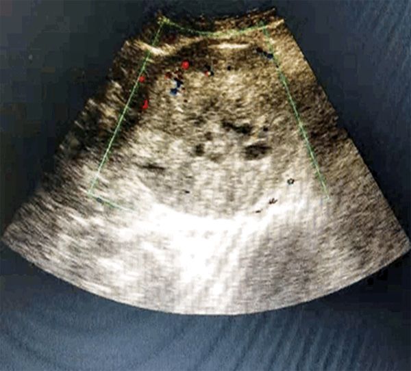
Advanced knowledge of SCCOHT

                                                                                 doid tumor of the ovary) due
                                                                                 to its resemblance to ATRT.
                                                                                 ATRT (atypical teratoid/rhab-
                                                                                 doid tumor) is a rare, malignant
                                                                                 CNS tumor that occurs mainly
                                                                                 in infants and children [12].
                                                                                 There are many similarities
                                                                                 between SCCOHT and ATRT
                                                                                 [3, 4]. First, in some SCCOHT
                                                                                 cases, large cells with rhab-
                                                                                 doid morphology can be de-
                                                                                 tected [13]. Second, the dual
                                                                                 loss of SMARCB1 and SMA-
                                                                                 RCA4 in ATRT closely resem-
                                                                                 bles those in SCCOHT [3, 14].
                                                                                 Third, both present with an
                                                                                 aggressive malignancy and
                                                                                 hypercalcemia in clinical fea-
                                                                                 tures. And last, their similari-
                                                                                 ties are shown by other clues
                                                                                 found in clinical examinations
                                                                                 and immunochemistry or in
Figure 4. Immunohistochemical features of the tumor. The proliferation in-       whole exome sequencing stud-
dex (Ki-67) of the tumor is about 70% positive. (magnification �����������
                                                                 × 100, ����
                                                                         mag-    ies [1, 14].
nification × 400, (A and B) Respectively). (C) CK is partially positive in the
tumor cells. (magnification, × 400). (D) SMARCA4 is negative which leads to      Moreover, studies of immuno-
the final diagnosis for SCCOHT (magnification, × 400).
                                                                                 therapy, like anti-PD1 immuno-
                                                                                 therapy, have also been en-
involved in the growth, differentiation, and infil-             couraged and focused on SCCOHT. Programmed
tration of various tumors [9, 10]. With the                     death 1 (PD-1) is a vital immune-checkpoint
genetic mutation of SMARCA4 in most cases,                      inhibitory receptor expressed by activated T
the loss of SMARCA2 may refer to the epigene-                   cells. It could be blocked in the peripheral tis-
tic silencing or mRNA degradation, confirmed                    sue by the immunosuppressive PD-1 ligands
by the lack of mutations or deletions involving                 PD-L1 and PD-L2, which are expressed by
SMARCA2 [1]. And with the mutually exclusive                    tumor cells, stromal cells, or both [15]. The
functions between SMARCA2 and SMARCA4                           interaction between PD-L1 and PD-1 could sig-
[8, 9], there may be an epigenetic recombina-                   nificantly suppress the infiltration of tumors
tion to support the residual functions of the                   and enhance T-cell responses in vitro [16, 17].
remaining SMI/SNF complex.                                      And it has been confirmed that the PD-L1
                                                                expressions and TILs (tumor-infiltrating lympho-
In addition, current research from Yibo et al.                  cytes) in tumors are associated with its muta-
demonstrated a remarkable susceptibility to                     tional burden, which also links the clinical
CDK4/6 (cyclin-dependent kinase 4/6) inhibi-                    responses to anti-PD1 immunotherapy [15,
tors in SCCOHT cell lines [11]. For the molecular               18].
mechanism of the susceptibility, Yibo declared
a deficiency of cyclinD1 and retinoblastoma                     Contrasted with its low burden in mutation,
(RB) phosphorylation, which are caused by the                   SCCOHT shows an unexpectedly high expres-
inactive mutation of SMARCA4 [11]. The “SM-                     sion of PD-L1 and is strongly statistically corre-
ARCA4-cyclin D1-CDK4/6” mechanism may                           lated with the infiltration of T cells, according to
give a proper interpretation of SCCOHT, but this                the experiments of Jelinic [18]. Also, the clinical
interpretation still needs more supporting evi-                 feasibility of anti-pd1 immunotherapy is also
dence in clinical and immunopathology.                          shown in his study, in which 3 of 4 cases didn’t
                                                                recur for over 1.5 years after additional anti-
Moreover, some researchers are predisposed                      pd1 immunotherapy [18]. Thus, it may be pos-
to reclassify SCCOHT as MRTO (malignant rhab-                   sible to treat patients diagnosed with SCCOHT

2169                                                                Int J Clin Exp Pathol 2019;12(6):2166-2172
Advanced knowledge of SCCOHT
with anti-PD1 immunotherapy [18, 19]. More Di-chen Li and Qun Xie (Department of
clinical analyses and further molecular studies Pathology, the Frist Affiliated Hospital of
are still needed. Bengbu Medical College) for their assistance
with the histopathological and immunohisto-
Regarding therapy, there are no targets or effi- chemical stain evaluations. This study was sup-
cient therapies now. A useful regimen for ported by the Natural Science Foundation of
SCCOHT is complete surgery followed by radia- Anhui Province (no. 1608085QH207) and the
tion, stem cell rescue, and high-dose chemo- Nature Science Key Program of Bengbu Medical
therapy [7, 20]. Furthermore, as indicated ab- College (BYKF1710, 1711).
ove, anti-PD1 immunotherapy such as CDK4/6
inhibitors may also prove favorable [18]. More Disclosure of conflict of interest
clinical care has also been shown to lead to a
better prognosis and treatment for patients. None.
First, it’s confirmed that ponatinib, a member of
RTK (receptor tyrosine kinase) inhibitors, could Address correspondence to: Drs. Nan Li and
delay tumor double time fourfold and decrease Zhenzhong Feng, Department of Pathology, The First
final tumor volumes by 50% and may also be Affiliated Hospital of Bengbu Medical College,
efficient in SMARCB1 mutations in ATRT tumors Bengbu Medical College, 287 Changhuai Road,
[21]. Secondly, the target epigenetic regulator Bengbu 233000, Anhui, P. R. China. Tel: +86-552-
TSA (the HDAC inhibitor trichostatin A) could 3070209; E-mail: linanangel100@sina.com (NL);
reactivate SMARCA2 expression in SCCOHT fzzapple1976@163.com (ZZF)
cell lines, which lead to a depression in tumors
References
[1]. In addition, it’s also been proved clinically
that patients are sensitive to the histone meth- [1] Karnezis AN, Wang Y, Ramos P, Hendricks WP,
yltransferase EZH2 [4, 10, 22]. Oliva E, D’Angelo E, Prat J, Nucci MR, Nielsen
TO, Chow C, Leung S, Kommoss F, Kommoss S,
Conclusion Silva A, Ronnett BM, Rabban JT, Bowtell DD,
Weissman BE, Trent JM, Gilks CB and
In general, SCCOHT is a rare but highly aggres- Huntsman DG. Dual loss of the SWI/SNF com-
sive disease in young women. The loss of plex ATPases SMARCA4/BRG1 and SMARCA2/
SMARCA4 alone or together with SMARCA2 is BRM is highly sensitive and specific for small
the only sensitive marker for its diagnosis, con- cell carcinoma of the ovary, hypercalcaemic
sidering that there is no specific clinical mani- type. J Pathol 2016; 238: 389-400.
[2] Scully RE. Tumors of the ovary and maldevel-
festation or other features. Druggable vulnera-
oped gonads. Atlas of Tumor Pathology 1979;
bility to CDK4/6 inhibitors may also be a molec-
2: 152-173.
ular mechanism for further study. SCCOHT’s [3] Foulkes WD, Clarke BA, Hasselblatt M,
resemblance to atypical teratoid rhabdoid Majewski J, Albrecht S and McCluggage WG.
tumors is also generally understood now. Up to No small surprise - small cell carcinoma of the
now, many studies have demonstrated that ovary, hypercalcaemic type, is a malignant
SCCOHT greatly resembles AT/RT. And current- rhabdoid tumour. J Pathol 2014; 233: 209-
ly, further studies have confirmed a notable 214.
expression of PD-L1 associated with the clini- [4] Chan-Penebre E, Armstrong K, Drew A,
cal efficacy of anti-PD1 immunochemotherapy. Grassian AR, Feldman I, Knutson SK, Kuplast-
Barr K, Roche M, Campbell J, Ho P, Copeland
Many other novel treatments like ponatinib,
RA, Chesworth R, Smith JJ, Keilhack H and
TSA, and EZH2 are also mentioned. Physicians Ribich SA. Selective killing of SMARCA2- and
and pathologists should have a new under- SMARCA4-deficient small cell carcinoma of the
standing of the SCCOHT’s etiology and clinical ovary, hypercalcemic type cells by inhibition of
therapy. EZH2: in vitro and in vivo preclinical models.
Mol Cancer Ther 2017; 16: 850-860.
Acknowledgements [5] Young RH, Oliva E, Scully RE. Small cell carci-
noma of the ovary, hypercalcemic type: a clini-
We would like to thank Editage for its help with copathological analysis of 150 cases. Am J
the English language editing. Thanks also to Surg Pathol 1994; 18: 1102-16.
2170 Int J Clin Exp Pathol 2019;12(6):2166-2172Advanced knowledge of SCCOHT
[6] Ramos P, Karnezis AN, Craig DW, Sekulic A, Schüller U, Schneppenheim R, Northcott PA,
Russell ML, Hendricks WP, Corneveaux JJ, Bar- Korbel JO, Siebert R, Frühwald MC, Lichter P,
rett MT, Shumansky K, Yang Y, Shah SP, Pren- Eils R, Gajjar A, Hasselblatt M, Pfister SM, Kool
tice LM, Marra MA, Kiefer J, Zismann VL, M. Atypical teratoid/rhabdoid tumors are com-
McEachron TA, Salhia B, Prat J, D’Angelo E, prised of three epigenetic subgroups with dis-
Clarke BA, Pressey JG, Farley JH, Anthony SP, tinct enhancer landscapes. Cancer Cell 2016;
Roden RB, Cunliffe HE, Huntsman DG and 29: 379-393.
Trent JM. Small cell carcinoma of the ovary, hy- [13] Wang L, Tan C, Tu X, Zhang Y, Li X and Chang B.
percalcemic type, displays frequent inactivat- Small cell carcinoma of ovary, hypercalcemic
ing germline and somatic mutations in SMAR- type: analysis of clinicopathologic characteris-
CA4. Nat Genet 2014; 46: 427-429. tics and the diagnostic utility of loss expres-
[7] Qin Q, Ajewole VB, Sheu TG, Donohue R and sion of SMARCA4 protein. Zhonghua Bing Li
Singh M. Successful treatment of a stage IIIC Xue Za Zhi 2015; 44: 859-863.
small-cell carcinoma of the ovary hypercalce- [14] Fahiminiya S, Witkowski L, Nadaf J, Carrot-
mic subtype using multi-modality therapeutic Zhang J, Goudie C, Hasselblatt M, Johann P,
approach. Ecancermedicalscience 2018; 12: Kool M, Lee RS, Gayden T, Roberts CW, Biegel
832. JA, Jabado N, Majewski J and Foulkes WD. Mo-
[8] Jelinic P, Schlappe BA, Conlon N, Tseng J, Ol- lecular analyses reveal close similarities be-
vera N, Dao F, Mueller JJ, Hussein Y, Soslow RA tween small cell carcinoma of the ovary, hyper-
and Levine DA. Concomitant loss of SMARCA2 calcemic type and atypical teratoid/rhabdoid
and SMARCA4 expression in small cell carci- tumor. Oncotarget 2016; 7: 1732-1740.
noma of the ovary, hypercalcemic type. Mod
[15] Topalian SL, Hodi FS, Brahmer JR, Gettinger
Pathol 2015; 29: 60.
SN, Smith DC, McDermott DF, Powderly JD,
[9] Fukumoto T, Magno E and Zhang R. SWI/SNF
Carvajal RD, Sosman JA, Atkins MB, Leming
complexes in ovarian cancer: mechanistic in-
PD, Spigel DR, Antonia SJ, Horn L, Drake CG,
sights and therapeutic implications. Mol Can-
Pardoll DM, Chen L, Sharfman WH, Anders RA,
cer Res 2018; 16: 1819-1825.
Taube JM, McMiller TL, Xu H, Korman AJ, Jure-
[10] Wang Y, Chen SY, Karnezis AN, Colborne S,
Kunkel M, Agrawal S, McDonald D, Kollia GD,
Santos ND, Lang JD, Hendricks WP, Orlando
Gupta A, Wigginton JM and Sznol M. Safety,
KA, Yap D, Kommoss F, Bally MB, Morin GB,
activity, and immune correlates of anti-PD-1
Trent JM, Weissman BE and Huntsman DG.
antibody in cancer. N Engl J Med 2012; 366:
The histone methyltransferase EZH2 is a thera-
peutic target in small cell carcinoma of the 2443-2454.
ovary, hypercalcaemic type. J Pathol 2017; [16] Shi J, Hou S, Fang Q, Liu X, Liu X and Qi H. PD-1
242: 371-383. controls follicular T helper cell positioning and
[11] Xue Y, Meehan B, Macdonald E, Venneti S, function. Immunity 2018; 49: 264-274, e264.
Wang XQD, Witkowski L, Jelinic P, Kong T, Mar- [17] Hamanishi J, Mandai M, Iwasaki M, Okazaki T,
tinez D, Morin G, Firlit M, Abedini A, Johnson Tanaka Y, Yamaguchi K, Higuchi T, Yagi H,
RM, Cencic R, Patibandla J, Chen H, Papadakis Takakura K, Minato N, Honjo T and Fujii S. Pro-
AI, Auguste A, de Rink I, Kerkhoven RM, Bertos grammed cell death 1 ligand 1 and tumor-infil-
N, Gotlieb WH, Clarke BA, Leary A, Witcher M, trating CD8+ T lymphocytes are prognostic fac-
Guiot MC, Pelletier J, Dostie J, Park M, Judkins tors of human ovarian cancer. Proc Natl Acad
AR, Hass R, Levine DA, Rak J, Vanderhyden B, Sci U S A 2007; 104: 3360-3365.
Foulkes WD, Huang S. CDK4/6 inhibitors tar- [18] Jelinic P, Ricca J, Van Oudenhove E, Olvera N,
get SMARCA4-determined cyclin D1 deficiency Merghoub T, Levine DA and Zamarin D. Im-
in hypercalcemic small cell carcinoma of the mune-active microenvironment in small cell
ovary. Nat Commun 2019; 10: 558. carcinoma of the ovary, hypercalcemic type:
[12] Johann PD, Erkek S, Zapatka M, Kerl K, Buch- rationale for immune checkpoint blockade. J
halter I, Hovestadt V, Jones DTW, Sturm D, Her- Natl Cancer Inst 2018; 110: 787-790.
mann C, Segura Wang M, Korshunov A, Rhyzo- [19] Pietzner K, Nasser S, Alavi S, Darb-Esfahani S,
va M, Gröbner S, Brabetz S, Chavez L, Bens S, Passler M, Muallem MZ and Sehouli J. Check-
Gröschel S, Kratochwil F, Wittmann A, Sieber L, point-inhibition in ovarian cancer: rising star or
Geörg C, Wolf S, Beck K, Oyen F, Capper D, van just a dream? J Gynecol Oncol 2018; 29: e93.
Sluis P, Volckmann R, Koster J, Versteeg R, von [20] Pautier P, Ribrag V, Duvillard P, Rey A, Elghis-
Deimling A, Milde T, Witt O, Kulozik AE, Ebinger sassi I, Sillet-Bach I, Kerbrat P, Mayer F, Lesoin
M, Shalaby T, Grotzer M, Sumerauer D, Zamec- A, Brun B, Crouet H, Barats JC, Morice P and
nik J, Mora J, Jabado N, Taylor MD, Huang A, Lhommé C. Results of a prospective dose-in-
Aronica E, Bertoni A, Radlwimmer B, Pietsch T, tensive regimen in 27 patients with small cell
2171 Int J Clin Exp Pathol 2019;12(6):2166-2172Advanced knowledge of SCCOHT
carcinoma of the ovary of the hypercalcemic [22] Wang Y, Chen SY, Colborne S, Lambert G, Shin
type. Ann Oncol 2007; 18: 1985-9. CY, Dos Santos N, Orlando KA, Lang JD, Hen-
[21] Lang JD, Hendricks WPD, Orlando KA, Yin H, dricks WPD, Bally MB, Karnezis AN, Hass R,
Kiefer J, Ramos P, Sharma R, Pirrotte P, Rau- Underhill TM, Morin GB, Trent JM, Weissman
pach EA, Sereduk C, Tang N, Liang WS, Wash- BE and Huntsman DG. Histone deacetylase in-
ington M, Facista SJ, Zismann VL, Cousins EM, hibitors synergizes with catalytic inhibitors of
Major MB, Wang Y, Karnezis AN, Sekulic A, EZH2 to exhibit anti-tumor activity in small cell
Hass R, Vanderhyden BC, Nair P, Weissman carcinoma of the ovary, hypercalcemic type.
BE, Huntsman DG, Trent JM. Ponatinib shows 2018.
potent antitumor activity in small cell carcino-
ma of the ovary hypercalcemic type (SCCOHT)
through multi-kinase inhibition. Clin Cancer
Res 2018; 24: 1932-1943.
2172 Int J Clin Exp Pathol 2019;12(6):2166-2172You can also read