Successful response to imatinib in two dogs with inoperable grade III infiltrating mast cell tumours: a case report
←
→
Page content transcription
If your browser does not render page correctly, please read the page content below
Veterinarni Medicina, 61, 2016 (8): 467–473                                                          Case Report

doi: 10.17221/87/2016-VETMED

Successful response to imatinib in two dogs
with inoperable grade III infiltrating mast cell tumours:
a case report
J.H. Kim1, H.J. Kim1, D.H. Kim2, J.H. Yim2, S.J. Lee2, K.H. Park2, H.Y. Yoon2
1
    Konkuk University Veterinary Medical Teaching Hospital, Seoul, Republic of Korea
2
    College of Veterinary Medicine, Konkuk University, Seoul, Republic of Korea

ABSTRACT: Two dogs were presented owing to sudden rapid growth of cutaneous masses greater than 10 cm
on the neck and axillary region, respectively. Based on history and results of physical examinations, blood work,
fine needle aspiration, histopathological examination, and computed tomography, inoperable grade III infiltrating
mast cell tumours were diagnosed. After the initiation of imatinib treatment, the masses markedly shrank and
became undetectable within 10 days in both dogs, although none of the tumour specimens showed evidence of
mutations in sequencing of c-kit exons 8 and 11. These results suggest that imatinib could be a therapeutic option
in patients with surgically inoperable canine mast cell tumours, even those that are histopathologically high grade
without c-kit exon 8 or 11 mutations.

Keywords: canine; KIT; oncogene; target therapy; tyrosine kinase inhibitor

  Mast cell tumours (MCTs) are one of the most             occur in higher-grade tumours and their presence
common cutaneous neoplasms in dogs and are of-             can be used to provide evidence of poor prognosis
ten aggressive (London and Seguin 2003; Yamada et          such as recurrence and metastasis (Downing et al.
al. 2011). In cases of aggressive MCTs, widespread         2002; Zemke et al. 2002). In the present report,
dissemination of the tumour or infiltration to sur-        however, treatment with imatinib successfully de-
rounding organs could occur, rendering the tumour          creased the size of inoperable grade III infiltrating
completely surgically non-resectable. In the past,         MCT masses in both of the cases even in the ab-
in such inoperable cases, chemotherapy and ra-             sence of mutations in exons 8 and 11of c-kit.
diotherapy were the possible therapeutic options.            In this report, we describe the clinical course and
However, unsatisfactory success rates and relatively       nucleotide sequencing of c-kit exon 8 and 11 using
high toxicity rates of systemic chemotherapy, as well      fine needle aspiration (FNA) samples of inoperative
as difficulties in access with radiotherapy, justify the   high-grade MCTs in two dogs, and the successful
exploration of new treatment options for MCT pa-           treatment of the tumours with imatinib.
tients (Cooper et al. 2009; Yamada et al. 2011).
  Recently, tyrosine kinase inhibitors (TKIs) have
begun to be used for canine MCTs as a novel tar-           Case description
geted therapy (Hahn et al. 2008; Isotani et al. 2008;
London et al. 2009; Yamada et al. 2011). Many pre-           Dog 1 was a 6-year-old, 39 kg castrated male
vious studies have demonstrated that response to           Golden Retriever presented for acute growth of
imatinib, which is a TKI, can be correlated with the       neck masses over the preceding 10 days. On initial
presence of mutations in c-kit oncogene exons 8            physical examination, dermal masses greater than
and 11 found in canine MCT (Kobayashi et al. 2012;         10 cm in diameter in the dorsal and ventral cervi-
Nakano et al. 2014). Furthermore, these mutations          cal region were detected. These masses were firm

                                                                                                               467
Case Report Veterinarni Medicina, 61, 2016 (8): 467–473
doi: 10.17221/87/2016-VETMED
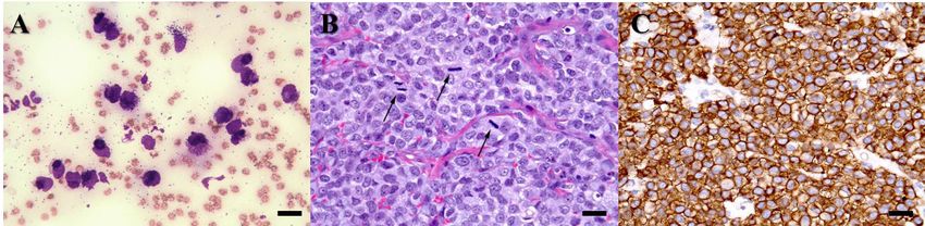
Figure 1. (A) Fine needle aspiration of the cervical masses detected in this dog reveals numerous degranulated round
cells with fine granules and with an increased nuclear-to-cytoplasmic ratio, prominent nucleoli, and anisokaryosis
indicating mast cell tumour (MCT) (Diff-Quik, × 400, scale bar = 20 µm). (B) Histopathology of the masses shows
moderate levels of degranulated mast cells with fine cytoplasmic granules having malignant features including large
dysmorphic nuclei with anisokaryosis and prominent nucleoli. Numerous mitotic figures are seen (arrows). Haema-
toxylin and eosin staining, × 400, scale bar = 20 µm. (C) Immunohistochemistry (IHC) shows that tumour cells are
immunolabelled with KIT (CD117), which shows a membrane and diffuse cytoplasmic staining pattern. Diffuse cyto-
plasmic staining, × 400, scale bar = 20 µm
and partially connected to each other. A complete the masses, computed tomography (CT) was per-
blood count (CBC) and serum biochemical pro- formed. CT images obtained using a LightSpeed
file showed no remarkable findings. Fine needle Plus apparatus (GE Medical Systems, Waukesha,
aspiration (FNA) biopsy performed on the masses WI, USA) revealed that the homogeneous mass in
demonstrated numerous degranulated round cells the cervical region had infiltrated adjacent muscle
with fine granules and with an increased nucle- and was moderately contrast-enhanced (Figure 3).
ar-to-cytoplasmic ratio, prominent nucleoli, and Due to infiltration to surrounding muscles, surgi-
anisokaryosis, indicating MCT (Figure 1A). In ad- cally complete excision of the mass could not be
dition, large numbers of mast cells were also found performed, but incisional biopsies were obtained.
in aspirated samples from the left submandibular The biopsy samples of the tumour revealed incom-
lymph nodes in this dog, suggesting metastasis. plete margins and aggressive histomorphological
However, mast cells were not identified on buffy features including an occasionally high mitotic in-
coat analysis. Survey radiographs revealed an dex (> 5/oil immersion field), moderate degranu-
asymmetrical soft-tissue density mass in the left lation with fine granules, and dysmorphic nuclei
cervical region without bone lysis (Figure 2A). with anisokaryosis (Figure 1B). These malignant
To determine the detailed anatomical location of tumour cells were densely proliferating and showed
Figure 2. Clinical course of Dog 1 as evaluated by changes in tumour size on X-rays. The patient was treated with
imatinib and radiographic examination showed gradual regression of the left cervical mass (dotted arrows). (A) On
presentation day, the soft-tissue density mass in the left cervical region was asymmetrical. (B) In Week 1, the cervical
mass had become undetectable after the initiation of imatinib therapy. (C) In Week 10, the mass was still undetect-
able and regression was sustained until first relapse at Week 25. The dog lived for 37 weeks after starting imatinib
administration
468Veterinarni Medicina, 61, 2016 (8): 467–473 Case Report
doi: 10.17221/87/2016-VETMED
Figure 3. (A) Transverse view of cervical region masses on computed tomographic examination. Note poorly demar-
cated and partially connected dorsal (dotted circle) and ventral (circle) masses. (B) Dorsal view of cervical dorsal
region shows a moderately contrast-enhanced large tumour mass in the left cervical region homogeneous with adja-
cent muscle; size: 8.7 × 18 cm. (C) Dorsal view of the cervical ventral region shows a moderately contrast-enhanced
homogeneous tumour mass medial to the mandibular salivary gland; size: 4.7 × 6.4 cm
invasion of dermis and muscle layers. Additionally, ons 8 and 11, the following primer sets developed
immunohistochemistry (IHC) for KIT (CD117) was by Nakano et al. (2014) were used for amplify-
performed, and the results revealed strong posi- ing c-kit exons 8 and 11: for exon 8, the forward
tive cell membrane staining, which was consist- primer was 5'-AGCCTTGGTGAGGTGTTCCA-3'
ent with undifferentiated mast cells (Figure 1C). and the reverse primer was 5'-CTACCCTGCT-
Histopathological evaluation demonstrated in- GTCCTTCCCT-3'; for exon 11, the forward primer
completely excised grade III MCTs with strong was 5'-CATTTGTTCTCTACCCTAAGTGCTA-3'
positive IHC results for KIT. To identify specific and the reverse primer was 5'-GTTCCCTAAAGT-
mutations, tumour cells were obtained from FNA CATTGTTACACG-3'. The amplicons of exons
and polymerase chain reaction (PCR) was per- 8 and 11 were 228 and 227 base pairs, respec-
formed following the consent of the dog’s owner. tively. Each PCR amplification reaction was per-
Tumour samples from FNA were washed three formed using 20 μl reaction mixtures consisting
times with phosphate buffered saline (pH 7.2, Difco of 10 μl of HotStart PCR premix (iMOD, SNC,
Laboratories Inc., Detroit, MI, USA), and genomic Seoul, Korea) and 0.5μM concentrations of prim-
DNA was isolated using the DNeasy Blood & Tissue ers. Thermocycling conditions were as follows:
Kit (Qiagen, Valencia, CA, USA) according to the 95 °C for 120 s, 35 cycles of 94 °C for 30 s, 60 °C
manufacturer’s protocol and eluted in 50 μl of Tris- for 30 s, and 72 °C for 30 s. Amplified products
EDTA buffer. To screen for mutations in c-kit ex- were visualised by electrophoresis on 2% agarose
Figure 4. Clinical course of Dog 2 as evaluated by changes in tumour size on X-rays. The patient was treated with
imatinib and radiographic examination showed gradual regression of the right axillary mass (dotted arrows). (A) On
presentation day, a soft-tissue density mass in the right axillary region was visible. (B) On Day 10, the right axillary
mass had become undetectable. (C) On Day 30, the mass was still undetectable and regression was sustained until
first relapse at Week 10, and the dog lived for 20 weeks after starting imatinib administration
469Case Report Veterinarni Medicina, 61, 2016 (8): 467–473
doi: 10.17221/87/2016-VETMED
Figure 5. Electrophoresis of polymerase chain reaction (PCR) products of exon 8 and 11 of c-kit from mast cell
tumour (MCT) samples from Dogs 1 and 2
L = 100 base pair (bp) ladder; 1 = Dog 1; 2 = Dog 2; + = positive PCR control (healthy dog); – = negative PCR control (water)
gels (Figure 5). Finally, DNA fragments were ex- abnormalities in haematological test results, includ-
cised for DNA purification. DNA was purified ing CBC and serum biochemistry, or physical exami-
using the QIAEX II Gel Extraction Kit (Qiagen) nation findings were noted in the dog. Ten weeks
according to the manufacturer’s protocol. The after presentation, the patient was still alive with no
extracted DNA fragments were sequenced using detectable masses (Figure 2C), and all medications
ABI BigDye v3.1 terminator sequencing chemis- were terminated due to good response and the own-
try according to the manufacturer’s instructions er’s financial concerns. From the time of presentation,
for a BigDye ® Terminator v3.1 Cycle sequencing there was no recurrence for 25 weeks. However, 15
kit (Applied Biosystems, Foster City, CA, USA) in weeks after discontinuation of the medication, the
an Applied Biosystems 3730xl DNA Analyzer at cervical mass recurred. At that time, the imatinib
Cosmo Genetech, Korea. For both exons 8 and 11, was re-administered and the mass regressed and was
DNA from this dog provided a clear single banding again undetectable within two weeks. The medication
pattern. Additionally, in sequence analysis of the was continued for 10 weeks in this patient. Thereafter,
bands, no mutations were found in either exon 8 the dog remained healthy for 22 weeks with no recur-
or 11 in this dog. rences and the dog lived for 37 weeks (nine months)
Based on the history and results of physical, cyto- after the initiation of imatinib. However, the patient
logical, and histopathological examinations, as well was subsequently euthanised due to acute pain of
as the PCR results, high-grade inoperative MCT was unknown origin with hind limb paralysis at a local
diagnosed and chemotherapy was recommended as animal hospital. Unfortunately, necropsy could not
an initial treatment. However, chemotherapeutic be performed due to the owner’s refusal.
drugs were not selected due to the owner’s request. Dog 2 was a 13-year-old, 29 kg castrated male
Finally, the dog was treated orally with imatinib Golden Retriever referred with a history of recur-
(Gleevec®; Novartis, Basel, Switzerland), which is rence of a previously excised cutaneous MCT with
the only available tyrosine kinase inhibitor in our infiltration to adjacent muscle in the right axillary
hospital, at a dose of 10 mg/kg daily for 10 weeks. In region that had developed over three months.
the first three weeks, prednisolone (Solondo®; Yahan During the previous surgery, the skin, fat, subcu-
Co., Seoul, Korea; from 1 to 0.5 mg/kg orally once taneous tissue, and fascia had been removed along
a day), chlorpheniramine (Peniramin®; Yahan Co., a 5 cm margin along the tumour edge. Additionally,
Seoul, Korea; 3 mg/kg orally once a day), and famo- the deep pectoral muscle adjacent to the mass was
tidine (Famotidine®; Hanmi Pharm., Seoul, Korea; partially resected. The surgically excised tumour
0.5 mg/kg orally twice a day) were given concomi- was histopathologically diagnosed as an incom-
tantly to the dog to decrease the inflammation asso- pletely resected infiltrating grade III MCT. No fur-
ciated with the tumour cells and protect against the ther therapy had been given at that time due to the
effects of histamine released from the tumour cells, owner’s request. At presentation, a newly formed
respectively. The cervical masses shrank rapidly and right axillary mass greater than 10 cm in diameter
were observed to have disappeared completely one was observed. CBC and serum biochemical profile
week after the start of oral medication (Figure 2B). showed no remarkable findings with the exception
During the period of treatment with imatinib, no of mild normocytic and normochromic anaemia
470Veterinarni Medicina, 61, 2016 (8): 467–473 Case Report
doi: 10.17221/87/2016-VETMED
(red blood cell count 5.31 × 10⁶/µl, reference in- al. 1999; Ma et al. 1999). These mutations make KIT
terval 5.5–8.5 × 10⁶/µl; haemoglobin concentration active without ligand binding, leading to constitu-
124 g/l, reference interval 130–200 g/l; HCT 34.1%, tive or amplified KIT signalling, which finally results
reference interval 40–55%). Additionally, on buffy in abnormal proliferation and survival of cells ex-
coat analysis, mast cells were not identified. Survey pressing these mutations (London et al. 1999; Ma
radiographs revealed an asymmetrical soft-tissue et al. 1999; Pryer et al. 2003). In canine MCT, many
density mass in the right axillary-to-humerus re- previous studies have identified specific mutations
gion without evidence of bone lysis (Figure 4A). in exons 8 and 11 of the c-kit oncogene and demon-
Cells in the tumour aspirate were almost entirely strated that these mutations occur in higher-grade
composed of mast cells and moderately to highly tumours and correlate with recurrence and metas-
granulated round cells. An additional FNA sample tasis (Downing et al. 2002; Zemke et al. 2002).
was obtained for PCR analysis of c-kit mutations. For the control of MCTs carrying c-kit exon 8 and
DNA preparation and sequencing of c-kit exons 8 11 mutations, several previous studies have shown
and 11 was performed in the same manner as for that TKIs can be effective, and imatinib has recently
Dog 1. As in Dog 1, no mutations were found in begun to be utilised for canine MCT as a novel
either exon 8 or 11 in this dog (Figure 5). Ultimately, targeted therapy (Kobayashi et al. 2012; Nakano et
this dog was diagnosed with recurrent inoperable al. 2014). Imatinib is a commercially available drug
grade III infiltrating MCT. targeting tyrosine kinases, and it inhibits down-
Based on the diagnosis, the patient was treat- stream signalling of KIT by competing with adeno-
ed with imatinib. Additionally, we administered sine triphosphate (ATP) for the ATP binding site
prednisolone, chlorpheniramine, and famotidine (Buchdunger et al. 2002). It has potent therapeutic
for palliative reasons, as described in the case of activity against canine MCT driven by constitutive-
Dog 1. After the initiation of medication, the vol- ly phosphorylated protein tyrosine kinases, an out-
ume of the mass rapidly shrank and became unde- come resulting from mutation of the corresponding
tectable within 10 days (Figure 4B), and regression c-kit gene (London et al. 1999; Ma et al. 1999; Liao
was sustained on follow-up at Day 30 (Figure 4C). et al. 2002). Mutations consisting of internal tan-
Although imatinib was administered without a dem duplication (ITD) within c-kit exons 8 and
break, recurrence of the mass in the axillary region 11, which causes ligand-independent phosphoryla-
was observed, and splenic metastasis occurred in tion of KIT, have been frequently found in high-
Week 10. At this point, chemotherapy was sug- grade MCT in dogs (London et al. 1999; Downing
gested, but the owner declined both chemother- et al. 2002; Zemke et al. 2002; Webster et al. 2006;
apy and any further examination due to the poor Kobayashi et al. 2012). It has been reported that
prognosis. However, the therapy with imatinib was canine MCT with these mutations in the exon 8
continued and the patient lived an additional 10 and exon 11 regions regressed after treatment with
weeks. On Week 20 (5 months), the dog died at imatinib in xenografted severe combined immu-
home. An autopsy could not be performed due to nodeficiency (SCID) mice and dogs with sponta-
the owner’s refusal. neous MCT (Kobie et al. 2007; Kobayashi et al.
2012; Nakano et al. 2014). Moreover, other studies
reported that several TKIs with different chemical
DISCUSSION AND CONCLUSIONS structures from imatinib induced objective tumour
regression in canine MCTs with these mutations
In canine MCT, the presence of molecular altera- (London et al. 2003; Hahn et al. 2008; London et
tions in the c-kit proto-oncogene is a well-studied al. 2009). However, several studies reported that
potential prognostic factor (Zavodovskaya et al. clinical activity of imatinib against MCT could not
2004). The protein product of c-kit is the receptor be predicted based on the presence of mutations in
tyrosine kinase (KIT), which is expressed on mast exon 8 or 11 of c-kit (Isotani et al. 2008; Bonkobara
cells and has important roles in mast cell activation, 2015) as in the cases described here.
differentiation, proliferation, and survival (Galli and During the therapy with imatinib, glucocorti-
Kitamura 1987; Galli et al. 1994). Activating muta- coid was concomitantly used in the initial three
tions in c-kit have been reported in both human weeks with tapering. Therefore, the initial success-
and canine cancers (Nagata et al. 1997; London et ful response in the present study could be partially
471Case Report Veterinarni Medicina, 61, 2016 (8): 467–473
doi: 10.17221/87/2016-VETMED
attributed to the concurrent glucocorticoid admin- genes as well as exons 8 and 11 of c-kit (Isotani et
istration. In particular, the local anti-inflammatory al. 2008; Gregory-Bryson et al. 2010).
effects of glucocorticoid can partially mask tumour In conclusion, we suggest that imatinib should be
recurrence. However, no tumour progression was considered as a therapeutic strategy in inoperable
observed over a period of seven weeks after termi- canine MCTs, even those without c-kit exon 8 or 11
nation of the use of glucocorticoid in both cases. mutations, and that the response to imatinib should
In the present cases, we prescribed the glucocor- not be predicted solely based on the presence or
ticoid for palliative reasons only, and we consider absence of these mutations. Furthermore, additional
that imatinib played a crucial role in the clinical studies are warranted to examine the entire nucleo-
responses in these dogs, as has also been shown tide sequence of c-kit DNA samples before and after
in previous studies (Isotani et al. 2004; Yamada et imatinib therapy to detect other mutations inducing
al. 2011). tumorigenesis and acquired second-site mutations
In Dog 2, although we continued treatment with inducing resistance in dogs with MCT.
imatinib, the MCT progressed after Week 10 of
treatment. Insensitivity to imatinib after an initial
favourable response has been described in human Acknowledgement
cancer, in which mutant KIT is a central patho-
genetic feature as in canine MCT (Antonescu This study was supported by Konkuk University
et al. 2005). That report demonstrated reactiva- Veterinary Medical Teaching Hospital, South Korea.
tion of KIT by emergence of a second-site mu-
tation in c-kit, which caused the acquisition of
resistance in tumours that initially responded References
to imatinib. Unfortunately, we were not able
to examine the entire nucleotide sequence of Antonescu CR, Besmer P, Guo T, Arkun K, Hom G, Koryo-
c-kit after the occurrence of insensitivity to towski B, Leversha MA, Jeffrey PD, Desantis D, Singer S,
imatinib therapy in this dog. Furthermore, other Brennan MF, Maki RG, DeMatteo RP (2005): Acquired re-
mechanisms such as decreased drug influx pumps, sistance to imatinib in gastrointestinal stromal tumor occurs
excessive binding of imatinib to interfering plas- through secondary gene mutation. Clinical Cancer Research
ma proteins, overexpressed multidrug-resistance 11, 4182–4190.
P-glycoprotein, or other signalling pathways might Bonkobara M (2015): Dysregulation of tyrosine kinases and
underlie the acquired insensitivity to imatinib in use of imatinib in small animal practice. Veterinary Journal
this case. In the present study, imatinib appeared 205, 180–188.
to be well tolerated and no adverse effects includ- Buchdunger E, O’Reilley T, Wood J (2002): Pharmacology of
ing hepatotoxicity were noted in either of the dogs imatinib (STI571). European Journal of Cancer 38, S28–S36.
during treatment with imatinib, although hepato- Cooper M, Tsai X, Bennett P (2009): Combination CCNU and
toxicity at a relatively higher dose was reported in vinblastine chemotherapy for canine mast cell tumours: 57
a study regarding the toxicity of imatinib in dogs cases. Veterinary and Comparative Oncology 7, 196–206.
(Druker and Lydon 2000). Downing S, Chien MB, Kass PH, Moore PF, London CA (2002):
On the basis of the successful responses of MCT Prevalence and importance of internal tandem duplications
to imatinib in the cases described here, we suggest in exons 11 and 12 of c-kit in mast cell tumors of dogs.
that the tumour cells possessed abnormally activat- American Journal of Veterinary Research 63, 1718–1723.
ed KIT resulting from mutations other than those Druker BJ, Lydon NB (2000): Lessons learned from the de-
in exons 8 and 11 of c-kit, which are commonly velopment of an abl tyrosine kinase inhibitor for chronic
reported in canine MCT. Furthermore, in Dog 1, myelogenous leukemia. The Journal of Clinical Investiga-
the result of IHC for KIT was strongly positive, sup- tion 105, 3–7.
porting the likelihood of aberrantly activated KIT. Galli SJ, Kitamura Y (1987): Genetically mast-cell-deficient
Considering the recent reports that canine MCT W/Wv and Sl/Sld mice. Their value for the analysis of the
can also be related to other mutations in exons 9, roles of mast cells in biologic responses in vivo. The Amer-
13, and 17 of c-kit, and the platelet-derived growth ican Journal of Pathology 127, 191–198.
factor receptor alpha (PDGFRA) gene, we suggest Galli SJ, Zsebo KM, Geissler EN (1994): The kit ligand, stem
that future studies should focus on analysis of these cell factor. Advances in Immunology 55, 1–96.
472Veterinarni Medicina, 61, 2016 (8): 467–473 Case Report
doi: 10.17221/87/2016-VETMED
Gregory-Bryson E, Bartlett E, Kiupel M, Hayes S, Yuzbasiyan- Michels GM (2009): Multi-center, placebo-controlled, dou-
Gurkan V (2010): Canine and human gastrointestinal stro- ble-blind, randomized study of oral toceranib phosphate
mal tumors display similar mutations in c-KIT exon 11. (SU11654), a receptor tyrosine kinase inhibitor, for the treat-
BMC Cancer 10, 559. ment of dogs with recurrent (either local or distant) mast
Hahn KA, Ogilvie G, Rusk T, Devauchelle P, Leblanc A, Leg- cell tumor following surgical excision. Clinical Cancer Re-
endre A, Powers B, Leventhal PS, Kinet JP, Palmerini F, Du- search 15, 3856–3865.
breuil P, Moussy A, Hermine O (2008): Masitinib is safe and Ma Y, Longley BJ, Wang X, Blount JL, Langley K, Caughey GH
effective for the treatment of canine mast cell tumors. Jour- (1999): Clustering of activating mutations in c-KIT’s jux-
nal of Veterinary Internal Medicine 22, 1301–1309. tamembrane coding region in canine mast cell neoplasms.
Isotani M, Ishida N, Tominaga M, Tamura K, Yagihara H, The Journal of Investigative Dermatology 112, 165–170.
Ochi S, Kato R, Kobayashi T, Fujita M, Fujino Y, Setoguchi Nagata H, Okada T, Worobec A, Semere T, Metcalfe D (1997):
A, Ono K, Washizu T, Bonkobara M (2008): Effect of ty- C-kit mutation in a population of patients with mastocyto-
rosine kinase inhibition by imatinib mesylate on mast cell sis. International Archives of Allergy and Immunology 113,
tumors in dogs. Journal of Veterinary Internal Medicine 184–186.
22, 985–988. Nakano Y, Kobayashi T, Oshima F, Fukazawa E, Yamagami T,
Kobayashi M, Sugisaki O, Ishii N, Yamada O, Ito K, Kuroki Shiraishi Y, Takanosu M (2014): Imatinib responsiveness in
S, Sasaki Y, Ono K, Washizu T, Bonkobara M (2012): Ca- canine mast cell tumors carrying novel mutations of c-KIT
nine intestinal mast cell tumor with c-kit exon 8 mutation exon 11. The Journal of Veterinary Medical Science 76,
responsive to imatinib therapy. Veterinary Journal 193, 545–548.
264–267. Pryer NK, Lee LB, Zadovaskaya R, Yu X, Sukbuntherng J,
Kobie K, Kawabata M, Hioki K, Tanaka A, Matsuda H, Mori Cherrington JM, London CA. (2003): Proof of target for
T, Maruo K (2007): The tyrosine kinase inhibitor imatinib SU11654 inhibition of KIT phosphorylation in canine
[STI571] induces regression of xenografted canine mast cell mast cell tumors. Clinical Cancer Research 9, 5729–5734.
tumors in SCID mice. Research in Veterinary Science 82, Webster JD, Yuzbasiyan-Gurkan V, Kaneene JB, Miller R,
239–241. Resau JH, Kiupel M (2006): The role of c-KIT in tumo-
Liao AT, Chien MB, Shenoy N, Mendel DB, McMahon G, rigenesis: evaluation in canine cutaneous mast cell tu-
Cherrington JM, London CA (2002): Inhibition of consti- mors. Neoplasia 8, 104–111.
tutively active forms of mutant kit by multitargeted in- Yamada O, Kobayashi M, Sugisaki O, Ishii N, Ito K, Kuroki
dolinone tyrosine kinase inhibitors. Blood 100, 585–593. S, Sasaki Y, Isotani M, Ono K, Washizu T, Bonkobara M
London CA, Seguin B (2003): Mast cell tumors in the dog. The (2011): Imatinib elicited a favorable response in a dog with
Veterinary Clinics of North America: Small Animal Practice a mast cell tumor carrying a c-kit c.1523A>T mutation via
33, 473–489. suppression of constitutive KIT activation. Veterinary Im-
London CA, Galli SJ, Yuuki T, Hu ZQ, Helfand SC, Geissler munology and Immunopathology 142, 101–106.
EN (1999): Spontaneous canine mast cell tumors express Zavodovskaya R, Chien MB, London CA (2004): Use of kit
tandem duplications in the proto-oncogene c-kit. Experi- internal tandem duplications to establish mast cell tumor
mental Hematology 27, 689–697. clonality in 2 dogs. Journal of Veterinary Internal Medi-
London CA, Hannah AL, Zadovoskaya R, Chien MB, Kollias- cine 18, 915–917.
Baker C, Rosenberg M, Downing S, Post G, Boucher J, She- Zemke D, Yamini B, Yuzbasiyan-Gurkan V (2002): Muta-
noy N, Mendel DB, McMahon G, Cherrington JM (2003): tions in the juxtamembrane domain of c-KIT are associ-
Phase I dose-escalating study of SU11654, a small molecule ated with higher grade mast cell tumors in dogs.
receptor tyrosine kinase inhibitor, in dogs with spontaneous Veterinary Pathology 39, 529–535.
malignancies. Clinical Cancer Research 9, 2755–2768.
London CA, Malpas PB, Wood-Follis SL, Boucher JF, Rusk Received: 2016–05–18
AW, Rosenberg MP, Henry CJ, Mitchener KL, Klein MK, Accepted after corrections: 2016–06–26
Hintermeister JG, Bergman PJ, Couto GC, Mauldin GN,
Corresponding Author:
Hun-Young Yoon, Konkuk University, College of Veterinary Medicine, Department of Veterinary Surgery,
20 Neungdong-ro, Gwangjin-gu, Seoul, 143-701, Republic of Korea
E-mail: yoonh@konkuk.ac.kr
473You can also read