Imaging: Molecular imaging/diagnostics in oncology Sanjay Tiwari - Department of Radiology - Uni Kiel
←
→
Page content transcription
If your browser does not render page correctly, please read the page content below
Imaging:

Molecular imaging/diagnostics in oncology

                  Sanjay Tiwari
              Department of Radiology

              Section Biomedical Imaging
        University Hospital Schleswig-Holstein
                     Campus Kiel
                stiwari@email.uni-kiel.de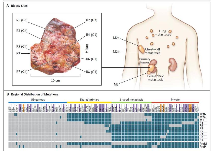
Outline of Presentation

Introduction: What is molecular imaging

Overview of different imaging modalities

Site-specific direct labelling of imaging probes

Principles and application of PET/Ultrasound/MRI

Principles and application of Optical Imaging
       - Indirect labelling using reporter genes
       - Functional imaging

Strategies to improve drug development.

The Molecular Imaging Facility in Campus Kiel
After the seminar the students should be able to:
• List the advantages and disadvantages of each imaging modality in terms
  of resolution, sensitivity and costs.

• Explain the advantages of molecular imaging

• Define the principal behind each imaging modality; namely positron
  emission tomography, ultrasound, magnetic resonance imaging
  and optical imaging.

• List the reasons why labelling imaging probes on lysine residues leads to
  suboptimal properties

• Identify the different reactive groups for site-directed conjugation of
  imaging probes

• Distinguish different strategies for functional imaging using reporter
  genes

• Describe three different strategies to reduce drug development failure
What is Molecular Imaging ?

 New Purpose: diagnose molecular abnormalities

 New Context: physiological environment

 Multidisciplinary: cellular & molecular biology, chemistry,
 pharmacology, physiology, medicine, medical physics, computer science.
Imaging Probes: are used to visualize molecular
           targets and processes in cancer

                                      Targeting       Contrast
                                      Moiety          agent

                    Target molecule

       Deregulated                Ligand
   cellular process in
      cell or tissue
                                Imaging probe with
                                  high specificity.

Targets
- cell types: e.g. tumor cells
- cell function: e.g.apoptosis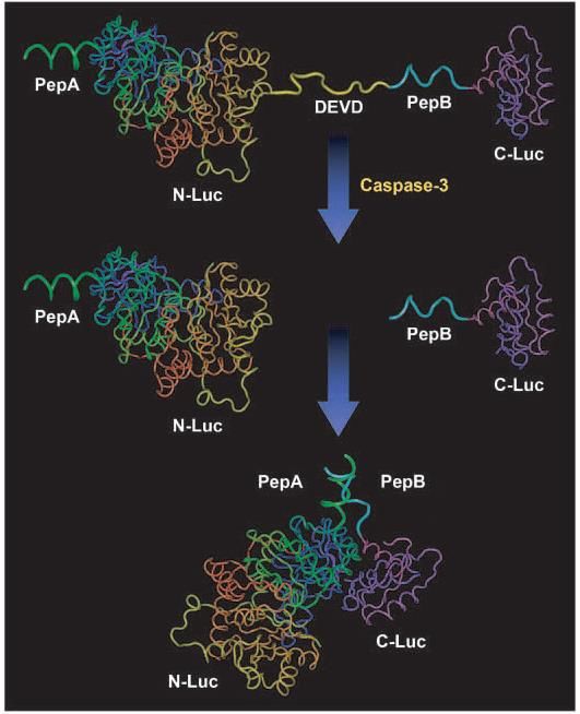
Molecular Imaging utilizes energy emission in specific
           regions of the electromagnetic spectrum

                                  Targeting   Contrast   99mTc, 123I, 111In
                                  Moiety      agent      18F, 64Cu, 68Ga

                Target molecule

    Deregulated
                              Ligand
cellular process in
   cell or tissue                                        NIR Dyes
                                                         Quantum Dots

                      Imaging probes with
                        high specificity.

                                                          Iron oxide
                                                          Nanoparticles
                                                          Carbon Nanotubes

                                                          microbubbles
Instruments which detect signals in specific regions of
                 the electromagnetic spectrum define modality

                       Optical                        Fluorescence

                                                                Magnetic Resonance Imaging
Positron Emission Tomography

                              Molecular Imaging
                                 Modalities
                 18F, 11C, 99mTc
                                                                     a paramagnetic atom, such as
                                                                     Gadolinium.

                                   Ultrasound

                                                      Bright: eg Diaphragm
                                                      Grey: eg Solid organs
                                                      Black: eg fluid

                                   Microbubbles: strongly reflect sound waves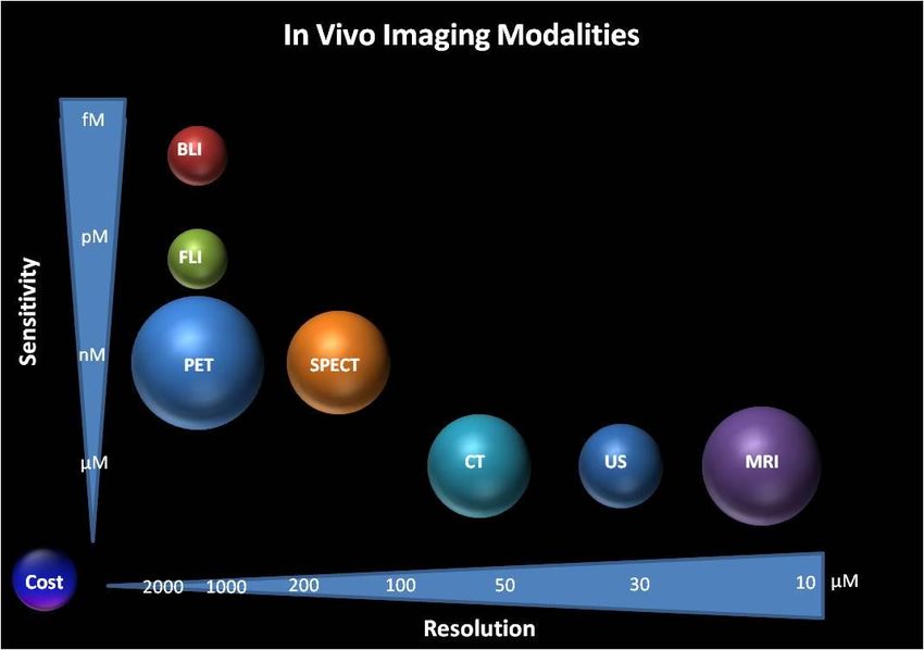
MRI: Anatomical Imaging -> Molecular Imaging
Identify subjects at risk for dementia or beginning disease allowing for clinical testing of preventive
strategies and early therapeutic intervention.
                                                                                      Molecular
                              Anatomical

            Normal                         Alzheimers                         Thioflavin-T derivative

                                                                        Binds to brain amyloid with high affinity.
                                                                        Yellow colors indicate high specific
                                                                        binding of 11-C-PiB to fibrillar Amyloid β-
                      Hippocampal atrophy                               deposits, which indicate a higher risk of
                                                                        progression to Alzheimer’s disease.

                                                                            KFSP Molecular Imaging Network Zurich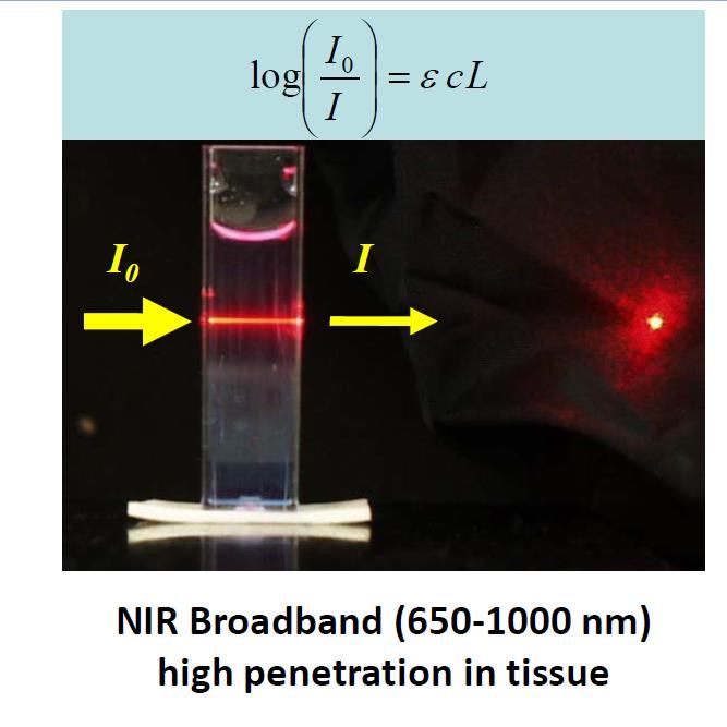
Molecular imaging in diagnostic of tumor subtypes
                         which impact therapy
                                                                                        64Cu-labeled

                                                                                  MRI   trastuzumab

Target                                                 PET Imaging Probe
Glucose Metabolism                                     FDG (18F-fluorodeoxyglucose)
Estrogen Receptor                                      16a-18F-fluoro-17b-estradiol
HER2 (Human Epidermal Growth Receptor-2)               64Cu-labeled   trastuzumab
EGFR (Epidermal Growth Factor Receptor)                89Zr-labeled   cetuximab
IGF-1R (Type 1. Insulin-like Growth Factor Receptor)   89Zr-labeled   R1507
VEGFR (Vascular Endothelial Growth Factor Receptor)    64Cu-DOTA-VEGF

Integrin                                               18F-galacto-RGD
UNIVERSITÄTSKLINIKUM
Schleswig-Holstein
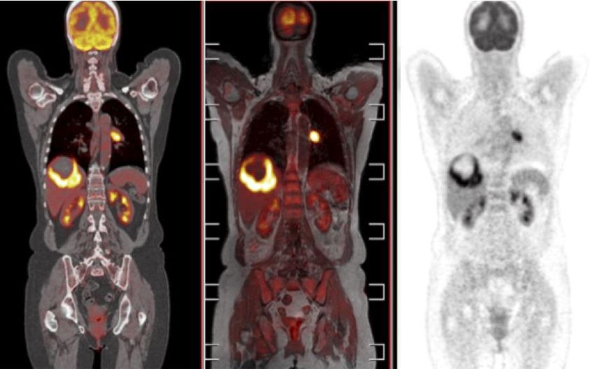
Resolution, Sensitivity and cost of different Imaging ModalitiesMulti modality imaging:Sensitivity/Resolution
PET/CT PET/MRI PET
PET/Optical methods: Sensitivity
combined with MRI/CT: Resolution
Sensitivity and specificity combined with
high resolution whole body morphologic
imaging.Quick Quizz - 1
Which of the following ranking is correct in terms of
sensitivity of imaging modality (most sensitive to least
sensitive)?
a) MRI>PET>Bioluminescence>Fluorescence
b) PET>Fluorescence>bioluminescence>MRI
c) Fluorescence> PET>Bioluminescence>MRI
d) Bioluminescence>Fluorescence>PET>MRI
Which combination of multimodal imaging offers high
sensitivity with high resolution
a) PET/Optical
b) MRI / Ultrasound
c) MRI / CT
d) PET/MRI
e) CT/UltrasoundAdvantages of Molecular Imaging
Understand disease mechanism in realistic in-vivo conditions
Optimize targeted drug therapy ie drug/antibody binding to
target cells, recruitment of immune cells in immunotherapy,
inhibition of a specific signal transduction pathway.
Detect disease prior to anatomical changes
Measure whole body therapeutic target expression
The presence or absence of a target eg HER2, ER
➡Select patients for a specific drug treatment
Measure early response to therapy
Early test of resistance (FDG 24hrs vs 2 mths tumor size)
Biomedizinische Bildgebung
Diagnostische RadiologieOutline of Presentation
Introduction: What is molecular imaging
Overview of different imaging modalities
Site-specific direct labelling of imaging probes
Principles and application of PET/Ultrasound/MRI
Principles and application of Optical Imaging
- Indirect labelling using reporter genes
- Functional imaging
Strategies to improve drug development.
The Molecular Imaging Facility in Campus KielTumor-Specific Antigens
• Tumor-specific antigens are absent on normal tissue and expressed or
overexpressed on tumor cells.
• These extremely rare targets are the „Holy Grail“ of cancer biology.
• Ideal targets for immunotherapy: minimizing autoimmune destruction and no
tolerance.
• Examples include:
– clonal surface IgG found on some B-cell lymphomas.
– EGFR vIII, an in frame deletion mutant found gliomas and other tumors.
• Above is partly true: Every cancer cell harbors at least a few mutations that
can be targeted BUT they are expressed only on individual patients. And thus
require next generation sequencing at the DNA and RNA level as well as
proteomics for individual patients to identify these potentially targeting
mutations.
MOIN SHTumor Associated Antigens
• Are the most common targets for antibody mediated detection.
• Are typically expressed at low levels in normal tissues and at
significantly higher levels in tumors (often 100,000 to 1,000,000
copies per cell).
• Examples include CEA
HER2
EGFR
CD20
PSMA
Folate Receptor
MOIN SHUNIVERSITÄTSKLINIKUM
Schleswig-Holstein
Labelling of contrast agent to reactive
groups
Primary amines (–NH2): This group exists at the N-terminus of
each polypeptide chain and in the side chain of lysine (Lys, K)
residues.
Carboxyls (–COOH): This group exists at the C-terminus of
each polypeptide chain and in the side chains of aspartic acid
(Asp, D) and glutamic acid (Glu, E).
Sulfhydryls (–SH): This group exists in the side chain of
cysteine (Cys, C). Often, as part of a protein's secondary or
tertiary structure, cysteines are joined together between their
side chains via disulfide bonds (–S–S–).
N-linked glycans on Asparagine residues: Glycosylation
occurs on a conserved asparagine residue in the Fc doamin.
Primary amines (–NH2):
Sulfhydryls (–SH):
Carboxyls (–COOH):
N-linked glycans on Asp
COO- COO-
Biomedizinische Bildgebung
Diagnostische RadiologiePotential problems by labelling lysine residues - Heterogeneity in (a) different degrees of labelling. Some antibodies will be labelled to 1 lysine residue, while other labelled to 5 residues. (b) The molecular location of the conjugation on the antibody. For example some conjuagtions will be in the CH3 region of the anitibody while others may be in VH and CH1 domains. (c) Conjuagtion in the antibody-binding domain impairing immunoreactivity of the antibody. These three factors lead to suboptimal pharmacokinetics, decreased accumulation in target tissues, and increased uptake in healthy tissues
Strategies for site-directed labelling of contrast agent

               Sulfhydryls (–SH): Create a labelled FAB antibody by digestion of
               antibody with pepsin and the subsequent reduction of the disulfides in
               the hinge region. The free sulfhydral groups are then available for
               binding to a maleimide-bearing contrast agent.

               N-linked glycans on Asparagine residues: Removal of terminal
               galactose and replacing it with galactose-azide group allows for direct
               conjugation with fluorescent or radioligands by click chemistry.
A. UNIVERSITÄTSKLINIKUM
Schleswig-Holstein
Example: Sulfhydral reactive labelling
A. Native disulfide bonds between cysteines must be cleaved using a reducing agent to free sulhydral groups.
IgG
F(AB)‘2 2 FAB
Immobilized Ficin
4mM cysteine
B. The maleimide group reacts specifically with sulfhydryl groups to form stable thioether linkage
2 FAB Cross-linked magnoxide with FAB
Iron Oxide with sulfhydral
reactive malemide group
S
+Example: N-linked glycan labelling
a
Removal of Terminal
Azide tagging DBCO-Cy7 ligation
Galactose
16hrs@RT
6hrs@370C 12h-16h@40C
Houghton-JL et al , PNAS | December 29, 2015 | vol. 112 | no. 52
https://www.thermofisher.com/de/de/home/references/newsletters-and-journals/bioprobes-journal-of-cell-biology-
applications/bioprobes-69/siteclick-antibody-labeling.htmlAdvantages of site-directed labelling of antibody and peptides •prevent the inadvertent attachment of labell to the antigen-binding domains of the antibody •More homogeneous immunoconjugates, simultaneously eliminating the problems of heterogeneity and irreproducibility created by random approaches. •more favorable pharmacokinetics, higher uptake in target tissues, and lower background accumulation in healthy tissues
Antibody Fragments Generated By
            Recombinant Technology

                               Advantages
      Pharmacokinetic                                      Clinical
     • Rapidly bind to target (ie tumor)     • Greater sensitivity
     • Rapidly cleared from the blood        • Diagnosis in outpatient clinic
     • Good penetration into solid tumors.   • Greater drug penetration
     • Reduced immunogenicity.               •   Minimize undesired side-effects

23
Peptide conjugate for visualization of glioblastoma
Ligands Near - Infrared Dyes:
Cy5.5-CTX
Chlorotoxin (CTX)
Cy5.5
36 amino acid peptide
→ Cl--Channel inhibitor λex = 678 nm
→ MMP-2
λem = 694 nm
Leiurus
quinquestriatus Glioma Fibroblasts
quinquestriatus,
Giant Yellow
Israeli Scorpion
Mandana Veiseh et al. Cancer Res 2007;67:6882-6888
Biomedizinische Bildgebung
Diagnostische RadiologieQuizz-2
(1) What are three reasons for suboptimal pharmacokinetics of an antibody imaging probe when
labelling of primary amines such as lysine residue is performed
(2). Name three other sites of an antibody that are used for site-specific labelling.Quizz-2
(1) What are three reasons for suboptimal pharmacokinetics of an antibody imaging probe when
labelling of primary amines such as lysine residue is performed
(a) different degrees of labelling. Some antibodies will be labelled to 1 lysine residue,
while other labelled to 5 residues.
(b) The molecular location of the conjugation on the antibody. For example some
conjuagtions will be in the CH3 region of the anitibody while others may be in VH
and CH1 domains.
(c) Conjuagtion in the antibody-binding domain impairing immunoreactivity of the
antibody.
(2). Name three other sites of an antibody that are used for site-specific labelling.
Sulfhydryls (–SH):
Carboxyls (–COOH):
N-linked Glycans on asparagine :Outline of Presentation
Introduction: What is molecular imaging
Overview of different imaging modalities
Site-specific direct labelling of imaging probes
Principles and application of PET/Ultrasound/MRI
Principles and application of Optical Imaging
- Indirect labelling using reporter genes
- Functional imaging
Strategies to improve drug development.
The Molecular Imaging Facility in Campus KielPrinciple of Positron emission tomography
Positron-emitting radioactive atom, such as fluorine-18, carbon-11, or oxygen-15.
When positrons, which combine with electrons it is converted into two photons, which are
emitted in opposite directions.
PET image acquisition is based on the simultaneous (coincidence) detection of these two
photons.
Decay
19Fl 18
9
Fl 18
8
O + e+ (positron)
9
(stable)
positron electron
8 e+ e-
9UNIVERSITÄTSKLINIKUM
Schleswig-Holstein
F18-Fluorodeoxyglucose as PET imaging
probe for tumor detection
Uptake of 18f-FDG
by tumor cellsPrinciple of Ultrasound Imaging: Echogenicity Tissues which strongly reflect sound waves give rise to bright dots (hyperechoic) e.g., diaphragm, gallstone, bone, pericardium. Weaker diffuse reflections produce grey dots (hypoechoic) e.g., solid organs. No reflection produces dark dots (anechoic) e.g., fluid and blood filled structures because the beam passes easily through these structures without significant reflection.
Ultrasound: Anatomical Imaging -> molecular imaging:
     Microbubbles conjugated with VEGFR ligand
Magnetic Resonance Imaging
Principles and Application
The single steps of an MR examination can be described
quite simply:
1) the patient is placed in a magnet,
2) a radio wave is sent in,
3) the radio wave is turned off,
4) the patient emits a signal, which is received and used for
5) reconstruction of the pictureHow do protons interact with a magnetic field
When a patient is placed within a magnetic field, it takes a certain
time (T1) for the hydrogen nuclei of water in tissues to be aligned
with or against the magnetic field.
Z= Along main magnetic field
X & Y = perpendicular to the fieldNext a Radio Frequency pulse is applied
perpendicular to the magnetic field.
•The protons tilt away from the magnetic field toward the traverse plane as the
individual spins take transit from a lower energy to a higher energy state.Next turn off radio frequency pulse. The nuclei go from an excited state to a relaxed state. There are two forms of relaxation. •T1 relaxation – The longitudinal relaxation time as flipped nuclei realign with the magnetic field. •T2 relaxation – The traverse relaxation time as the flipped nuclei start off all spinning together, but quickly become incoherent (out of phase) as they lose energy. This has the effect of reducing the over all magnetisation vector in the XY plane.
• Although it is typically water that is detected, water in different tissues has different relaxation times. • By making the image acquisition sensitive to differences in T1 and T2, contrast can be generated. The voxel intensity of a given tissue type (i.e. white matter vs grey matter) depends on the proton density of the tissue reduced by the T1 and T2 relaxation components. A contrast agent can further enhance images by selectively shortening either the longitudinal (T1) or transverse (T2) relaxation time of nearby water protons thereby enhancing the contrast of the image.
Quick Quizz-3

     Which of the following statements is false

     a) Ultrasound imaging depends on echogenicity of sound waves to
        generate contrast
     b) Positron emission tomography utilizes the X-Ray region of the
        electromagnetic spectrum.
     c) Positron emission tomography is based on the simultaneous
        detection of two photons.
     d) Fluid and blood-filled structures are dark in ultasound imaging
     e) In MRI imaging different tissues have different relaxation times
        depending on the density of the protons in that area.
Optical Imaging Principles and Application
Optical imaging uses non-ionizing radiation,
which includes visible, ultraviolet, and infrared light
Imaging using absorption of light

   The intensity is reduced due to absorption properties
                     of a given molecule

   Beer-Lambert Law: There is a linear relationahip between absorbance and
                   concentration of an absorbing species
Absoption Spectra The absorption spectrum of a sample is a plot of the attenuation (decrease in intensity) of the sample as a function of the illumination wavelength For a given molecule, some wavelengths of light are strongly absorbed and others are weakly absorbed.
Near Infra Red light (700-900nm) penetrates tissue to greater depth.

In the NIR region (700-900 nm), the absorption of light by hemoglobin, melanin,
lipids, and other compounds present in living tissue are at a minimum.
Diffuse Optical Spectroscopy
Diffuse optical spectroscopy (DOS) combines near-infrared light using time domain technology to quantify
tissue absorption and scattering spectra from 650nm to 1000 nm. DOS allows quantitative analysis of tissue
chromophore concentrations of oxyhemoglobin, deoxyhemoglobin, methemoglobin, water and lipid.
UNIVERSITÄTSKLINIKUM
                   Advances in Imaging Technology                    Schleswig-Holstein

The SoftScan optical breast imaging device uses near-infrared
light, combined with time domain technology, to obtain 3D
images of the breast. A laser sends brief pulses of light into the
breast and detectors measure photon migration through the
breast. This information is converted into clinically significant,
physiological parameters such as scattering, oxy- and deoxy-
hemoglobin.
Direct labelling of targeting moiety for tumor resection
RAFT-RGD-Alexa Fluor 700
Veiseh et al., Cancer Research 67, 6882-6888, July 15, 2007
Group Probe Target Tumor
Fox Chase Cancer Prosense Cathepsin activity Ovarian
Center and
VisenMedical
TransMolecular Inc. Synthetic Chlorotoxin MMP2-associated target Glioma
TM-601-
Iodine-131Intraoperative Imaging
Chlorotoxin:Cy5.5 Konjugat ( 500x sensitiver als MRT)
Glioma Fibroblast
Darstellung der Ausbreitung von
Krebszellen (intraoperativ)
Cancer Research 67, 2007Direct Labelling by ‚Smart Probes‘: Activated only
in the presence of their intended target.
A. Activity Based Probe
The main backbone is made up of PEGylated poly-lysine modified with fluorophores that
are quenched by close proximity (gray stars). Upon cleavage by a protease at free lysine
residues, smaller fragments containing the unquenched fluorophore (blue stars) are
released.Direct labelImaging Probes
DIRECT INDIRECT
Probes specific for cell surface receptors,
intracellular molecules or gene expression.
Disadvantages solution
Advantages
Short term labelling
reporter gene
Direct labelling agents such as
certain radionucleotides, SPIOs, False positives
and ICG are already approved for
clinical use.Outline of Presentation
Introduction: What is molecular imaging
Overview of different imaging modalities
Site-specific direct labelling of imaging probes
Principles and application of PET/Ultrasound/MRI
Principles and application of Optical Imaging
- Indirect labelling using reporter genes
- Functional imaging
Strategies to improve drug development.
The Molecular Imaging Facility in Campus KielUNIVERSITÄTSKLINIKUM
Schleswig-Holstein
Indirect labelling using Reporter genesUNIVERSITÄTSKLINIKUM
Schleswig-Holstein
Utilization of a promoter to drive a reporter gene expressionPromoters which are always active are used for
cellular tracking
CMV fluorescent
CMV luciferase
Grimm/Tannos et al., Nat Methods (2006)UNIVERSITÄTSKLINIKUM
Schleswig-Holstein
Methods for getting reporter gene into the cell.REPORTER GENE EXPRESSION ALLOWS DETECTION OF TUMOR CELLS IN THE ANIMAL
B. Expression
A. Transfection Fluorescence Phase
C. DetectionIndirect Labelling: Red Fluorescent Protein as a Reporter Gene
A
A spectrum of genetically encoded fluorescent reporters for molecular imaging made by creating mutations in the
chromophore of Jellyfish, corals and bacterial phytochrome.Genetically Encoded Fluorescent Proteins
DsRed2 TdTomato mCherry mPlum Katushka mKate2 IFP iRFP
ex 590 nm Ex 684nm ex 690
ex 560nm ex 550nm ex 590nm ex 590 nm ex 590 nm
em 630 nm Em 708nm em 713
em 580nm em 580nm em 610nm em 650 nm em 650 nm
3 fold brighter QY 0.07 120% brighter
QY 0.55 QY 0.69 QY 0.22 QY 0.1 QY 0.33
than katushka, than IFPOptical Imaging using
emission of light
Fluorescence BioluminescenceIndirect labelling: Luciferase as a reporter gene
UNIVERSITÄTSKLINIKUM
                                                                       Schleswig-Holstein

             Genetically Encoded Bioluminescent Proteins

Firefly Luciferase          Click Beetle Luciferase   Renilla Luciferase           Gaussia luciferase
Em: 610nm.                      Em: Green; 540nm      Em: 480nm                     Em: 480nm
                                Em: Red; 610nm

             D-Luciferin                                          Coelenterazine
                                                                   ATP independent
                                                                   Small size ca. 1kb
                                                                   Gaussia is secreted
         Ideal for deep tissue imaging
Testing of drug efficacy: Sutent in Ovarian Cancer
Day14 Day28 Day42 Day56 Day62
Luminescence signal photons per second (ph/sec), red treatment
group (40mg SU11248/kg bodyweight.
Bauerschlag DO, Schem C, Tiwari S, Egberts JH, Weigel MT, Kalthoff H, Jonat W, Maass N, Meinhold-Heerlein I., Anticancer Res. 2010 Sep;30:3355-60UNIVERSITÄTSKLINIKUM
Schleswig-Holstein
In Vivo sensitivity of fluorescence vs bioluminescence
Luc2-codon optimized firefly luciferase
Detection of a single cell implanted
subcutaneousQuick Quizz - 4
Which of the following reporter gene is the most optimal for
in vivo optical imaging
a) GFP emission maximum: 512
b) RFP: emission maximum: 580
c) mCherry: emission maximum: 610nm
d) mKate2;: emission maximum: 630nm
e) iRFP: emission maximum: 713nm
(1) An advantage of fluorescent imaging compared to
bioluminescent imaging is
a) it is more sensitive
b) it has lower background signal
c) it can be used for intraoperative imaging
d) it is cheaperUNIVERSITÄTSKLINIKUM
Schleswig-Holstein
Functional Imaging: The imaging of gene expression and
products to give information on biological processes.Functional Imaging: Split Firefly Reporter Strategy for imaging enzyme activity in vivo.
Noninvasive Imaging of Apoptosis Noninvasive Imaging of Epidermal Growth Factor Receptor
Kinase Activation.
Coppola et al. Clin. Can Res. 2008
Li et al. Cancer Res. 2008Functional Imaging: The imaging of gene expression and
activity to give information on biological processes.
CMV GFPGFP
Promoter Species Specificity
B29 Human B cells
CD14 Human Monocytic cells
CD43 Human Leukocytes & platelets
CD45 Human Haematopoietic cells
CD68 Human Macrophages
Tie2 Human Endothelial Precurser Cells
Endoglin Human Endothelial cells
Flt-1 Human Endothelial cells
ICAM-2 Human Endothelial cells
GFAP Human Astrocytes
GPIIb Human Megakaryocytes
Mb Human Muscle
NphsI Human Podocytes
SP-B Human Lung
SYN1 Human Neuron
WASP Human Hematopoietic cells
Promoters can be inducible so that function could be imaged during a specific
biological process. The functional activation of the reporter is then imaged
accordingly based on the selected protein.
Grimm/Tannos et al., Nat Methods (2006)Circulating Tie-2 expressing monocytes are a distinct monocyte
population which home to tumors and promote angiogenesis.
Tie2 expressing cells can be targets for cancer therapy and tools to deliver anti-cancer agents to tumors
De Palma et al., 2007 Trends in Immunology
Biomedizinische Bildgebung
Diagnostische RadiologieFunctional imaging with inducible promoters- Osterix
Detection of osteoblasts using Cre-Lox recombination
Osx Cre (A)n pROSA26
X
stop pTurbo635 (A)n
loxP loxP
Cre expressing cells Non-Cre expressing cells
eg osteoblast
pROSA26 pTurbo635 (A)n pROSA26
X
stop pTurbo635 (A)n
loxP loxP
Biomedizinische Bildgebung
Diagnostische RadiologieQuick Quizz - 5
To track and image a specific cell type in vivo (eg
endothelial precursor cells), which of the following imaging
strategy is optimal
a) utilization of an inducible promoter
b) utilization of a split-firefly strategy
c) utilization of a smart-probe
d) utilization of a constitutive active promoter
Which of the following imaging approaches is not
considered to be functional imaging
a) utilization of an inducible promoter
b) utilization of a constitutive active promoter
c) utilization of a smart-probe
d) utilization of a split-firefly strategy
e) utilization of reporter mice using cre-lox technologyOutline of Presentation
Introduction: What is molecular imaging
Overview of different imaging modalities
Site-specific direct labelling of imaging probes
Principles and application of PET/Ultrasound/MRI
Principles and application of Optical Imaging
- Indirect labelling using reporter genes
- Functional imaging
Strategies to improve drug development.
The Molecular Imaging Facility in Campus KielUNIVERSITÄTSKLINIKUM
Schleswig-Holstein
Perspectives: Improve drug development and clinical trials
-More relevant mouse models
-Stratify patients that express therapeutic target
-Assess responders vs non-responders earlyPerspective of moleucular imaging: Where we need to improve
Oncology drug development: Low success rates at every
stage of clinical development
Data for 1991-2000 for 10 largest pharmaceutical companies
Kola & Landis, Nature Reviews Drug Discovery 2004. Andrea Pirzkall, MD GenetechUNIVERSITÄTSKLINIKUM
Schleswig-Holstein
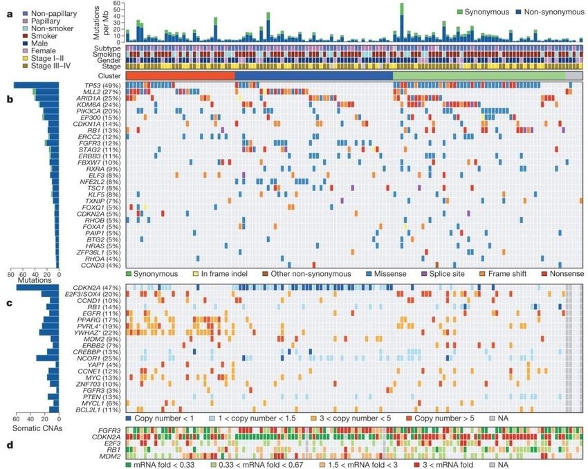
The genomic landscape of bladder cancer.
The Cancer Genome Atlas Research Network Nature 507, 315-322 (2014) doi:10.1038/nature12965Genomic Sequencing
•High degree of intertumor (between two tumors in same patient) genetic
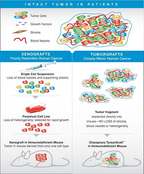
variability and intratumor variability (different cells in same tumor)2. Cell line xenografts vs Patient derived xenograft
The PDX collection at EuroPDX
            Characterised by transcriptome arrays, CGH,
                    WES, targeted sequencing
A‚Roadmap‘ to precision medicine in cancer
To leverage patient genomic tumor heterogeneity as predictors of drug sensitivity for testing in PDX mouse
models.
Building Blocks to the ‚Roadmap‘
• Patient stratification
• In silico determination of drug
combinations
• Selection of PDX model that matches
patient stratification.
• Establish preclinical in vivo efficacyApproach to Patient Stratification
• Patient stratification
• In silico determination of drug
combinations
• Selection of PDX model that matches
patient stratification.
• Establish preclinical in vivo efficacyStratification
Patient stratification is the grouping of patients with
shared biological characteristics by using molecular
diagnostic
testing to select the most optimal therapy in order to
achieve the best possible medicinal outcomes for that
group.Paradigm change in patient stratification.
1947 Sydney Farber achieves remission in pediatric acute
leukemia patients following treatment with an antimetabolite but
clinical resistance emerges.
Tumor type
1958 Following successful combination therapy in turberculosis,
Emil Frei III combines chemotherapeutic agents in patients.
1993 Vogelstein, Fearon, Kinzler proposed
distinct oncogenic mutations drive cancer
malignancy
Several deregulated genes
2001 Gleevac (Imatinib), the first-ever molecularly
targeted cancer treatment in CML
2003. First generation cDNA microarray
identified four major intrinsic gene
signatures.
2012. The Cancer Genome Atlas Network
sequencing entire genome of Global genome deregulation
10,000-tumors from 20
different cancer types.Biomarkers The National Institute of Health defined a biomarker as: ‚ a characteristic that is objectively measured and evaluated as an indicator of normal biological processes, pathological processes or responses (pharamacologic or otherwise) to a therapeutic intervention‘. Biomarkers Definition Working Group 2001
Breast cancer biomarkers based on receptor expression
                                are prognostic
Stratification              Biomarkers                                                Median Duration of
                                                                                      survival from time of
                                                                                      first distant
                                                                                      metastases
Luminal A                   Estrogen Receptor-positive and/or Progesterone Receptor   2.2 years
                            positive, HER2 negative and Ki67low
Luminal B                   Estrogen Receptor –positive and/or Progesterone           1.6 years
                            Receptor positive, HER2 negative and Ki67 high
Luminal-HER2                Estrogen Receptor-positive and/or Progesterone Receptor   1.3 years
                            positive and HER2-positive
HER2-enriched               Estrogen Receptor-negative, Progesterone Receptor         0.7 years
                            negative, HER2-positive
Basal-like                  Estrogen Receptor negative                                0.5 years
                            Progesterone Receptor negative
                            HER2 negative
                            EGFR positive or
                            Cytokeratin 5/6 positive
Triple-negative non basal   Estrogen Receptor negative                                0.9 years
                            Progesterone Receptor negative
                            HER2 negative
                            EGFR negative
                            Cytokeratin 5/6 negative
Cancer Biomarker
Diagnostic
Biomarkers Diagnosis
Treatment
Predictive
Biomarkers
Prognostic
Biomarkers Outcome
Imaging surveys the entire patient and can therefore assess the entire disease burden and directly measures the
heterogeneity of both target expression and therapeutic response, which are increasingly recognized as key
factors in therapeutic resistance. Especially assess sites challenging to biopsy and assay, such as bone.Role of molecular imaging in biomarker driven therapy
Imaging surveys the entire patient
- disease burden
- heterogeneity of both target expression and therapeutic
response.Example 1. Predictive Biomarker: Is the target
present?Progressive diseaseThe role of molecular imaging in precision medicine
Imaging surveys the entire patient
- disease burden
- heterogeneity of both target expression and therapeutic
response.Take home messages • List the advantages and disadvantages of each imaging modality in terms of resolution, sensitivity and costs. • Explain the advantages of molecular imaging • Define the principal behind each imaging modality; namely positron emission tomography, ultrasound, magnetic resonance imaging and optical imaging. • List the reasons why labelling imaging probes on lysine residues leads to suboptimal properties • Identify the different reactive groups for site-directed conjugation of imaging probes • Distinguish different strategies for functional imaging using reporter genes • Describe three different strategies to improve drug development
THANK YOU FOR YOUR
    ATTENTION
You can also read