Molecular Correlation between Larval, Deutonymph and Adult Stages of the Water Mite Arrenurus (Micruracarus) Novus - MDPI
←
→
Page content transcription
If your browser does not render page correctly, please read the page content below
life

Article
Molecular Correlation between Larval, Deutonymph
and Adult Stages of the Water Mite Arrenurus
(Micruracarus) Novus
Pedro María Alarcón-Elbal 1 , Ricardo García-Jiménez 2 , María Luisa Peláez 2 , Jose Luis Horreo 3, *
and Antonio G. Valdecasas 2
 1    Instituto de Medicina Tropical & Salud Global (IMTSAG), Universidad Iberoamericana (UNIBE),
      22333 Santo Domingo, Dominican Republic; Pedro.Alarcon@uv.es
 2    Museo Nacional de Ciencias Naturales, CSIC. C/José Gutiérrez Abascal, 2, 28006 Madrid, Spain;
      rgarcia@mncn.csic.es (R.G.-J.); maaller@yahoo.es (M.L.P.); valdeca@mncn.csic.es (A.G.V.)
 3    UMIB Research Unit of Biodiversity (UO, CSIC, PA), C/Gonzalo Gutiérrez de Quirós s/n, 33600 Mieres, Spain
 *    Correspondence: horreojose@gmail.com; Tel.: +34-985-10-30-00 (ext. 5943)

 Received: 27 May 2020; Accepted: 7 July 2020; Published: 9 July 2020

 Abstract: The systematics of many groups of organisms has been based on the adult stage.
 Morphological transformations that occur during development from the embryonic to the adult stage
 make it difficult (or impossible) to identify a juvenile (larval) stage in some species. Hydrachnidia
 (Acari, Actinotrichida, which inhabit mainly continental waters) are characterized by three main
 active stages—larval, deutonymph and adult—with intermediate dormant stages. Deutonymphs
 and adults may be identified through diagnostic morphological characters. Larvae that have not
 been tracked directly from a gravid female are difficult to identify to the species level. In this work,
 we compared the morphology of five water mite larvae and obtained the molecular sequences of
 that found on a pupa of the common mosquito Culex (Culex) pipiens with the sequences of 51 adults
 diagnosed as Arrenurus species and identified the undescribed larvae as Arrenurus (Micruracarus)
 novus. Further corroborating this finding, adult A. novus was found thriving in the same mosquito
 habitat. We established the identity of adult and deutonymph A. novus by morphology and by
 correlating COI and cytB sequences of the water mites at the larval, deutonymph and adult (both
 male and female) life stages in a particular case of ‘reverse taxonomy’. In addition, we constructed the
 Arrenuridae phylogeny based on mitochondrial DNA, which supports the idea that three Arrenurus
 subgenera are ‘natural’: Arrenurus, Megaluracarus and Micruracarus, and the somewhat arbitrary
 distinction of the species assigned to the subgenus Truncaturus.

 Keywords: Acari Actinotrichida; COI; cytochrome B; genetic identification; Hydrachnidia; Culicidae;
 reverse taxonomy; species identification

1. Introduction
     The systematics of many groups of organisms has traditionally been based on the adult stage [1].
For species showing a deep morphological transformation from the embryonic to the adult stage, due to
a change in habitat or different behavior (parasitic and free-living stages) it may be difficult, if not
impossible, to identify the juvenile stage without a previous correlation with the corresponding adult.
     Members of the clade Hydrachnidia (Acari Actinotrichida) mainly inhabit continental waters [2]
and are characterized by three main active life stages—larval, deutonymph and adult—with
intermediate dormant stages [3]. Deutonymphs and adults share a characteristic morphology and may
be identified through diagnostic characters. Larval morphology is very different from that of the two
other stages. The standard procedure to match the larvae of a species with its corresponding adult

Life 2020, 10, 108; doi:10.3390/life10070108                                          www.mdpi.com/journal/life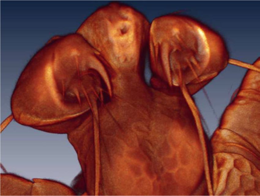
Life 2020, 10, 108 2 of 14
is to track them directly from a fertilized female. In short, a fertilized female is kept in a small vial
with a strip of paper or another substrate until they oviposit eggs and larvae emerge [4,5]. It is hard to
identify the species level of larvae that have not been tracked in this way with certainty.
The water mite genus Arrenurus has a worldwide distribution, except for the perpetual
snow regions, but there are no cosmopolitan species; rather, each main area upholds its own set of species.
There are 152 Arrenurus in Europe and, of these, about 30% have a wide distribution [6–8]. As in
other Hydrachnidia genera, Arrenurus deutonymphs and adults share diagnostic characters, however,
the larval stage has a distinct morphology and requires independent morphological characterization.
Water mite larvae usually attach to insects at preimaginal stages before they hatch, using adults as
vector to disperse [9]. Adults and larvae of the same species, therefore, may be found in very different
locations in their life cycle. Some Arrenurus species have occasionally been recorded as an ectoparasite
of aquatic insects at the juvenile stage, either nymphs or larvae [2]. Some have even been recorded
in insect exuviae [6,10]. Given these challenges, species identification of isolated Arrenurus larvae is
fraught with uncertainty.
In this work, we characterized Arrenurus sp. larvae that were attached to larvae and a pupa
of the common mosquito Culex (Culex) pipiens and cross-correlate their sequences with those of 51
adult Arrenurus species. One of these species, Arrenurus (Micruracarus) novus George, 1884, was in the
same mosquito habitat from which the Arrenurus larvae were isolated. We compare cytochrome C
oxidase subunit I (COI) and cytochrome B (cytB) DNA sequences of the water mite larvae with those of
A. novus deutonymphs and adults (male and female) and other Arrenurus species, in order to establish
the identity of the water mite larvae in a particular case of ‘reverse taxonomy’ [11].
2. Materials and Methods
2.1. Taxon Sampling
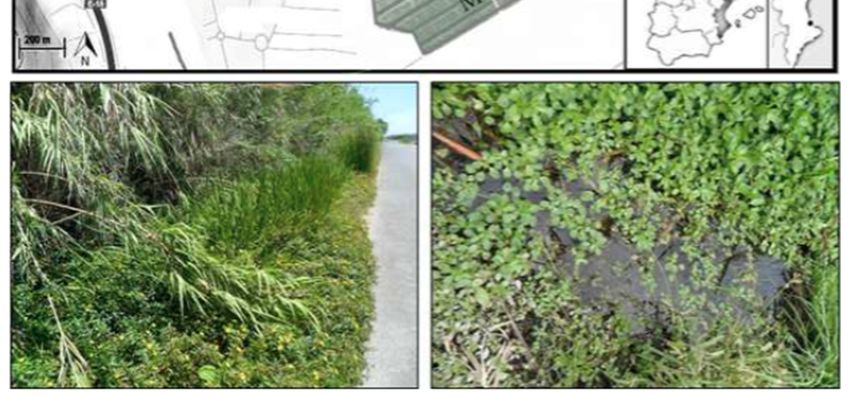
Water mite and mosquito specimens were both collected during a survey of mosquitoes at
preimaginal stages carried out every two weeks between May and October of 2012 at Acequia del
Caminàs (UTM 30S 741855.92 E 4324431.48), a semiartificial irrigation channel within the marsh waters
of the Xeraco and Xeresa wetlands (Valencia, Spain; Figure 1). For more details on the characteristics
of the area, see Alarcón-Elbal et al. [12]. Further collection efforts were carried out in May 2019
without success.
We used a quick dipping technique [13] with a standard 500-mL mosquito dipper (BioQuip
Products Inc., Rancho Dominguez, CA, USA). Culicids at preimaginal stages were sorted manually at
the laboratory and then transferred to a 60 ◦ C water bath for a minute and then to absolute ethanol.
Culicid specimens with ectoparasites and free-living water mites in the same sample were both
preserved in absolute ethanol and stored at −20 ◦ C.
Preservation in absolute ethanol ensures that both microscopic and molecular techniques can
be used on any water mite specimen collected from potential hosts or directly from the field [14,15].
Both the culicids and the water mite specimens were morphologically identified, and subsequently
used for DNA extraction.
Life 2020, 10, 108 3 of 14
Life 2020, 10, x FOR PEER REVIEW 3 of 15
Figure 1.
Figure Map of
1. Map of the
the sampling
sampling site
site (Xeraco,
(Xeraco, Valencia,
Valencia, Spain).
Spain).
2.2. Morphological Identification of Culicids
2.2. Morphological Identification of Culicids
Morphological identification of culicids at preimaginal stages were made to the species level
Morphological identification of culicids at preimaginal stages were made to the species level
based on external morphology and diagnostic characters related with chaetotaxy, as described by
based on external morphology and diagnostic characters related with chaetotaxy, as described by
Schaffner et al. [16]. Specimens were examined under a Nikon SMZ-1B Stereozoom microscope.
Schaffner et al. [16]. Specimens were examined under a Nikon SMZ-1B Stereozoom microscope.
2.3. Morphological Identification of Water Mites
2.3. Morphological Identification of Water Mites
2.3.1. Rearing of Adult Female Water Mites
2.3.1. Rearing of Adult Female Water Mites
In an attempt to rear larvae, six live adult Arrenurus sp. females were collected in the field
In ankept
and then attempt
under to laboratory
rear larvae,conditions
six live adult Arrenurus transparent
in individual sp. females were collected
poliestirene in thecontainers
plastic field and
then kept under laboratory conditions in individual transparent poliestirene
(diameter 10–15 mm, height 15 mm) with a piece of paper and mineral water at room temperature for plastic containers
(diameter
14–15 days10–15 mm, height
[4]. Following the15 mm) withperiod,
monitoring a piecetheof paper
females and mineral
were waterinatabsolute
preserved room temperature
ethanol for
for 14–15 days [4]. Following the monitoring
morphological identification of the species level. period, the females were preserved in absolute ethanol
for morphological identification of the species level.
2.3.2. Morphological Identification of Water Mite Adults, Deutonymphs and Larvae
2.3.2. Morphological Identification of Water Mite Adults, Deutonymphs and Larvae
Morphological identification of the adult and deutonymph water mite specimens collected from
Morphological
the field identification
site was performed usingof thediagnostic
the adult andcharacters
deutonymph water mite
originally specimens
described collected
by George [17]from
and
the field site was
subsequently performed
revised by Vietsusing the diagnostic
[18] and Gerecke [2]. characters originally described by George [17] and
subsequently
The studyrevised
of larvalby water
Viets [18]
miteand Gereckewas
specimens [2]. based on four specimens found anchored on the
bodyTheof a study
culicidoflarva
larval waterinmite
(L1–L4 specimens
Table 1) and onewas based
larva foundononfour specimens
a culicid pupafound anchored
(L5). The culicidon the
larva,
body and
pupa of a attached
culicid larva
water(L1–L4 in Table
mite larvae were1)examined
and one larva founda Bausch
first under on a culicid pupa stereomicroscope
and Lomb (L5). The culicid
larva,
and thenpupa
withandlaserattached
scanningwater mite
confocal larvae were
microscopy (LSCM,examined firstSPE).
Leica TCS under
Thea host
Bausch and Lomb
attachment site,
stereomicroscope
where the water mite andlarval
then mouthparts
with laser scanning confocal was
were embedded, microscopy
recorded(LSCM,
and theLeica
larvaeTCS
wereSPE). The host
individually
attachment
and carefullysite, whereusing
detached the water mite larval
fine tweezers and mouthparts
transferred towere embedded,
a drop of glycerolwas
on arecorded
microscope andslide
the
larvae
for LSCM were individually
stack acquisition.and carefully
After imaging,detached
the five using
larvae fine
weretweezers and transferred
then processed to aanalyses.
for molecular drop of
glycerol on a microscope slide for LSCM stack acquisition. After imaging, the five larvae were then
processed for molecular analyses.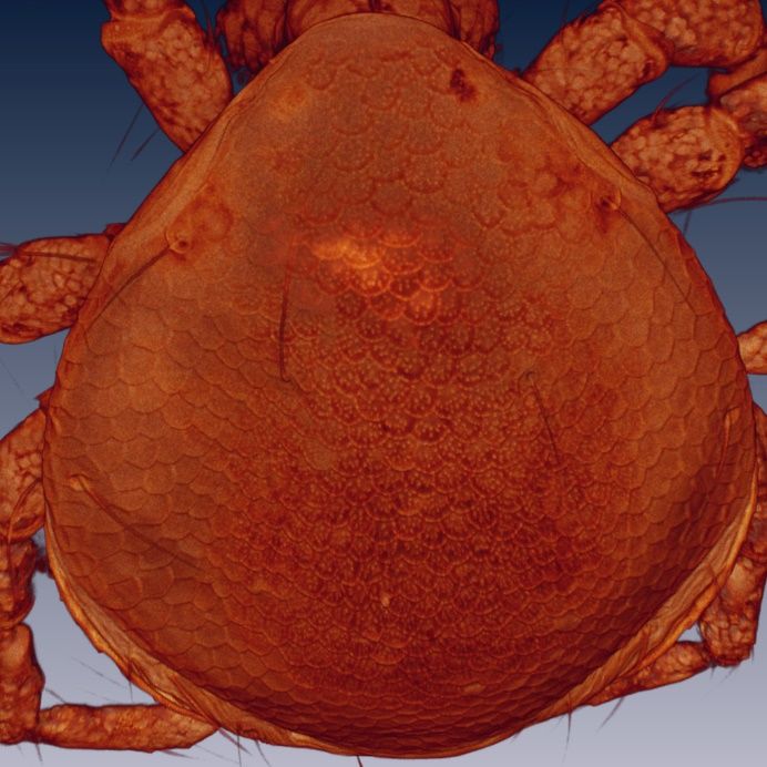
Life 2020, 10, 108                                                                                         4 of 14

      Table 1. Standard measurements (see [8]) of the larval body morphology (in µm; only an approximation
      due to specimen orientation on the slide).

                                          L1             L2             L3            L4             L5
  Body length                             197           182            190            204            200
  Body width                              175           168            168            164            156
  Dorsal plate length                     190           186            190            185            185
  Dorsal plate width                      171           152            160            148            147
  CpI medial margin length                 57            55             61             57             62
  CpII medial margin length                27            30             30             27             26
  CpIII medial margin length               38            42             44             40             42
  Distance between C1 and CpI
                                          21             19             23             18            20
  median margin
  Distance between C4 and CpIII
                                          28             27             24             27            29
  median margin
  Distance between C1 and C2              40             36             40             38            39
  Excretory pore plate length             25             27             26             22            29
  Excretory pore plate width              32             30             32             30            31
  Distance between Exp and Expp
                                          11             15             15             12            13
  posterior margin
  PIII length                             27             21             26             31            31
  Length of PIV claw                      27             23             14             16            17

2.3.3. Laser Scanning Confocal Microscopy Imaging
     Laser scanning confocal microscopy (LSCM, Leica TCS SPE) was employed to acquire z-stack
images with the following objectives: 10×/0.30 NA, 20×/0.70 NA, 40×/1.25 NA oil immersion and
63x/1.30 NA glycerol immersion. The samples were subjected to an excitation wavelength of 488 nm
and an emission range between 520 and 660 nm. For further information on the stack acquisition
procedure, see Valdecasas and Abad [14]. Serial images were processed with Amira (ver. 5.4.3) and
ImageJ/Fiji to obtain maximum 2D projections and Voltex volume renderings. Different parameters
of visualization were used with Voltex to best highlight the characters of interest. Fiji/ImageJ were
used to take all measurements directly from the image stacks. As all the material was used for the
subsequent molecular analyses, no morphological vouchers could be kept. Therefore, a photographic
voucher comprised of the full set of original unaltered stacks is stored in the confocal collection at the
National Museum of Natural Sciences of Spain (CSIC–MNCN).

2.4. Molecular Analyses
      A total of nine water mite samples—three adults, one deutonymph and five larvae—were
processed for molecular analyses. DNA extraction, PCR amplification and sequencing were performed
as previously described) [15]. The COI gene was amplified using the primer pair LCO1490
and HC02198 [19]. For the larval water mite samples (n = 5), the internal primer COI-AV-F
(50 -ATAAGATTTTGACTTCTYCC-30 ) and HC02198 (360 basepairs length) were used because the
complete amplification of the gene was not successful. CytB was amplified using the primer pair
COB-F/R [20]. Sequencing reactions were performed using ABI BigDye® v3.1 Cycle Sequencing Kit
(Applied Biosystems) and run on an ABI 3100 Genetic Analyzer. Sequence chromatograms were
checked for accuracy and edited using Sequencher® version 5.0 (Gene Codes Corporation, Ann Arbor,
MI, USA). DNA sequences were deposited in GenBank (accession numbers MT598102 to MT598106 for
COI and MT607634 to MT607637 for cytB).
      The sequences of both gene fragments were analyzed for all three life cycle stages in order
to molecularly correlate the undescribed larvae with the diagnosed adults and deutonymphs.
Genetic distances were estimated using the Kimura-2-Parameter (K2P) distance model [21],
as implemented in MEGA v.7 [22].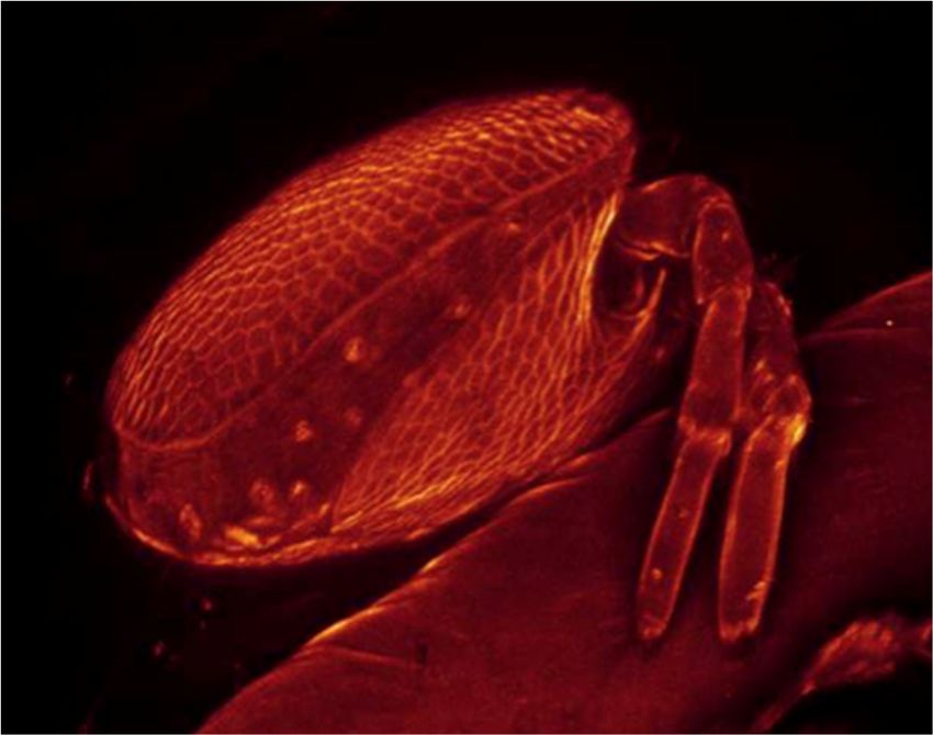
Life 2020, 10, x FOR PEER REVIEW 5 of 15
The sequences of both gene fragments were analyzed for all three life cycle stages in order to
molecularly correlate the undescribed larvae with the diagnosed adults and deutonymphs. Genetic
Life 2020, 10, 108 5 of 14
distances were estimated using the Kimura-2-Parameter (K2P) distance model [21], as implemented
in MEGA v.7 [22].
Additionally, COI COIandandcytB
cytB sequences
sequences of Arrenurus
of Arrenurus species
species were were searched
searched in GenBank
in GenBank (https:
//www.ncbi.nlm.nih.gov/genbank/;
(https://www.ncbi.nlm.nih.gov/genbank/; accessed accessed
21 May21 May
2020) in 2020)
order in order to the
to estimate estimate
genetic the genetic
distances
distances
among allamong all sequences,
available available sequences,
including including the new
the new ones ones
of this of this
study. Thestudy. The of
distances distances of the
the GenBank
GenBank sequences
sequences were calculated
were calculated using MEGA.using A MEGA. A Maximum-Likelihood
Maximum-Likelihood phylogeneticphylogenetic
tree of COItree of COI
sequences
sequences
was was constructed
constructed with theweb
with the IQ-TREE IQ-TREE
serverweb server (http://iqtree.cibiv.univie.ac.at)
(http://iqtree.cibiv.univie.ac.at) with the
with the following
followingautomatic
settings: settings: detection
automaticofdetection of substitution
substitution model, perturbation
model, perturbation strength
strength of 0.5, IQ-TREE of stopping
0.5, IQ-TREErule
stopping
of rule ofbootstrap
100, ultrafast 100, ultrafast bootstrap
analysis analysis (1000
(1000 replicates) replicates)
and SH-aLRT and SH-aLRT
single branch testssingle
(1000branch tests
replicates).
(1000
For replicates).
this For this tree,
tree, two outgroup two outgroup
sequences sequences
were used: werelundbladi
Torrenticola used: Torrenticola
(GenBanklundbladi
accession (GenBank
number
accession number JX629050) and Lebertia
JX629050) and Lebertia maderigena (KX421869). maderigena (KX421869).
3. Results
3.1. Morphological Identification
3.1. Morphological Identification of
of Culicids
Culicids
All
All of
of the culicid specimens
the culicid specimens were
were identified as Culex
identified as (Culex) pipiens
Culex (Culex) pipiens Linnaeus,
Linnaeus, 1758. The principal
1758. The principal
and
and reliable
reliablecharacters
charactersforfordiagnosis at at
diagnosis thethe
larval stage
larval are the
stage are syphon index,index,
the syphon the syphon/saddle index,
the syphon/saddle
the branch number of seta one on abdominal segments III–IV, 1a–S tuft, 1b–S tuft and syphon
index, the branch number of seta one on abdominal segments III–IV, 1a–S tuft, 1b–S tuft and syphon shape [23].
One
shapeculicid larvaculicid
[23]. One and one pupa
larva were
and oneparasitized
pupa were with four and
parasitized onefour
with water
andmite
onelarvae,
water respectively
mite larvae,
(Figures 2–4).
respectively (Figures 2–4).
Figure 2. Culex pipiens larva with Arrenurus sp. larvae attached (arrows) and detail.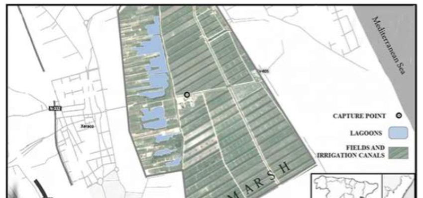
Life 2020, 10, 108                                                                                      6 of 14
Life 2020,
Life 2020, 10,
           10, x
               x FOR
                 FOR PEER
                     PEER REVIEW
                          REVIEW                                                                      66 of
                                                                                                         of 15
                                                                                                             15

                   Figure 3.
                   Figure 3. Culex
                             Culex pipiens
                                   pipiens pupa
                                           pupa with
                                                with Arrenurus
                                                     Arrenurus sp.
                                                               sp. larva
                                                                   larva attached
                                                                         attached (white
                                                                                  (white dot).
                                                                                         dot).

     Figure 4. Culex
     Figure    Culex pipiens pupa with Arrenurus sp. larva
                                                     larva attached
                                                           attached (laser
                                                                    (laser scanning confocal microscopy
     (LSCM) image).

3.2. Morphological
3.2. Morphological and
                   and Molecular
                       Molecular Identification
                                 Identification of
                                                of Water
                                                   Water Mites
                                                         Mites
3.2.1. Rearing of Adult Female Water Mites
3.2.1. Rearing of Adult Female Water Mites
      Six Arrenurus females were maintained for 14–15 days, but no eggs or larvae were observed
     Six Arrenurus females were maintained for 14–15 days, but no eggs or larvae were observed
during this period. We were not able to ascertain whether the females were gravid and if the lack of
during this period. We were not able to ascertain whether the females were gravid and if the lack of
eggs and larvae was due to unsuitable laboratory conditions.
eggs and larvae was due to unsuitable laboratory conditions.
3.2.2. Larval Description
3.2.2. Larval Description
      The description of the water mite larvae is preliminary: confocal microscopy could not resolve
someThe    description
       of the extremelyoffine
                          the morphological
                              water mite larvae     is preliminary:
                                                 details,            confocal
                                                          an issue faced      microscopy
                                                                         by other         could
                                                                                  researchers of not resolve
                                                                                                 water  mite
some   of the extremely  fine morphological      details, an issue faced by other researchers of water
larvae [7]. Therefore, scanning electron microscopy analyses are necessary for a complete description.  mite
larvae [7]. Therefore, scanning electron microscopy analyses are necessary for a complete description.
The terminology used here follows that of Zawal [7]. Figures 5 and 6 show a Voltex projection of the
The terminology
ventral            used habitus
         and the dorsal  here follows   that ofrespectively.
                                  of a larva,    Zawal [7]. Figures  5 and 6 show
                                                              The projections     a Voltex
                                                                              do not        projection
                                                                                     indicate          of the
                                                                                              morphometric
ventral and the dorsal habitus of a larva, respectively. The projections do not indicate morphometric
distances, and the scales are only an approximation of size. Tables 1 and 2 include some standard
distances, and the scales are only an approximation of size. Tables 1 and 2 include some standard
morphometric data taken independently of the Voltex projections. Figure 7 shows a schematic of the
approximate distribution of setae on the leg segments.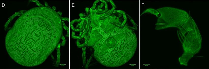
Life 2020, 10, 108                                                                                         7 of 14

morphometric data taken independently of the Voltex projections. Figure 7 shows a schematic of the
Life 2020, 10, x FOR PEER REVIEW                                                                          7 of 15
approximate distribution of setae on the leg segments.
     Arrenurus novus larvae (see molecular analysis below) may be distinguished from other previously
                    Table 2. Standard measurements of the larval leg morphology in µm.
described Micruracarus larvae [7] by the following set of characters: dorsal plate ovoid, narrowed
                    Larvae ratio lateral
anteriorly; coxas distinct,          Trochanter       Femur
                                          length of coxal   plateGenu
                                                                  I, II andTibia    Tarsus
                                                                            III: 5:3:4; a hexagonal excretory
plate perimeter and the excretory
                               leg1 pore in25
                                            line or slightly
                                                        21 above29E2 setae42(Figure 55  8). Total length of Leg1
< Leg II = Leg III.
                      L1       leg2        30           25        30        46          57
                                     leg3 measurements
                           Table 2. Standard 30        of 25
                                                          the larval 30      46
                                                                     leg morphology in 57
                                                                                       µm.
                                    leg1         23           21        29       40        49
                     Larvae                 Trochanter   Femur        Genu        Tibia         Tarsus
                              L2    leg2         23           25        30       44        59
                                   leg1        25          21           29            42          55
                      L1             leg3
                                   leg2        3027        25 23        25
                                                                        30       48 46     57     57
                                   leg3        30          25           30
                                    leg1         30           23        30       38 46     49     57
                                   leg1        23          21           29          40            49
                              L3     leg2        30           32        27       42        57
                      L2           leg2        23          25           30          44            59
                                     leg3
                                   leg3        2734        23 34        27
                                                                        25       44 48     57     57
                                     leg1
                                   leg1        3029        23 23        27
                                                                        30       44 38     55     49
                      L3           leg2        30          32           27          42            57
                              L4     leg2        21           25        29       46        53
                                   leg3        34          34           27          44            57
                                    leg3
                                   leg1        29
                                                 32        23
                                                              29        29
                                                                        27
                                                                                 46 44     59     55
                      L4             leg1
                                   leg2        2124        25 23        27
                                                                        29       40 46     41     53
                                   leg3        32          29           29            46          59
                              L5    leg2         29           22        26       45        60
                                   leg1        24          23           27          40            41
                      L5
                                     leg3
                                   leg2        2930        22 25        30
                                                                        26       40 45     31     60
                                   leg3        30          25           30          40            31

                           Figure 5. Arrenurus
                                     Arrenurus (Micruracarus) novus larva, ventral view (LSCM).
Life 2020, 10, 108 8 of 14
Life2020,
Life 2020,10,
10,xxFOR
FORPEER
PEERREVIEW
REVIEW 88 of
of 15
15
Life 2020, 10, x FOR PEER REVIEW
VIEW
REVIEW 8 of815
of 15
Figure6.
Figure Arrenurus(Micruracarus)
6.Arrenurus (Micruracarus)novus
novuslarva,
larva,dorsal
dorsalview
view(LSCM).
(LSCM).
Figure 6. Arrenurus (Micruracarus) novus larva, dor
re 6. Arrenurus
gure (Micruracarus)
6. Arrenurus novus
(Micruracarus) larva,
novus dorsal
larva, view
dorsal (LSCM).
view (LSCM).
Figure
Figure 7. 7. ASchematicshowing
7. Schematic
Schematic showingthe thedistribution
distributionofofsetae setae on
on the
the leg
leg segments
segments of of an
an Arrenurus
of an Arrenurus
Figure showing the distribution of setae on the leg segments Arrenurus
(Micruracarus)novus
(Micruracarus)
(Micruracarus) novus larva.
novuslarva. Tr:
larva.Tr: trochanter;
Tr:trochanter; Fe:
trochanter;Fe: femur;
Fe:femur;
femur;Ge: Ge:genu;
Ge: genu;
Figure
genu; Ti:7.
Ti:
Ti: tibia;
tibia;
tibia; Ta: tarsus.
Schematic
Ta: tarsus.
s Round
Round
Round setae;
howing
setae;
setae; the distr
solenidum;
solenidum; other setae.
other setae. segments of an Arrenurus (Micruracarus) novus larva. Tr: trochanter
s s
tic howing
howing thesolenidum;
distr
the distr otheribution
setae.
ibutionof setae on on
of setae the the
leg leg
tarsus. Round setae; solenidum; other setae.
urus
renurus(Micruracarus) novus
(Micruracarus) larva.
novus Tr: Tr:
larva.
Arrenurus trochanter; Fe: Fe:
trochanter;
novus larvaefemur;
(seeGe:
femur; genu;
Ge: Ti: tibia;
genu;
molecular Ti: Ta: Ta:below) may be distinguished from other
tibia;
analysis
Arrenurus novus larvae (see molecular analysis below) may be distinguished from other
ae; solenidum;
setae; solenidum; other setae.
other
previouslysetae.described Micruracarus larvae [7] by the following set of novus
Arrenurus characters:
larvaedorsal
(see plate ovoid,analysis below)
molecular
previously described Micruracarus larvae [7] by the following set of characters: dorsal plate ovoid,
narrowedanteriorly;
narrowed anteriorly;coxas
coxasdistinct,
distinct,;;ratio
ratiolateral
laterallength
length ofcoxal
coxaldescribed
previously
of plateI,II
plate I,IIand
and III:5:3:4;
III: 5:3:4;aalarvae
Micruracarus hexagonal
[7] by the following se
hexagonal
arvae (see(see
larvae molecular
molecular analysis
excretory platebelow)
analysis below)
perimetermay maybe the
and distinguished
be distinguished
excretory from
pore from
in other
lineother
or slightly above E2 setae (Figure 8). Total
excretory plate perimeter and the excretory pore in line narrowed anteriorly;
or slightly above E2coxas distinct,
setae (Figure; ratio lateral length of coxal
8). Total
icruracarus
Micruracarus larvae
larvae[7] [7]
lengthby by
theLeg1
of following
the LegIIIIset
following of characters:
set ofIII.
characters: dorsal
dorsalplate ovoid,
plate ovoid,
length of Leg1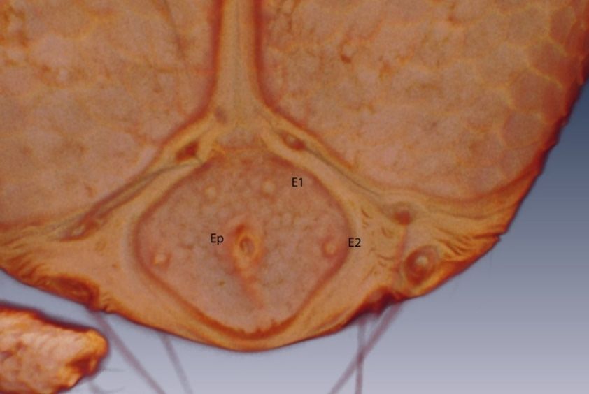
Life 2020, 10, x FOR PEER REVIEW                                                                                  9 of 15

       The number of setae on the pedipalp segments are in agreement with Zawal’s description of
Arrenurus larvae [7]: none on P-I; one on P-II; two on P-III, one long and thick and the other short;
four
Life   on 10,
     2020, P-IV,  three
              x FOR PEERthin and one thick; and a solenoid and seven setae on P-V (Figure 9).
                          REVIEW                                                               9 of 15
Life 2020, 10, 108                                                                                                9 of 14

     The number of setae on the pedipalp segments are in agreement with Zawal’s description of
Arrenurus larvae [7]: none on P-I; one on P-II; two on P-III, one long and thick and the other short;
four on P-IV, three thin and one thick; and a solenoid and seven setae on P-V (Figure 9).

                      Figure 8. Arrenurus (Micruracarus) novus larva, excretory plate (LSCM).

     The number of setae on the pedipalp segments are in agreement with Zawal’s description of
Arrenurus larvae [7]: none on P-I; one on P-II; two on P-III, one long and thick and the other short;
four on P-IV, threeFigure
                    thin and  one thick;
                          8. Arrenurus   and a solenoid
                                       (Micruracarus) novusand seven
                                                            larva,     setae plate
                                                                   excretory on P-V   (Figure 9).
                                                                                   (LSCM).

                         Figure 9. Arrenurus (Micruracarus) novus larva, pedipalp (LSCM).

3.2.3. Morphological Identification of Adult and Deutonymph Water Mites
      Seven male and six female adults plus two deutonymphs were identified as A. novus, a species
                        Figure 9. Arrenurus    (Micruracarus)
                                   ArrenurusEurope
                                               (Micruracarus)   novus larva, pedipalp (LSCM).
found in standing waters       throughout               [2]. Among      the main diagnostic characters identifying
A. novus
3.2.3.     adults are the
       Morphological       petiole in the
                        Identification    ofmale
                                             Adult (see
                                                      andarrow    in FigureWater
                                                            Deutonymph       10A), the setation of male and female
3.2.3. Morphological    Identification    of Adult   and   Deutonymph        Water Mites
                                                                                     Mites
palps, and the shape and length of cauda (Figure 10). The deutonymph diagnosis was based on palp
shape Seven
      Seven   male and
              male andand
        and setation     six habitat
                        six  female adults
                             female   adults   plus two
                                               plus
                                      co-occurrence  two(Figure
                                                           deutonymphs
                                                           deutonymphs       were identified
                                                                             were
                                                                    11): approximateidentified  as A. novus,
                                                                                                as A.
                                                                                         body length: novus,  aa species
                                                                                                         526 µm; species
                                                                                                                   body
found
found
width:inin standing
           standing
         467         waters
             µm; P-1waters
                      without  throughout
                               throughout     Europe
                                              Europe
                                 setae; P-II with       [2]. Among
                                                        [2].medial
                                                    three    Among      the main
                                                                        the main
                                                                     setae;        diagnostic
                                                                                   diagnostic
                                                                            P-III with         characters
                                                                                               characters
                                                                                        one medial          identifying
                                                                                                     setae; identifying
                                                                                                            PIV uncate
A.
A.
and novus
   novus    adults
           adults
     a strong      aresetae
                  are
               distal   thepetiole
                      the    petiole
                             and       in
                                       thethe
                                    in with
                                   PV        malemale   (see
                                                   (seefine
                                              a basal    arrow arrow    in Figure
                                                                  in Figure
                                                             setae.          10A),10A),    the setation
                                                                                    the setation  of maleofandmale   and
                                                                                                                 female
femaleand
palps,   palps,
             theand  theand
                 shape    shape   andof
                              length   length
                                          caudaof(Figure
                                                    cauda (Figure
                                                             10). The10).   The deutonymph
                                                                        deutonymph      diagnosisdiagnosis
                                                                                                   was based wasonbased
                                                                                                                    palp
on  palp  shape  and  setation   and  habitat   co-occurrence      (Figure  11): approximate
shape and setation and habitat co-occurrence (Figure 11): approximate body length: 526 µm;      body   length:  526body
                                                                                                                     µm;
body width:
width:  467 µm;467P-1µm;   P-1 without
                      without              setae;
                                 setae; P-II withP-II
                                                    threewith   three
                                                           medial       medial
                                                                     setae; P-IIIsetae; P-IIImedial
                                                                                  with one    with one
                                                                                                    setae;medial   setae;
                                                                                                            PIV uncate
PIV  uncate   and a strong   distal setae   and  PV  with
and a strong distal setae and PV with a basal fine setae.    a basal  fine setae.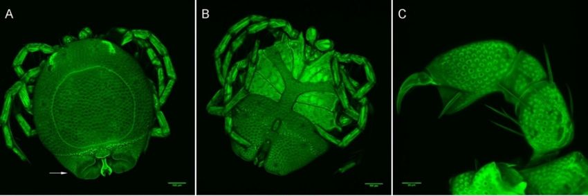
Life 2020, 10, 108 10 of 14
Life 2020, 10, x FOR PEER REVIEW 10 of 15
Life 2020, 10, x FOR PEER REVIEW 10 of 15
Figure
Figure 10.
10. Arrenurus
Arrenurus (Micruracarus)
(Micruracarus) novus
novus adults
adults (LSCM).
(LSCM). (A)
(A) male,
male, dorsal;
dorsal; (B)
(B) male,
male, ventral;
ventral; (C)
(C) male,
male,
Figure 10. Arrenurus (Micruracarus) novus adults (LSCM). (A) male, dorsal; (B) male, ventral; (C) male,
palp; (D) female,
palp; (D) female, dorsal;
dorsal; (E)
(E) female,
female, ventral;
ventral; (F)
(F) female,
female, palp.
palp.
palp; (D) female, dorsal; (E) female, ventral; (F) female, palp.
Figure
Figure 11. Ventral
Ventral view of Arrenurus (Micruracarus) novus deutonymph.
Figure 11. Ventral view of Arrenurus (Micruracarus) novus deutonymph.
3.3. Molecular
3.3. Molecular Analyses
Analyses
3.3. Molecular Analyses
GenBank identified
GenBank identified 990 Arrenurus barcoding
990 Arrenurus barcoding COICOI sequences
sequences but no Arrenurus
but no Arrenurus cytBcytB sequences
sequences
GenBank
(probably identified
because cytB 990
is notArrenurus
generally barcoding
used for COI
speciessequences but
delimitation). no Arrenurus
These cytB sequences
sequences belonged
(probably because cytB is not generally used for species delimitation). These sequences belonged to
(probably because cytB is not generally
A. novus, used forsequence
species delimitation). These sequences belonged to
51 species of the genus plus A. novus, whose sequence is reported here for the first time (genetic
to 51 species of the genus plus whose is reported here for the first time (genetic
51 speciesamong
distances of the genus are plus A. novus, whose sequence is reported here for the
12). first time (genetic
distances among species
species areshown shown in in
Table S1 and
Table phylogenetic
S1 and tree in
phylogenetic Figure
tree in Figgure The12).
meanThegenetic
mean
distances
distances among
among species
the 52 are
Arrenurusshown in
species Table
was S1
16.4%and phylogenetic
(standard tree
deviation =in Figgure
4.1). The 12). The mean
here-amplified
genetic distances among the 52 Arrenurus species was 16.4% (standard deviation = 4.1). The here-
genetic distances among the 52 Arrenurus species was 16.4%were(standard deviation = 4.1). The here-
COI sequences
amplified from
COI sequences all thefrom all theA.different
different novus life-cycle
A. novusstages all thewere
life-cycle stages same. all the same.
amplified COI
Very few sequences from all the different A. novus life-cycle stages were all the same.
Very few phylogenetic
phylogenetic information
information about
about Arrenurus
Arrenurus currently
currently exist
exist [24].
[24]. The
The here-constructed
here-constructed
Very few
COI phylogenetic phylogenetic
phylogenetic tree information about Arrenurus currently exist [24]. The here-constructed
COI tree (Figure
(Figure 12,12, Material
Material S1)
S1) may
may bebe divided
divided into
into three
three main
main arms: the upper
arms: the upper branch
branch
COI
is phylogenetic
dominated by tree (Figure
species belonging12, Material
to the S1) may Micruracarus,
subgenus be divided into three
with main arms:
occasional the upper
incursions of branch
species
is dominated by species belonging to the subgenus Micruracarus, with occasional incursions of species
is dominated by species belonging to the subgenus Micruracarus, with occasional incursions of species
belonging
belonging to the
to subgenera Arrenurus,
the subgenera Arrenurus, Truncaturus,
Truncaturus, Megaluracarus
Megaluracarus andand Micrarrenurus.
Micrarrenurus. In In this
this upper
upper
belonging to the subgenera Arrenurus, Truncaturus, Megaluracarus and Micrarrenurus. In this upper
branch is located Arrenurus (Micruracarus) novus. The intermediate branch of the tree is composed
branch is located Arrenurus (Micruracarus) novus. The intermediate branch of the tree is composedLife 2020, 10, 108 11 of 14
Life 2020, 10, x FOR PEER REVIEW 11 of 15
exclusively
branch isof species
located of the subgenus
Arrenurus Arrenurus,
(Micruracarus) novus.except for a sequence
The intermediate belonging
branch to is
of the tree subgenus
composed
Truncaturus. The
exclusively oflower section
species of subgenus
of the the tree consists exclusively
Arrenurus, except of species
for belonging
a sequence to the sub-genus
belonging to subgenus
Megaluracarus,
Truncaturus.with
The European andof
lower section American representatives.
the tree consists exclusively of species belonging to the sub-genus
Megaluracarus, with European and American representatives.
Figure
Figure 12.12. MaximumLikelihood
Maximum Likelihoodphylogenetic
phylogenetic tree Arrenurus
treeofofthethe C oxidase
Arrenurus subunit
C oxidase I (COI)I sequences
subunit (COI)
employed
sequences in this in
employed study and the
this study andoutgroups (Torrenticola
the outgroups lundbladi
(Torrenticola lundbladi Lebertia
and and maderigena).
Lebertia Node
maderigena).
numbers
Node show
numbers showSH-aLRT
SH-aLRT (left) and
(left) bootstrap
and (right)
bootstrap support
(right) values
support (1000
values replicates).
(1000 Letters
replicates). Lettersat at
right
subgenus acronyms: Arrenurus (A), Micruracarus (Mi), Truncaturus
right show subgenus acronyms: Arrenurus (A), Micruracarus (Mi), Truncaturus (T),and
show (T), Megaluracarus (Me)
Micrarrenurus(Me)
Megaluracarus (Ma).and Micrarrenurus (Ma).
The 360-bp COI fragment was successfully amplified only from one larvae; however, no genetic
The 360-bp COI fragment was successfully amplified only from one larvae; however, no genetic
variation was found among the sequences of this region (two adults, one deutonymph and one larvae).
variation was found among the sequences of this region (two adults, one deutonymph and one
Genetic differences were, however, observed in the 330-bp cytB fragment (Table 3). One of the adults
larvae). Genetic differences were, however, observed in the 330-bp cytB fragment (Table 3). One of
and all five larvae shared the same cytB haplotype, whereas the deutonymph and the other adult
the adults and all five larvae shared the same cytB haplotype, whereas the deutonymph and the other
shared a different haplotype (Table 3).
adult shared a different haplotype (Table 3).
Table 3. K2P distances among the adult, deutonymph and larval Arrenurus sp. cytB sequences analyzed
Table 3. K2P
in this studydistances among
(alignment: the adult, deutonymph and larval Arrenurus sp. cytB sequences
330 bp).
analyzed in this study (alignment: 330 bp).
K2P Adult 1 Adult 2 Nymph Larvae
K2P Adult 1 Adult 2 Nymph Larvae
Adult 1 -
Adult 2 Adult 1
0.006 - -
Nymph 0.006
Adult 2 0.006 -0.000 -
Larvae 0.000 0.006 0.006 -
Nymph 0.006 0.000 -
Larvae 0.000 0.006 0.006 -
4. Discussion
4. Discussion
The number of molecular sequences available for Hydrachnidia has increased substantially in
recent years. GenBank
The number list 6450
of molecular Hydrachnidia
sequences available nucleotide sequences
for Hydrachnidia has (as of 21 May
increased 2020). Weindid
substantially
recent years. GenBank list 6450 Hydrachnidia nucleotide sequences (as of 21 May 2020). We did notLife 2020, 10, 108 12 of 14
not search the BOLD Systems database for two main reasons: many of the Arrenurus sequences are
privately held, and it was recently shown that some water mite sequences do not agree with their
species assignment [25]. Of the GenBank sequences, almost 70% are identified to the generic level.
Some new species have been described with morphological and molecular data [15,26]. For the genus
Arrenurus, there are 1071 sequences (990 of them belonging to COI gene) but only 198, representing
51 taxa, are at the species level. These were employed to the genetic distance calculations. All of the
Arrenurus sequences in GenBank identified to the species level are based on the adult stage; sequences
obtained from larvae are only given a generic rank and are frequently singled out as operational
taxonomic units (e.g. [27]). There is a need for more species-level sequences based on different life
cycle stages to widen the usefulness of this type of data to address ecological, biogeographical and
evolutionary questions. We identified the adult and deutonymph water mite specimens as A. novus,
a broadly distributed species found from the Western Palearctic to the Afrotropic, commonly in
standing waters [2,25]. However, according to Gerecke et al. [2], it has been “little reported in Europe”.
In the present study, we combined morphological and molecular techniques to associate
undescribed larvae with corresponding adults and deutonymphs for the first time in water mites.
Associations among life cycle stages are especially relevant for this genus given that less than 20%
of arrenurid larvae are known [28]. To link undescribed larval specimens with other life stages
(i.e., deutonymph and adult), we employed a DNA-based identification method in which sequences
are considered as standardized comparative characters that can be used to support and integrate
morphological datasets [29]. Even in cases with very small genetic distances, these data can be useful
for species identification [30]. The results of our analyses of two mitochondrial genes (COI and cytB),
which proved to be informative, clearly support the same conclusion: the larvae belong to the species
A. novus. The larval COI sequences were compared with 52 adult Arrenurus species sequences (51 were
from GenBank; the A. novus sequences, from one adult male, two adult females, one deutonymphs and
one larvae, are provided for the first time here). Genetic distances among the three A. novus life-cycle
stages were 0.000 (the same DNA sequence), confirming the co-specificity of the larvae, deutonymph
and adults. The distance of A. novus with other Arrenurus species ranged between 9.8 with A. setiger
and 24.3 with A. megalurus (see their phylogenetic relationships in Figure S1). The cytB sequences of
the larvae and one of the adults were identical and highly similar to those of the other adult and the
deutonymph (which were identical; see Table 3), strongly supporting the conclusion that they belong
to the same species. It is important to note here that we are studying only mitochondrial DNA, thus,
in the case of hybrids, we would be only describing the maternal species of the larvae. In addition,
the phylogenetic tree based on COI mitochondrial information (Figure 12) supports the idea that three
Arrenurus subgenera are ‘natural’—Arrenurus, Megaluracarus and Micruracarus—and the somewhat
arbitrary distinction of the species assigned to the subgenus Truncaturus. This topology differs from
the only other Arrenurus phylogeny published [24] and points to the need for more molecular data in
order to clarify their phylogenetic relationships.
This study demonstrates the usefulness of an integrative approach to resolve the taxonomic
uncertainty of water mites at the larval stage. Furthermore, it appears to be a promising approach for
identifying larvae at the species level in terms of both reliability and speed, in comparison with rearing
or hatching approaches. With these data, we can also begin to elucidate host–parasite interactions
between a specific water mite species and its host(s) with greater detail. To date, a host of A. novus
larvae was unknown [2]. Indeed, only a few studies have described a host-parasite interaction
between a Culex species and an Arrenurus species (e.g. [6,10,31,32]). Arrenurus is the genus most
reported to parasitize mosquitoes, evidence of its flexibility regarding host specificity [33]. Despite
this, recent findings suggest that these mites prefer Culex species [34]. Aside from reducing host
fitness by piercing their exoskeleton to feed on hemolymph, each host–parasite association has its own
characteristics, which are determined mainly by the size of the partners, intrinsic defence mechanisms
and environmental conditions [35]. Parasitic mites may play a significant role in the biological control of
mosquitos in wetlands, especially of adult populations. Therefore, more observations and experimentalLife 2020, 10, 108 13 of 14
data of water mite species are needed to better understand their host–parasite interactions, as well as
to incorporate the use of molecular techniques in their identification, particularly at the larval stage.
Supplementary Materials: The following are available online at http://www.mdpi.com/2075-1729/10/7/108/s1,
Table S1: K2P distances among the COI sequences of the 52 Arrenurus species included in the analysis. The final
alignment was 659 bp. Arrenurus novus is indicated in bold. Material S1: Maximum Likelihood phylogenetic
tree of the Arrenurus COI sequences employed in this study and the outgroups (Torrenticola lundbladi and Lebertia
maderigena) in newick format.
Author Contributions: Field data sampling and culicids identification, P.M.A.-E.; Bright field microscopy and
Laser Scanning Confocal Microscopy imaging, and identification of water mites, A.G.V.; Molecular extraction,
amplification and sequencing R.G.-J.; Genbank searches and downloading and molecular analysis, M.L.P. and J.L.H.;
all the authors contributed to the writing and reviewed the last version of the manuscript. All authors have read
and agreed to the published version of the manuscript.
Funding: J.L.H. was supported by the Regional Government of Asturias (reference SV-16-UMB-1).
Acknowledgments: We thank Annie Machordom for access to the Systematics Molecular Laboratory of the MNCN.
Conflicts of Interest: The authors declare no conflict of interest. The funders had no role in the design of the
study; in the collection, analyses, or interpretation of data; in the writing of the manuscript, or in the decision to
publish the results.
References
1. Thorp, J.H.; Rogers, D.C.; Dimmick, W.W. Introduction to invertebrates of inland waters. In Freshwater
Invertebrates, 4th ed.; Elsevier BV: Amsterdam, The Netherlands, 2015; pp. 1–19.
2. Gerecke, R.; Gledhill, T.; Pešić, V.; Smit, H. Süßwasserfauna von Mitteleuropa, Bd. 7/2-3 Chelicerata; Springer
Spektrum: Heidelberg, Germany, 2016.
3. Smith, I.M.; Cook, D.R.; Smith, B.P. Water mites (Hydrachnidiae) and other arachnids. In Ecology and
Classification of North American Freshwater Invertebrates; Elsevier BV: Amsterdam, The Netherlands, 2010;
pp. 485–586.
4. Cook, W.J.; Smith, B.P.; Brooks, R.J. Allocation of reproductive effort in female Arrenurus spp. water mites
(Acari: Hydrachnidia; Arrenuridae). Oecologia 1989, 79, 184–188. [CrossRef]
5. Tuzovsky, P. A new water mite species of the genus Arrenurus Dugès, 1834 (Acariformes: Hydrachnidia:
Arrenuridae) from Eastern Palaearctic. Acarina 2012, 20, 173–179.
6. Zawal, A. Phoresy and parasitism: Water mite larvae of the genus Arrenurus (Acari: Hydrachnidia) on
Odonata from lake Binowskie (NW Poland). Biol. Lett. 2006, 43, 257–276.
7. Zawal, A. Morphological characteristics of water mite larvae of the genus Arrenurus Duges, 1834, with notes
on the phylogeny of the genus and an identification key. Zootaxa 2008, 1765, 1–75. [CrossRef]
8. Zawal, A. Morphology of the larval stages of Arrenurus affinis Koenike, 1887, A. neumani Piersig, 1895,
and A. vietsi Koenike, 1911 (Acari: Hydrachnidia). Genus 2008, 19, 161–169.
9. Bohonak, A.J.; Smith, B.P.; Thornton, M. Distributional, morphological and genetic consequences of dispersal
for temporary pond water mites. Freshw. Boil. 2004, 49, 170–180. [CrossRef]
10. Lanciani, C.A. Sexual bias in host selection by parasitic mites of the mosquito anopheles crucians (Diptera:
Culicidae). J. Parasitol. 1988, 74, 768. [CrossRef]
11. Markmann, M.; Tautz, D. Reverse taxonomy: An approach towards determining the diversity of meiobenthic
organisms based on ribosomal RNA signature sequences. Philos. Trans. R. Soc. B: Boil. Sci. 2005, 360,
1917–1924. [CrossRef]
12. Alarcón-Elbal, P.M.; Murillo, J.M.S.; Estrella, S.D.; Ruiz-Arrondo, I.; Prieto, R.P.; Curdi, J.L. Asociación de
vector del VNO e hidrófito invasor: Culex pipiens Linnaeus, 1758 y Ludwigia grandiflora (Michaux) Greuter
and Burdet en el marjal de Xeraco-Xeresa, Valencia. Anales Biol. 2013, 35, 17–27. [CrossRef]
13. Service, M.W. A critical review of procedures for sampling populations of adult mosquitoes.
Bull. Entomol. Res. 1977, 67, 343–382. [CrossRef]
14. Valdecasas, A.G.; Abad, A. Morphological confocal microscopy in arthropods and the enhancement of
autofluorescence after proteinase K extraction. Microsc. Microanal. 2010, 17, 109–113. [CrossRef] [PubMed]
15. Pešić, V.; Valdecasas, A.G.; García-Jimenez, R. Simultaneous evidence for a new species of Torrenticola
Piersig, 1896 (Acari, Hydrachnidia) from Montenegro. Zootaxa 2012, 3515, 38–50. [CrossRef]Life 2020, 10, 108 14 of 14
16. Schaffner, E.; Angel, G.; Geoffroy, B.; Hervy, J.P.; Rhaiem, A.; Brunhes, J. Les Moustiques d’Europe: Logiciel
d’Identification et d’Enseignement = The Mosquitoes of Europe: An Identification and Training Programme;
IRD Editions & EID Méditerranée: Montpellier, France, 2001.
17. George, C.F. The British fresh-water mites. Hardwicke’s Sci. 1884, 20, 80–81.
18. Viets, K. Zur kenntnis dert Hydracarinen-Fauna von Spanien. Arch. Hydrobiol. 1930.
19. Folmer, O.; Black, M.; Hoeh, W.; Lutz, R.; Vrijenhoek, R. DNA primers for amplification of mitochondrial
cytochrome c oxidase subunit I from diverse metazoan invertebrates. Mol. Mar. Biol. Biotechnol. 1994, 3,
294–299. [CrossRef] [PubMed]
20. Ernsting, B.R.; Edwards, D.D.; Aldred, K.J.; Fites, J.S.; Neff, C.R. Mitochondrial genome sequence of
Unionicola foili (Acari: Unionicolidae): A unique gene order with implications for phylogenetic inference.
Exp. Appl. Acarol. 2009, 49, 305–316. [CrossRef]
21. Kimura, M. A simple method for estimating evolutionary rates of base substitutions through comparative
studies of nucleotide sequences. J. Mol. Evol. 1980, 16, 111–120. [CrossRef]
22. Kumar, S.; Stecher, G.; Tamura, K. MEGA7: Molecular evolutionary genetics analysis version 7.0 for bigger
datasets. Mol. Boil. Evol. 2016, 33, 1870–1874. [CrossRef]
23. Azari-Hamidian, S.; Harbach, R.E. Keys to the adult females and fourth-instar larvae of the mosquitoes of
Iran (Diptera: Culicidae). Zootaxa 2009, 2078, 1–33. [CrossRef]
24. Wieçel, M. Effects of the Evolution of Intromission on Courtship Complexity and Male and Female
Morphology: Water Mites of the Genus Arrenurus (Acari; Hydrachnida) from Europe and North America.
Ph.D. Thesis, Adam Mickiewicz University, Poznan, Poland, 2016.
25. Valdecasas, A.G.; García-Jiménez, R.; Marín, M. Two rare water mite species (Acari, Parasitengona,
Hydrachnidia) new for the Iberian Peninsula. Rev. Ibérica Aracnol. 2019, 35, 33–37.
26. Pešić, V.; Smit, H. Neumania kyrgyzica sp. nov. a new water mite from Kyrgyzstan based on morphological
and molecular data (Acari, Hydrachnidia: Unionicolidae). Syst. Appl. Acarol. 2017, 22, 885. [CrossRef]
27. Mlynarek, J.J.; Knee, W.; Forbes, M.R. Explaining susceptibility and resistance to a multi-host parasite.
Evol. Boil. 2013, 41, 115–122. [CrossRef]
28. Martin, P. Wassermilben (Hydrachnidia, Acari) und Insekten: Ein Überblick über eine selten betrachtete
Beziehung. Entomol. Heute 2008, 20, 45–75.
29. Scorrano, S.; Aglieri, G.; Boero, F.; Dawson, M.N.; Piraino, S. Unmasking Aurelia species in the Mediterranean
Sea: An integrative morphometric and molecular approach. Zool. J. Linn. Soc. 2016, 180, 243–267. [CrossRef]
30. Horreo, J.L.; Ardura, A.; Pola, I.G.; Martinez, J.L.; Garcia-Vazquez, E. Universal primers for species
authentication of animal foodstuff in a single polymerase chain reaction. J. Sci. Food Agric. 2012, 93, 354–361.
[CrossRef] [PubMed]
31. Mullen, G.R. Water mites of the subgenus Truncaturus (Arrenuridae, Arrenurus) in North America.
Ithaca Agric. Entomol. 1976, 6, 1–35.
32. Kirkhoff, C.J.; Simmons, T.W.; Hutchinson, M.; Simmons, T.W. Adult mosquitoes parasitized by larval water
mites in Pennsylvania. J. Parasitol. 2013, 99, 31–39. [CrossRef] [PubMed]
33. Dos Santos, E.B.; Favretto, M.A.; Dos Santos Costa, S.G.; Navarro-Silva, M.A. Mites (Acari: Trombidiformes)
parasitizing mosquitoes (Diptera: Culicidae) in an Atlantic Forest area in southern Brazil with a new mite
genus country record. Exp. Appl. Acarol. 2016, 69, 323–333. [CrossRef] [PubMed]
34. Atwa, A.A.; Bilgrami, A.L.; Al-Saggaf, A.I. Host–parasite interaction and impact of mite infection on mosquito
population. Rev. Bras. Entomol. 2017, 61, 101–106. [CrossRef]
35. Gerson, U.; Smiley, R.L.; Ochoa, R. Mites (Acari) for Pest Control; Blackewell: Malden, MA, USA, 2003.
© 2020 by the authors. Licensee MDPI, Basel, Switzerland. This article is an open access
article distributed under the terms and conditions of the Creative Commons Attribution
(CC BY) license (http://creativecommons.org/licenses/by/4.0/).You can also read


