Relation of antivimentin antibodies to anticardiolipin antibodies in systemic lupus erythernatosus
←
→
Page content transcription
If your browser does not render page correctly, please read the page content below
Ann Rheum Dis: first published as 10.1136/ard.47.9.708 on 1 September 1988. Downloaded from http://ard.bmj.com/ on January 18, 2021 by guest. Protected by
Annals of the Rheumatic Diseases, 1988; 47, 708-716

Relation of antivimentin antibodies to anticardiolipin
antibodies in systemic lupus erythernatosus
MARTIN A BLASCHEK,' MICHAEL BOEHME,4 JEAN JOUQUAN,'-
ANNE-MARIE SIMITZIS,3 SPYROS FIFAS,' PAUL LE GOFF,2
AND PIERRE YOUINOU'
From the Departments of 'Immunology, 2Rheumatology, and @Microbiology, Brest University Medical
School, Brest, France; the 4Abteilung fur Innere Medizin, Universitatsklinik, Ulm, West Germany; and the
5French National Institute of Marine Biology, Brest, France

SUMMARY Tests for antivimentin antibodies (AVA) were performed on 50 systemic lupus
erythematosus (SLE) and 63 control sera by indirect immunofluorescence and enzyme linked
immunosorbent assay (ELISA). The prevalence was significantly raised in SLE (38% and 50%/o of
sera positive for IgM-AVA and IgG-AVA, respectively, by immunofluorescence; 36% and 64%
of sera positive for IgM-AVA and IgG-AVA, respectively, by ELISA) in comparison with the
control sera. A significant correlation existed between IgM-AVA, on the one hand, and
anticardiolipin antibodies (ACA) and anti-single-stranded DNA (ssDNA), on the other. A
stepwise principal component analysis demonstrated that IgM-AVA and IgG-AVA accounted
for 71% of the total variance in SLE (50 patients x 5 parameters=total variance). Twenty ACA

                                                                                                                                                                                          copyright.
positive serum samples from patients with syphilis were therefore tested for the presence of
AVA, but hardly any were found to be positive. IgM-AVA from patients with SLE were
inhibited by cardiolipin and absorbed with ssDNA. An association between AVA positivity and
arthralgia was also shown in SLE.
Key words: anti-single-stranded DNA antibody.

Intermediate sized filaments are polymeric fibrous                 As part of our studies on anticytoskeletal
structures.' Together with microfilaments and micro- antibodies)             "' we looked for the presence of anti-
tubules they form part of the extensive cytoplasmic             vimentin antibodies (AVA) in SLE sera, using an
network called the cytoskeleton. The collective term            indirect immunofluorescence technique and an
intermediate sized filaments refers to those fila-              enzyme linked immunosorbent assay (ELISA). The
ments whose diameter is approximately 10 nm, and                second objective of the present work was to elicit
therefore intermediate between that of microfila-               information concerning the cross reacting specifici-
ments (7 nm) and microtubules (25 nm). Most cells               ties and the interrelationship between AVA, on the
contain only one type of intermediate sized fila-               one hand, and anticardiolipin antibodies (ACA).
ment. For example, epithelial cells contain keratin,              anti-double-stranded (ds) and anti-single-stranded
whereas cells of mesenchymal origin, e.g., fibro-                 (ss) DNA antibodies, on the other.
blasts, contain only vimentin.
   Vimentin has been shown to be a target for an                  Patients and methods
autoimmune reaction, not only in bacterial,2
parasitic,3 and viral4 infections, but also in rheuma-            PATI ENTS
tic diseases,"8 especially systemic lupus erythema-               Fifty serum samples from patients with SLE were
tosus (SLE).                                                      obtained from the Brest University Hospital. The
                                                                  group included eight men and 42 women, ranging in
                                                                  age from 10 to 71 years (mean 40.8), and meeting
Accepted for publication 19 February 1988.                        the Amerian Rheumatism Association preliminary"
Correspondence to Professor Pierre Youinou, Department of         and revised'2 criteria for SLE. Patients were
Immunology, Brest University Medical School. BP 824, F 29285,
Brest Ccdex, France                                               selected because they were new patients at their first
                                                            708
Ann Rheum Dis: first published as 10.1136/ard.47.9.708 on 1 September 1988. Downloaded from http://ard.bmj.com/ on January 18, 2021 by guest. Protected by
Anitivitnentin antibodies in SLE 709
encounter with us in the hospital (department of cine treated cells (Fig. lb). A titre of at least 1/10
rheumatology and internal medicine). The serum was considered positive.
was drawn at the same time that the clinical
evaluation was performed. Stored sera from 20 ISOLATION AND PURIFICATION OF
patients with syphilis, as demonstrated by strongly VIMENTIN
positive Venereal Disease Research Laboratory Vimentin intermediate sized filaments were isolated
slide flocculation and Treponema pallidum im- from human skin fibroblasts and purified by the
mobilisation tests, were also examined. The control method of Steinert et at. 14 Briefly, the cells were
sera were from 25 men and 38 women with ages lysed with PBS containing 0-6 M KCl, 1% Triton
ranging from 23 to 79 years (mean 34-2). They were X-100, 10 mM MgC12, 0-5 mM phenylmethylsul-
members of the medical staff or residents of a home phonyl fluoride, 1 mg/ml N-p-tosyl-L-arginine methyl
for the elderly. All the serum samples were stored at ester HCI (TAME), and 0-5 mg/ml DNAse 1 (Sigma
-70°C until tested. Chemical Co, St Louis, MO). The pelleted filaments
were resuspended (1 mg/ml total protein) in 5 mM
CELL LINE trometamol (TRIS) HCI, 1 mM dithiothreitol, 1 mM
IMR-33, derived from a gerbil fibroma, was orig- ethyleneglycol-bis (N,N'-tetra-acetic acid), and 1
inally obtained from the American Type Culture mg/ml TAME, homogenised and dialysed against
Collection (Rockville, MD), and cultured in mini- 1000 volumes of PBS for 16 hours at 4°C. Centrifu-
mal essential medium 199 supplemented with 20% gations at 40 000 g for 15 minutes and 250 000 g for
heat inactivated fetal calf serum apd antibiotics. one hour were performed to clarify the solution.
Cells were seeded onto multispot slides after brief Vimentin reassembled within six hours at 4°C upon
trypsinisation of stock cultures. Some of them were addition of 2-5 M KCI to a final concentration of
treated with colchicine (20 Rg/ml) for 18 hours 0 17 mol/l. It was then purified by two further cycles
before use. After washing the slides with phosphate of disassembly-reassembly.
buffered saline (PBS), pH 7.4, the cells were fixed in
copyright.
methanol at -20°C for 10 minutes, rinsed for ENZYME LINKED IMMUNOSORBENT ASSAY
15 seconds in acetone prechilled to -20°C, and An ELISA'5 was performed by a modification of the
thoroughly washed in PBS before serum samples microtitre technique of Voller et al, 16 which will be
were applied. described in detail elsewhere (Boehme and
Blaschek, manuscript in preparation). Aliquots of
INDIRECT IMMUNOFLUORESCENCE vimentin intermediate sized filaments were put into
The slides were incubated for 45 minutes at 20°C in a polyvinylchloride microtitre plates (10 [ig/ml, 300
moisture chamber with the sera diluted 1:10 in PBS ,tl/well), and the plates were incubated at 37°C for
containing 0-1% NaN3 to prevent contamination, three hours. After three washes with PBS containing
washed three times with PBS, and further incubated 0-05% Tween 20 (PBS-Tween) the plates were incu-
for 45 minutes at 20°C with a second layer of two bated with PBS containing 0-3% gelatin for five
antibodies mixed 50:50 together. These were an minutes to allow blocking of any free binding sites.
affinity purified, fluorescein conjugated goat F(ab')2 Test sera, diluted 1:500 in PBS-Tween, were then
antihuman IgM, R chain specific, and an affinity incubated in triplicate wells for three hours at room
purified, Texas red conjugated goat F(ab')2 anti- temperature. After washing the wells three times
human IgG, y chain specific (Jackson Immuno- with PBS-Tween a mixture 50:50 of peroxidase con-
research Laboratories, Avondale, PA). After exten- jugated, rabbit antihuman IgM (Medac, Hamburg,
sive washing with PBS the final preparation was West Germany) and alkaline phosphatase-
covered with a glycerol mounting solution contain- conjugated, goat antihuman IgG (Dako, Glostrup,
ing 25 g/l diazobicyclo-octane to prevent fading.1' Denmark), both 1:500 in the same diluent as used
Cells were examined under a Leitz Dialux 22 for the sera, was added, then after a three hour
fluorescence microscope equipped with epifluore- incubation the plates were washed three times in
scence illumination and the appropriate filters for PBS-Tween. o-Phenylenediamine and p-nitrophenyl
fluorescein and Texas red. Pictures were taken on phosphate were used as substrates for anti-IgM and
Fujichrome film with an oil immersion objective anti-IgG antibodies respectively. The absorbance at
Leitz Neofluar (SOx). Cytoskeletal structures were 492 and at 405 nm was read in a Titertek multi-
identified by using a panel of commercially available scanner (Flow Laboratories, McLean, VA).
(Boehringer, Mannheim, West Germany) mono- Standard curves were prepared with a reference
clonal antibodies as markers. Positive sera stained serum, previously tested on several cell line prepara-
cytoplasmic arrays of filaments in untreated cells tions for AVA by indirect immunofluorescence
(Fig. la) and perinuclear filamentous coils in colchi- assay and found to be strongly positive.Ann Rheum Dis: first published as 10.1136/ard.47.9.708 on 1 September 1988. Downloaded from http://ard.bmj.com/ on January 18, 2021 by guest. Protected by
710 Blaschek, Boehme, Jouquan, Simitzis, Fifas, Le Goff, Youinou
copyright.
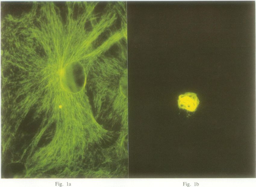
Fig. 1 Identification of IgM-antivimentin antibody (AVA) by indirect immunofluorescence. (a) Fluorescence pattern due
to a systemic lupus erythematosus serum on IMR-33 cell line. (b) The coiling of A VA around the nucleus after treatment
with colchicine.
AVA INHIBITION BY CARDIOLIPIN bromide (CNBr) activated Sepharose 4B (Pharma-
Ethanol was evaporated from the cardiolipin stock cia, Upsala, Sweden) by means of pOly-L-lysine at
solution (Sigma Chemical Co) under a nitrogen 20°C by the method of Kubota et al. 7
stream, and the dried phospholipid was then dis- Sepharose human Cohn fraction II (CFII) and
solved in PBS-Tween to a concentration of 800 Sepharose bovine serum albumin (BSA) were pre-
[ig/ml under sonication. Three AVA positive and pared by coupling CFII and BSA (Sigma Chemical
ACA positive sera from patients with SLE and three Co) respectively, to a concentration of 5 mg
AVA-negative/ACA-positive sera from patients protein/ml Sepharose. Serum samples from five
with syphilis were diluted 1:250 in PBS-Tween. Ali- patients with SLE were diluted 1:2, passed through
quots were incubated in a 1:1 ratio with varying the ssDNA, CFII, or BSA column and reconcen-
amounts of the cardiolipin inhibitor (100, 200, 400, trated until the original sample volume was
and 800 gig) for one hour at 20°C and one hour at obtained. The effluents were diluted 1:500 and
4°C. Sera were then centrifuged at 3000 g for 15 tested for AVA.
minutes at 22°C, and supernatants were tested for
AVA by ELISA according to the method outlined OTHER SEROLOGICAL TESTS
above. ACA were determined by the ELISA technique
described by Harris et al.'8 Alkaline phosphatase
AVA ABSORPTION WITH SINGLE conjugated, goat antihuman immunoglobulins (IgG,
STRANDED DNA IgM, and IgA; Sigma Chemical Co) diluted 1:500
Salmon sperm DNA (Sigma Chemical Co) was were used. Fifty microlitres of 1 mg/ml p-
denatured by heating at 90°C for 10 mintues and fast nitrophenyl phosphate in diethanolamine buffer, pH
cooling to 0°C, and ssDNA was fixed to cyanogen 9-8, was added to each well. After 45 minutes theAnn Rheum Dis: first published as 10.1136/ard.47.9.708 on 1 September 1988. Downloaded from http://ard.bmj.com/ on January 18, 2021 by guest. Protected by
Antivimentin antibodies in SLE 711
reaction was stopped by adding 50 tl aliquots of 3 M a system of five orthogonal axes (one for each of the
sodium hydroxide to each well. Absorbance was variables). The percentage of the total variance (the
read at 405 nm. Standard positive sera were supplied total variance is the variance of the five variables
by Dr E N Harris, St Thomas's Hospital, London. multiplied by the number of individuals, i.e., 50) is
Anti-dsDNA antibodies were tested by a com- accounted for by each of the axes.
mercial ELISA kit purchased from Cordis Labor-
atories (Miami, Flor) and the results reported in Results
international units/ml (IU) traceable to the World Analysis of the 50 serum samples from patients with
Health Organisation antinuclear antibody serum. SLE and 63 from normal controls by indirect
To set up the ELISA test for anti-ssDNA anti- immunofluorescence assay (Table 1) showed that 19
bodies, dsDNA (Sigma Chemical Co) was dena- (38%) and 11 (17%), respectively, had significantly
tured as previously described, coated to discs raised titres of IgM-AVA whereas eight (16%) and
(Cordis Laboratories Inc), and processed as four (6%), respectively, had significantly raised
above.'9 titres of IgG-AVA. With the ELISA (Fig. 2), of the
STATISTICAL ANALYSIS * 2,021 !8
All tests were performed in triplicate and the data i 1,75'4
* 1,35:13
o
2,849
z .1,330
were averaged. In the five ELISA tests the upper a . 929
limit of normality was taken as two standard * 8711
*820 9
* 829
*800
deviations above mean normal control levels. The * 3
5 I . 750
sensitivity and the specificity of the tests for SLE 8 667
(i . 705S
were calculated using the formulas established by
Youden.2t1 Specificity is the number of subjects 5001
without presumed SLE with a negative assay divided
by the number of subjects without presumed SLE,
X
copyright.
i.e., the true negative rate. Sensitivity is the number I.
of patients with SLE with a positive assay divided by
I
the number of patients with SLE, i.e., the true I._
positive rate. * T
BX0''~~~0
The analysis of these data was carried out using *+ 250
the Statistical Package for the Biological Sciences on
the Person 1600 IBM compatible at the French
National Institute of Marine Biology computing
centre. The relation between autoantibodies and
certain clinical features was examined using x2 t T
analysis with Yates's correction for small numbers.
Correlations between the different autoantibodies
were established by Spearman's correlation coeffi- 0
cient. A stepwise principal component analysis was Controis Patients
performed. This method of analysis allows the Fig. 2 Optical densities obtained with antivimentin
evaluation of the statistical weight of each of the five antibody by an enzyme linked immunosorbent assay in
quantitative variables (ACA, IgM-AVA, IgG- serum samples from 50 patients with systemic lupus
AVA, anti-dsDNA, and anti-ssDNA antibodies) in erythematosus and from 63 normal controls.
Table 1 Prevalence of IgM- and IgG-antivimentin antibodies by indirect immunofluorescence and enzyme linked
immunosorbent assay in serum samples from 50 patients with systemic lupus erythematosus and from 63 normal controls
lip ELISA IgM-A VA IgG-A VA
No of No of No of No of
patients controls patients controls
+ + 13 1 6 0
+ 6 10 2 4
+ 5 1 26 3
-- 26 51 16 56
*IIF=indirect immunofluorescence: ELISA=enzyme linked immunosorbcnt aissay: AVA=antivimcntin antibody.Ann Rheum Dis: first published as 10.1136/ard.47.9.708 on 1 September 1988. Downloaded from http://ard.bmj.com/ on January 18, 2021 by guest. Protected by
712 Blaschek, Boehme, Jouquan, Simitzis, Fifas, Le Goff, Youinou
63 serum samples from normal controls, the range of Correlations were sought between IgM-AVA,
absorbance at 492 nm, representing the results for IgG-AVA. ACA, anti-dsDNA antibodies, and anti-
IgM-AVA, was 0-010-0(507 (mean (SD) 0-214 ssDNA antibodies (Table 3). Statistically significant
(0-113)), and the upper limit of normal was set at correlations were found between both isotypes of
0-440 optical density (OD) units (mean + 2SD). The AVA, between IgM-AVA and ACA/anti-ssDNA
range of absorbance at 405 nm, representing the antibodies, and between ACA and both anti-DNA
results for IgG-AVA, was 0-009{-375 (mean (SD) antibodies. Because this study was devoted to AVA
0 172 (0 087)), and the upper limit of normal was set we focused on the relation of IgM-AVA to ACA
at 0-346 OD units (mean + 2SD). Eighteen (36%) (r=0-52) and to anti-ssDNA antibodies (r=0-46).
of the 50 SLE sera contained raised levels of IgM- Twentv ACA positive sera from patients with
AVA and 32 (64%) raised levels of IgG-AVA.
Comparison of the indirect immunofluorescence
assay and ELISA methods of measuring AVA Table 4 Results of the stepwise principal component
(Table 1) showed that the latter was more specific analysis
and more sensitive than the former technique
(specificity: 97% v 83% for IgM-AVA, and 950O li Axis Variables associated % Of the total variance*
94% for IgG-AVA; sensitivity: 38% 1 38%O for IgM- accounted for bs
AVA, and 52% v 16% for IgG-AVA). Therefore, each of the axes
for convenience, only the ELISA results will be 1 IgM-AVA 48-551
referred to in the following part. 2 IgG-AVA 22-583
Table 2 shows the number of serum samples from 3 ACA 14-449
4 anti-dsDNA Ab 7-854
patients and controls in which increased activity 5 anti-ssDNA Ab 6-563
against vimentin, cardiolipin, dsDNA, and ssDNA
was detected. *Total variance=50 patients x 5 variables.
For abbreviations see Tables 1 and 2.
copyright.
Table 2 Results of various serological investigations in 50 1800-
patients with systemic lupus erythematous and in 63 normal
controls --0
--
0 ----
No /) p)oslitiSc'
Patietstz (ni=50) Controls (n=63) -0-
1350- - -0
AVA* 33 (66) 4 (6)
ACA* 27 (54) 1 (2)
Anti-dsDNA Ab' 44 (88) 2 (3) 0
Anti-ssDNA Ab" 43 (86) 3 (5)
C)
*AVA=antivimentin antibody; ACA=anticardiolipin antibody; x
anti-dsDNA Ab=anti-double-stranded DNA antibody; anti-ssDNA
Ab=anti-single-stranded DNA antibody.
.o N
0
Table 3 Correlation of antivimentin, anticardiolipin, and 1.50-
anti-DNA antibodies
ACA IgM-A VA IgG-A VA CLsDNA
IgM-AVA r =0-52
p0- 10 p>- 1 Fig. 3 Inhibition of IgM-antivimentin antibody by
ssDNA r =0-77 r =0-46 r= 0-12 r =0-25
p)-05 cardiolipin (CL). Systemic lupus erythematosus serum
inhibited by CL=-- or by bovine serum albumin
,
Correlations established using Spcarman's correlation coefficient. (BSA)-=- - Syphilis serum inhibited by CL or
-@.
For abbreviations see Tables I and 2. BSA=- *.Ann Rheum Dis: first published as 10.1136/ard.47.9.708 on 1 September 1988. Downloaded from http://ard.bmj.com/ on January 18, 2021 by guest. Protected by
Antivimentin antibodies in SLE 713
Before After syphilis were examined for the presence of IgM-
1,800 AVA and IgG-AVA. Three (15%) and two (10%),
respectively, were found to be positive.
-0 Inhibition experiments with cardiolipin liposomes
were carried out for three SLE and three syphilis
sera (Fig. 3), and the same sera were extensively
absorbed with ssDNA (Fig. 4). The binding was
readily inhibited by cardiolipin liposomes, and to a
lesser degree absorbed with ssDNA, whereas BSA
and human CFII were inefficient.
1,350 . Table 4 and Fig. 5 show the results of the stepwise
principal component analysis. It can be seen that
axis 1 (IgM-AVA) accounts for 48-6% and axis 2
(IgG-AVA) for 22*6% of the total variance (total
variance=the variance of five variables multiplied
by 50 patients). Thus axes 1 and 2 appear to be the
0 most important axes as together they account for
v-
C 71-2% of the total variance. This is correct when
considering the patients group as a whole, but is
'in 9001. incorrect when considering any given individual.
-0 Careful review of the charts of these 50 patients
0. with SLE (Table 5) showed that IgG-AVA were
0
significantly associated with arthralgia in SLE.
copyright.
Discussion
In the present study serum samples from patients
450[ with SLE were shown to react with cytoskeletal
proteins. There is a six year difference in the
average age of the patients with SLE and the normal
controls, and the range is much wider in the former
than in the latter group. There are no data on the
variability of these autoantibodies with age, how-
ever. Despite the close association in cells between
vimentin and tubulin we can exclude the latter
-L.
F - protein. Indeed, the pattern of indirect immuno-
fluorescence assay obtained in cells treated with
Fig. 4 Absorption of IgM-antivimentin antibody with
colchicine, an inhibitor of microtubule polymerisa-
single stranded DNA (ssDNA). Systemic lupus tion, indicates that the fibrous pattern is due to
erythematosus serum absorbed with ssDNA =0-*, or vimentin, and the molecular weight of the protein
by bovine serum albumin (BSA)=*- - -. Syphilis serum used to coat the wells in the ELISA test (Boehme
absorbed with ssDNA or BSA = * *. and Blaschek, manuscript in preparation) has been
Table 5 Clinical features associated with IgM and IgG antivimentin antibodies (A VA). The number of patients suffering
from a given clinical symptom is in parentheses on the left. Of these, some have IgM-A VA or IgG-A VA, or both
Clinical 1gM-A VA IgG-A VA
features Positive Negative yIf p Value Positive Negative x p Value
Arthralgia (n=40) 16 24 0-66 0-42 30 10 8-25 0-1
Renal disease (n=22) 6 16 1 41 0-24 15 7 0(35 0-55
CNS* discase (n=9) 5 4 0-93 0-34 7 2 0-32 0-57
Rash (n=44) 17 27 0-66 0-42 30 14 1-38 0(24
*CNS=central nervous system.
tx2 with Yates's correction for small numbers.Ann Rheum Dis: first published as 10.1136/ard.47.9.708 on 1 September 1988. Downloaded from http://ard.bmj.com/ on January 18, 2021 by guest. Protected by
714 Blaschek, Boehme, Jouquan, Simitzis, Fifas, Le Goff, Youinou
/gG-AVA
M-AVA
.
0 0 .
0 0
.
. .0 0
S 0 .
0 0
0
Axis 1 0 a a
0
a o v . 0
(48-6% of the variance) 0
0
0 ACA
. .
0
0
0
0 0 0 Anti-ss
DNA Ab
.
Anti-ds
DNA Ab
copyright.
Axis 2
(22*6% of the variance)
Fig. 5 Stepwise principal component analysis. Distribution of 50 patients with systemic lupus erythematosus according to
axis 1 (IgM-A VA) and axis 2 (IgG-A VA). This combination of axes accounts for 48-6+22-6=712% of the total variance
(see text).
shown to be 54 000 by sodium dodecyl sulphate- We showed a significant association between AVA,
polyacrylamide gel electrophoresis.22 Detection of ACA, and anti-DNA antibodies, especially between
AVA with class specific antisera to human IgG or IgM-AVA, on the one hand, and ACA and anti-
IgM suggests that AVA are composed of both ssDNA antibodies, on the other. Further, the
classes of immunoglobulins in the sera from patients stepwise principal component analysis showed that
with SLE. An effect of detergent in PBS-Tween on IgM-AVA and IgG-AVA accounted for 71% of the
the cardiolipin liposome is possible, but as a differ- total variance (variance of 50 patients x 5 para-
ence was shown between SLE and syphilis, this is meters). Although the statistical correlations do not
probably not significant. prove that a given antibody may necessarily bind to
Osung et al found that the prevalence of AVA was two different antigens, they make it more likely.
10% in sera from patients with SLE,23 whereas Prompted by this observation, we sought AVA in 20
Alcover et al,5 Kurki et al,6 and Senecal et a!7 ACA positive sera from patients with syphilis, but
reported that 38-53% of SLE sera contained AVA. none of them reacted with cytoskeletal proteins.
Our immunofluorescence results agree with these IgM-AVA from patients with SLE were inhibited by
findings. In an attempt to resolve the discrepancies cardiolipin and absorbed with ssDNA.
between reports we performed further experiments These results are not surprising, given the pre-
with an ELISA, a technique claimed to be more viously described cross reactions, 22 Murine27 and
sensitive than indirect immunfluorescence assay. human28 monoclonal anti-DNA antibodies can bind
The prevalence of IgM-AVA was 36%, and that of to cardiolipin, and human monoclonal anti-DNA
IgG-AVA 64%, in SLE sera. antibodies from patients with SLE" and from
Statistical analysis of the serological results from normals30 can bind to cytoskeleton proteins. Any
50 patients with SLE showed correlations between relation between ACA and AVA has never been
AVA and other non-organ-specific autoantibodies. unequivocally shown. Several indirect argumentsAnn Rheum Dis: first published as 10.1136/ard.47.9.708 on 1 September 1988. Downloaded from http://ard.bmj.com/ on January 18, 2021 by guest. Protected by
Antivimentin antibodies in SLE 715
may be put forward. For example, patients with Avila J. Antibodies to vimentin intermediate filaments in sera
from patients with systemic lupus erythematosus. Arthritis
Behqet's disease have been shown to be capable of Rheum 1984; 27: 922-8.
making antibodies to cardiolipin31 and antibodies to 6 Kurki P, Helve T, Virtanen 1. Antibodies to cytoplasmic inter-
the cytoskeleton.32 ACA3' and AVA97 23 exist in mediate filaments in rheumatic diseases. J Rheumatol 1983,, 10:
normal sera. We showed hardly any AVA positive 558-62.
serum in syphilis despite the presence of ACA in 7 Senecal J L, Oliver J M, Rothfield N. Anticytoskeletal auto-
antibodies in the connective tissue diseases. Arthritis Rheumn
this disease. This may be due to differences in 1985; 28: 889-98.
epitope specificity. ACA from patients with SLE are 8 Blaschek M A. Nicht-organspezifische antizytoplasmatische
more likely to bind to phosphatidylserine than to Autoantikorper. Immunoserologischer Nachweis und diag-
phosphatidylethanolamine, and vice versa with nostische Relevanz. Wien Med Wochenschr 1987; 13: 303-9.
9 Youinou P, Le Goff P, Miossec P, Moineau M P, Ferec C.
syphilis serum.33 The binding of syphilitic antibodies Untersuchungen zur Beziehung zwischen anti-perinuklearen
to cardiolipin was readily inhibited by cardiolipin Faktoren, Anti-Keratin-Antikorpern und dem agglutinierenden
liposomes, whereas this pattern of reactivity was und nicht-agglutinierenden Rheumafaktor bei der chronischen
noted in only one of four sera from patients with Polyarthritis. Z Rheumatol 1983; 42: 36-9.
10 Youinou P, Le Goff P, Colaco C B, et al. Antikeratin anti-
acute infection by Vaarala et al.34 bodies in serum and synovial fluid show specificity for rheuma-
The successful inhibition of AVA from SLE with toid arthritis in a study of connective tissue diseases. Ann
cardiolipin may mean that the epitope shared by Rheum Dis 1985; 44: 450-4.
vimentin and phospholipids is related to 11 Cohen A S. Reynolds W E, Franklin E C, et al. Preliminary
criteria for the classification of systemic lupus erythematosus.
phosphatidylserine.27 Some of the ACA, however, Bull Rheum Dis 1971; 21: 643-8.
may react with the cytoskeletal epitopes that are 12 Tan E M, Cohen A S, Fries J F, et al. The 1982 revised criteria
shared with vimentin.35 for the classification of systemic lupus erythematosus. Arthritis
Analysis of the clinical details showed an associ- Rheum 1982; 25: 1271-7.
13 Johnson G D, Davidson R S, McNamee K C. Russell G,
ation between AVA positivity and arthralgia. One Goodwin D, Holborow E J. Fading of immunofluorescence
explanation of this finding is that proliferating during microscopy: a study of the phenomenon and its remedy.
synovial linin6g cells contain vimentin as a prominent J Immunol Methods 1982; 55: 231-42.
copyright.
constituent. 3Thus a vicious circle may be set off 14 Steinert P, Zackroff R, Aynardi-Whitman M, Goldman R D.
Isolation and characterization of intermediate filaments.
once joints are involved. Methods Cell Biol 1982; 24: 399-418.
The cross reaction between vimentin, cardiolipin, 15 Engvall E, Perlmann P. Enzyme-linked immunosorbent assay,
and DNA raises the important question of polyre- ELISA. III. Quantitation of specific antibodies by enzyme-
active autoantibodies. The likelihood that DNA is labelled anti-immunoglobulin in antigen-coated tubes.
J Immunol 1972; 109: 129-35.
the immunogen in SLE is extremely question- 16 Voller A, Bidwell D, Huldt G, Engvall E. A microplate method
able 226 37 of enzyme-linked immunosorbent assay and its application to
malaria. Bull WHO 1974; 51: 209-11.
17 Kubota T, Akatsuka T, Kanai Y. DNA affinity column
We are indebted to Professor E J Holborow (London, UK) and to chromatography: application in the isolation of distinct
Dr M J Fritzler (Calgary, Canada) whose advice enabled us to set antibody populations from SLE sera. Clin Exp Immunol 1985;
up this study. The technical expertise of Miss Nelly Jezequel and 62: 321-8.
Mrs Simone Forest has been greatly appreciated. We are most 18 Harris E N. Gharavi A D, Patel S P, Hughes G R V. Evaluation
grateful to Specia Pharmaceutical Company (Paris) for financial of the anti-cardiolipin antibody test: report of an international
support. This study was supported by a grant from Le Ministere de workshop held 4 April 1986. Clin Exp Immunol 1987; 68:
la Recherche et de la Technologie, and by a grant from Le Comite 215-22.
Departemental de la Ligue Nationale contre le Cancer. MAB was 19 Klotz J L, Minami R M, Teplitz R L. An enzyme-linked
supported by a doctoral fellowship from the Studienstiftung des immunosorbent assay for antibodies to native and denatured
deutschen Volkes. DNA. J Immunol Methods 1979; 29: 155-65
20 Youden W J. Index for rating diagnostic test. Cancer 1950; 3:
References 32-5.
21 Osborn M, Franke W W, Weber K. Direct demonstration of
I Lazarides E. Intermediate filaments as mechanical integrators the presence of two immunologically distinct intermediate-sized
of cellular space. Nature 1980; 283: 249-56. filament systems in the same cell by double immunofluores-
2 Bretherton L, Toh B H, Jack I. IgM autoantibody to cence microscopy: vimentin and cytokeratin fibers in cultured
intermediate filaments in Mycoplasma pneumoniae infections. epithelial cells. Exp Cell Res 1980; 125: 37-46.
Clin Immunol Immunopathol 1981; 18: 425-30. 22 Lazarides E. Intermediate filaments. A chemically hetero-
3 Boehme M W J, Evans D A, Miles M A, Holborow E J. geneous developmentally regulated class of proteins. Ann Rev
Occurrence of autoantibodies to intermediate filament proteins Biochem 1982; 51: 219-50.
in human visceral leishmaniasis and their induction by ex- 23 Osung D A, Chandra M, Holborow E J. Antibody to
perimental polyclonal B-cell activation. Immunology 1986; 59: intermediate filaments of the cytoskeleton in rheumatoid
583-8. arthritis. Ann Rheum Dis 1982; 41: 69-73.
4 Kataaha P K, Mortazavi-Milani S M, Russell G, Holborow E J.
Anti-intermediate filament antibodies, antikeratin antibody, 24 Eilat D. Anti-DNA antibodies: problems in their study and
and antiperinuclear factor in rheumatoid arthritis and infectious interpretation. Clin Exp Immunol 1986; 65: 215-22.
mononucleosis. Ann Rheum Dis 1985; 44: 446-9. 25 Emlen W, Pisetoky D S, Taylor R P. Antibodies to DNA, a
5 Alcover A, Molano J, Renart J, Gil-Aguado A, Nieto A, perspective. Arthritis Rheum 1986; 29: 1417-26.Ann Rheum Dis: first published as 10.1136/ard.47.9.708 on 1 September 1988. Downloaded from http://ard.bmj.com/ on January 18, 2021 by guest. Protected by
716 Blaschek, Boehme, Jouquan, Simitzis, Fifas, Le Goff, Youinou
26 Isenberg D, Shoenfeld Y. The origin and significance of anti- 32 Akoglu T. Kozakoglu H. Akoglu E. Antibody to internmcdiatc
DNA antibodies. Itninutiol Today 1987; 8: 279-82. filaments of the cytoskelcton in patients with Behcct's discase.
27 Lafer M, Rauch J, Andrzejewski C Jr, et al. Polyspecific Cliti lhnrnutiol Immunopatiol 1986; 41: 427-32.
monoclonal lupus autoantibodies reactive with both polynuc- 33 Colaco C B, Male D K. Anti-phospholipid antibodics in syphilis
leotides and phospholipids. J E.xp Med 1981; 153: 897-909. and a thrombotic subset of SLE: distinct profiles of epitope
28 Shoenfeld Y, Rauch J. Massicotte H. et al. Polvspecificity of spccificity. Clini Exp lhtzinl,uol 1985; 59: 449-56.
monoclonal lupus autoantibodies produced by human-human 34 Vaatralat 0. Palosuo T. Kleemole M. Aho K. Anticardiolipin
hybridomas. N Engl J Med 1983; 308: 414-2(. responses in acute infections. Cliii lImzinunlol lhttnintiopatlol
29 Andre-Schwartz J, Katta S K. Shoenfeld Y. Isenberg D A. 1986; 41: 8-15.
Stollar B D, Schwartz R S. Binding of cytoskeletal proteins by 35 Pruss R M. Mirskv R. Raff M C. All classes of intermedilte
monoclonal anti-DNA lupus autoantibodies. Cliii Itninuniol filaments sharc a common antigcnic dcterminant defined bs a
Immunopathol 1984; 31: 261-71. monoclonal antibody. Cell 1981; 27: 419-28.
30 Cavins E. Komar R. Bell D A. Cvtoskeletal binding of 36 Osung 0 A. Chandra M. Holboross E J. Intermediatc filamcnts
monoclonal anti-DNA antibodics derived from tonsillar lym- in synovial lining cells in rheumatoid arthritis and other
phoid cells of a normal subject. Arthritis Rlheuirn 1986; 29: arthritides are of vimentin tvpe. Anntz Rlheumti Dis 1982; 41: 74-7.
1351-8. 37 Jacob L. Lety M A. Bach J F. Louvard D. Human systemic
31 Hull R G, Harris E N, Gharavi A E, et al. Anticardiolipin lupus crythcmatosus scra contain antibodics against cclI-surface
antibodies: occurrence in Behqet's syndrome. Annti Rlielutn Dis protein(s) that sharc(s) epitopc(s) ssith DNA. Proc Nad Acad
1984; 43: 746-8. Sci USA 1986; 83: 697(0-4.
copyright.You can also read