Absence of pontine perforators in vertebrobasilar dolichoectasia on ultrahigh resolution cone- beam computed tomography
←
→
Page content transcription
If your browser does not render page correctly, please read the page content below
J NeuroIntervent Surg: first published as 10.1136/neurintsurg-2020-016818 on 21 October 2020. Downloaded from http://jnis.bmj.com/ on October 14, 2021 by guest. Protected by copyright.
Neuroimaging
Original research
Absence of pontine perforators in vertebrobasilar
dolichoectasia on ultra-high resolution cone-beam
computed tomography
Tomas Dobrocky ,1 Eike I Piechowiak ,1 Johannes Goldberg,2
Enrique Barvulsky Aleman,1 Patrick Nicholson,3 Jeremy Lynch,3 David Bervini,2
Johannes Kaesmacher ,1 Ronit Agid,3 Timo Krings,3 Andreas Raabe,2 Jan Gralla,1
Vitor M Pereira,3 Pasquale Mordasini1
1
Department of Diagnostic and ABSTRACT diagnosed incidentally. The natural course of the
Interventional Neuroradiology, Background Vertebrobasilar dolichoectasia (VBDE) is disease is generally poor, with mortality rates of
Inselspital, University of Bern,
Bern, Switzerland a rare type of non-saccular intracranial aneurysm, with 40% after 7 years follow-up reported in a large
2
Department of Neurosurgery, poor natural history and limited effective treatment meta-analysis including 440 patients.1 In particular,
Inselspital, University of Bern, options. Visualizing neurovascular microanatomy in patients presenting with compressive symptoms
Bern, Switzerland patients with VBDE has not been previously reported, tend to have an exceedingly poor outcome, with
3
Division of Neuroradiology,
but may yield insight into the pathology, and provide 7/13 (54%) patients dying due to severe compres-
Department of Medical Imaging
and Division of Neurosurgery, important information for treatment planning. sion, and 8/13 (62%) experiencing worsening of the
Department of Surgery, Objective To carry out a retrospective analysis of mass effect during follow-up.2
University Health Network ultra-high resolution cone-beam computed tomography Despite technical advances in the endovascular
- Toronto Western Hospital, (UHR-CBCT) in patients with fusiform basilar aneurysms,
Toronto, Ontario, Canada approach the treatment of VBDE remains a chal-
visualizing neurovascular microanatomy of the lenge, with higher complications rates than with
posterior circulation with a special focus on the pontine saccular aneurysms. Endovascular reconstruction
Correspondence to
Dr Tomas Dobrocky, Department perforators. of the fusiform vessel segment with stent-assisted
of diagnostic and interventional Methods UHR-CBCT was performed in seven patients coiling, stent placement alone, flow diverters,
Neuroradiology, Inselspital (mean age 59 years; two female) with a VBDE, and in 14 and flow reversal with vertebral artery occlusion
Universitatsspital Bern, Bern control patients with unrelated conditions.
3010, Switzerland; tomas. have been reported in small case series and case
dobrocky@insel.ch Results The mean maximum diameter of the fusiform reports.3–5 Ischemic complications due to throm-
vessel segment was 28 mm (range 19–36 mm), and bosis—namely, perforating arteries supplying the
Received 31 August 2020 the mean length of the segment was 39 mm (range brainstem, remain the major concern irrespective
Revised 23 September 2020 15–50 mm). In all patients with VBDE, UHR-CBCT
Accepted 26 September 2020 of the technique, but their visualization in vivo
demonstrated an absence of perforating arteries in the
Published Online First remains a challenge and has not been previously
21 October 2020 fusiform arterial segment and a mean of 3.7 perforators
reported in VBDE.6
arising from the unaffected vessel segment. The network
Recently, a new generation ultra-high resolution
of interconnected superficial circumferential pontine
cone-beam computed tomography (UHR- CBCT)
arteries (brainstem vasocorona) were draping around the
has been introduced, enabling visualization of the
aneurysm sac. In controls, a mean of 3.6, 2.5, and 1.2
neurovascular microanatomy in vivo. The main
perforators were demonstrated arising from the distal,
mid-, and proximal basilar artery, respectively. goal of our study was the retrospective analysis of
Conclusions The absence of pontine perforators in the UHR-CBCT in patients with fusiform basilar aneu-
fusiform vessel segment of VBDE is counterbalanced by rysms, visualizing neurovascular microanatomy of
recruitment of collateral flow from pontine perforators the posterior circulation, with a special focus on the
arising from the unaffected segment of the basilar artery, pontine perforators.
as well as collaterals arising from the anterior inferior
cerebellar artery/posterior inferior cerebellar artery and
MATERIAL AND METHODS
superior cerebellar artery. These alternative routes supply
Institutional review board approval was obtained
the superficial brainstem arteries (brainstem vasocorona)
for this dual-center study. Informed consent was
and sustain brainstem viability. Our findings might have
waived owing to the retrospective nature of the
implications for further treatment planning.
© Author(s) (or their study.
employer(s)) 2021. Re-use All patients with a VBDE, in whom a UHR-
permitted under CC BY-NC. No
commercial re-use. See rights CBCT of the posterior circulation between March
and permissions. Published INTRODUCTION 2019 and May 2020 was visualized using either the
by BMJ. Vertebrobasilar dolichoectasia (VBDE) is a rare type new angiographic system (Artis icono; Siemens,
To cite: Dobrocky T, of intracranial, non-saccular aneurysm, which may Erlangen, Germany) or between February 2018
Piechowiak EI, Goldberg J, become clinically apparent due to compression of and March 2020 using an established angiographic
et al. J NeuroIntervent Surg the brainstem and cranial nerves, hemorrhagic or system (AlluraClarity, Philips Healthcare, Best,
2021;13:580–584. ischemic symptoms, and, less frequently, may be Netherlands), were included.
Dobrocky T, et al. J NeuroIntervent Surg 2021;13:580–584. doi:10.1136/neurintsurg-2020-016818 1 of 6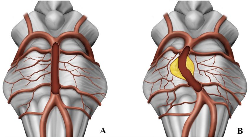
J NeuroIntervent Surg: first published as 10.1136/neurintsurg-2020-016818 on 21 October 2020. Downloaded from http://jnis.bmj.com/ on October 14, 2021 by guest. Protected by copyright.
Neuroimaging
VBDE was defined using the definitions proposed by Flem- patient over 200 degrees to create a circumferential run of the
ming et al and had to meet at least one of the following imaging region of interest. In each run, 500 images with a resolution of
definitions: (1) fusiform aneurysmal dilatation: 1.5×normal 0.14 mm were obtained. The raw data were then transferred to
diameter without a definable neck involving a portion of an a dedicated workstation.
arterial segment (either vertebral or basilar) with any degree
of tortuosity, (2) dolichoectasia: uniform aneurysmal dilatation 3D VasoCT for Philips AlluraClarity
of the artery >1.5× normal involving either the entire basilar A slightly different protocol was used with the Philips angio.
or vertebral artery or both with any degree of tortuosity, or To create a circumferential run the C-arm rotates around the
(3) transitional: uniform aneurysmal dilatation of the artery patient over 207 degrees in 20.7 s, acquiring 622 images of the
>1.5× normal involving the vertebral or basilar artery, or both region of interest (22 cm field of view). The injector was coupled
with a superimposed dilatation of a portion of the involved to the acquisition system, with a fixed 3 s delay. For good vessel
arterial segment.7 Acute dissecting aneurysms in two young opacification during the run (20.7 scan +3 delay) a mixture of
adults presenting with an extensive subarachnoid hemorrhage non-ionic contrast agent and sterile saline solution (1:3) was
and a normal caliber basilar artery were excluded. Patients in injected with a flow of 3 mL/s (total volume 75 mL).
whom a UHR-CBCT was performed due to other indications,
mainly angiography performed to evaluate patients with non-
Data reconstruction and analysis
aneurysmal or CT angiography (CTA)- negative intracranial
Transversal flat-detector CT reconstructions (arterial mode)
hemorrhage, served as controls.
were reconstructed at the discretion of the operator. Multiplanar
reconstructions with a slice thickness of 0.5 mm and maximum
Angiography intensity projections with section thicknesses between 5 and 10
Selective intra-arterial digital subtraction angiography was mm as well as volume rendering reconstructions were sent to
performed under local anesthesia on a biplane, high- resolution the local Picture Archiving and Communicating System (PACS)
angiographic system (Artis icono or AlluraClarity) using Iopa- (R11.4.1, 2009; Philips, Best, Netherlands; Sectra, Linkoping,
miro 300 (Iopamidol, Bracco, Switzerland) for vessel opacifica- Sweden; or Coral, Toronto, Canada) and used for perforator
tion. The prototype of the Artis icono system was installed in analysis.
one of the contributing institutions in March 2019 and provides The clinical information was retrieved from locally main-
improved and higher spatial resolution using the as40HDR tained databases, and all imaging findings were reviewed by a
flat-panel detector with a 49 cm diagonal entrance plane and board-certified neuroradiologist with 9 years of experience (TD)
an active imaging size of 398×293 and active matrix size of and neuroradiology fellow with 2 years of experience (EBA).
2586×1904.
On both systems, digital subtraction angiography images were
obtained using a standard program with 2.5 frames per second in
RESULTS
the arterial phase, a focal spot size of 0.3 mm, and edge enhance- Vertebrobasilar dolichoectasia group
ment reconstruction algorithm. A flat-detector zoom format of In total, UHR-CBCT was available in seven patients with VBDE.
32×32 cm in each plane was used. Their mean age was 59.0 years (±6.7), and two patients were
After puncture of the common femoral artery a 5 French (Fr) female. All patients with VBDE demonstrated severe brain-
sheath (Terumo, Terumo Medical, Tokyo, Japan) was inserted to stem compression with a mean minimal AP distance of 8 mm
secure the access. For angiography of the supra-aortic vessels a measured on axial images (range 3–12 mm; figures 1A,B and
5 Fr diagnostic catheter was advanced over a 0.035" hydrophilic 2A,B). Surrounding T2 hyperintense brainstem edema was noted
guide wire (Terumo). In all patients a four- vessel angiogram on MRI in five patients. The mean maximum diameter of the
was obtained to evaluate the intracranial circulation including fusiform vessel segment was 28 mm (range 19–36 mm), and
anteroposterior (AP), lateral, and spin angiograms. the mean length of the segment was 39 mm (range 15–50 mm;
table 1). The basilar artery demonstrated an elongated course
in all patients.Intramural hematoma was noted in all but one
3D micro DynaCT technique for Artis icono
patient (figure 2C), In total, one patient demonstrated a small
The diagnostic catheter was placed at the origin of the dominant
hyperintense lesion in the brainstem on diffusion weighted
vertebral artery. The catheter was connected to double- head
imaging, indicating acute ischemia. One patient presented infra-
contrast agent injector (Accutron HP- D, Medtron AG, Saar-
tentorial superficial siderosis at the 1- year follow-up, indicative
brücken, Germany) using a designated Y-pressure line with two
of interval hemorrhage.
check valves (Medtron AG) after having purged all air bubbles
In all patients with VBDE, UHR- CBCT demonstrated an
with sterile saline solution. In intubated patients, the contrast
absence of perforating arteries in the fusiform arterial segment
injection was performed under apnea. Awake patients were
and a mean of 3.7 perforators arising from the unaffected vessel
instructed to hold their breath immediately prior to initiation of
segment. Owing to the mass effect of the aneurysm sac the super-
the rotational run to reduce artifacts.
ficial brainstem arteries were displaced and were draping around
During image acquisition a mixture of non-ionic contrast agent
the aneurysm.
(15 mL) and sterile saline solution (30 mL) was injected with a
In one patient the anterior inferior cerebellar artery (AICA)
flow of 3 mL/s (total volume 45 mL, contrast agent concentra-
was noted to arise from the fusiform vessel segment, in the
tion 33%, injection time 15 s). The amount of contrast agent was
remaining patients the AICA territory was supplied by a domi-
adapted to allow adequate vessel opacification during the entire
nant posterior inferior cerebellar artery (PICA-AICA variant).
run and prevent any beam hardening artifacts. Continuous fluo-
roscopic monitoring at a rate of 1 frame/s was used for contrast
appearance in the basilar artery (BA) to trigger the rotational Control group
run. On appropriate opacification a dedicated UHR-CBCT run In total, 14 patients with a mean age of 54.5 years (±7.9) in
with a scan time of 14 s, focusing on the posterior circulation, whom a UHR-CBCT was performed due to unrelated indications
was performed. During the run the C-arm rotates around the were included for reference (n=11 CTA-negative intracranial
2 of 6 Dobrocky T, et al. J NeuroIntervent Surg 2021;13:580–584. doi:10.1136/neurintsurg-2020-016818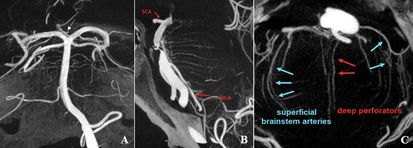
J NeuroIntervent Surg: first published as 10.1136/neurintsurg-2020-016818 on 21 October 2020. Downloaded from http://jnis.bmj.com/ on October 14, 2021 by guest. Protected by copyright.
Neuroimaging
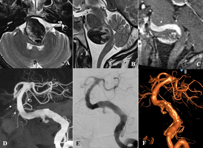
Figure 1 (A) Axial T2-weighted image demonstrating a large
vertebrobasilar dolichoectasia with severe mass effect leading to
brainstem compression and displacement. (B) Sagittal T1-weighted
enhanced image demonstrating the longitudinal extension of the
aneurysm and extensive intramural hematoma (black arrows). (C-E)
Ultra-high resolution cone-beam computed tomography with maximal
Figure 2 (A, B) Axial and mid-sagittal T2-weighted image
intensity reconstructions demonstrating an irregular margin of the
demonstrating a large vertebrobasilar dolichoectasia with severe
fusiform vessel segment in the mid- and lower basilar artery with
compression of the brainstem and edema. (C) Sagittal T1-weighted
absence of pontine perforators originating from the fusiform vessel
enhanced image demonstrating the opacified vessel lumen in the
segment. Multiple perforators arising from the basilar artery distal to
cranial part of the aneurysm (white arrows), and extensive intramural
the aneurysmal segment may be appreciated (white arrows). Note
hematoma in the inferior portion (black arrow). (D) Ultra-high resolution
the circumferential pontine arteries (black arrows) which have been
cone-beam computed tomography (UHR-CBCT) with coronal maximal
displaced due to the mass effect, and seem to be draping around
intensity reconstructions demonstrating a fusiform aneurysm with
the aneurysm; their patency is maintained by collateral flow from
incorporation of a 40 mm segment of the lower and mid-basilar
perforators arising from the unaffected segment of the basilar artery, as
trunk. Note the irregular entry zone of contrast (black arrow) into the
well as the anterior inferior cerebellar artery/posterior inferior cerebellar
thrombosed portion of the aneurysm. There is no evidence of pontine
artery and superior cerebellar artery.
perforators within the fusiform vessel segment. Multiple perforators
arising from the distal basilar artery may be appreciated (white arrows).
(E) Digital subtraction angiography run in posterior-anterior projection
hemorrhage, n=1 symptomatic basilar artery stenosis; n=1 and (F) three-dimensional rotational angiography for comparison show
basilar tip aneurysm, n=1 hemorrhagic dural arteriovenous the findings but with significantly less detail and clarity than on UHR-
fistula at the craniocervical junction). In controls, a mean of 3.6, CBCT.
2.5, and 1.2 perforators were demonstrated arising from the
distal, mid, and proximal basilar artery, respectively (figure 3). In
most of them multiple circumferential pontine arteries coursing In general, pontine perforators are poorly appreciated on stan-
along the surface of the pons and small interconnecting branches dard cross-sectional imaging. including CTA, and conventional
(‘brainstem vasocorona’) were appreciated. 1.5 and 3 Tesla MRA techniques. Recently, compelling results
on ultra-high field 7 Tesla MRI have been published, reporting
DISCUSSION a mean number of 7.14±2.79 perforator arteries arising from
The improved spatial resolution available with new-generation the basilar artery.9 However, ultra-high field MRI is not readily
angiographic systems incorporating cone-beam CT technology available in clinical routine.
provides valuable insight into the neurovascular microanatomy Alternatively, flat-detector CT, first described by Kyriakou et al
of the posterior fossa. It demonstrates a complete absence of in 2007,10 has been used to depict in vivo vascular microanatomy
pontine perforators in the fusiform vessel segment of patients in various intracranial pathologies.11 12 The continuing evolution
with VBDE. Their absence is counterbalanced by recruitment of of flat-detector technology has enabled the current rotational
collateral flow from pontine perforators arising from the unaf- angiography systems to provide high acquisition speed coupled
fected segment of the BA, as well as collaterals arising from the with excellent resolution isotropic datasets, and good recon-
AICA/PICA and superior cerebellar artery (SCA). These alterna- struction quality. Lescher et al reported that three-dimensional
tive routes supply the superficial brainstem arteries and sustain rotation angiography and flat-detector CT reconstructions were
brainstem viability. superior to 2D digital subtraction angiography in demonstrating
Perforating arteries arising from the BA play a crucial role in anatomic patterns of circumferential arteries and direct pontine
the supply of the brainstem, and their compromise may lead to perforators. Furthermore, the authors reported that no zone of
ischemia affecting respiratory and cardiac function, as well as the basilar artery is free from important side branches, which is in
relay of major motor and sensory functions. Clinicopathologic line with the findings in our control population.13 On the other
studies in patients with VBDE have demonstrated circumferen- hand, according to our results the absence of perforators arising
tial fragmentation of the internal elastic lamina, intimal hyper- from the fusiform vessel segment seems to be a characteristic
plasia, and angiogenesis of vasa vasorum, which may result in feature in patients with VBDE.
repetitive intramural hemorrhages and thrombus formation.8 Lasjaunias et al regarded the arterial anatomy of the posterior
According to our results we believe that these destructive vessel fossa as a transitional pattern between the ‘simple’ arrangement
wall changes in the fusiform segment progressively lead to oblit- of the spinal cord and the more ‘complex’ neocortical pattern.
eration of perforating arteries arising in this vessel segment, and During the embryological development, the BA below the trigem-
when occurring gradually are balanced by collateral pathways. inal point is formed by the fusion of the paired longitudinal
Dobrocky T, et al. J NeuroIntervent Surg 2021;13:580–584. doi:10.1136/neurintsurg-2020-016818 3 of 6J NeuroIntervent Surg: first published as 10.1136/neurintsurg-2020-016818 on 21 October 2020. Downloaded from http://jnis.bmj.com/ on October 14, 2021 by guest. Protected by copyright.
Neuroimaging
Table 1 Demographic information on patients with vertebrobasilar dolichoectasia
VBDE max. diameter VBDE length Intramural hematoma Brainstem compression Min. brainstem width Oedema Hemorrhage Siderosis Ischemia Ectasia ICA
1 36 45 1 1 12 1 1 1 0 1
2 34 40 1 1 3 1 0 0 0 1
3 30 50 1 1 5 1 0 0 0 0
4 33 38 1 1 10 1 0 0 0 0
5 21 43 1 1 15 1 0 0 1 1
6 19 15 0 1 11 0 0 0 0 0
7 26 45 1 1 6 0 0 0 0 0
All measurements are provided in millimeters (mm). 1=Yes, 0=No.
ICA, internal carotid artery; VBDE, vertebrobasilar dolichoectasia.
neural arteries—similar to the formation of the anterior spinal As reported by Lindsberg in patients with acute basilar artery
artery, whereas the BA cranial to the trigeminal point is formed occlusion, the PICA is a strong collateral to the AICA and SCA
by the fusion of the paired caudal divisions of the embryologic and thus to the brainstem perforating arterioles which may
internal carotid artery (ICA). Thus, the junction between the two maintain brainstem vitality for significant periods if the clot does
systems (simplified spinal system and complex cortical system) is not gradually extend to block the perforating arteries.17 Likewise
near the trigeminal remnant site on the BA. Being thus a transi- in patients with VBDE the gradual obliteration of perforating
tional artery between the two systems, there is a certain degree arteries at the level of the fusiform vessel segment is probably
of resemblance to the ASA system, and the pontine perforators compensated by a considerable network of anastomoses (‘brain-
may be regarded as counterparts of the coronary and sulcocom- stem vosocorona’), recruiting collaterals arising in the unaffected
missural arteries.14 The BA, vertebrobasilar confluence, and the vessel segments of the basilar artery as well as AICA/PICA and
PICA give rise to a considerable number of perforators supplying SCA (figure 4). In patients in whom the AICA is incorporated
the brainstem and the pontomedullary junction. Conversely, within the fusiform segment, based on our data the thrombotic
there is a paucity of perforators arising from the distal vertebral risks after flow diverter deployment remain unclear.
artery between its entry through the dura mater and the PICA In a large meta- analysis looking at the natural history of
origin. Distal vertebral perforators are reported in only 4% of VBDE, including 827 patients with 5093 patient- years, the
individuals in an anatomical study by Mercier et al.15 authors reported an overall annual mortality rate of 13%/year
Detailed anatomical studies have demonstrated a considerable (95% CI 8% to 19%). Patients with fusiform aneurysms had a
amount of artery- to-
artery anastomoses between the superfi- higher mortality rate than those with dolichoectatic aneurysmal
cial brainstem arteries (‘brainstem vosocorona’),16 and may be dilatation (12 vs 8%, p=0.11). The overall growth rate was 6%/
considered the equivalent of the rich arterial network coursing on year (95% CI 4% to 13%), and the overall rupture rate was
the surface of the spinal cord known as the arterial vasocorona. 3%/year (95% CI 1% to 5%).18 As stated previously, patients
presenting with compressive symptoms, in particular, tend to
show exceedingly poor outcome. Chen et al reported that a BA
diameter ≥5.3 mm was independently associated with increased
Figure 3 (A) A patient presenting with thunderclap headache and
perimesencephalic subarachnoid hemorrhage on CT (not shown).
Ultra-high resolution cone-beam computed tomography (UHR-CBCT)
acquired during injection of the left vertebral artery, Maximum
intensity projection images in the coronal plane demonstrating multiple
perforating arteries originating from the mid- and distal basilar artery.
Note: artery of Percheron (arrow) originating as a solitary arterial trunk
from the P1 segment on the left supplying the paramedian thalami Figure 4 (A) Illustration of a normal basilar artery with several
and parts of the rostral midbrain bilaterally. (B) UHR-CBCT in the circumferential pontine arteries coursing along the surface of the
sagittal projection in a patient presenting with acute pontine ischemia pons and small interconnecting branches ('brainstem vasocorona';
(not shown) due to an underlying high-grade mid-basilar stenosis. arrowheads). (B) A large fusiform basilar artery aneurysm with
Multiple deep perforators and several circumferential pontine arteries circumferential intramural hematoma. Note the absence of pontine
coursing along the surface of the pons and small interconnecting perforators within the fusiform vessel segment. Obliteration of
branches ('brainstem vasocorona') may be appreciated. (C) UHR-CBCT perforating arteries at the level of the fusiform vessel segment is
of the same patient in the axial plane demonstrating the course of compensated by the brainstem vosocorona, recruiting collaterals arising
circumferential pontine arteries running along the surface of the pons in the unaffected vessel segments of the basilar artery as well as from
and deep perforating branches running in the midline. AICA, anterior the anterior internal cerebral artery/posterio internal cerebral artery and
inferior cerebellar artery; SCA, superior cerebellar artery. superior cerebellar artery.
4 of 6 Dobrocky T, et al. J NeuroIntervent Surg 2021;13:580–584. doi:10.1136/neurintsurg-2020-016818J NeuroIntervent Surg: first published as 10.1136/neurintsurg-2020-016818 on 21 October 2020. Downloaded from http://jnis.bmj.com/ on October 14, 2021 by guest. Protected by copyright.
Neuroimaging
stroke recurrence in patients with VBDE on multivariate anal- Provenance and peer review Not commissioned; externally peer reviewed.
ysis (HR=4.744; 95% CI, 1.718 to 13.097; p=0.003).19 This Data availability statement Data are available upon reasonable request. Raw
is mainly thought to relate to compromise of basilar perforating data of all patients included in this study can be made available upon request to the
arteries, either due to thrombosis or due to their compression corresponding author and after clearance by the local ethics committee.
or stretching. Open access This is an open access article distributed in accordance with the
Initially the anatomic aspects and the large number of perfora- Creative Commons Attribution Non Commercial (CC BY-NC 4.0) license, which
permits others to distribute, remix, adapt, build upon this work non-commercially,
tors were believed to preclude neurointerventional treatment of
and license their derivative works on different terms, provided the original work is
VBDE. However, promising results of endovascular reconstruc- properly cited, appropriate credit is given, any changes made indicated, and the use
tion of the fusiform segment with flow diverters (FDs) and stent- is non-commercial. See: http://creativecommons.org/licenses/by-nc/4.0/.
assisted coiling have been reported in multiple case reports and
case series.20–24 Fiorella et al reported treatment of a giant mid- ORCID iDs
Tomas Dobrocky http://orcid.org/0000-0002-6167-3343
basilar trunk aneurysm with seven serially placed, telescoping Eike I Piechowiak http://orcid.org/0000-0001-5609-0998
FDs with complete occlusion.25 Nevertheless, in a recent meta- Johannes Kaesmacher http://orcid.org/0000-0002-9177-2289
analysis the overall complication rate was higher for FDs than
with stent-assisted coiling (18% vs 6%), including higher chance
for stroke (13% vs 5%, p=0.04).26 The safety and efficacy of REFERENCES
endovascular treatment of patients with VBDE is dependent on 1 Shapiro M, Becske T, Riina HA, et al. Non-saccular vertebrobasilar aneurysms and
dolichoectasia: a systematic literature review. J Neurointerv Surg 2014;6:389–93.
the size of the lesion, the patient’s overall clinical condition, and
2 Wang J, Jia L, Yang X, et al. Outcomes in symptomatic patients with vertebrobasilar
the treatment modality used. dolichoectasia following endovascular treatment. Front Neurol 2019;10:610.
Further development of UHB-CBCT has enabled visualization 3 Wu X, Xu Y, Hong B, et al. Endovascular reconstruction for treatment of
of the neurovascular microanatomy in vivo unprecedented by vertebrobasilar dolichoectasia: long-term outcomes. AJNR Am J Neuroradiol
previous techniques. It thus leads to a better understanding of 2013;34:583–8.
4 Tan LA, Moftakhar R, Lopes DK. Treatment of a ruptured vertebrobasilar fusiform
the pathology and as such has important implications on therapy. aneurysm using pipeline embolization device. J Cerebrovasc Endovasc Neurosurg
Its implementation into clinical routine in selected pathologies 2013;15:30.
may be helpful. Nevertheless, VBDE continues to be a chal- 5 Bhogal P, Pérez MA, Ganslandt O, et al. Treatment of posterior circulation non-
lenging pathology for endovascular therapy, but based on our saccular aneurysms with flow diverters: a single-center experience and review of 56
results, the risk of compromising perforators in the fusiform patients. J Neurointerv Surg 2017;9:471–81.
6 Klisch J, Turk A, Turner R, et al. Very late thrombosis of flow-diverting constructs
segment itself seems to be low. Attention has to be paid to the after the treatment of large fusiform posterior circulation aneurysms. AJNR Am J
perforators arising from the adjacent normal proximal and distal Neuroradiol 2011;32:627–32.
basilar artery segments. 7 Flemming KD, Wiebers DO, Brown RD, et al. Prospective risk of hemorrhage in
The major strength of our study is a meticulous description of patients with vertebrobasilar nonsaccular intracranial aneurysm. J Neurosurg
a protocol for improved in vivo visualization of microanatomy 2004;101:82–7.
8 Nakatomi H, Segawa H, Kurata A, et al. Clinicopathological study of intracranial
using UHB-CBCT and presentation of illustrative case examples fusiform and dolichoectatic aneurysms : insight on the mechanism of growth. Stroke
of patients with a VBDE. The limitation of this study is the small 2000;31:896–900.
number of patients, its retrospective nature, and its multicenter 9 Kang C-K, Park C-A, Kim K-N, et al. Non-Invasive visualization of basilar artery
design. perforators with 7T Mr angiography. J Magn Reson Imaging 2010;32:544–50.
10 Kyriakou Y, Struffert T, Dörfler A, et al. Grundlagen Der Flachdetektor-CT (FD-CT).
Radiologe 2009;49:811–9.
CONCLUSION 11 Amano T, Sato M, Matsumaru Y, et al. Intra-arterial contrasted cone-beam computed
In patients with VBDE the fusiform vessel segment is character- tomography assessment of vessels distal from occluded site in acute ischemic stroke
with major vessel occlusion. Neurol Med Chir 2017;57:292–8.
ized by the absence of pontine perforators, which is in contrast 12 Safain MG, Rahal JP, Patel S, et al. Superior performance of cone-beam CT
to controls, in whom no zone of the basilar artery is free of angiography in characterization of intracranial atherosclerosis. J Neurosurg
perforators. In patients with VBDE, perforating arteries arising 2014;121:441–9.
in the unaffected segment of the basilar artery, as well as AICA/ 13 Lescher S, Samaan T, Berkefeld J. Evaluation of the pontine perforators of the basilar
artery using digital subtraction angiography in high resolution and 3D rotation
PICA, and SCA, supply the network of superficial brainstem
technique. AJNR Am J Neuroradiol 2014;35:1942–7.
arteries which maintain brainstem viability. UHR-CBCT leads to 14 Lasjaunias P, Berenstein A, ter Brugge KG. Clinical vascular anatomy and variations.
a better understanding of the pathology, and its implementation 2nd ed. Springer, 2001.
into clinical routine has important implications and may help to 15 Mercier PH, Brassier G, Fournier HD, et al. Vascular microanatomy of the
guide further therapies. pontomedullary junction, posterior inferior cerebellar arteries, and the lateral spinal
arteries. Interv Neuroradiol 2008;14:49–58.
16 Duvernoy HM. Human brain stem vessels including the pineal gland and information
Acknowledgements We thank Anja Giger, Department of Neurosurgery, on brain stem infarction. 2nd ed. Springer Berlin: Berlin, 1999.
University of Bern, Inselspital, for the provided illustrations. We thank the Neuroangio 17 Lindsberg PJ, Pekkola J, Strbian D, et al. Time window for recanalization in basilar
teams from Bern and Toronto, in particular our radiology technicians Marco artery occlusion: speculative synthesis. Neurology 2015;85:1806–15.
Matzinger, Thierry Horisberger, and Nicole Cancelliere. 18 Nasr DM, Flemming KD, Lanzino G, et al. Natural history of vertebrobasilar
Contributors TD, VMP, PM designed the study concept. TD, EIIP, EBA, PM were dolichoectatic and fusiform aneurysms: a systematic review and meta-analysis.
responsible for data acquisition. TD, EBA performed image analysis. The lead author Cerebrovasc Dis 2018;45:68–77.
(TD) wrote the first draft of the manuscript. All authors were involved in data 19 Chen Z, Zhang S, Dai Z, et al. Recurrent risk of ischemic stroke due to vertebrobasilar
interpretation and revising the manuscript for intellectual content. dolichoectasia. BMC Neurol 2019;19:4–11.
20 Natarajan SK, Lin N, Sonig A, et al. The safety of pipeline flow diversion in fusiform
Funding The authors have not declared a specific grant for this research from any vertebrobasilar aneurysms: a consecutive case series with longer-term follow-up from
funding agency in the public, commercial or not-for-profit sectors. a single us center. J Neurosurg 2016;125:111–9.
Competing interests None declared. 21 Siddiqui AH, Abla AA, Kan P, et al. Panacea or problem: flow diverters in the treatment
of symptomatic large or giant fusiform vertebrobasilar aneurysms. J Neurosurg
Patient consent for publication Not required. 2012;116:1258–66.
Ethics approval Institutional review board approval was obtained for this dual- 22 Jia L, Wang J, Zhang L, et al. Pediatric patient with a giant vertebrobasilar dissecting
center study. Informed consent was waived owing to the retrospective nature of the aneurysm successfully treated with three pipeline embolization devices. Front Neurol
study. 2020;11:1–4.
Dobrocky T, et al. J NeuroIntervent Surg 2021;13:580–584. doi:10.1136/neurintsurg-2020-016818 5 of 6J NeuroIntervent Surg: first published as 10.1136/neurintsurg-2020-016818 on 21 October 2020. Downloaded from http://jnis.bmj.com/ on October 14, 2021 by guest. Protected by copyright.
Neuroimaging
23 Pumar JM, Garcia-Dorrego R, Nieto A, et al. Vascular reconstruction of a 25 Fiorella D, Kelly ME, Albuquerque FC, et al. Curative reconstruction of a giant
fusiform basilar aneurysm with the silk embolization system. J Neurointerv Surg midbasilar trunk aneurysm with the pipeline embolization device. Neurosurgery
2010;2:242–4. 2009;64:212–7.
24 He X, Duan C, Zhang J, et al. The safety and efficacy of using large woven stents to 26 Domingo RA, Tripathi S, Perez-Vega C, et al. Treatment of posterior circulation
treat vertebrobasilar dolichoectasia. J Neurointerv Surg 2019;11:1162–6. non-saccular aneurysms with flow diversion versus stent-assisted coiling: a
systematic review and meta-analysis. J Neurointerv Surg 2020. doi:10.1136/
neurintsurg-2020-016294. [Epub ahead of print: 10 Jul 2020].
6 of 6 Dobrocky T, et al. J NeuroIntervent Surg 2021;13:580–584. doi:10.1136/neurintsurg-2020-016818You can also read