C-Type Lectin-Like Molecule-1 as a Biomarker for Diagnosis and Prognosis in Acute Myeloid Leukemia: A Preliminary Study
←
→
Page content transcription
If your browser does not render page correctly, please read the page content below
Hindawi
BioMed Research International
Volume 2021, Article ID 6643948, 9 pages
https://doi.org/10.1155/2021/6643948

Research Article
C-Type Lectin-Like Molecule-1 as a Biomarker for Diagnosis and
Prognosis in Acute Myeloid Leukemia: A Preliminary Study

          Jinghua Wang ,1 Weida Wang,2 Hao Chen ,3 Wenmin Li,4 Tian Huang,1 Weiya Zhang,1
          Wei Ling,1 Peilong Lai,1 Yulian Wang,1 Suxia Geng ,1 Minming Li,1 Xin Du ,1
          and Jianyu Weng 1
          1
            Department of Hematology, Guangdong Provincial People’s Hospital, Guangdong Academy of Medical Sciences, Guangzhou, China
          2
            Department of Hematology Oncology, State Key Laboratory of Oncology in South China, Collaborative Innovation Center for
            Cancer Medicine, Sun Yat-sen University Cancer Center, Guangzhou, China
          3
            Department of Gastroenterology, Guangdong Provincial People’s Hospital, Guangdong Academy of Medical Sciences,
            Guangzhou, China
          4
            Department of Laboratory Medicine, Guangdong Provincial People’s Hospital, Guangdong Academy of Medical Sciences,
            Guangzhou, China

          Correspondence should be addressed to Xin Du; miyadu@hotmail.com and Jianyu Weng; wengjianyu1969@163.com

          Received 30 November 2020; Revised 29 January 2021; Accepted 7 February 2021; Published 12 March 2021

          Academic Editor: Dorota Formanowicz

          Copyright © 2021 Jinghua Wang et al. This is an open access article distributed under the Creative Commons Attribution License,
          which permits unrestricted use, distribution, and reproduction in any medium, provided the original work is properly cited.

          Objective. AML is a heterogeneous disease both in genomic and proteomic backgrounds, and variable outcomes may appear in the
          same cytogenetic risk group. Therefore, it is still necessary to identify new antigens that contribute to diagnostic information and to
          refine the current risk stratification. Methods. The expression of C-type lectin-like molecule-1 (CLL-1) in AML blasts was examined
          in 52 patients with newly diagnosed or relapsed/refractory AML and was compared with two other classic markers CD33 and CD34
          in AML, in order to assess the value of CLL-1 as an independent biomarker or in combination with other markers for diagnosis in
          AML. Subsequently, the value of CLL-1 as a biomarker for prognosis was assessed in this malignant tumor. Results. The results
          showed that CLL-1 was expressed on the cell surface of the majority of AML blasts (78.8%) and also expressed on leukemic
          stem cells in varying degree but absent on normal hematopoietic stem cells. Notably, CLL-1 was able to complement the classic
          markers CD33 or CD34. After dividing the cases into CLL-1high and CLL-1low groups according to cutoff 59.0%, we discovered
          that event-free survival and overall survival (OS) of the CLL-1low group were significantly lower than that of the CLL-1high
          group, and low CLL-1 expression seems to be independently associated with shorter OS. Conclusions. These preliminary
          observations identified CLL-1 as a biomarker for diagnosis and prognosis of AML.

1. Introduction                                                           mia blasts [2]. Evidence in the literature suggests that the
                                                                          leukemia-associated immunophenotype varies greatly from
Acute myeloid leukemia (AML) is the most common form of                   patient to patient and is not necessarily stable throughout
acute leukemia in adults, which is characterized by the accu-             the course of the disease [3]. Therefore, it is still necessary
mulation of immature myeloid cells in the bone marrow                     to identify new antigens that contribute to diagnostic infor-
(BM) leading to hematopoietic dysfunction [1]. AML is a                   mation. Cytogenetic and molecular genetic abnormalities
heterogeneous disease both in clinical manifestation and                  are considered to be important factors affecting the prognosis
reaction to therapy. Underlying this is a similar heterogeneity           of AML and are increasingly guiding the risk stratification
of genomic and proteomic backgrounds, with respect to the                 and treatment selection of AML [4]. However, patients in
latter which is reflected in the immunophenotype of leuke-                 the same risk group may show variable outcomes with a
2                                                                                                 BioMed Research International

considerable degree of heterogeneity. Consequently, addi-           and blasts with Auer rods; absence of extramedullary leuke-
tional prognostic markers are needed to refine the current           mia; absolute neutrophil count > 1:0 × 109 /L; platelet count
risk stratification.                                                 > 100 × 109 /L. This study was approved by the Ethics Com-
     Human C-type lectin-like molecule-1 (CLL-1; also               mittee of the Guangdong General Hospital and Sun Yat-sen
known as CLEC12A, CD371, myeloid inhibitory C-type                  University Cancer Center.
lectin-like receptor (MICL), dendritic cell-associated lectin
2 (DCAL2), or killer cell lectin like receptor-1 (KLRL1)) is        2.2. Sample Preparation. BM samples were collected at diag-
emerging as a surface marker of blasts in AML these years.          nosis after informed consent from 52 AML patients. In two
It is a type II transmembrane glycoprotein which plays a role       cases, BM was not available, and PB was used. Five control
in immune regulation as an inhibitory receptor [5–10],              normal BM samples were obtained from healthy volunteers
though the ligand of this receptor needs to be determined.          in the setting of BM harvest for allogeneic BM transplanta-
The physiological expression of CLL-1 is restricted in hema-        tion after informed consent. The majority of the samples
topoietic cells; it is present on almost all the granulocytes and   were analyzed freshly. Red blood cells were lysed afterward
monocytes and on parts of myeloid progenitors in normal             by 10 minutes of incubation on ice, using 10 mL lysis buffer
BM and peripheral blood (PB), while absent in nonhemato-            (155 mM NH4Cl, 10 mM KHCO3, 0.1 mM Na2EDTA,
logical tissues. Quite apart from that, CLL-1 is lack of expres-    pH 7.4), and washed with phosphate-buffered saline (PBS)
sion on normal hematopoietic stem cells (HSCs) in the               containing 0.1% fetal calf serum (FBS). The frozen samples
CD34+/CD38– compartment. In the setting of AML, CLL-1               were prepared using red blood cell lysis buffer, then frozen
is detected on the majority of AML blasts varying from              in RPMI 1640 media (Gibco, Grand Island, NY, USA) con-
77.5% to 92% at diagnosis. More importantly, CLL-1 is also          taining 20% FBS and 10% dimethylsulfoxide and subse-
present on leukemic stem cells (LSCs), which is the main            quently stored in liquid nitrogen. When analysis was
cause of treatment failure and leukemia relapse [5, 11–15].         needed, the cells were thawed and suspended in prewarmed
Accordingly, its differential characteristics may make CLL-1         RPMI with 10% FBS at 37°C and enabled to recover for
a useful tool for diagnosis and follow-up settings and also         45 min. The prepared samples were then used to perform
an ideal therapeutic target for AML. Indeed, therapeutic use        multiparameter flow cytometry (FCM) subsequently.
of monoclonal antibody or chimeric antigen receptor-
modified T cell targeting CLL-1 has demonstrated to be effec-
tive in reducing AML burden in preclinical and clinical trials      2.3. Flow Cytometry. Cells were stained with appropriate
[14, 16–23]. However, the relationship between the expres-          fluorochrome-conjugated anti-human monoclonal antibod-
sion of CLL-1 and other AML classical markers is still              ies as follows: CD45-PerCP, CD34-FITC, CD38-PE/cy7,
unclear, and the prognostic value of CLL-1 expression in            CD33-PE, and CLL1-APC. All the antibodies were purchased
AML patients is still rarely reported.                              from Biolegend (San Diego, CA, USA). PBS was used as a
     In this study, we first examined the expression of CLL-1        negative control, because for these specific antibodies, isotype
in samples of newly diagnosed or relapsed/refractory AML            controls provided the same results. The gating strategy has
patients and compared it with other classic markers in              been described in detail before [14]. In short, the blast cell
AML, in order to assess the value of CLL-1 as an independent        population was gated based on low side scatter versus
biomarker or in combination with other markers for diagno-          CD45dim expression. For the LSCs subsets, CD34+ blasts were
sis in AML and subsequently assessed the value of CLL-1 as a        identified in blasts, and then, CD34+CD38- compartment
biomarker for prognosis in this malignant tumor.                    was defined (Supplementary Figure 1(A, B)). FCM were
                                                                    performed on a BD Fortesa FCM, and results were analyzed
                                                                    using the software FlowJo7.6.5.
2. Materials and Methods
2.1. Patient Enrollment and Characteristics. From April 2015        2.4. Statistical Analysis. We evaluated the predictive value of
to March 2020, a total of 52 prospectively accrued diagnostic       CLL-1 expression level using the area under the receiver-
patients with de novo or relapsed AML from the Department           operating characteristic (ROC) curve and determined the
of Hematology, Guangdong General Hospital, and Sun Yat-             cutoff point to maximize the sum of the sensitivity and spec-
sen University Cancer Center were enrolled in this study.           ificity. Categorical variables were compared using the chi-
The patients who simultaneously suffered from other malig-           square test. The Student’s t test (two tailed) was used for con-
nant tumor were excluded from this study. All samples and           tinuous variables. Survival analysis was performed using the
medical information were collected with informed patient            Kaplan-Meier method, and survival between groups was
consent and in accordance with the Declaration of Helsinki.         compared using log-rank tests. Univariate and multivariate
Diagnosis of patients was based on morphology using the             analyses were used to estimate the prognostic impact of
French-American-British (FAB) classification, immunophe-             different variables in overall survival (OS) and event-free
notyping, cytogenetics, and molecular genetics. Relapse risk        survival (EFS) using the Cox regression model. OS was mea-
for acute promyelocytic leukemia (APL) was classified                sured from the date of diagnosis to the date of death from
according to Sanz score [24]. Genetic risk for non-APL              any cause or the last follow-up. EFS was measured from
AML was classified according to 2017 European Leukemia-              the date of diagnosis to the date of disease progression
Net recommendations [25]. The complete response (CR)                or relapse or death from any cause or the last follow-up.
was defined as BM blasts < 5%; absence of circulating blasts         Analyses were performed using SPSS version 19 statistical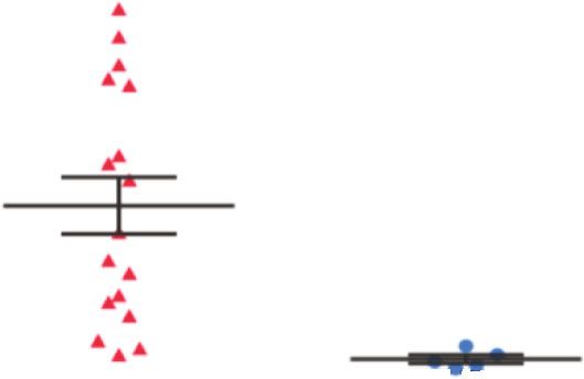
BioMed Research International 3
p < 0.05
NS
n = 52 n = 50 n = 47
100
80
% cells positive
60
40
20
0
CLL-1 CD33 CD34
(b)
p < 0.05
n = 17 n=5
60
50
% cells positive
40
30
20
10
100 101 102 103 104
0
CLL-1 AML NBM
(a) (c)
Low High
Adult urinary bladder
Adult adrenal gland
Adult frontal cortex
Sample
Adult gallbladder
Adult spinal cord
Adult esophagus
Adult pancreas
Adult prostate
Adult rectum
CD4+ T cells
CD8+ T cells
Adult kidney
Adult retina
Adult ovary
Adult colon
Adult heart
Adult testis
Fetal ovary
Monocytes
Fetal brain
Adult lung
Adult liver
Fetal heart
Fetal testis
Gene
Fetal liver
Fetal gut
Placenta
NK cells
Platelets
B cells
CLEC12A
GAPDH
(d)
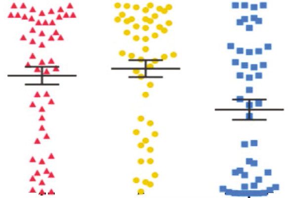
Figure 1: Expression of CLL-1 on AML and stem cells. (a) Variant CLL-1 expression levels among primary AML samples. CLL-1 staining
from ten representative samples is shown. (b) Distribution of CLL-1+, CD33+, and CD34+ cells in primary AML samples. (c) CLL-1
expression on CD34+CD38- stem cells in AML and NBM. (d) Expression profile of CLL-1 in normal tissue at protein level was assessed
by utilizing publicly available databases for mass-spectrometry proteomic analysis (Human Proteome Map: CLL-1). AML: acute myeloid
leukemia; NBM: normal bone marrow; NS: not significant.
software, and two-tailed values of p < 0:05 were considered the bulk of the population of blast cells showed no clear sepa-
significant. ration in populations with positive and negative blasts.
Since CD33 and CD34 were classic markers for AML, we
3. Result measured the expression of these two markers in combina-
tion with CLL-1 on primary AML samples. We found that
3.1. Validation of CLL-1 as an AML Marker. To analyze the CD33 and CD34 antigens were expressed on >20% of blast
expression profile of CLL-1 on AML blasts, we defined posi- cells in 43 of 50 (86.0%) and 28 of 47 (59.6%) AML samples,
tive antigen expression as the expression of the antigen in respectively. Interestingly, of the 7 CD33-negative samples, 3
more than 20% of the sample cells. In the prospectively col- were positive for CLL-1 antigen. Furthermore, of the 19
lected test set, 41 of 52 (78.8%) patients stained CLL-1 positive CD34-negative samples, 15 were positive for CLL-1 antigen.
with different intensities, consistent with our previous report When we compared the antigen expression levels, we found
[14]. Representative examples of CLL-1 expression on primary that CLL-1 was more frequently expressed than CD34
AML samples have been shown in Figure 1(a). In most cases, (p < 0:05), but no difference with CD33 (Figure 1(b)).4 BioMed Research International
Table 1: Patient characteristics.
Characteristic CLL-1low patients CLL-1high patients p value
Total, no. (%) 19 (36.5) 33 (63.5)
Gender, no. (%) 0.76
Male 11 (57.9) 17 (51.5)
Female 8 (42.1) 16 (48.5)
Age at diagnosis, years, median (range) 38.0 (13-75) 50.0 (7-75) 0.44
Disease status, no. (%) 0.26
De novo 14 (73.7%) 29 (87.9%)
Secondary 5 (26.3%) 4 (12.1%)
WBC count at diagnosis, 109/L, median (range) 17.3 (0.5-345.6) 31.7 (0.8-373.9) 0.33
Bone marrow blasts at diagnosis, %, median (range) 30.0 (10.1-91.5) 73.0 (9.5-96.0) 0.03
FAB classification, no. (%) 0.39
M1 2 (10.5) 1 (3.0)
M2 5 (26.3) 9 (27.3)
M3 1 (5.3) 4 (12.1)
M4 0 (0) 3 (9.1)
M5 9 (47.4) 8 (24.2)
M6 0 (0) 1 (3.0)
M7 0 (0) 0 (0)
Not done 2 (10.5) 7 (21.2)
Risk group, no. (%) 0.44
Favorable 1 (5.3) 2 (6.1)
Intermediate 3 (15.8) 11 (33.3)
Unfavorable 7 (36.8) 9 (27.3)
Not done 8 (42.1) 11 (33.3)
CR reached after cycle 1, no. (%) 0.03
Yes 6 (35.3) 22 (73.3)
No 11 (64.7) 8 (26.7)
Allogeneic stem cell transplantation, no. (%) 1.00
Yes 2 (10.5) 5 (15.2)
No 17 (89.5) 28 (84.8)
CLL-1: C-type lectin-like molecule-1; WBC: white blood cells; FAB: French-American-British; CR: complete response.
We next investigated the expression of CLL-1 on CD34+- and 19 CLL-1low cases (59.0%) tic relevance of CLL-1 was investigated. As depicted inBioMed Research International 5
1.0 1.0
0.8 0.8
Event free survival (%)
Overall survival (%)
0.6 0.6
0.4 0.4
0.2 0.2
p = 0.048 p = 0.012
0.0 0.0
0 10 20 30 40 50 60 0 10 20 30 40 50 60
Time (months) Time (months)
CLL-1low CLL-1low-censored CLL-1low CLL-1low-censored
CLL-1high CLL-1high-censored CLL-1high CLL-1high-censored
(a) (b)
1.0 1.0
0.8 0.8
Event free survival (%)
Overall survival (%)
0.6 0.6
0.4 0.4
0.2 0.2
p = 0.013 p = 0.03
0.0 0.0
0 10 20 30 40 50 0 10 20 30 40 50
Time (months) Time (months)
CLL-1low CLL-1low-censored CLL-1low CLL-1low-censored
CLL-1high CLL-1high-censored CLL-1high CLL-1high-censored
(c) (d)
1.0 1.0
0.8 0.8
Event free survival (%)
Overal survival (%)
0.6 0.6
0.4 0.4
0.2 0.2
p = 0.001 p = 0.001
0.0 0.0
0 10 20 30 40 50 60 0 10 20 30 40 50 60
Time (months) Time (months)
CLL-1low CLL-1low-censored CLL-1low CLL-1low-censored
CLL-1high CLL-1high-censored CLL-1high CLL-1high-censored
(e) (f)
Figure 2: Patient survival. Probability of EFS (a) and OS (b) in AML patients according to CLL-1 expression (entire cohort). EFS (c) and OS
(d) in AML patients with poor outcome according to CLL-1 expression. EFS (e) and OS (f) in AML patients according to treatment response.
EFS and OS were estimated using the Kaplan-Meier method, and the log-rank test was used for comparison of the survival curves. EFS: event-
free survival; OS: overall survival; AML: acute myeloid leukemia; CR: complete response.6 BioMed Research International
Table 2: Univariate and multivariate survival analysis.
Event-free survival Overall survival
Clinical factors Univariate Univariate Multivariate
p HR (%95 CI) p HR (%95 CI) p HR (%95 CI)
Gender (male) 0.702 0.834 (0.329-2.115) 0.200 1.757 (0.743-4.159) — —
Age (>50) 0.848 0.911 (0.351-2.362) 0.159 1.817 (0.792-4.167) — —
WBC (>100 ∗ 109 /l) 0.218 0.036 (0.000-7.056) 0.820 0.890 (0.325-2.435) — —
BM blasts (>50%) 0.146 0.494 (0.191-1.278) 0.922 0.960 (0.422-2.186) — —
Risk stratification (poor) 0.388 1.383 (0.662-2.889) 0.395 1.407 (0.640-3.094) — —
CLL-1 (BioMed Research International 7
be identified. Consequently, our coexpression analysis sug- AML, further supporting its high value in prognosis. Besides,
gested that combinatorial detecting approaches might some studies have compared the expression of CLL-1 on
enhance diagnosing efficiency in AML, and combinatorial AML samples at diagnosis, treatment, and relapse and found
targeting approaches might enhance therapeutic efficacy in no significant difference [13, 15, 29–31]. This suggested that
AML that should be validated in the future. Besides, CLL-1 could be used as a reliable marker for disease follow
CD34, a stem cell-specific marker in AML, is widely used up/detection of MRD.
to monitor minimal residual disease (MRD). The addition Concerning the mechanism of low CLL-1 expression in
of CLL-1 maybe valuable in increasing the likelihood of leukemic blasts as a predictor of poor prognosis, it remains
upfront MRD marker identification by FCM in the CD34- unclear. It was previously reported that CLL-1 might play a
subgroup. Notably, we are the first to compare the antigen role in the control of cell maturation [7], so we consider that
expression levels of CLL-1, CD33, and CD34 simulta- the loss of CLL-1 expression may prevent leukemia cell pro-
neously on AML bulk cells. liferation and keep it in a relatively static state, thus reducing
We next analyzed the relationship between CLL-1 the sensitivity to chemotherapy. In addition, we discovered
expression level and clinicopathological features. We found that the proportion of CLL-1low group in poor-risk stratifi-
that CLL-1 was nonrandomly expressed on AML samples cation was higher than that of CLL-1high group, which
throughout the different FAB subtypes and risk groups. After may also partly explain the poorer prognosis of patients with
having established the definition of CLL-1high and CLL-1low low CLL-1 expression. However, further studies will be
according to cutoff 59.0%, we found that the low expression required to understand the mechanism underlying the
level of CLL-1 was significantly correlated with lower BM impact of CLL-1 expression for prognosis.
blast percentage, and what is more, the CR rate after cycle 1 Taken together, we have demonstrated that CLL-1 is one
of the CLL-1low group was significantly lower than the of the promising surface molecules for AML diagnosis;
CLL-1high group (p < 0:05). We know that low remission meanwhile, CLL-1 is easy to measure in clinical practice
rates often indicate poor prognosis, and we did demonstrate and thus can be incorporated into the routine practice of
that both the EFS and OS were significantly worse among most clinical laboratories. Furthermore, we have proved that
patients who did not reach CR than those did. Meanwhile, CLL-1 is an effective tool to predict the survival of AML
no CR was demonstrated to be an independent factor associ- patients, so it can be used as a supplement to the current
ated with EFS and OS in multivariate Cox regression model. AML prognostic risk stratification system and may optimize
However, a statement on the possible correlation with other the clinical management of AML. However, the relatively
clinicopathological features, such as gender, age, disease sta- small number of patients and the short follow-up time lim-
tus, WBC count at diagnosis, FAB subtype, and risk group, ited us to draw a robust conclusion in our study, and cytoge-
cannot be presently made probably due to the relatively small netic and molecular genetic profiles were not complete for all
number of patients. patients. Further and larger-scale studies are required to
When we compared the survival between CLL-1high and more clearly define the significance of CLL-1 expression in
CLL-1low groups, we discovered that EFS and OS of the AML and also to elucidate the underlying mechanisms in
CLL-1low group were significantly lower than the CLL-1high the future.
group (p < 0:05). Furthermore, in univariate Cox regression
model analysis, we found that the CLL-1low group was an 5. Conclusions
independent prognostic value associated with OS, and multi-
variate Cox regression model analysis showed that CLL-1low In summary, we report that CLL-1 was expressed on the cell
was still independent from other well-established factors. surface of the majority of AML blasts (78.8%) and also
However, the low expression level of CLL-1 did not maintain expressed on leukemic stem cells in varying degree but absent
its value in predicting EFS. Taking into consideration that on normal hematopoietic stem cells. Notably, CLL-1 was able
risk stratification is critical in AML, we analyzed the impact to complement the classic markers CD33 or CD34. In addi-
of CLL-1 expression in each risk subgroup. The results tion, we discovered that EFS and OS of the CLL-1low group
showed that the CLL-1high patients had better EFS and OS were significantly lower than the CLL-1high group, and low
than the CLL-1low patients (p < 0:05) in the poor-risk group. CLL-1 expression seems to be independently associated with
This implied that CLL-1high patients in the poor-risk group shorter OS. These results suggested that CLL-1 may serve as a
had a more intermediate prognosis comparing to poor-risk biomarker for diagnosis and prognosis of AML.
CLL-1low patients, who actually have a worse prognosis. As
a result, the CLL-1 expression should be incorporated into Data Availability
future risk-adapted therapy and prognosticating relapse risk
in this subset of poor-risk AML patients. We did not find The data used to support the findings of this study are avail-
similar results in other risk subgroups probably due to the able from the corresponding author upon request.
small numbers. This study agreed with Wang et al., who also
demonstrated that CLL-1low indicated poor prognosis Conflicts of Interest
(p < 0:001 for EFS and OS) in patients with AML. Neverthe-
less, they detected the CLL-1 expression only in de novo The authors declare that they have no known competing
CD34+ Non-M3 AML, while we evaluated the CLL-1 expres- financial interests or personal relationships that could have
sion in nonselected patients including de novo and relapsed appeared to influence the work reported in this paper.8 BioMed Research International
Authors’ Contributions tion of a novel human myeloid inhibitory C-type lectin-like
receptor (MICL) that is predominantly expressed on granulo-
Jinghua Wang, Weida Wang, and Hao Chen contributed cytes and monocytes,” The Journal of Biological Chemistry,
equally to this work. vol. 279, no. 15, pp. 14792–14802, 2004.
[8] Y. Han, M. Zhang, N. Li et al., “KLRL1, a novel killer cell lec-
Acknowledgments tinlike receptor, inhibits natural killer cell cytotoxicity,” Blood,
vol. 104, no. 9, pp. 2858–2866, 2004.
This study was supported by the High-level Hospital Con- [9] V. R. Wiersma, M. de Bruyn, C. Shi et al., “C-type lectin-like
struction Project (Nos. DFJH201923 and DFJH201803), the molecule-1 (CLL1)-targeted TRAIL augments the tumoricidal
Medical Scientific Research Foundation of Guangdong Prov- activity of granulocytes and potentiates therapeutic antibody-
ince (No. A2019063), the Fundamental Research Funds for dependent cell-mediated cytotoxicity,” MAbs, vol. 7, no. 2,
the Central Universities (No. D2180600), National Natural pp. 321–330, 2015.
Science Foundation of China (Nos. 81300279 and [10] A. S. Marshall, J. A. Willment, E. Pyż et al., “Human MICL
81741067), and the Special Fund for Science and Technology (CLEC12A) is differentially glycosylated and is down-
of Guangdong Provincial People’s Hospital/Guangdong regulated following cellular activation,” European Journal of
Academy of Medical Sciences (No. 2018bq01). Immunology, vol. 36, no. 8, pp. 2159–2169, 2006.
[11] S. Haubner, F. Perna, T. Köhnke et al., “Coexpression profile of
Supplementary Materials leukemic stem cell markers for combinatorial targeted therapy
in AML,” Leukemia, vol. 33, no. 1, pp. 64–74, 2019.
Supplementary Figure 1: the gating strategy of AML blasts [12] X. Zhao, S. Singh, C. Pardoux et al., “Targeting C-type lectin-
and LSCs. (A) Initially, cells were gated based on forward like molecule-1 for antibody-mediated immunotherapy in
and side scatter properties. Subsequently, AML blasts were acute myeloid leukemia,” Haematologica, vol. 95, no. 1,
selected based on low-side scatter versus CD45dim expres- pp. 71–78, 2010.
sion. (B) After AML blasts were selected, CD34+ cells were [13] H. Larsen, A. S. Roug, T. Just, G. D. Brown, and P. Hokland,
then gated. Finally, CD38- cells were recognized as LSCs. “Expression of the hMICL in acute myeloid leukemia-a highly
AML: acute myeloid leukemia; LSC: leukemic stem cells. reliable disease marker at diagnosis and during follow-up,”
Supplementary Figure 2: the ROC curve analysis for the opti- Cytometry Part B, Clinical Cytometry, vol. 82B, no. 1, pp. 3–
8, 2012.
mal cut-off point of CLL-1 expression. The most discrimina-
tive cut-off value for CLL-1 was 59% with a sensitivity of [14] J. Wang, S. Chen, W. Xiao et al., “CAR-T cells targeting CLL-1
as an approach to treat acute myeloid leukemia,” Journal of
60.9% and a specificity of 79.4%; the area under ROC curve
Hematology & Oncology, vol. 11, no. 1, p. 7, 2018.
is 0.694 (p = 0:017). (Supplementary Materials)
[15] A. van Rhenen, G. A. M. S. van Dongen, A. Kelder et al., “The
novel AML stem cell associated antigen CLL-1 aids in discrim-
References ination between normal and leukemic stem cells,” Blood,
[1] C. J. Eaves and R. K. Humphries, “Acute myeloid leukemia and vol. 110, no. 7, pp. 2659–2666, 2007.
the Wnt pathway,” The New England Journal of Medicine, [16] B. Zheng, S. F. Yu, G. del Rosario et al., “An anti-CLL-1
vol. 362, no. 24, pp. 2326-2327, 2010. antibody-drug conjugate for the treatment of acute myeloid
[2] D. Grimwade and R. K. Hills, Independent prognostic factors leukemia,” Clinical Cancer Research, vol. 25, no. 4, pp. 1358–
for AML outcome, Hematology American Society of Hematol- 1368, 2019.
ogy Education Program, 2009. [17] Y. P. Jiang, B. Y. Liu, Q. Zheng et al., “CLT030, a leukemic stem
[3] D. Voskova, C. Schoch, S. Schnittger, W. Hiddemann, cell-targeting CLL1 antibody-drug conjugate for treatment of
T. Haferlach, and W. Kern, “Stability of leukemia-associated acute myeloid leukemia,” Blood Advances, vol. 2, no. 14,
aberrant immunophenotypes in patients with acute myeloid pp. 1738–1749, 2018.
leukemia between diagnosis and relapse: comparison with [18] T. J. Hutten, S. Thordardottir, H. Fredrix et al., “CLEC12A-
cytomorphologic, cytogenetic, and molecular genetic find- mediated antigen uptake and cross-presentation by human
ings,” Cytometry Part B, Clinical Cytometry, vol. 62, no. 1, dendritic cell subsets efficiently boost tumor-reactive T cell
pp. 25–38, 2004. responses,” Journal of Immunology, vol. 197, no. 7, pp. 2715–
[4] E. Papaemmanuil, M. Gerstung, L. Bullinger et al., “Genomic 2725, 2016.
classification and prognosis in acute myeloid leukemia,” New [19] P. F. van Loo, B. N. Hangalapura, S. Thordardottir et al.,
England Journal of Medicine, vol. 374, no. 23, pp. 2209–2221, “MCLA-117, a CLEC12AxCD3 bispecific antibody targeting
2016. a leukaemic stem cell antigen, induces T cell-mediated AML
[5] A. B. Bakker, S. van den Oudenrijn, A. Q. Bakker et al., “C-type blast lysis,” Expert Opinion on Biological Therapy, vol. 19,
lectin-like molecule-1: a novel myeloid cell surface marker no. 7, pp. 721–733, 2019.
associated with acute myeloid leukemia,” Cancer Research, [20] H. Lu, Q. Zhou, V. Deshmukh et al., “Targeting human C-type
vol. 64, no. 22, pp. 8443–8450, 2004. lectin-like molecule-1 (CLL1) with a bispecific antibody for
[6] C. H. Chen, H. Floyd, N. E. Olson et al., “Dendritic-cell-asso- immunotherapy of acute myeloid leukemia,” Angewandte
ciated C-type lectin 2 (DCAL-2) alters dendritic-cell matura- Chemie (International Ed. in English), vol. 53, no. 37,
tion and cytokine production,” Blood, vol. 107, no. 4, pp. 9841–9845, 2014.
pp. 1459–1467, 2006. [21] H. Ma, I. S. Padmanabhan, S. Parmar, and Y. Gong, “Targeting
[7] A. S. Marshall, J. A. Willment, H. H. Lin, D. L. Williams, CLL-1 for acute myeloid leukemia therapy,” Journal of Hema-
S. Gordon, and G. D. Brown, “Identification and characteriza- tology & Oncology, vol. 12, no. 1, p. 41, 2019.BioMed Research International 9
[22] E. Laborda, M. Mazagova, S. Shao et al., “Development of a
chimeric antigen receptor targeting C-type lectin-like
molecule-1 for human acute myeloid leukemia,” International
Journal of Molecular Sciences, vol. 18, no. 11, p. 2259, 2017.
[23] H. Tashiro, T. Sauer, T. Shum et al., “Treatment of acute mye-
loid leukemia with T cells expressing chimeric antigen recep-
tors directed to C-type lectin-like molecule 1,” Molecular
Therapy, vol. 25, no. 9, pp. 2202–2213, 2017.
[24] M. A. Sanz, F. Lo Coco, G. Martín et al., “Definition of relapse
risk and role of nonanthracycline drugs for consolidation in
patients with acute promyelocytic leukemia: a joint study of
the PETHEMA and GIMEMA cooperative groups,” Blood,
vol. 96, no. 4, pp. 1247–1253, 2000.
[25] H. Döhner, E. Estey, D. Grimwade et al., “Diagnosis and man-
agement of AML in adults: 2017 ELN recommendations from
an international expert panel,” Blood, vol. 129, no. 4, pp. 424–
447, 2017.
[26] B. Löwenberg, Acute myeloid leukemia: the challenge of captur-
ing disease variety, Hematology American Society of Hematol-
ogy Education Program, 2008.
[27] A. S. Lima, M. R. de Mello, E. Fernandes et al., “Clinical out-
comes of patients with acute myeloid leukemia: evaluation of
genetic and molecular findings in a real-life setting,” Blood,
vol. 126, no. 15, pp. 1863–1865, 2015.
[28] M. Bill, P. B. van Kooten Niekerk, P. S. Woll et al., “Mapping
the CLEC12A expression on myeloid progenitors in normal
bone marrow; implications for understanding CLEC12A‐
related cancer stem cell biology,” Journal of Cellular and
Molecular Medicine, vol. 22, no. 4, pp. 2311–2318, 2018.
[29] E. Coustan-Smith, G. Song, S. Shurtleff et al., “Universal mon-
itoring of minimal residual disease in acute myeloid leukemia,”
JCI Insight, vol. 3, no. 9, 2018.
[30] D. S. Eissa, E. Z. Kandeel, and M. Ghareeb, “Human myeloid
inhibitory C-lectin: a highly specific and stable acute myeloid
leukemia marker,” Hematological Oncology, vol. 35, no. 4,
pp. 814–820, 2017.
[31] A. van Rhenen, B. Moshaver, A. Kelder et al., “Aberrant
marker expression patterns on the CD34+CD38− stem cell
compartment in acute myeloid leukemia allows to distinguish
the malignant from the normal stem cell compartment both at
diagnosis and in remission,” Leukemia, vol. 21, no. 8,
pp. 1700–1707, 2007.You can also read