High Intensity Focused Electromagnetic Therapy Evaluated by Magnetic Resonance Imaging: Safety and Efficacy Study of a Dual Tissue Effect Based ...
←
→
Page content transcription
If your browser does not render page correctly, please read the page content below
Lasers in Surgery and Medicine

High Intensity Focused Electromagnetic Therapy
Evaluated by Magnetic Resonance Imaging: Safety and
Efficacy Study of a Dual Tissue Effect Based Non-Invasive
Abdominal Body Shaping
Brian M. Kinney, MD, MSME, FACS1 and Paula Lozanova,           MD
                                                                  2
1
  USC School of Medicine, Hills Beverly, California
2
  Paula Fines Center, Sofia BG, Europe

Objectives: This study introduces an initial evaluation of      INTRODUCTION
a novel High-Intensity Focused Electromagnetic (HI-                The popularity of non-invasive body shaping procedures has
FEM) technology. The primary goal is to quantify any            been growing rapidly—the number of procedures performed in
effects the treatments may have on abdominal tissues, as        the US more than doubled between 2012 and 2016 [1].
well as to establish hypotheses for future research of this     Cryolipolysis, radiofrequency, low level laser therapy and
technology.                                                     focused ultrasound [2] are most widely used for treating
Methods: Twenty-two patients received four abdominal            patients’ fat bulges, and their efficacy has been demonstrated
treatments using the EMSCULPT device (BTL Industries            in multiple previous studies. Similar to every aesthetic
Inc., Boston, MA). Anthropometric evaluations were              procedure, these technologies have also certain limitations.
recorded and digital photographs were taken at baseline,        All current non-invasive fat removal treatments are based on
at 2 months, and at 6 months post-treatments. The MRI           thermal effects and as such, they may bring about various cold
without contrast determined by vertertebras T12 and S1          or heat related side effects. More importantly, all these
(FIESTA and FSPRG sequences) was used to measure                modalities are designed to address only fat tissue.
dimensions in coronal cross-sectional images of abdominal          Subcutaneous fat is an important factor affecting
muscle and fatty tissues, in order to assess any anatomical     patient’s body contours as it comprises approximately
changes induced by the application.                             25% [3] of human body composition. However, muscle
Results: Analysis of the same MRI slices verified by tissue     tissue comprises even a larger portion of the human body
artefacts showed a statistically significant (all P < 0.0001)   composition (42% male/36% female [4]) and depending on
average 18.6% reduction of adipose tissue thickness, 15.4%      individual characteristics, the condition of patient’s
increase in rectus abdominis muscle thickness, and 10.4%        muscle can play either an equal or even more important
reduction in rectus abdominus separation (diastasis recti)      role in defining the overall aesthetic appearance. Still,
as measured from the medial border of the muscle 2 months       physical workout is currently the only generally available
post-treatment. More significant improvements were              method for natural strengthening of one’s muscles.
observed in patients with BMI 18.5–24.9 (classified as             The use of magnetic stimulation has a proven track
“normal”). MRI data from 6-month follow-up suggest the          record when treating various medical indications, ranging
changes can be preserved in longer term. Tape measure-          from neurology [5–7], psychiatry [8], physiotherapy [9–12],
ments showed on average 3.8 cm subumbilical circumfer-          to treating urinary incontinence in women [13]. Further-
ence reduction. The weight of the subjects did not change       more, due to the non-thermal and non-ionizing nature of
significantly (average 0.5 lb; P > 0.05). No adverse events
were reported.
Conclusions: MRI, considered as a highly precise                   This is an open access article under the terms of the Creative
diagnostic method, revealed simultaneous muscle growth,         Commons Attribution-NonCommercial-NoDerivs License, which
                                                                permits use and distribution in any medium, provided the
fat reduction and reduced abdominal separation at               original work is properly cited, the use is non-commercial and
2 months and at 6 months post treatments, unrelated             no modifications or adaptations are made.
                                                                   Conflict of Interest Disclosures: All authors have completed
with dieting. Further research should investigate the exact     and submitted the ICMJE Form for Disclosure of Potential
physiological processes which stand behind the tissue           Conflicts of Interest and have disclosed the following: Brian
changes observed in this study. Lasers Surg. Med. © 2018        Kinney MD is a medical advisor to BTL. Paula Lozanova MD has
                                                                no conflicts to declare.
The Authors. Lasers in Surgery and Medicine Published by
                                                                     Correspondence to: Brian M. Kinney MD, FACS, USC School
Wiley Periodicals, Inc.                                         of Medicine, Hills Beverly, CA.
                                                                E-mail: brian@briankinneymd.com
                                                                   Accepted 14 September 2018
Key words: diastasis recti; fat reduction; HIFEM;                  Published online in Wiley Online Library
magnetic technology; muscle growth                                 (wileyonlinelibrary.com).
                                                                   DOI 10.1002/lsm.23024

ß 2018 The Authors. Lasers in Surgery and Medicine Published by Wiley Periodicals, Inc.
2                                                  KINNEY AND LOZANOVA

the technology, its application is considered relatively           caloric intake was calculated in cooperation with a
safe [8]. Even though the technology is highly effective, it is    professional nutritionist. All patients were asked to
not as widely used as electrical stimulation [14].                 maintain their routine diet and activity level without
  This study brings an initial evaluation of a novel High-         any modifications until study completion. Afterwards,
Intensity Focused Electro-Magnetic (HIFEM) technology              patients received four treatments (spaced by 2–5 days)
applied to the abdominal area, in order to assess the              using a HIFEM technology device (EMSCULPT, BTL
physiological response in treated patients. The primary            Industries, Boston, MA) as per the IRB-approved protocol.
goal is to quantify any effects the treatments may have on
abdominal tissues, as well as to establish hypotheses for          EMSCULPT Procedure
future research of this technology. The outcomes of the study        During the application, patients did not receive any
are expected to suggest if HIFEM can be potentially used as a      anesthesia and were lying in a supine position. All
new technology for non-invasive body shaping treatments.           procedures were applied to the abdomen and each session
                                                                   included exactly 30 minutes of continuous application. One
MATERIALS AND METHODS
                                                                   applicator (see Fig. 1) was placed on the skin at the
Study Population                                                   umbilical level. The center of the magnetic coil was placed
  Twenty-two subjects (10 females and 12 males) partici-           exactly above the navel. The applicator was affixed by a
pated in this prospective, multi-center, non-randomized,           disinfected fixation belt to minimize movements during the
pilot study. The average age of the participants was               procedure. The stimulation intensity started at 0% and
39.4  10.2 with a mean BMI prior to the treatments of             within 60 seconds to the treatment it was slowly increased
25.7  2.4 kg/m2. The exclusion criteria included preg-            by the operator until reaching patient’s tolerance thresh-
nancy, breastfeeding, any medical condition contraindicat-         old. The tolerance threshold was continuously challenged
ing the application of an electromagnetic field, heart             during the course of the treatments. A dual feedback
disorders, unhealed wound in abdominal area, and any               principle was applied, with the operator visually checking
concomitant medication known to cause bloating or affect           the intensity and homogeneity of the muscle contractions
weight. See Table 1 for the baseline demographic profile.          across the abdomen, as well as regularly asking the patient
Patients were not financially incentivized for either              about feedback regarding the level of comfort and the
participation or completion of the study; an informed              balance of contractions between different abdominal areas.
consent was obtained from all of them. The study was
conducted in compliance with applicable ethical standards          Evaluation Methodology
and used an IRB approved protocol.                                    A complete evaluation of the patients was performed at
                                                                   baseline and 2 months after their last treatment, and
Study Design                                                       included a brief medical history and examination, mag-
  Prior to the treatments, each subject was inquired about         netic resonance imaging (MRI) scan, weight and waist
his/her physical activity habits and an approximate daily          circumference measurements, digital photography, and
                                                                   monitoring of any adverse events. Due to financial
                                                                   constraints, only four randomly selected patients were
                                                                   scheduled for a 6-month follow-up to gain an insight into
TABLE 1. Baseline             Demographic     Profile   of   the
                                                                   the tendencies the result may have in the long term.
Subjects
                                                                      MRI scans were used to observe changes in abdominal
                                       Count                 %     fat and muscle tissues of the treated patients. The scanned
                                                                   body volume was defined by T12 and S1 vertebrae and the
Age                                                                array coil system was set up in such a way to minimize any
  50                                  5                     23
BMI
  30.0 (Obese)                        1                     5
Gender
  Female                               10                    45
  Male                                 12                    55
Ethnicity
  Caucasian                            22                    100
  Deliveries (10 patients)             1.6a
a
    Average nr. of childbirths.                                    Fig. 1. Scheme of the EMSCULPT applicator.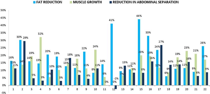
MRI EVALUATION OF ELECTROMAGNETIC THERAPY                                                   3

epiumbilical slices of the same sequence and of the same         observed in all three measurements when comparing the
bodily section were extracted in cooperation with a              2-month follow-up to the baseline—a reduction in adipose
qualified radiologist (experienced in reading abdominal          tissue thickness ( 18.6%), an increase in rectus abdominis
scans), and the thickness of subcutaneous adipose tissue as      thickness (þ15.4%) and a reduction in abdominal separa-
well as rectus abdominis were measured (InVesalius 3.1).         tion ( 10.4%). In total 91 % (n ¼ 20) of patients improved in
The measurements were taken in multiple points which             all three facets simultaneously. The analysis did not show
were laid out laterally in the range between patient’s iliac     any non-responding patients who would not have any
crests. Direct umbilical area was excluded from evaluation       changes in the tissue at all. No other structural changes in
due to absence of the muscle structure (linea alba) and          the tissues were observed.
adipose layer (the navel). Furthermore, the size of                 An increase in the abdominal muscle mass was observed
abdominal separation was measured from the same MRI              in 95% (n ¼ 21) of patients; one subject did not show any
slices.                                                          change. The muscle growth was relatively consistent, with
   Gulick II spring-loaded tape assisted measurements            majority of patients showing an increase in the range of
were taken 5 cm below umbilicus; patients had the most           10–20% (see Fig. 3). The changes were calculated across
distinctive fat bulges in this region prior to the treatments.   both sides of the muscle; the difference in growth between
Frontal and lateral digital photography was taken; a             the right and left rectus abdominis was insignificant.
positioning mat was used to ensure consistency.                  However, the distance (separation) between the left and
   All data collected prior to the treatments were compared      right abdominal muscles decreased in 91% (n ¼ 20) of
with the follow-up data; all results were tested for             patients; one patient did not show any change and for
significance with a two-sample paired t-test. Descriptive        another patient the distance marginally increased
data were presented as the mean and SD.                          (þ0.26 mm or þ2.4%). Contrary to our expectations, a
                                                                 subgroup of women who had previously been pregnant
RESULTS                                                          (n ¼ 9) did not have higher values of abdominal separation
The Procedures                                                   before treatments (average 14.9 mm compared to 17.8 mm
                                                                 in other patients). They however did trend toward slightly
   All 22 subjects completed the entire study. On average,
                                                                 greater proportional improvement (average reduction was
12.6  2.5 days and 57.1  8.6 days elapsed between the
                                                                 11.0% compared to 10.0% in the rest of the cohort). The
baseline and the last procedure, and between the last
                                                                 percentage change in abdominal separation was indepen-
procedure and the follow-up evaluation, respectively. Most
                                                                 dent of its severity (size) before treatments. Also, statistical
patients tolerated stimulation intensities ranging between
                                                                 analysis confirmed that the changes in muscle thickness
90 and 100% already by the end of their first session or
                                                                 and changes in abdominal separation were two highly
during their second session, depending on individual
                                                                 independent effects (P > 0.05; correlation coefficient
sensitivity. Minimum tolerable intensity was 74% (a
                                                                   0.31). MRI of subjects with major muscle growth thus
patient with BMI 19.7), 17 out of 22 patients tolerated
                                                                 did not necessarily reveal a major reduction in abdominal
100% intensity. Higher BMI patients tended to tolerate
                                                                 separation.
slightly higher intensity settings. No adverse events
                                                                    Measurements of the fat tissue revealed an opposite
occurred. The only noticed side effect was mild muscle
                                                                 trend, with the average thickness decreasing in all
soreness 1 day after the first treatment reported by six
                                                                 patients. The reduction was slightly more variable than
patients; in all cases the soreness resolved itself within the
                                                                 changes in the muscle (coefficient of var. 58.1%); this was
next 24 hours. Overall the patients did not change their
                                                                 primarily driven by two positive extremities. In total 82%
lifestyle or dietary intake significantly.
                                                                 (n ¼ 18) of patients had the fat layer reduced by more than
                                                                 10% at the follow-up. More significant absolute changes
MRI Evaluation of Abdominal Tissues                              were observed in subumbilical MRI cuts opposed to
  The study average and individual patient changes in            epiumbilical cuts.
abdominal fat, abdominal muscle and diastasis are                   For both the reduction in fat and reduction in abdominal
presented in Table 2 and Figure 2, respectively. On              separation, slightly more significant improvements were
average a statistically significant improvement was              seen in patients with BMI classified as “normal” (18.5–

TABLE 2. Average Changes in Abdominal Tissues in Treated Subjects

Measurement                                Baseline                2-Month FU                 Difference                P-value

Muscle thickness [mm]                      11.1  3.1               12.7  3.3                  1.6  0.7              P < 0.001
Fat thickness [mm]                         23.6  8.2               19.3  7.6                  4.3  2.5              P < 0.001
Abdominal separation [mm]                  16.6  7.2               14.9  6.7                  1.8  1.5              P < 0.001
Waist circumference [cm]                   95.3  6.6               91.5  7.4                  3.8  2.1              P < 0.001
Weight [lb]                               175.8  24.8             175.2  24.3                 0.5  2.5              P > 0.05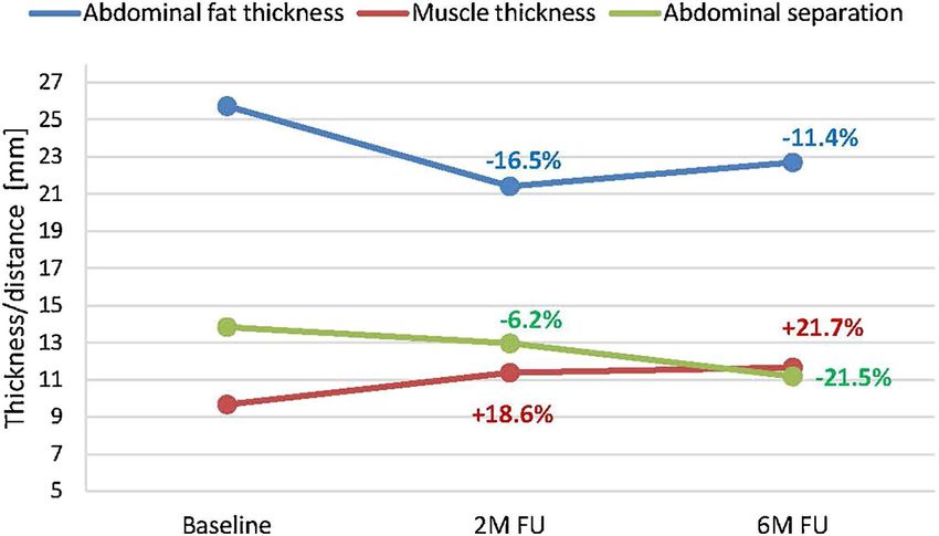
4                                                KINNEY AND LOZANOVA

               Fig. 2. Changes (%) in abdominal tissues in individual patients. Reduction in subcutaneous adipose
               tissue thickness (light blue), growth in rectus abdominis thickness (dark blue), and reduction in
               abdominal separation (gray) are presented.

24.9 kg/m2). Their subcutaneous fat mass decreased on            11.65 mm (20.5% increase at 6 months). The abdominal
average by 20.6%, and the size of diastasis decreased by         separation further improved from average of 12.95 mm (2
11.7%. For “overweight” patients (25.0–29.9 kg/m2) the           months) to 11.18 mm (6 months). In the same patients, the
same measurements averaged 18.1% and 10.0%,                      average thickness of subcutaneous fat was on average
respectively.                                                    3.03 mm lower (22.69 mm) at 6 months compared to the
                                                                 baseline (25.72 mm), see Figure 4.
6-Month Data
  Based on MRI evaluation, the muscle thickness contin-          Other Evaluation
ued to grow and the abdominal separation continued to               Compared to the baseline, the average subumbilical
shorten in all four randomly selected patients when              circumference of patients decreased by 3.8  2.1 cm at the
compared to the 2-month follow-up. The average thickness         2-month follow-up. The change was statistically indepen-
in these patients evolved from 9.67 mm (baseline), to            dent of weight variations; the average weight remained
11.38 mm (17.7% increase at 2 months), and on to                 stable. Digital photographs showed distinctive aesthetic

               Fig. 3. Plots show the median value, quartile values, as well as the maximum and minimum sample
               value with regards to changes in abdominal tissues of treated patients calculated from MRI scans.
               The changes represent a comparison between the baseline and the 2-month follow-up.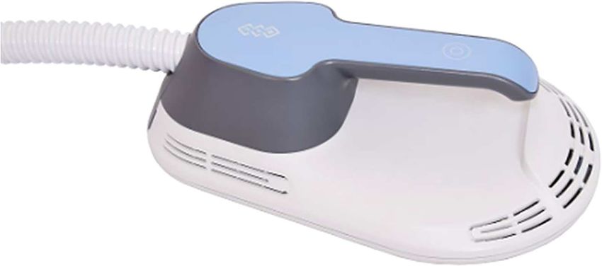
MRI EVALUATION OF ELECTROMAGNETIC THERAPY                                                  5

                                                                 strengthening of the muscular foundations. Currently,
                                                                 the only way to strengthen the core is a physical workout
                                                                 plan. The investigated device uses HIFEM technology to
                                                                 induce almost 20 thousand pulses in one 30-minute
                                                                 session. Such frequency of nerve stimuli leads to supra-
                                                                 maximal muscle contractions which are not achievable
                                                                 voluntarily. The muscle tissue is forced to adapt to this
                                                                 stress, resulting in muscle thickening. The principle of
                                                                 muscle hypertrophy and hyperplasia induced by intensive
                                                                 muscle contractions has already been proven in previous
                                                                 studies [15–19]. The 6 month data suggest that the muscles
                                                                 continue to improve in longer term, both in terms of their
                                                                 overall mass and lateral separation, yet further investiga-
Fig. 4. Average results of MRI evaluation at 6 months post-      tion is necessary to better understand the exact physiology.
treatments.
                                                                    Research on high intensity muscle training has shown
                                                                 that a lipolytic reaction takes plan in fat tissue adjacent to
                                                                 the contracting muscle [20]. The MRI scans presented
improvements in all patients except for one. Examples of         herein show a reduction in adipose tissue not immediately
digital photographs linked with corresponding MRI                after the treatments, but 2 months after the last procedure.
images are shown in Figures 5 and 6.                             A possible explanation for the lasting reduction in fat is
                                                                 that the lipolytic reaction is so intense, releasing large
DISCUSSISON                                                      amount of free fatty acids (FFA) which intoxicate the
  The findings from MRI scans presented herein have              adipocytes and trigger their death. This cell reaction has
shown that application of the HIFEM technology on the            already been shown in multiple studies in other fields of
abdomen can cause three different simultaneous changes           medicine [21–24]. A recent histology study reported a
in abdominal tissues non-invasively. Visual improvement          significant increase in the apoptotic index of adipocytes
in patients’ appearance observed 2 months after their last       after one HIFEM treatment on pigs (Weiss R, presented at
treatment very much resemble the effects of non-invasive         ASLMS, Dallas TX, April 2018). Their observation was
heat or cold-based fat reduction treatments combined with        coupled with an increased presence of mRNA pro-apoptotic
an extremely intensive physical workout.                         markers in molecular biochemistry results, as well as with
  The majority of therapeutic approaches aim at reducing         an increased concentration of FFA in blood serum. In
the subcutaneous fat layer (either surgically or non-            addition, an increase of 91.7% (from 18.8 to 35.9) in the
invasively), yet none of the previous ones deal with             apoptotic index was calculated from 120 histological

                Fig. 5. Magnetic resonance and digital images of Subject ID2 before (left) and 2 months post-
                treatments (right). Male (30), BMI 24.8 kg/m2 (before) and 24.5 kg/m2 (2 months), weight 2.2 lb
                ( 1.2 %), subcutaneous fat 30.3% (white markings), muscle thickness þ13.7% (green markings),
                abdominal separation 24.9% (red markings), circumference 3 cm. Combination of the effects
                produced an overall visual improvement in patient’s abdominal area.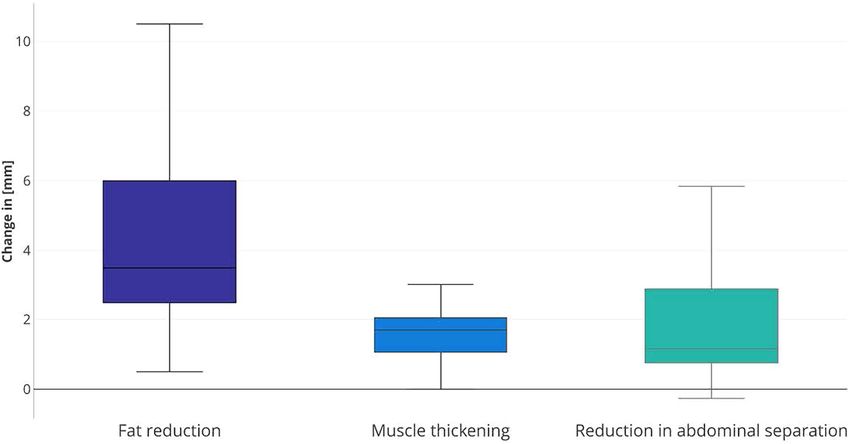
6                                                 KINNEY AND LOZANOVA

                Fig. 6. Magnetic resonance and digital images of Subject ID16 before (left) and 2 months post-
                treatments (right). Female (52), BMI 25.1 kg/m2 (before) and 24.4 kg/m2 (2 months), weight 4.4 lb
                ( 2.9%), subcutaneous fat 32.9%, muscle thickness þ19.4 %, abdominal separation 15.9%,
                circumference 5.7 cm. Combination of the effects produced an overall visual improvement in
                patient’s abdominal area.

samples. This again suggests a potential relationship             between the coil and the motor neurons responsive to the
between FFA released after the muscle contractions and            current will tend to be much larger due to the interspacing
fat apoptosis, however this hypothesis requires further           fat deposits. Such patients might not achieve as intensive
research as its validation was not the purpose of our study.      muscle contractions compared to normal BMI individuals.
  The third major observation, a reduction in abdominal           Despite the fact that inductive effects of HIFEM taper with
separation, was rather variable. An 84.8% coefficient of          distance, they can be felt from a distance of more than 7 cm
variation shows that the response in patients differed            from the actual applicator. Data from our study suggest
significantly, from very little change to more dramatic           that ideal candidates might be patients with less than an
reduction in the muscle distance. At the baseline, only one       inch (2.5 cm) of a pinchable subcutaneous fat. Our 6-month
patient suffered from actual diastasis recti as per the           data suggest the tissue changes may last. However, due to
medical definition (i.e., gap >2.7 cm) [25]. Still 91% of         the absence of any guidance in the literature on performing
subjects showed an improvement. This suggests that the            longer follow-up studies, 4 to 6 months seem to be a
application can not only help severely affected individuals,      reasonable time window for re-invitation of patients to
but is effective on most individuals regardless of their          assess if any additional procedures may or may not be
condition. This concept of reducing abdominal separation          beneficial.
by using a magnetic field technology would deserve further
investigation. In addition, there may be some role in             CONCLUSION
prevention by intervention prior to reaching the medical             The aim of this study was not to establish conclusive
definition of diastasis, although this would deserve further      evidence for the efficacy of the investigated device. To the
study as well.                                                    best of our knowledge, no peer-reviewed study has
  Although the sample is not large enough for a detailed          investigated a potential use of the HIFEM technology for
statistical analysis of fragmented sub-groups, the data           non-invasive body shaping. The data presented herein
indicate that neither gender nor age affect the outcomes of       show an initial evaluation on 22 patients, and suggest
the treatments. The fact that slightly more significant           possible physiological responses of the human body to the
changes in abdominal tissues were observed in thinner             treatments. We may well conclude that the results have
rather than overweight patients can most likely be                established a hypothesis of three simultaneous abdominal
attributed to the intensity of the magnetic field which           tissue effects induced as a direct result of the treatments,
decreases with an increasing distance from the actual             yet additional research is necessary to validate this in a
magnetic coil. For higher BMI patients, the distance              larger controlled study, as well as in a histological study
MRI EVALUATION OF ELECTROMAGNETIC THERAPY 7
that would help further cast light on the exact mechanism augment resistance training. J Funct Morphol Kinesiol
of action that would explain our observations. If confirmed, 2016;1:328–342.
13. Galloway NTM, El-Galley RES, Sand PK, Appell RA,
the technology would represent a completely new approach Russell HW, Carlan SJ. Extracorporeal magnetic innerva-
to non-invasive body shaping, bringing the additional tion therapy for stress urinary incontinence. Urology
muscle effects to the already established fat removal 1999;53:1108–1111.
market. 14. Szecsi J, Schiller M, Straube A, Gerling D. A comparison of
functional electrical and magnetic stimulation for propelled
cycling of paretic patients. Arch Phys Med Rehabil 2009;90:
REFERENCES 564–570.
1. The American Society for Aesthetic Plastic Surgery (2016). 15. Charette SL, et al. Muscle hypertrophy response to resistance
Cosmetic surgery national data bank statistics. http://www. training in older women. J Appl Physiol 1991;70:1912–1916.
surgery.org/sites/default/files/ASAPS-Stats2016.pdf. Ac- 16. Schoenfeld BJ. The mechanisms of muscle hypertrophy and
cessed April 25, 2017. their application to resistance training. J Strength Cond Res
2. Bernstein D, Farberg AS, Khorasani H, Kriegel D. Noninva- 2010;24:2857–2872.
sive body contouring: Literature review and summary of 17. Seynnes OR, de Boer M, Narici MV. Early skeletal muscle
objective data. SKIN J Cutan Med 2017;1:18–31. hypertrophy and architectural changes in response to high-
3. Muth ND. What are the guidelines for percentage of body fat intensity resistance training. J Appl Physiol 2007;102:
loss. Am Counc Exerc ACE Ask Expert Blog 2009. https:// 368–373.
www.acefitness.org/education-and-resources/lifestyle/blog/ 18. Alway SE, Grumbt WH, Gonyea WJ, Stray-Gundersen J.
112/what-are-the-guidelines-for-percentage-of-body-fat-loss. Contrasts in muscle and myofibers of elite male and female
4. Wallis MC, Davies EA, Thalib L, Griffiths S. Pelvic static bodybuilders. J Appl Physiol Bethesda Md 1985 1989;67:
magnetic stimulation to control urinary incontinence in older 24–31.
women: A randomized controlled trial. Clin Med Res 2012; 19. Marconnet P, Komi P. Muscular Function in exercise and
10:7–14. training. 3rd International symposium on biological sciences
5. Ziemann U. Cortical threshold and excitability measurements. in sport, nice, October/November 1986. Med Sport Sci
In: Eisen A, editor. Handbook of Clinical Neurophysiology. vol. 1987;26:67–89.
4. Supplement C vols. Amsterdam, The Netherlands: Elsevier; 20. Stallknecht B, Dela F, Helge JW. Are blood flow and lipolysis
2004. pp 317–335. in subcutaneous adipose tissue influenced by contractions in
6. Nitsche MA, Paulus W. Excitability changes induced in the adjacent muscles in humans? Am J Physiol Endocrinol Metab
human motor cortex by weak transcranial direct current 2007;292:E394–E399.
stimulation. J Physiol 2000;527:633–639. 21. Hardy S, El-Assaad W, Przybytkowski E, Joly E, Prentki M,
7. Badawy RAB, Loetscher T, Macdonell RAL, Brodtmann A. Langelier Y. Saturated fatty acid-induced apoptosis in MDA-
Cortical excitability and neurology: Insights into the patho- MB-231 breast cancer cells. A role for cardiolipin. J Biol Chem
physiology. Funct Neurol 2012;27:131–145. 2003;278:31861–31870.
8. Rossi S, Hallett M, Rossini PM, Pascual-Leone A. Safety, 22. Gunduz F, Aboulnasr FM, Chandra PK, et al. Free fatty acids
ethical considerations, and application guidelines for the use induce ER stress and block antiviral activity of interferon
of transcranial magnetic stimulation in clinical practice and alpha against hepatitis C virus in cell culture. Virol J
research. Clin Neurophysiol 2009;120:2008–2039. 2012;9:143.
9. Bogataj U, Gros N, Kljajic M, Acimovic R, Malezic M. The 23. Guo W, Wong S, Xie W, Lei T, Luo Z. Palmitate modulates
rehabilitation of gait in patients with hemiplegia: A comparison intracellular signaling, induces endoplasmic reticulum
between conventional therapy and multichannel functional stress, and causes apoptosis in mouse 3T3-L1 and rat primary
electrical stimulation therapy. Phys Ther 1995;75:490–502. preadipocytes. Am J Physiol Endocrinol Metab 2007;293:
10. Currier DP, Mann R. Muscular strength development by E576–E586.
electrical stimulation in healthy individuals. Phys Ther 24. Zhang Y, Xue R, Zhang Z, Yang X, Shi H. Palmitic and linoleic
1983;63:915–921. acids induce ER stress and apoptosis in hepatoma cells. Lipids
11. Han T-R, Shin H-I, Kim I-S. Magnetic stimulation of the Health Dis 2012;11:1.
quadriceps femoris muscle: Comparison of pain with electrical 25. Benjamin DR, Van de Water ATM, Peiris CL. Effects of
stimulation. Am J Phys Med Rehabil 2006;85:593–599. exercise on diastasis of the rectus abdominis muscle in the
12. Abulhasan JF, Rumble YLD, Morgan ER, Slatter WH, Grey antenatal and postnatal periods: A systematic review.
MJ. Peripheral electrical and magnetic stimulation to Physiotherapy 2014;100:1–8.You can also read