Alteration of the abundance of Parvimonas micra in the gut along the adenoma carcinoma sequence
←
→
Page content transcription
If your browser does not render page correctly, please read the page content below
ONCOLOGY LETTERS 20: 106, 2020

              Alteration of the abundance of Parvimonas micra in
                the gut along the adenoma‑carcinoma sequence
                           JUN XU1, MIN YANG2, DONGYAN WANG2, SHUILONG ZHANG2,
                                SU YAN1, YONGLIANG ZHU3 and WEICHANG CHEN1

                    1
                     Department of Gastroenterology, The First Affiliated Hospital of Soochow University,
                     Suzhou, Jiangsu 215006; 2Suzhou Precision Gene Biotechnology Co., Ltd., Suzhou,
                        Jiangsu 215000, P.R. China; 3Precision Gene, Inc., Fremont, CA 95134, USA

                                        Received March 7, 2020; Accepted July 10, 2020

                                                   DOI: 10.3892/ol.2020.11967

Abstract. Parvimonas micra (P. micra) is reported to be asso‑      CRC, was found in all four public sequencing datasets. These
ciated with colorectal cancer (CRC). However, its association      results suggested that P. micra was closely associated with,
with colorectal adenoma (CRA) and its role in the initiation of    and may serve as a diagnostic marker for, CRC but not CRA.
colorectal tumors remain unknown. The present study aimed          Moreover, it was indicated that P. micra may be an opportu‑
to clarify the relationship between P. micra and CRA and           nistic pathogen of CRC, which may promote CRC development
CRC by exploring the changes of P. micra abundance in an           but serve a limited role in tumorigenesis.
adenoma‑carcinoma sequence in a new cohort and 4 public
sequencing datasets. To investigate the alterations of P. micra    Introduction
abundance in the gut along the adenoma‑carcinoma sequence,
quantitative PCR (qPCR) was conducted to measure the rela‑         Colorectal cancer (CRC) is the third most common cancer in
tive abundance of P. micra in fecal samples from 277 subjects      the world with >1.3 million cases diagnosed every year, and
(128 patients with CRA, 66 patients with CRC and 83 healthy        the incidence of CRC worldwide is predicted to increase to
individuals, as controls) who underwent colonoscopy as             2.5 million new cases a year in 2035 (1). Furthermore, CRC
outpatients. Then, the relative abundance of P. micra was          accounts for ~10% of all annually diagnosed cancer types and
analyzed in fecal samples from 596 subjects (185 healthy           cancer‑related mortalities worldwide (1,2). Several of the risk
controls, 158 CRC, 253 CRA) in four public 16S rRNA                factors of CRC, such as obesity, physical activity, smoking
sequencing datasets. The qPCR results demonstrated that the        and alcohol use, easily affect the metabolic environment of
CRA group had an abundance of P. micra (P=0.2) similar to          the host, leading to alterations in the intestinal microbial
that of the healthy control group, while the CRC group had         community that may directly or indirectly cause gut micro‑
a significantly increased abundance (P=8.2x10 ‑11). The level      biota dysbiosis and trigger the development of adenoma
of P. micra effectively discriminated patients with CRC from       and CRC (3‑6). It has been reported that ~1014 bacteria live
healthy controls, while it poorly discriminated patients with      within the human intestinal tract, which maintain a healthy
CRA from healthy controls; with an area under the receiver         gastrointestinal system for regulating processes such as
operating characteristic curve of 0.867 for patients with CRC      immune regulation, microbial metabolism and host‑derived
and 0.554 for patients with CRA. The same pattern of the           chemical productions (5,7). Compared with healthy controls,
alteration of P. micra abundance, which was low in healthy         patients with CRC have an abnormal gut microbiome
controls and patients with CRA but elevated in patients with       structure (8). For example, patients with CRC can be distin‑
                                                                   guished from healthy individuals using specific microbial
                                                                   markers, including Fusobacterium nucleatum (F. nucleatum),
                                                                   Peptostreptococcus stomatis, Parvimonas micra (P. micra)
Correspondence to: Professor Weichang Chen, Department             and Solobacterium moorei (2). It has also been revealed that
of Gastroenterology, The First Affiliated Hospital of Soochow      transplanting fecal bacteria from patients with CRC into sterile
University, 899 Pinghai Road, Suzhou, Jiangsu 215006, P.R. China   mice results in the formation of tumors (9). Therefore, these
E‑mail: weichangchen@126.com                                       studies suggest a causal relationship between the presence of
Dr Yongliang Zhu, Precision Gene, Inc., 2904 Orchard Parkway       specific microorganisms and the development of cancer.
San Jose, Fremont, CA 95134, USA                                       P. micra is a fastidious, anaerobic, gram‑positive coccus
E‑mail: zhuyongliang@precisiongene.cn                              that is found in healthy human oral and gastrointestinal
                                                                   flora (10). Previous studies have reported that P. micra is
Key words: Parvimonas micra, colorectal cancer, colorectal         involved in lung abscesses, iliopsoas abscesses, gastric carci‑
adenoma, 16S RNA sequencing, opportunistic pathogen                nogenesis and infections of the periodontal area, soft tissue,
                                                                   bone and joints (11‑14). Currently, based on metagenomic
                                                                   or 16S RNA sequencing analysis, numerous studies have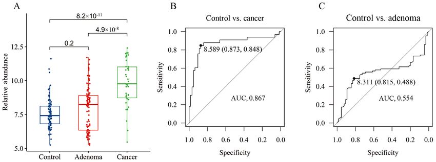
2 XU et al: P. micra ALONG THE ADENOMA-CARCINOMA SEQUENCE
revealed the relationship between P. micra and CRC (15‑17). sample collection. The clinical variables included age, sex and
By analyzing the 16S rRNA gene sequence data of 509 fecal BMI (kg/m 2). All 277 participants (age range, 26‑88 years)
samples from ethnically different cohorts, including those had been local residents of Suzhou city for >5 years prior to
from China and Austria, Yu et al (2) observed that the detec‑ the study. In total, one fecal sample was self‑collected prior
tion rate and abundance of P. micra were significantly higher to bowel preparation the day before colonoscopy from each
in patients with CRC compared with controls, and these results patient or healthy subject. Samples were transported to the
were further validated using quantitative PCR (qPCR) in 309 laboratory within 24 h after collection.
subjects (18). By analyzing the metagenomics sequencing All individuals provided written informed consent prior to
results from 778 (including 386 samples from patients with participating in the study. All procedures were performed in
CRC and 392 controls) and 969 (meta‑analysis of five publicly accordance with, and were approved by, the ethical standards
available databases and two new cohorts with validation of of the institutional and/or the national research committee
the findings of two additional cohorts) stool samples, two [the Ethics Committee of The First Affiliated Hospital of
research groups discovered that CRC‑related microbial Soochow University; approval no. + 056 (2016)], and with the
markers, including P. micra, could be consistently detected 1964 Helsinki declaration and its later amendments or compa‑
among different populations, regardless of the detection rable ethical standards.
techniques, diet, geographical environment, genetics and other
factors (19,20). These results demonstrate that P. micra has an Nucleic acid extraction and storage. Stool samples were imme‑
important relationship with CRC, and may be involved in the diately frozen in liquid nitrogen and stored at ‑80˚C. DNA was
development of CRC. extracted using a TIANamp Stool DNA kit (Tiangen Biotech
Most cancer types arise from adenoma, and colorectal Co., Ltd.) according to the manufacturer's protocols (27). The
adenoma (CRA) is a critical precursor of CRC (21,22). The integrity of DNA was measured via 2% (w/v) agarose gel
process of CRC development begins with an aberrant crypt, electrophoresis. Purified nucleic acids were quantified using
which evolves into a polyp or adenoma and eventually a Qubit 3.0 instrument (Thermo Fisher Scientific, Inc.), and
progresses to CRC over an estimated 10‑15 year period (1). stored at ‑80˚C. Nucleic acids were extracted from all stool
Currently, the microbiota associated with CRA have not been samples in a single batch by one operator to avoid inter‑batch
consistently identified, and the association between P. micra variation.
and CRA remains elusive (23‑25). Therefore, the present study
aimed to investigate the association between P. micra and CRA qPCR. All reactions were performed in a 96‑well optical PCR
by measuring the changes in the relative abundance of P. micra plate. Each reaction contained 40 ng extracted fecal DNA,
in stool samples obtained along the adenoma‑carcinoma 250 nM primers and 2X ChamQ Universal SYBR qPCR
sequence using a qPCR method. Furthermore, the alteration Master Mix (Vazyme Biotech Co., Ltd.) in 20 µl reaction
pattern of the relative abundance of P. micra were evaluated in volume. Amplification and detection of DNA was performed
patients with CRC or CRA by analyzing four public 16S rRNA with the Applied Biosystems 7500 Fast Real‑Time PCR
datasets. System (Applied Biosystems; Thermo Fisher Scientific, Inc.)
with the following reaction conditions: Initial denaturation at
Patients and methods 50˚C for 2 min and 95˚C for 2 min, followed by 40 cycles at
95˚C for 15 sec and 60˚C for 1 min. The primers sequences
Patient recruitment and sample collection. An observational (V3‑V4) were as follows: P. micra forward, 5'‑GTCACTACG
case‑control study was conducted between January 2017 GAAGAAT TTGTC‑3' and reverse, 5'‑GGCT TGAGCGAT
and March 2019 at The First Affiliated Hospital of Soochow AATA ACT TC‑3'; and total bacterial DNA forward, 5'‑GTG
University. Stool samples were collected prior to colonoscopy. STGCAYGGYTGTCGTCA‑3' and reverse, 5'‑ACGTCRTCC
All patients with CRC (37 males and 29 females) and CRA MCACCTTCCTC‑3'. Each sample was assayed three times.
(66 males and 62 females) were first diagnosed via colonoscopy Results were analyzed using 2‑ΔΔCq method (28).
screening, and the diagnosis was later confirmed by pathology.
The pathological diagnosis was performed by two profes‑ Meta‑analysis of datasets from publications. A systematic
sionals. Inclusion criteria were as follows: i) Age ≥18 years old; PubMed search with the terms 16S, colorectal cancer or
and ii) colonoscopy. The exclusion criteria for all participants adenoma and gut microbiome was performed to identify
included the use of the following medicines: Antibiotics within studies involving 16S rRNA sequencing of stool samples from
1 month of study participation, non‑steroidal anti‑inflamma‑ patients with CRC or CRA, and healthy controls. Available
tory drugs or probiotics. Individuals who reported chronic data was only found in four studies: Zeller et al (29) (acces‑
bowel disorders, food allergies or dietary restrictions were sion no. ERP005534); Zackular et al (8) (http://www.mothur.
also excluded from the study. Additional exclusion criteria for org/MicrobiomeBiomarkerCRC); Baxter et al (15) (accession
patients with CRC included chemotherapy or radiation treat‑ no. SRP062005); and Mori et al (30). All four datasets were
ment prior to surgery. All patients were categorized according obtained from samples from patients with CRA or CRC, and
to histopathological features on the basis of the TNM clas‑ healthy subjects as controls (Table I).
sification of malignant tumors after surgery (26). A total of
83 healthy subjects (43 males and 40 females) were selected Bioinformatics and sequence analysis. During data
as controls by volunteering during a physical examination, processing, short overlapping forward and reverse reads
and none of the healthy subjects had gastrointestinal tract from the same fragment were joined together using
disorders or any antibiotics treatments in the 3 months before PANDAseq (v0.21.1) to form overlapping sequences of the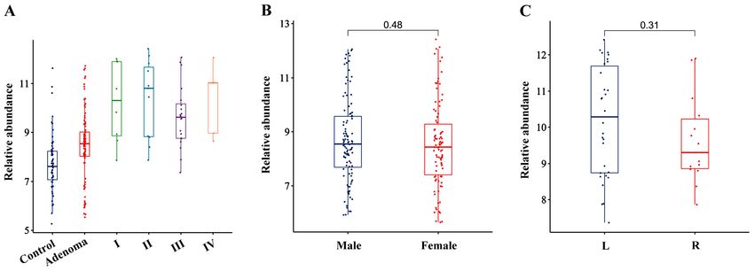
ONCOLOGY LETTERS 20: 106, 2020                                                  3

Table I. Characteristics of the datasets included in this study.

Named             Author               Country          Healthy    CRA       CRC       Region of 16S rRNA           Seq platform

crc2        Zeller et al (29)       France                50         38        41                V4                Illumina MiSeq
crc4        Zackular et al (8)      USA                   30         30        30                V4                Illumina MiSeq
crc45       Baxter et al (15)       USA+Canada            87        147        79                V4                Illumina MiSeq
crc49       Mori et al (30)         Italy                 18         38         8                V4                Illumina MiSeq

CRC, colorectal cancer; CRA, colorectal adenoma.

                                                                   Table II. Demography of patients.
V3‑V4 16S region (31). After joining, the resulting fragments
were trimmed using Trimmomatic (v0.30) (32). The average           Group            Healthy       CRA           CRC        P‑value
probability of a base being called in error was
4 XU et al: P. micra ALONG THE ADENOMA-CARCINOMA SEQUENCE
Table III. Diagnostic performance of Parvimonas micra. Discussion
CRC vs. CRA vs. The present study used qPCR to measure the relative abun‑
Value Group healthy healthy dance of P. micra in healthy individuals, patients with CRA
and patients with CRC, and demonstrated that the relative
Actual value abundance of P. micra was similar in the healthy and CRA
Healthy 19 19 groups, but significantly increased in the CRC group. The
CRC/CRA 10 26 fecal level of P. micra could effectively distinguish patients
Predicted value Healthy 18 17 with CRC from healthy controls (AUC, 0.867) but could only
CRC/CRA 7 10 poorly distinguish patients with CRA from healthy controls
False positive rate 0.3 0.615 (AUC, 0.554). The same alteration pattern in fecal P. micra
False negative rate 0.053 0.105 abundance, which was low in healthy controls and patients
with CRA, but elevated in patients with CRC, was identified
CRC, colorectal cancer; CRA, colorectal adenoma. in all four public 16S RNA sequencing datasets. These results
suggested that P. micra was closely associated with, and may
serve as a diagnostic marker of, CRC but not CRA. To the best
of our knowledge, the present study was the first to demon‑
Table IV. A correlation between the relative abundance of strate the association of P. micra with CRC but not CRA in a
Parvimonas micra and the characteristics of patients. large number of cases and using two different methods. The
present study had some limitations, such as the mechanism of
Factors r P‑value Method
P. micra in CRC initiation and development was not clear and
whether changes in its abundance are influenced by a number
Age, years 0.2812 0.00006 Pearson
of host extrinsic factors, including diet medications and other
BMI, kg/m2 ‑0.0319 0.6759 Pearson
lifestyle components, such as exercise, smoking, and sleep
Sex 0.4845 Mann‑Whitney cycles was not assessed. The aforementioned points should be
Tumor stage, I, II, III, IV ‑0.0720 0.5383 Kendall explored in future studies.
Early screening is essential for the prevention of
r: 0‑0.3, uncorrelated; 0.3‑0.5, weakly correlated; 0.5‑0.8, moderately
CRC and the survival of patients with CRC, as the 5‑year
correlated; >0.8, strongly correlated.
survival rate >90% if CRC is detected at an early stage but
decreases to 10% if it is discovered at an advanced metastatic
stage (24). Currently, the methods for CRC screening are the
fecal occult blood test (FOBT), fecal DNA test, detection of
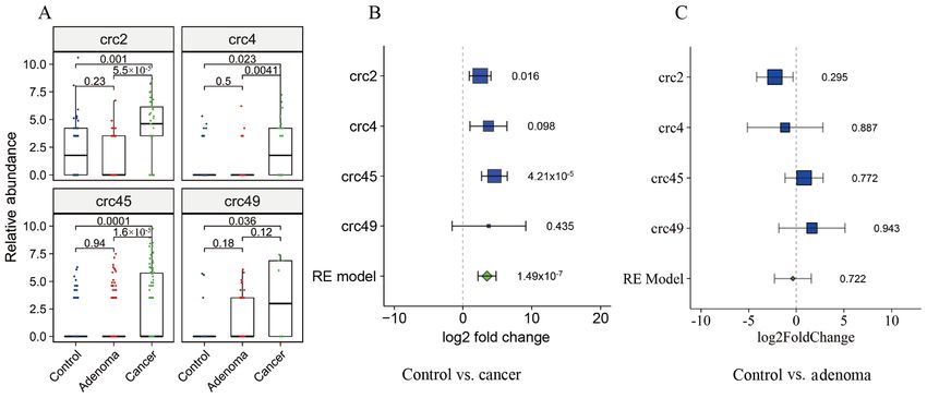
FNR (false negative rate) of 0.3 (Table III). However, the CRA tumor markers and colonoscopy. However, these methods
samples were poorly distinguished from the healthy control suffer from high costs, invasiveness and/or low sensi‑
samples (AUC, 0.554 at a cutoff of 8.311) with an FPR of 0.105 tivity (8,25). The FOBT is currently the standard non‑invasive
and an FNR of 0.615 (Fig. 1C; Table III). These results screening test, which has limited sensitivity and specificity for
suggested that P. micra may serve as a diagnostic marker for CRC and does not reliably detect precancerous lesions (29).
CRC, but not for CRA. A previous study indicated that the accuracy of fecal micro‑
It was also identified that P. micra was predominantly biota detection was similar to that of the standard FOBT, and
enriched in stages I/II and III/IV of CRC compared with when both approaches were combined, the sensitivity can
the healthy controls (Fig. 2A), and the relative abundance be ≤45% while maintaining the specificity of FOBT (29). In
of P. micra was not affected by the sex, BMI or the site of addition, combining a fecal immunochemical test (FIT) with
cancer origin (right and left) of the patients but was affected the detection of diagnostic markers, such as F. nucleatum,
by age when a Pearson correlation was used (Fig. 2B and C; Peptostreptococcus anaerobius and P. micra, can signifi‑
Table IV). cantly increase the detection rate for CRC with a sensitivity
of 92.3% and a specificity of 93.0% (18). The combined test
Meta‑analysis of 16S rRNA sequencing datasets. The asso‑ identifies >75% of the CRC samples missed by the stand‑alone
ciation between P. micra and CRC was analyzed in four FIT (18). Similarly, the present results suggested that the fecal
public datasets. A total of 596 samples, including 158 CRC, level of P. micra can effectively distinguish patients with CRC,
253 CRA and 185 healthy control samples, were included indicating that P. micra can be used as a diagnostic marker for
in the analysis after quality filtering. Compared with the CRC screening.
healthy controls, the relative abundance of P. micra in patients To evaluate the role of the gut microbiota in CRC initia‑
with CRC was significantly higher in all four datasets (crc2, tion and development, researchers have proposed a number
P=0.001; crc4, P=0.023; crc45, P=0.0001; crc49, P=0.036) of models (45-47), including the ‘driver‑passenger’ model,
but was not different in patients with CRA (P=0.18‑0.94) first suggested by Tjalsma et al (48). In the ‘driver‑passenger’
(Fig. 3A). Furthermore, there were significant increases in the model, the ‘drivers’ are defined as microbial species that
fold changes in the relative abundance of P. micra in the CRC increase in abundance in the early stage of CRC, such as
group compared with the healthy control group in all four adenoma, while the ‘passengers’ are defined as those species
datasets, while there were few changes between the healthy that increase in abundance in the late stage of CRC (46).
control and CRA groups (Fig. 3B and C). Drivers are the primary pathogens that cause the initiationONCOLOGY LETTERS 20: 106, 2020 5 Figure 1. Quantitative detection of fecal P. micra in samples from healthy controls, patients with CRC and patients with CRA in the Suzhou cohort. (A) Boxplot of P. micra relative abundances in the healthy control, CRA and CRC groups. Receiver operating characteristic curve of P. micra for the discrimination of patients with (B) CRC and (C) CRA from healthy control subjects. AUC, area under the curve; P. micra, Parvimonas micra. Figure 2. Association between the relative abundance of Parvimonas micra and the characteristics of patients. Association between the relative abundance of Parvimonas micra and (A) cancer progression, the (B) sex of patients and the (C) site of cancer origin. L, Left; R, Right. Figure 3. Meta‑analysis of P. micra relative abundance in four publicly available datasets. (A) Boxplot of the relative abundance of P. micra in healthy control, CRA and CRC samples. Forest plot of the fold changes in the P. micra relative abundance in the form of the ratios of the values for (B) patients with CRC over healthy controls and those for (C) patients with CRA over healthy controls. P. micra, Parvimonas micra; CRC, colorectal cancer; CRA, colorectal adenoma; RE model, random effect model.
6                                 XU et al: P. micra ALONG THE ADENOMA-CARCINOMA SEQUENCE

of tumors, and passengers are more suited to survive in the          JX, MY and DW participated in the experimental study and
gut microenvironment resulting from tumorigenesis (46). An           data analysis. JX, MY and SZ participated in data collection
example of a passenger is F. nucleatum, which is enriched in         and statistical analysis. JX, YZ and WC conceived the idea of,
CRC but not in CRA cases (49). The present study identified          and designed the study. All authors read and approved the final
a significant elevation of P. micra in CRC but not in CRA            manuscript.
cases. Consistent with these findings, it has been shown that
P. micra is predominantly enriched in stages I/II and III/IV,        Ethics approval and consent to participate
and its abundance is decreased after tumor resection, indi‑
cating that P. micra is not the cause of carcinogenesis but          All individuals provided written informed consent prior to
is adapted to the CRC microenvironment (50,51). Therefore,           participating in the study. All procedures were performed in
P. micra may be a passenger in the driver‑passenger model.           accordance with and were approved by the ethical standards
     P. micra is a component of the healthy commensal flora of       of the institutional and/or the national research committee [the
the gastrointestinal tract, and an opportunistic pathogen (10). As   Ethics Committee of The First Affiliated Hospital of Soochow
types of periodontal bacteria, P. micra and F. nucleatum have        University; approval no. + 056 (2016)], and with the 1964
synergistic effects on biofilm formation, which is important         Helsinki declaration and its later amendments or comparable
for the colonization by these two species of apical periodon‑        ethical standards.
titis lesions (52). P. micra significantly enhances the activity
of gingipains, which are virulence factors in Porphyromonas          Patient consent for publication
gingiva that are important in periodontal disease (53). P. micra
may also promote cancer development, although the exact              Not applicable.
mechanism it yet to be fully elucidated. Moreover, P. micra may
contribute to the pathogenesis of periodontitis by stimulating       Competing interests
Toll‑like receptor 4, nucleotide binding oligomerization domain
containing (NOD)1 and NOD2 (54). It has also been reported that      The authors declare that they have no competing interests.
P. micra may be involved in gut bacterial translocation and the
upregulation of interleukins in the tumor microenvironment (55).     References
A previous study demonstrated that APC Min/+ mice gavaged
with P. micra exhibited a significantly higher tumor burden and       1. Dekker E, Tanis PJ, Vleugels JLA, Kasi PM and Wallace MB:
                                                                         Colorectal cancer. Lancet 394: 1467‑1480, 2019.
tumor load, and cell proliferation was significantly higher in the    2. Yu J, Feng Q, Wong SH, Zhang D, Liang QY, Qin Y, Tang L,
colon tissues of P. micra gavaged germ‑free mice compared with           Zhao H, Stenvang J, Li Y, et al: Metagenomic analysis of faecal
control mice (56). Furthermore, the tumor promoting effect of            microbiome as a tool towards targeted non‑invasive biomarkers
                                                                         for colorectal cancer. Gut 66: 70‑78, 2017.
P. micra has been reported to be associated with altered immune       3. Louis P, Hold GL and Flint HJ: The gut microbiota, bacterial
responses and increased inflammation in the gut (50,56). These           metabolites and colorectal cancer. Nat Rev Microbiol 12:
findings indicate that P. micra is primarily adapted to the CRC          661‑672, 2014.
                                                                      4. Niederreiter L, Adolph TE and Tilg H: Food, microbiome and
microenvironment and could contribute to a pro‑tumoral inflam‑           colorectal cancer. Dig Liver Dis 50: 647‑652, 2018.
matory environment in patients susceptible to developing CRC.         5. Saus E, Iraola‑Guzmán S, Willis JR, Brunet‑Vega A and
     In conclusion, the present study identified that P. micra was       Gabaldón T: Microbiome and colorectal cancer: Roles in carcino‑
                                                                         genesis and clinical potential. Mol Aspects Med 69: 93‑106, 2019.
associated with CRC and may serve as a diagnostic marker              6. De Almeida CV, de Camargo MR, Russo E and Amedei A: Role
for CRC. In addition, P. micra was not enriched in patients              of diet and gut microbiota on colorectal cancer immunomodula‑
with CRA, suggesting that it serves a limited role in the                tion. World J Gastroenterol 25: 151‑162, 2019.
                                                                      7. Gagnière J, Raisch J, Veziant J, Barnich N, Bonnet R, Buc E,
tumorigenesis of CRA.                                                    Bringer MA, Pezet D and Bonnet M: Gut microbiota imbalance
                                                                         and colorectal cancer. World J Gastroenterol 22: 501‑518, 2016.
Acknowledgements                                                      8. Zackular JP, Rogers MA, Ruffin MT IV and Schloss PD: The
                                                                         human gut microbiome as a screening tool for colorectal cancer.
                                                                         Cancer Prev Res (Phila) 7: 1112‑1121, 2014.
Not applicable.                                                       9. Wong SH, Zhao L, Zhang X, Nakatsu G, Han J, Xu W,
                                                                         Xiao X, Kwong TNY, Tsoi H, Wu WKK, et al: Gavage of
                                                                         fecal samples from patients with colorectal cancer promotes
Funding                                                                  intestinal carcinogenesis in germ‑free and conventional mice.
                                                                         Gastroenterology 153: 1621‑1633.e6, 2017.
This study was supported by grants from the National Natural         10. Baghban A and Gupta S: Parvimonas micra: A rare cause of
                                                                         native joint septic arthritis. Anaerobe 39: 26‑27, 2016.
Science Foundation of China (grant no. 81672372).                    11. Khan MS, Ishaq M, Hinson M, Potugari B and Rehman AU:
                                                                         Parvimonas micra bacteremia in a patient with colonic carci‑
Availability of data and materials                                       noma. Caspian J Intern Med 10: 472‑475, 2019.
                                                                     12. Yun SS, Cho HS, Heo M, Jeong JH, Lee HR, Ju S, Kim JY,
                                                                         You JW, Cho YJ, Jeong YY, et al: Lung abscess by actinomyces
The datasets used and/or analyzed during the current study are           odontolyticus and Parvimonas micra co‑infection presenting as
available from the corresponding author on reasonable request.           acute respiratory failure: A case report. Medicine (Baltimore) 98:
                                                                         e16911, 2019.
                                                                     13. Coker OO, Dai Z, Nie Y, Zhao G, Cao L, Nakatsu G, Wu WK,
Authors' contributions                                                   Wong SH, Chen Z, Sung JJY and Yu J: Mucosal micro‑
                                                                         biome dysbiosis in gastric carcinogenesis. Gut 67: 1024‑1032, 2018.
                                                                     14. Sawai T, Koga S, Ide S, Yoshioka S, Matsuo N and Mukae H: An
JX, MY and SY analyzed and interpreted the patient data from             iliopsoas abscess caused by Parvimonas micra: A case report.
patients with CRC. DW, YZ and WC wrote the manuscript.                   J Med Case Rep 13: 47, 2019.
ONCOLOGY LETTERS 20: 106, 2020 7
15. Baxter NT, Koumpouras CC, Rogers MA, Ruffin MT IV and 35. Edgar RC: Search and clustering orders of magnitude faster than
Schloss PD: DNA from fecal immunochemical test can replace BLAST. Bioinformatics 26: 2460‑2461, 2010.
stool for detection of colonic lesions using a microbiota‑based 36. Quast C, Pruesse E, Yilmaz P, Gerken J, Schweer T, Yarza P,
model. Microbiome 4: 59, 2016. Peplies J and Glöckner FO: The SILVA ribosomal RNA gene
16. Zhang Y, Yu X, Yu E, Wang N, Cai Q, Shuai Q, Yan F, Jiang L, database project: Improved data processing and web‑based tools.
Wang H, Liu J, et al: Changes in gut microbiota and plasma Nucleic Acids Res 41 (Database Issue): D590‑D596, 2013.
inflammatory factors across the stages of colorectal tumorigen‑ 37. Griffen AL, Beall CJ, Firestone ND, Gross EL, Difranco JM,
esis: A case‑control study. BMC Microbiol 18: 92, 2018. Hardman JH, Vriesendorp B, Faust RA, Janies DA and Leys EJ:
17. Baxter NT, Ruffin MT IV, Rogers MA and Schloss PD: CORE: A phylogenetically‑curated 16S rDNA database of the
Microbiota‑based model improves the sensitivity of fecal immu‑ core oral microbiome. PLoS One 6: e19051, 2011.
nochemical test for detecting colonic lesions. Genome Med 8: 37, 38. Liang JQ, Li T, Nakatsu G, Chen YX, Yau TO, Chu E, Wong S,
2016. Szeto CH, Ng SC, Chan FKL, et al: A novel faecal lachnoclos‑
18. Wong SH, Kwong TNY, Chow TC, Luk AKC, Dai RZW, tridium marker for the non‑invasive diagnosis of colorectal
Nakatsu G, Lam TYT, Zhang L, Wu JCY, Chan FKL, et al: adenoma and cancer. Gut 69: 1248‑1257, 2020.
Quantitation of faecal Fusobacterium improves faecal immuno‑ 39. Paulson JN, Stine OC, Bravo HC and Pop M: Differential
chemical test in detecting advanced colorectal neoplasia. Gut 66: abundance analysis for microbial marker‑gene surveys. Nat
1441‑1448, 2017. Methods 10: 1200‑1202, 2013.
19. Wirbel J, Pyl PT, Kartal E, Zych K, Kashani A, Milanese A, 40. Viechtbauer W: Conducting meta‑analyses in R with the metafor
Fleck JS, Voigt AY, Palleja A, Ponnudurai R, et al: Meta‑analysis package. J Stat Softw 36: 1‑48, 2010.
of fecal metagenomes reveals global microbial signatures that 41. Love MI, Huber W and Anders S: Moderated estimation of fold
are specific for colorectal cancer. Nat Med 25: 679‑689, 2019. change and dispersion for RNA‑seq data with DESeq2. Genome
20. Thomas AM, Manghi P, Asnicar F, Pasolli E, Armanini F, Biol 15: 550, 2014.
Zolfo M, Beghini F, Manara S, Karcher N, Pozzi C, et al: 42. Robin X, Turck N, Hainard A, Tiberti N, Lisacek F, Sanchez JC
Metagenomic analysis of colorectal cancer datasets identifies and Müller M: pROC: An open‑source package for R and S+ to
cross‑cohort microbial diagnostic signatures and a link with analyze and compare ROC curves. BMC Bioinformatics 12: 77,
choline degradation. Nat Med 25: 667‑678, 2019. 2011.
21. Castellarin M, War ren RL, Freeman JD, Dreolini L, 43. Wickham H: ggplot2: Elegant Graphics for Data Analysis.
Krzywinski M, Strauss J, Barnes R, Watson P, Allen‑Vercoe E, Springer‑Verlag New York, NY, 2016.
Moore RA and Holt RA: Fusobacterium nucleatum infection 44. Alboukadel K: ggpubr: ‘ggplot2’ based publication ready plots.
is prevalent in human colorectal carcinoma. Genome Res 22: R package version 0.4.0, 2018.
299‑306, 2012. 45. Sears CL and Pardoll DM: Perspective: Alpha‑bugs, their
22. Valcz G, Sipos F, Krenács T, Molnár J, Patai AV, Leiszter K, microbial partners, and the link to colon cancer. J Infect Dis 203:
Tóth K, Solymosi N, Galamb O, Molnár B and Tulassay Z: 306‑311, 2011.
Elevated osteopontin expression and proliferative/apoptotic ratio 46. Vogelstein B and Kinzler KW: The multistep nature of cancer.
in the colorectal adenoma‑dysplasia‑carcinoma sequence. Pathol Trends Genet 9: 138‑141, 1993.
Oncol Res 16: 541‑545, 2010. 47. Fearon ER: Molecular genetics of colorectal cancer. Annu Rev
23. Sze MA and Schloss PD: Leveraging existing 16S rRNA gene Pathol 6: 479‑507, 2011.
surveys to identify reproducible biomarkers in individuals with 48. Tjalsma H, Boleij A, Marchesi JR and Dutilh BE: A bacterial
colorectal tumors. mBio 9: e00630‑18, 2018. driver‑passenger model for colorectal cancer: Beyond the usual
24. Zhang B, Xu S, Xu W, Chen Q, Chen Z, Yan C, Fan Y, Zhang H, suspects. Nat Rev Microbiol 10: 575‑582, 2012.
Liu Q, Yang J, et al: Leveraging fecal bacterial survey data to 49. Amitay EL, Werner S, Vital M, Pieper DH, Höfler D, Gierse IJ,
predict colorectal tumors. Front Genet 10: 447, 2019. Butt J, Balavarca Y, Cuk K and Brenner H: Fusobacterium and
25. Shah MS, DeSantis TZ, Weinmaier T, McMurdie PJ, Cope JL, colorectal cancer: Causal factor or passenger? Results from
Altrichter A, Yamal JM and Hollister EB: Leveraging a large colorectal cancer screening study. Carcinogenesis 38:
sequence‑based faecal microbial community survey data to 781‑788, 2017.
identify a composite biomarker for colorectal cancer. Gut 67: 50. Mizutani S, Yamada T and Yachida S: Significance of the gut
882‑891, 2018. microbiome in multistep colorectal carcinogenesis. Cancer
26. Russo E, Bacci G, Chiellini C, Fagorzi C, Niccolai E, Taddei A, Sci 111: 766‑773, 2020.
Ricci F, Ringressi MN, Borrelli R, Melli F, et al: Preliminary 51. Yachida S, Mizutani S, Shiroma H, Shiba S, Nakajima T,
comparison of oral and intestinal human microbiota in patients Sa kamoto T, Watanabe H, Masuda K, Nishimoto Y,
with colorectal cancer: A pilot study. Front Microbiol 8: 2699, Kubo M, et al: Metagenomic and metabolomic analyses reveal
2018. distinct stage‑specific phenotypes of the gut microbiota in
27. Sheng Q, Du H, Cheng X, Cheng X, Tang Y, Pan L, Wang Q colorectal cancer. Nat Med 25: 968‑976, 2019.
and Lin J: Characteristics of fecal gut microbiota in patients with 52. Horiuchi A, Kokubu E, Warita T and Ishihara K: Synergistic biofilm
colorectal cancer at different stages and different sites. Oncol formation by Parvimonas micra and Fusobacterium nucleatum.
Lett 18: 4834‑4844, 2019. Anaerobe 62: 102100, 2020.
28. Livak KJ and Schmittgen TD: Analysis of relative gene expres‑ 53. Neilands J, Davies JR, Bikker FJ and Svensäter G: Parvimonas micra
sion data using real‑time quantitative PCR and the 2(‑Delta Delta stimulates expression of gingipains from Porphyromonas gingi‑
C(T)) method. Methods 25: 402‑408, 2001. valis in multi‑species communities. Anaerobe 55: 54‑60, 2019.
29. Zeller G, Tap J, Voigt AY, Sunagawa S, Kultima JR, Costea PI, 54. Marchesan J, Jiao Y, Schaff RA, Hao J, Morelli T, Kinney JS,
Amiot A, Böhm J, Brunetti F, Habermann N, et al: Potential of Gerow E, Sheridan R, Rodrigues V, Paster BJ, et al: TLR4,
fecal microbiota for early‑stage detection of colorectal cancer. NOD1 and NOD2 mediate immune recognition of putative
Mol Syst Biol 10: 766, 2014. newly identified periodontal pathogens. Mol Oral Microbiol 31:
30. Mori G, Rampelli S, Orena BS, Rengucci C, De Maio G, 243‑258, 2016.
Barbieri G, Passardi A, Casadei Gardini A, Frassineti GL, 55. Ge W, Hu H, Cai W, Xu J, Hu W, Weng X, Qin X, Huang Y,
Gaiarsa S, et al: Shifts of faecal microbiota during sporadic Han W, Hu Y, et al: High‑risk stage III colon cancer patients iden‑
colorectal carcinogenesis. Sci Rep 8: 10329, 2018. tified by a novel five‑gene mutational signature are characterized
31. Masella AP, Bartram AK, Truszkowski JM, Brown DG and by upregulation of IL‑23A and gut bacterial translocation of the
Neufeld JD: PANDAseq: Paired‑end assembler for illumina tumor microenvironment. Int J Cancer 146: 2027‑2035, 2020.
sequences. BMC Bioinformatics 13: 31, 2012. 56. Yu J, Zhao LY, Zhao RS, Long XH, Coker O and Sung JJY:
32. Bolger AM, Lohse M and Usadel B: Trimmomatic: A flex‑ The role of Parvimonas micra in intestinal tumorigenesis in
ible trimmer for Illumina sequence data. Bioinformatics 30: germ‑free and conventional APCmin/+ mice. J Clin Oncol 37
2114‑2120, 2014. (Suppl 4): S531, 2019.
33. Rognes T, Flouri T, Nichols B, Quince C and Mahé F: VSEARCH:
A versatile open source tool for metagenomics. PeerJ 4: e2584, This work is licensed under a Creative Commons
2016. Attribution-NonCommercial-NoDerivatives 4.0
34. Caporaso JG, Kuczynski J, Stombaugh J, Bittinger K, International (CC BY-NC-ND 4.0) License.
Bushman FD, Costello EK, Fierer N, Peña AG, Goodrich JK,
Gordon JI, et al: QIIME allows analysis of high‑throughput
community sequencing data. Nat Methods 7: 335‑336, 2010.You can also read