Effectiveness of Cardiac Scintigraphy and Standard Eco-Cardiograph in the Detection and Evaluation of Cardiac Disorders, Including Ischemic Heart ...
←
→
Page content transcription
If your browser does not render page correctly, please read the page content below
Journal of Biosciences and Medicines, 2021, 9, 90-109
                                                                                               https://www.scirp.org/journal/jbm
                                                                                                         ISSN Online: 2327-509X
                                                                                                           ISSN Print: 2327-5081

Effectiveness of Cardiac Scintigraphy and
Standard Eco-Cardiograph in the Detection and
Evaluation of Cardiac Disorders, Including
Ischemic Heart Diseases

Saeed M. Bafaraj

Diagnostic Radiology Department, Faculty of Applied Medical Sciences, King Abdulaziz University, Jeddah, KSA

How to cite this paper: Bafaraj, S.M.         Abstract
(2021) Effectiveness of Cardiac Scintigra-
phy and Standard Eco-Cardiograph in the       Background: Cardiac disorders are the leading causes of morbidity and mor-
Detection and Evaluation of Cardiac Dis-      tality globally. Aim: The current study aimed to compare the effectiveness of
orders, Including Ischemic Heart Diseases.
                                              echocardiographic and scintigraphy investigation in cardiovascular disorders
Journal of Biosciences and Medicines, 9,
90-109.                                       detection and evaluation, including ischemic heart disease (IHD) diagnosis at
https://doi.org/10.4236/jbm.2021.94007        King Abdulaziz University in Saudi Arabia. Methods: 157 patients (male/
                                              female: 68%/32%; mean age 64 ± 0.83 years) were included in the report in
Received: January 23, 2021
                                              the current study. All subjects underwent gated myocardial perfusion scinti-
Accepted: April 16, 2021
Published: April 19, 2021                     graphy and standard Echocardiography. Results: About 84% of cases were
                                              diagnosed with any types of cardiac disorders by Echocardiography, whereas
Copyright © 2021 by author(s) and             through scintigraphy, 61% of patients were detected cardiac disorders. The
Scientific Research Publishing Inc.
                                              age stratification did not impact the prevalence of cardiac disorders detected
This work is licensed under the Creative
Commons Attribution International             by them. Scintigraphy showed a higher detection rate (59%), while only 29%
License (CC BY 4.0).                          of cases were detected with IHD by eco-cardiograph. Conclusions: In con-
http://creativecommons.org/licenses/by/4.0/   clusion, both techniques, namely standard Echocardiography, and myocardial
               Open Access
                                              scintigraphy, are useful in the evaluation and detection of cardiac disorders in
                                              patients having any type of cardiac problems. Both investigations showed a
                                              differential pattern in cardiac disorders diagnosis with a particular focus on
                                              IHD. Gender differences and age stratification also contributed to this diffe-
                                              rential pattern of diagnosis. Trial Registration: The research doesn’t include
                                              experiments in humans or animals. It is a retrospective study for data record
                                              review as an observational study, so no trial registration is required.

                                              Keywords
                                              Cardiac Disorders, Evaluation, Comparison, Standard Echocardiography,
                                              Cardiac Scintigraphy

DOI: 10.4236/jbm.2021.94007 Apr. 19, 2021                        90                         Journal of Biosciences and Medicines
S. M. Bafaraj

                              1. Introduction
                              Cardiovascular diseases are the most common cause of disability and mortality
                              worldwide, with a prevalence of 126 million persons affected globally (1655 per
                              100,000) and 17.9 million mortalities making it 31% of all global deaths [1] [2].
                              The term “heart disease or cardiac disorders” refers to several conditions where
                              heart functions or structures are impaired and abnormal [3]. Characterized by
                              atherosclerosis in the arteries, coronary artery disease (CAD) is the most com-
                              mon heart disease which is also the most frequent cause of heart attack [2]. Oth-
                              er significant cardiac disorders are valvular disorders, congenital heart diseases,
                              and others. More than 600,000 Americans die from cardiac or heart disease
                              every year, leading to one in every four deaths at the statistical level. Though the
                              efforts of prevention of cardiovascular disease at primary, secondary, and ter-
                              tiary levels are mandatory, diagnostic modalities at this regard need to be
                              re-visited and re-evaluated from various contexts. A lot of arrangements should
                              be and can be made at different stages of health care services to combat this life
                              killer through early diagnosis and consequent therapeutic and preventive strate-
                              gies.
                                CAD is regarded as the most common fatal disease all over the world [4]. The
                              identification of patients with myocardial ischemia is an integral part of the di-
                              agnosis of CAD [1] [2]. To date, Echocardiography is a first-line investigation to
                              detect cardiac disorders. Echocardiography is the potential in providing in-depth
                              and thorough comprehensive profiling of both the structural, morphological,
                              anatomical, and hemodynamic changes which are induced and aggravated by
                              either acute or chronic CAD [5] [6]. Thus, over a long time standard, Echocar-
                              diography occupies a vital position in evaluating the CAD in saving a hundred
                              thousand lives. Standard Echocardiography, the most widespread investigation
                              in cardiology, has numerous objectives in the vulnerable cardiac population, in-
                              cluding the valuation of myocardial performance and function, valvular diseases,
                              congenital cardiac abnormalities, hemodynamics [5] [6].
                                Indeed, Echocardiography, including both two-dimensional and, in recent
                              times, three-dimensional speckle tracking Echocardiography, exhibit reliable
                              methods for the detection and assessment of global as well as regional myocardi-
                              al dysfunction has as often seen various cardiovascular diseases [7] [8]. Thus,
                              these unique and innovative echocardiographic imaging modalities have broa-
                              dened the knowledge of LV and RV mechanics, physiology, pathology, and
                              morphology. A striking drawback of this study’s valuation and interpretation is
                              the subjective visual analysis of endocardial motion and wall thickening that is
                              only semiquantitative needing more quantitative techniques. Indeed, further
                              advancement has already taken place to the incorporation of new indices in the
                              analysis of regional wall motion.
                                On the other hand, myocardial perfusion scintigraphy (MPS) is regarded as an
                              accurate and sensitive modality in diagnosing the ischemia affected regions in
                              the myocardial tissue [9] [10] [11]. Myocardial perfusion single-photon emission

DOI: 10.4236/jbm.2021.94007                        91                          Journal of Biosciences and Medicines
S. M. Bafaraj
computed tomography is considered to be the most potential and commonly
performed cardiac non-invasive imaging technologies. Scintigraphy is unique in
playing a key and vital role in the diagnosis and detection of cardiovascular dis-
orders, with assessing the disease prognosis, establishing therapeutic effective-
ness and contributes to evaluating myocardial viability [9] [11]. In this regard,
other potential diagnostics can be into consideration, such as cardiac computed
tomography scan or cardiac magnetic resonance imaging [12] [13]. Despite all
these advances, scintigraphy is still the most essential and sensitive modality in
showing and viewing the ischemia. Indeed, scintigraphy is taken as a reference
gold standard method in several recent studies in which various stress Echo
protocols are introduced [14] [15].
The present study intended to compare the effectiveness and contribution of
standard eco-cardiograph and nuclear medicine scintigraphy in detecting and
evaluating cardiac disorders at a tertiary level hospital in Saudi Arabia. The ob-
jective of the study also includes to find their effectiveness based on gender and
age of patients. This is the first effort to compare and contrast the detection of
cardiac diseases in Saudi patients using two popular and standard diagnostic
methodologies used in the field of cardiology. Here, the patients diagnosed hav-
ing cardiac disorders without further stratification were evaluated.
2. Aim
The aim of the study was to compare the effectiveness of echocardiographic and
scintigraphy investigation in cardiovascular disorders detection and evaluation,
including ischemic heart disease (IHD) diagnosis at a tertiary level hospital in
Saudi Arabia.
3. Materials and Methods
3.1. Study Population
One hundred sixty-six subjects underwent echocardiography and nuclear medi-
cine imaging at the hospital. These patients took both Echocardiography and
nuclear imaging scintigraphy. In this study, clinical data records of patients with
cardiac problems were analyzed, while cancer patients were excluded. Here
broadly, the patients diagnosed with cardiac disorders were included (such as
with ischemia, infarction, IHD and other); there were no specific criteria for
cardiac disorders patient selection in this study. Adult patients with both gend-
ers were included and were later characterized based on gender and age less than
or greater than 50 years. For IHD, they needed to be identified by Echo findings.
The study protocol has been reviewed and approved by the Institutional Eth-
ics Committee. All patients have provided written informed consent. As this
study involved human participants, therefore it was in accordance with the Hel-
sinki declaration and standard ethic standard of the Institutional Ethics Com-
mittee. Specific purpose, objective, and risks of the nuclear imaging technique
have been clearly explained to study subjects.
DOI: 10.4236/jbm.2021.94007 92 Journal of Biosciences and MedicinesS. M. Bafaraj
3.2. Study Materials
The patients underwent nitrate-enhanced rest scintigraphy with techne-
tium-99mmethoxy isobutyl isonitrile (99mTc-MIBI). Technetium-99m-sestamibi
is a myocardial perfusion radiotracer that has been commercially available in the
United States since 1990 [16] [17] [18]. Since the recent introduction of techne-
tium-99m-labeled radiotracers for myocardial perfusion imaging, a variety of
protocols for the performance of this procedure have been developed [16] [17].
3.3. Echocardiographic Examination
The EC was accomplished by Vivid 7 instruments (GE Medical Systems, Mil-
waukee, WI, USA), with a 2.5-MHz transducer and harmonic imaging. Echocar-
diographic assessments were done on the patients with lying in the left lateral
decubitus position under the recommendations and protocols of the American
Society of Echocardiography [19]. Two-dimensional images were captured at the
apical four- and five-chambers and the long axis. Indeed, both the apical
four-chamber and two-chamber views are regarded as necessary in Echocardio-
graphy to assess wall motion disorders. The left ventricular systolic and diastolic
diameters (LVS, LVD) were obtained by M-mode echocardiography. The
LV-ejection fraction (LV-EF) was determined by applying the altered version of
biplane Simpson’s method.
Strain (S) and strain rate (SR) were tracked by the speckle tracking. Tissue
Doppler-derived LVS longitudinal S and SR rates were attained in two- and
four-chamber apical views. Through Echocardiography 16 segments were as-
sessed, namely basal, middle, and apical parts of the septum, inferior, lateral, and
anterior walls, and basal and middle segments of the anterior septum and post-
erior wall. Recordings were taken at the end of the expiration phase continuing
three consecutive cycles. The well-trained specialized doctors and supporting
staffs have conducted the echocardiographic assessment.
3.4. Nuclear Medicine Scintigraphy Imaging Protocol
The study was conducted at the Department of Nuclear Medicine by specialty
medical doctors, who were wholly blinded to the other diagnostics and risk fac-
tors data. All enrolled participants had to undergo a rest protocol with 99mTc-MIBI.
Patients are prohibited from having anything orally (except oral administration
of medicine or water) after midnight before the examination to restrict gut activ-
ity as it may affect the assessment of the inferior wall of the LV. Patients must be
dressed comfortably for the exercise part of the investigation. Medicines com-
prising methylxanthines or caffeine and caffeine enriched food-beverage should
be kept away for at least 12 - 24 hours. Metal or other potential attenuators
should be taken out before imaging to prevent attenuation artifacts. Before ad-
ministering 99mTc-MIB, registered patients were provided with 1 - 2 tablets of
sublingual nitroglycerin (0.4 mg) with a duration of 5 min apart. Finally, 740
MBq of 99mTc-MIBI was injected through the intravenous route while resting,
DOI: 10.4236/jbm.2021.94007 93 Journal of Biosciences and MedicinesS. M. Bafaraj
and scintigraphy investigation was performed after 45 min.
99mTcsestamibi and 99mTc-tetrofosmin are the bulk utilized radiopharma-
ceuticals in cardiac scintigraphy. These radiopharmaceuticals show extraction
efficiency of around 65% in the physiological flow range; the absorption is in
relative equilibrium to flow. As compared to the hepatobiliary excretion of
99mTctetrofosmin, the hepatobiliary excretion of sestamibi is less, and evalua-
tion of the inferior wall is less challenging. The conventional protocol of
201Tl-chloride comprises stress imaging and consequently redistribution imag-
ing past 3 or 4 hours. When delayed, imaging is attained after 24 hours to assess
fixed faults (primarily to evaluate the myocardium feasibility). The second
201Tl-chloride procedure includes the performance of the elective delayed im-
ages on the same day, following an injection of a little dose of 201Tl-chloride in
patients with a fixed defect. The third 201Tl-chloride protocol includes acquir-
ing the 3 or 4-hour images followed by the injection of a small amount of
201Tl-chloride, and delayed images at 24 hours are still performed when re-
quired.
99mTc utilizes four protocols. The original protocol consists of two injections
of the radiopharmaceuticals on two different days. The original procedure was
altered for the convenience of patients, preferring a low dose and high dose on
the same day, initiating with either the most commonly used, rest and then
stress (second protocol) or, stress than rest (third protocol). In the fourth pro-
tocol, the so-called dual isotopes, 201Tl-chloride images are first taken by resting
injection, followed by which and 99mTc radiopharmaceutical is injected during
stress. Above mentioned protocols have their proponent, that highlights the lo-
gistical, practical consequences, as available scientific evidence has not estab-
lished the clarity of any one of these protocols. Patients with CAD and severe LV
dysfunction, can provide resting imaging to determine normal or near-normal
uptake of either 99mTc agents or 201Tl-chloride and, consequently, myocardial
viability [15].
We identified ischemic heart disease in scintigraphy by myocardial perfusion
imaging as a nuclear stress test and via pharmacologic stress.
3.5. Definition Criteria of the Positive Diagnostic Imaging Tool
Positive stress echocardiography is defined as the presence of abnormal cardiac
wall motion on either exercise or pharmacologic stress echocardiography.
A positive nuclear medicine means the heart’s blood flow is insufficient, may
occur only during the exercise phase of the stress test.
3.6. Statistical Analysis
Statistical analysis was done with SPSS 20 software package (version 17, SPSS
Inc., Chicago, IL, USA). The study parameters were expressed as mean ± stan-
dard error or as a percentage. We used the crosstabs chi-square test for categor-
ical and independent variables. Samples T-Test was used for continuous va-
DOI: 10.4236/jbm.2021.94007 94 Journal of Biosciences and MedicinesS. M. Bafaraj
riables.
4. Results
4.1. Basic Profiles of Study Participants
Although 166 patients were recruited for the current study, because of the pres-
ence of some missing data, 157 data for the final analysis were included (Figure
1).
Table 1 demonstrated the basic profiles of study participants. The mean age
was 64.0 ± 0.83 years; the ratio of male and female participants was 107:50.
Figure 1. Participants data.
Table 1. Patients characteristics.
Variables Total (n = 157) Male (n = 107) Female (n = 50) P value
Demographics
Age, years 64.0 ± 0.83 64.3 ± 1.03 63.5 ± 1.43 0.641
Age ≥ 50 years 145 (92.4) 98 (91.6) 47 (94.0) 0.596
Age < 50 years 12 (7.6) 9 (8.4) 3 (6.0)
Echocardiography
Echocardiography (positive) 131 (83.4) 86 (80.4) 45 (90.0) 0.131
IHD (identified by Echo findings) 46 (29.3) 37 (34.6) 9 (18.0) 0.033
Nuclear medicine
Nuclear medicine (positive) 96 (61.1) 71 (66.4) 25 (50.0) 0.050
Ischemia 35 (22.3) 22 (20.6) 13 (26.0) 0.068
Infarction 30 (19.1) 26 (24.3) 4 (8.0)
Ischemia and Infarction 29 (18.5) 22 (20.6) 7 (14.0)
Others 6 (3.8) 4 (3.7) 2 (4.0)
IHD (identified by NM findings) 94 (59.9) 70 (65.4) 24 (48.0) 0.038
N = 157, Values are mean ± standard error or n (%), IHD: ischemia heart diseases. Based on T-test for con-
tinuous variable and Pearson Chi-square test for the categorical variable.
DOI: 10.4236/jbm.2021.94007 95 Journal of Biosciences and MedicinesS. M. Bafaraj
There was a 7.6% study participant who was less than 50 years old. All patients
were studied at rest and stress.
Among the study patients who underwent standard echo-cardiograph, 83.4%
had been diagnosed with cardiac disorders while with scintigraphy, and this
percent was 61.1% (cardiac disorder is any disorder that affects/involves the
heart or its blood vessels). Here 29.3% of participants were diagnosed with IHD
by eco-cardiograph. In contrast, the detection rate of IHD was higher by scinti-
graphy, and that was 59%, more than two-fold higher than that of Echocardio-
graphy. The prevalence of IHD is higher in the male group than the female
groups by both technologies. The prevalence of cardiac disorders in the age
group more than 50 years was 10-fold higher than the age group below 50 by
both diagnostics. Looking at the subtypes of IHD, a very close prevalence rate of
isolated ischemia, isolated infarction, and both are found. A bit more detail of
cardiac abnormalities detected by both techniques were demonstrated in Table
2(A), Table 2(B).
4.2. Comparison by Both Technologies for Cardiac Disorders
The various abnormalities detected in echo-cardiography and scintigraphy of
current study participants have been listed in Table 3(A), Table 3(B). As shown
Table 2. (A) Cardiac disorder diagnosed by echocardiography; (B) Cardiac disorder di-
agnosed by nuclear medicine (scintigraphy).
(A)
Name of cardiac disorder Total number Disorder number Percentage (%)
Echocardiography (positive) 157 131 83.4
IHD (identified by Echo findings) 157 46 29.3
Normal global LV systolic function 157 53 33.8
Mildly decreased global LV systolic function 157 9 5.73
Moderately decreased global LV systolic function 157 7 4.46
Severely decreased global LV systolic function 157 2 1.27
Hypertensive heart disease 157 4 2.55
IHD: ischemia heart diseases; LV: left ventricular.
(B)
Name of cardiac disorder Total number Disorder number Percentage (%)
Nuclear medicine (positive) 157 96 61.1
Ischemia 157 35 22.3
Infarction 157 30 19.1
Ischemia and Infarction 157 29 18.5
IHD (identified by NM findings) 157 94 59.9
Others 157 6 3.8
IHD: ischemia heart diseases.
DOI: 10.4236/jbm.2021.94007 96 Journal of Biosciences and MedicinesS. M. Bafaraj
Table 3. (A) Echocardiography positive findings; (B) Nuclear medicine (scintigraphy)
positive findings.
(A)
Mildly decreased LV systolic function
Impaired relaxation pattern pf LV diastolic filling
Severely dilated LA
Mild LV dysfunction
Borderline concentric LV hypertrophy
Mildly dilated LA
Moderately to severely decreased Normal global LV systolic function
Mildly increased LV internal cavity size
Sclerosed aortic valve
Mild LV systolic dysfunction
Difficult subcostal view
Mild thickening and calcification of the posterior mitral valve leaflet
Mild mitral annular calcification
Mild aortic regurgitation
Mild concentric LV hypertrophy
Trivial pericardial effusion
Mildly elevated pulmonary artery systolic pressure
Mildly to moderately decreased global LV systolic function
Multiple segmental abnormalities exist
RCA distribution is abnormal
Mild to moderately mitral valve regurgitation.
Mild-moderate tricuspid regurgitation.
Moderately dilated LA
Severe mitral valve regurgitation
Mid and apical anterior wall, basal and mid anterior septum, basal inferoseptal segment, and apex
are abnormal
Positive stress echo for coronary angiography
Mild to moderate mitral valve regurgitation
Pulmonary artery systolic pressure is normal with a decrease in severity of tricuspid regurgitation
Mild mitral valve regurgitation mild tricuspid regurgitation
Mild thickening of the anterior and posterior mitral valve leaflet
Normal LV systolic function
Mild mitral stenosis
Rheumatic heart disease
Mild mitral valve regurgitation, moderate aortic regurgitation
Trivial tricuspid regurgitation with normal PASP
Moderately decreased global LV systolic function
Mild aortic valve sclerosis
Moderately enlarged RV
Moderately elevated pulmonary artery systolic pressure
Non-dilated LV with good systolic function
Trivial tricuspid regurgitation
Mildly decreased global LV systolic function
Fair LV systolic function
Difficult echo study, suboptimal echo windows
Trace mitral regurgitation, trace tricuspid regurgitation, trace pulmonary regurgitation
Mild thickening and calcification of the anterior mitral valve leaflet
Diastolic dysfunction
Trivial mitral regurgitation
Concentric LV hypertrophy
Moderate mitral valve regurgitation
Moderate LV systolic dysfunction
Mild pulmonary hypertension
Poor echo window
DOI: 10.4236/jbm.2021.94007 97 Journal of Biosciences and MedicinesS. M. Bafaraj
Continued
Nondilated LV with overall good systolic function
Trivial aortic regurgitation
Sclerocalcific changes of mitral and aortic valve
Mild aortic stenosis
Mildly decrease global LV systolic function
Type 3 LV diastolic dysfunction
Regional wall motion abnormality
Dilated LV with severely impaired systolic function
Moderately decrease global LV systolic function
Mildly increase LV internal cavity size
Aortic valve disease with mild aortic regurgitation
Mitral valve prolapses with travail mitral regurgitation
Dilated ischemic cardiomyopathy
Impaired relaxation pattern of LV diastolic filling
Small secundum arterial septal defect with predominantly right to left across the atrial septum
Mildly dilated RA
Moderate tricuspid regurgitation
IHD, S\P CABG,
Regional wall motion abnormalities
Mild thickening aortic valve with maximum SPG and no aortic regurgitation
Mildly mitral valve regurgitation
Severe LV systolic dysfunction
Trivial pulmonary regurgitation
Trivial mitral valve regurgitation
S\P CABG, IHD
RWMA
LV dysfunction EF = 40%
(B)
Ischemia
Infarction
Ischemia and Infarction
Infarction, likely dilated cardiomyopathy with a low ejection fraction
Ischemia, low ejection fraction at stress
Old infarction
Low ejection fraction at stress, no ischemia no infarction
Left bundle branch block
Ischemic cardiomyopathy
in Figure 2(A), 81 participants were diagnosed cardiac disorders by both tech-
niques, 50 subjects were by eco-cardiograph only, 15 subjects were by scintigra-
phy only, and 11 subjects did not show any cardiac disorders by both investiga-
tions.
When male population was considered (Figure 2(B)), 58 subjects showed
cardiac disorders in both diagnostic methodologies; 28 patients were by Echo-
cardiography only; 13 cases were by scintigraphy only, and eight subjects were
not diagnosed cardiac disorders by both assessments. When only the female
subjects were considered (Figure 2(C)), 23 subjects showed cardiac disorders in
both diagnostic methodologies; 22 patients were by Echocardiography only; 2
cases were by scintigraphy only, and three subjects were not diagnosed cardiac
disorders by both assessments.
When the study subjects aged less than 50 years were considered (Figure 2(D)),
DOI: 10.4236/jbm.2021.94007 98 Journal of Biosciences and MedicinesS. M. Bafaraj
Figure 2. Venn diagrams for diagnosis of cardiac disorder.
three subjects showed cardiac disorders in both diagnostic methodologies; 8 pa-
tients were by Echocardiography only; 1 case was by scintigraphy only, and no
subjects were found not diagnosed cardiac disorders by both assessments.
When the study, subjects aged more than 50 years were considered (Figure
2(E)), 78 subjects showed cardiac disorders in both diagnostic methodologies; 42
patients were by echocardiography only; 15 cases were by scintigraphy only, and
ten subjects were found not diagnosed cardiac disorders by both assessments.
4.3. Characteristics of Cardiac Scintigraphy
As shown in Figure 3(A), among the study subjects who are male and less than
50 years age, the prevalence of cardiac disorders by scintigraphy is half in com-
parison to total participants in this study group.
As shown in Figure 3(B), among the study subjects who are female and less
than 50 years age, the prevalence of cardiac disorders by scintigraphy was zero;
no case of cardiac disease was detected by scintigraphy. Among the study sub-
jects who are male and more than 50 years age, the prevalence of cardiac disord-
ers by scintigraphy was near around 2.4 times higher in comparison to the
number of participants without the cardiac disorder in this study group (Figure
3(C)). As shown in Figure 3(D), among the study subjects who are female and
more than 50 years of age, the prevalence of cardiac disorders by scintigraphy
was 53%, and the percentage of subjects without cardiac disorder was 46%. Fig-
ure 3(E) demonstrated that, among the study subjects who are male, the number
of IHD by scintigraphy was almost double without the IHD patient group.
Among the study subjects who are female, the number of IHD by scintigraphy
was almost the same as that of without IHD (Figure 3(F)).
DOI: 10.4236/jbm.2021.94007 99 Journal of Biosciences and MedicinesS. M. Bafaraj
Figure 3. Prevalence of cardiac disorder including IHD diagnosed by nuclear medicine (Scintigraphy).
As shown in Figure 3(G) and Figure 3(H), among both gender study sub-
jects, the IHD patients were almost all who were more than 50 years of age.
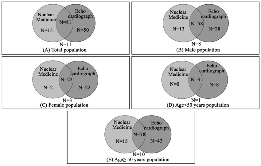
Comparison by both technologies for ischemic heart diseases (IHD). As shown
in Figure 4(A), 36 participants were diagnosed IHD by both technologies, ten
subjects were by eco-cardiograph only, 36 subjects were by scintigraphy only,
and 53 subjects did not show features of IHD by both investigations. When only
male participants were considered (Figure 4(B)), 29 subjects demonstrated by
both diagnostic methodologies; 8 patients were by Echocardiography only; 41
cases were by scintigraphy only, and 29 did not have IHD with both technolo-
gies.
When only female subjects were considered (Figure 4(C)), seven patients had
IHD in both diagnostic methodologies; 2 patients were by Echocardiography
only; 17 cases were diagnosed IHD by scintigraphy only; 24 participants had no
IHD in either of these investigations.
When the study subjects aged less than 50 years were considered (Figure
4(D)), only one subject had IHD in both technologies; 2 patients were by Echo-
cardiography only; 1 case was by scintigraphy only, and eight subjects were
found not diagnosed IHD by both assessments.
In the analysis of the patients more than 50 years of age (Figure 4(E)), 35
subjects showed IHD in both diagnostic methodologies; 8 patients were by
Echocardiography only; 57 cases were by scintigraphy only, and 45 subjects were
found not diagnosed IHD by both assessments.
Characteristics of Echocardiography
As shown in Figure 5(A), among the study subjects who are male and less
than 50 years of age, the prevalence of cardiac disorders by Echocardiography is
about eight times higher. In contrast, no echo-negative subject of cardiac disease
was found in the same age group of female (Figure 5(B)). Among the study
DOI: 10.4236/jbm.2021.94007 100 Journal of Biosciences and MedicinesS. M. Bafaraj
Figure 4. Venn diagrams for ischemic heart disease (IHD).
Figure 5. Prevalence of cardiac disorder including IHD diagnosed by Echocardiography.
DOI: 10.4236/jbm.2021.94007 101 Journal of Biosciences and MedicinesS. M. Bafaraj
subjects who are male and more than 50 years age, the prevalence of cardiac dis-
orders by Echocardiography was 4 times higher in comparison to the number of
participants without cardiac disorder in this study group (Figure 5(C)); while in
the same age group of female, the prevalence of positive cases of cardiac disorder
was about 9 times higher (Figure 5(D)).
Figure 5(E) demonstrated that, among the study subjects who are male, the
number of IHD by Echocardiography was near around half in comparison to the
number of subjects who were IHD negative. In the female group, the prevalence
of IHD positive vs. negative was 18% vs. 82% (Figure 5(F)). As shown in Figure
5(G) and Figure 5(H), among both gender study subjects, the IHD patients
were almost 90% to 100% who were in the age groups more than 50 years of age.
The validity of Echocardiography:
As shown in Table 4, sensitivity was 0.84 (0.80 - 0.89); specificity was 0.18
(0.11 - 0.25); PPV: positive predictive value was 0.61 (0.58 - 0.65); NPV: negative
predictive value was 0.42 (0.26 - 0.59); LR+: likelihood ratio for positive results
was 1.02 (0.90 - 1.19); LR−: likelihood ratio for negative results was 0.86 (0.43 -
1.75) when scintigraphy was considered as reference.
5. Discussion
The present study demonstrated the significance and evaluation of cardiac dis-
orders detection by standard Echocardiography and the nuclear medicine scin-
tigraphy at a tertiary levels hospital with the patients having any types of cardiac
problems. While conventional Echocardiography detected the greater number of
positive cases of cardiac disorders; the percentage of IHD by myocardial scinti-
graphy was near about two-fold higher than that of Echocardiography. The car-
diac disorders were ten-fold more elevated in the patients aged more than 50
years. Also, the cardiac disorders diagnosed by myocardial scintigraphy were
almost the patient of IHD.
Transthoracic Echocardiography, a non-invasive (the skin is not pierced)
technology, has gained intense popularity as the most standard imaging modali-
ty worldwide [5] [7]. In the current study, some of the common types of echo-
cardiography are M-mode echocardiography, Doppler echocardiography, Color
Doppler, and 2-D (2-dimensional) Echocardiography was used [20]. It deals
Table 4. Validity of echocardiography considering scintigraphy as reference.
Variable Total number of data (N = 157) Male (N = 107) Femail (N = 50)
Sensitivity 0.84 (0.80 - 0.89) 0.81 (0.76 - 0.87) 0.92 (0.84 - 0.97)
Specifieity 0.18 (0.11 - 0.25) 0.22 (0.12 - 0.33) 0.12 (0.04 - 0.17)
PPV 0.61 (0.58 - 0.65) 0.67 (0.63 - 0.72) 0.51 (0.47 - 0.54)
NPV 0.42 (0.26 - 0.59) 0.38 (0.21 - 0.56) 0.60 (0.24 - 0.87)
LR+ 1.02 (0.90 - 1.19) 1.05 (0.88 - 1.30) 1.04 (0.89 - 1.18)
LR− 0.86 (0.43 - 1.75) 0.82 (0.38 - 1.81) 0.66 (0.13 - 3.16)
DOI: 10.4236/jbm.2021.94007 102 Journal of Biosciences and MedicinesS. M. Bafaraj
with the information on overall cardiac function, presence of many types of
heart disease, follow up the prognosis of heart valve disease over time, evaluation
of the effectiveness of medical or surgical treatments and hemodynamics. This
technique is mainly useful in diagnosis, assessing prognosis, and determining
optimal therapy for several indications, including heart failure, ischemic heart
disease, and valve disease [7]. It occupies the top third position in the diagnosis
of cardiac disorders next to electrocardiography and chest X-ray [7]. In the
present study, more than 83% of enrolled patients were diagnosed with any types
of cardiac disorders by Echocardiography. The rate is quite high, there was no
drastic difference in the rate of cardiac disorders in the present study in context
to gender, but for IHD, it was about two-fold higher in male than female. In fact,
in a significant number of cardiovascular diseases, gender differences are evident
in the context of pathophysiology, prevalence rate, clinical presentation, man-
agement, and finally, gender-specific outcomes of cardiac disorders
(Supplementary Table S1).
Both sex and gender differences are essential in the diagnosis and manage-
ment of cardiovascular diseases. The expert’s panels of institutions and societies
from the European Union and the United States have urged and demanded
more robust reporting of gender-specific CVD outcomes. In the Euro Heart
Survey, Echocardiography was used less frequently in women [21]. Physicians
should be informed about this potential bias to reduce it [21]. On the other
hand, the male gender is an independent predictor of echocardiographic assess-
ment [22]. Echocardiography also has a potential prediction capability of first
cardiovascular events, thus enhances risk stratification for primary prevention in
the community [22]. These past study findings are consistent with the current
results.
In the present study, most of the study participants were more than 50 years
of age; there was no significant difference to be stated in the efficacy of Echocar-
diography in the detection of cardiac disorders based on age stratification
(Supplementary Table S2). IHD has been reported to be developed at a later age
for women compared to men, and accordingly, the present study also found that
the percentage of cardiac disorders are less in women than men as assessed by
Echocardiography (Supplementary Table S3) [23].
MPS is admittedly an integral diagnostic modality and management of CHD.
It helps not only in diagnosis but also guides in the decision making of the
treatment options. The scintigraphy protocol used in the present study is well
unitized [18]. In the current study, the scintigraphy was performed under the
supervision of professional guidelines. In the present study, about 61% of par-
ticipants were detected cardiac disorders by myocardial scintigraphy with a
three-fold higher proportion in the male gender. The total percentage detection
rate for cardiac disorders is almost near to that of the percentage of having IHD.
This implies that through scintigraphy, the diagnosed cardiac disorders were
almost IHD patients. But for echocardiographic assessment, the rate of positivity
of cardiac disorders in total and that of IHD are different (83% vs. 29%). Al-
DOI: 10.4236/jbm.2021.94007 103 Journal of Biosciences and MedicinesS. M. Bafaraj
though any comment could not be made on the ability of scintigraphy in the de-
tection of asymptomatic patients in the present study, recent evidence showed
that myocardial scintigraphy could also detect asymptomatic ischemia prevailing
in early CAD family history and patients with erectile dysfunction, chronic renal
failure, and type 2 diabetes. For such case detection, the recommendation to go
through scintigraphy investigation is not so popular to date (Supplementary
Tables S4-S6) [24].
Through the scintigraphy, the present study could also differentiate the cases
of myocardial ischemia, infarction, or having both; whereas, through Echocar-
diography, this sub-classificationwas not evident in this analysis. The current
results again complement the finding that myocardial scintigraphy has a unique
diagnostic capability for IHD. Thus, Echocardiography has the potential in di-
agnosing a wide range of cardiac disorders, whereas scintigraphy is mainly con-
fined to IHD diagnosis, although both of these technologies are already proven
beneficial for the detection of CAD. A further recommendation can be made
from the present results because myocardial scintigraphy should be performed at
a clinical setting in patients with high pretest probability, although even when
typical symptoms for ischemia are not evident.
The significant limitations of the study are the lack of data on other potential
risk factors and lifestyle factors for heart diseases. The present study included
broadly the patients diagnosed with cardiac disorders. Therefore, more stratified
analysis based on disease category was not possible in the current study setting.
A more sensitive echocardiographic protocol could be used. The data were taken
only at the one-time point, therefore difficult to interpret these technologies in
the evaluation of treatment modalities and prognostic factors. The current study
also lacked biochemical data and data on the presenting feature of cardiac dis-
orders that the participants represented. Besides, the present study did not com-
pare both resting echocardiographic examination and resting nuclear examina-
tion to determine which one has higher accuracy in predicting coronary lesions.
In general concept, to best determine whether one technique is better than the
other, a comparison with a standard gold test such as coronary angiography is
necessary to exclude the presence of false positive and false negative examina-
tions in both techniques. Nevertheless, the present study lacked the data from a
gold-standard technique for the vital comparison, and this issue must be ad-
dressed in future studies. Future studies should also re-evaluate the validity of
Echocardiography in the current study setting.
6. Conclusion
Both the techniques, namely standard echocardiography and myocardial scinti-
graphy, are useful in evaluating and detecting cardiac disorders in patients hav-
ing any type of cardiac problems. While the percentage of cardiac disorders de-
tection capacity is higher in echocardiography than scintigraphy, for diagnosis of
IHD, the positive case detection rate is more significant in scintigraphy. Al-
DOI: 10.4236/jbm.2021.94007 104 Journal of Biosciences and MedicinesS. M. Bafaraj
though the current study setting has limited us to conclude the concrete supe-
riority with statistical standpoint between the technologies used, a more syste-
matic and well-designed study plan awaits the clear conclusion for the study is-
sue investigated here.
Declarations
Ethics Approval and Consent to Participate
This study has been reviewed and approved by the Faculty of Applied Medical
Sciences, King Abdulaziz University, on 19 September 2019.
Consent for Publication
Not applicable.
Availability of Data and Materials
All data generated or analyzed during this study are included in this published
article and its supplementary information files.
Funding
There were no sources of funding.
Conflicts of Interest
The author declares that they have no competing interests.
Acknowledgements
The author is very thankful to all the associated personnel and departments in
any reference that contributed in/for the purpose of this research.
References
[1] World Health Organization (2021) Cardiovascular Diseases (CVDs) (Online).
https://www.who.int/news-room/fact-sheets/detail/cardiovascular-diseases-(cvds)
[2] Khan, M.A., Hashim, M.J., Mustafa, H., et al. (2020) Global Epidemiology of
Ischemic Heart Disease: Results from the Global Burden of Disease Study. Cureus,
12, e9349. https://doi.org/10.7759/cureus.9349
[3] Virani, S.S., Alonso, A., Benjamin, E.J., et al. (2020) Heart Disease and Stroke Statis-
tics—2020 Update: A Report from the American Heart Association External Icon.
Circulation, 141, e139-e596.
[4] Naya, M., Murthy, V.L., Blankstein, R., et al. (2011) Quantitative Relationship be-
tween the Extent and Morphology of Coronary Atherosclerotic Plaque and Down-
stream Myocardial Perfusion. Journal of the American College of Cardiology, 58,
1807-1816. https://doi.org/10.1016/j.jacc.2011.06.051
[5] Esmaeilzadeh, M., Parsaee, M. and Maleki, M. (2013) The Role of Echocardiography
in Coronary Artery Disease and Acute Myocardial Infarction. The Journal of Te-
hran University Heart Center, 8, 1-13.
[6] Alizadehasl, A. and Sadeghpour, A. (2018) Echocardiography in Coronary Artery
DOI: 10.4236/jbm.2021.94007 105 Journal of Biosciences and MedicinesS. M. Bafaraj
Disease and Acute Myocardial Infarction. In: Case-Based Textbook of Echocardio-
graphy, Springer, Berlin, 315-321. https://doi.org/10.1007/978-3-319-67691-3_25
[7] Steeds, R.P. (2011) Echocardiography: Frontier Imaging in Cardiology. British
Journal of Radiology, 84, S237-S245. https://doi.org/10.1259/bjr/77730594
[8] Mordi, I.R., Badar, A.A., Irving, R.J., et al. (2017) Efficacy of Non-Invasive Cardiac
Imaging Tests in Diagnosis and Management of Stable Coronary Artery Disease.
Vascular Health and Risk Management, 13, 427-437.
https://doi.org/10.2147/VHRM.S106838
[9] Underwood, S.R., Anagnostopoulos, C., Cerqueira, M., et al. (2004) Myocardial
Perfusion Scintigraphy: The Evidence. The European Journal of Nuclear Medicine
and Molecular Imaging, 31, 261-291. https://doi.org/10.1007/s00259-003-1344-5
[10] Gudmundsson, P., Shahgaldi, K., Winter, R., et al. (2010) Parametric Quantification
of Myocardial Ischaemia Using Real-Time Perfusion Adenosine Stress Echocardio-
graphy Images, with SPECT as Reference Method. Clinical Physiology and Func-
tional Imaging, 30, 30-42. https://doi.org/10.1111/j.1475-097X.2009.00901.x
[11] Beller, G.A. and Zaret, B.L. (2000) Contributions of Nuclear Cardiology to Diagno-
sis and Prognosis of Patients with Coronary Artery Disease. Circulation, 101,
1465-1478. https://doi.org/10.1161/01.CIR.101.12.1465
[12] Kazakauskaitė, E., Žaliaduonytė-Pekšienė, D., Rumbinaitė, E., et al. (2018) Positron
Emission Tomography in the Diagnosis and Management of Coronary Artery Dis-
ease. Medicina, 54, 47. https://doi.org/10.3390/medicina54030047
[13] Schinkel, A.F., Bax, J.J., Geleijnse, M.L., et al. (2003) Non-Invasive Evaluation of
Ischaemic Heart Disease: Myocardial Perfusion Imaging or Stress Echocardiogra-
phy? European Heart Journal, 24, 789-800.
https://doi.org/10.1016/S0195-668X(02)00634-6
[14] Chou, R. (2015) Cardiac Screening with Electrocardiography, Stress Echocardio-
graphy, or Myocardial Perfusion Imaging: Advice for High-Value Care from the
American College of Physicians. Annals of Internal Medicine, 162, 438-447.
https://doi.org/10.7326/M14-1225
[15] Fathala, A. (2011) Myocardial Perfusion Scintigraphy: Techniques, Interpretation,
Indications and Reporting. The Annals of Saudi Medicine, 31, 625-634.
https://doi.org/10.4103/0256-4947.87101
[16] Berman, D.S., Kiat, H.S., Train, K.F., et al. (1994) Myocardial Perfusion Imaging
with Technetium-99m-Sestamibi: Comparative Analysis of Available Imaging Pro-
tocols Journal of Nuclear Medicine, 35, 683-688.
[17] Kiat, H., Berman, D.S. and Maddahi, J.A. (1993) Myocardial Perfusion Imaging
Using Technetium-99m Radiopharmaceuticals. Radiologic Clinics of North Ameri-
ca, 31, 795-815.
[18] DePuey, E.G. and Rozanski, A. (1995) Using Gated Technetium-99m-Sestamibi
SPECT to Characterize Fixed Myocardial Defects as Infarct or Artifact. Journal of
Nuclear Medicine, 36, 952-955.
[19] Lang, R.M., Bierig, M., Devereux, R.B., et al. (2005) Recommendations for Chamber
Quantification: A Report from the American Society of Echocardiography’s Guide-
lines and Standards Committee and the Chamber Quantification Writing Group,
Developed in Conjunction with the European Association of Echocardiography, a
Branch of the European Society of Cardiology. Journal of the American Society of
Echocardiography, 18, 1440-1463. https://doi.org/10.1016/j.echo.2005.10.005
[20] Mor-Avi, V., Lang, R.M., Badano, L.P., et al. (2017) Current and Evolving Echocar-
DOI: 10.4236/jbm.2021.94007 106 Journal of Biosciences and MedicinesS. M. Bafaraj
diographic Techniques for the Quantitative Evaluation of Cardiac Mechanics:
ASE/EAE Consensus Statement on Methodology and Indications Endorsed by the
Japanese Society of Echocardiography. Journal of the American Society of Echocar-
diography, 24, 277-313. https://doi.org/10.1016/j.echo.2011.01.015
[21] Cleland, J.G. (2003) Study Group on Diagnosis of the Working Group on Heart
Failure of the European Society of Cardiology, the Euro Heart Failure Survey Pro-
gramme—A Survey on the Quality of Care among Patients with Heart Failure in
Europe. European Heart Journal, 24, 442-463.
[22] Tsang, T.S., Barnes, M.E., Gersh, B.J., et al. (2003) Prediction of Risk for First
Age-Related Cardiovascular Events in an Elderly Population: The Incremental Val-
ue of Echocardiography. Journal of the American College of Cardiology, 42, 1199-1205.
https://doi.org/10.1016/S0735-1097(03)00943-4
[23] Aggarwal, N.R., Bond, R.M. and Mieres, J.H. (2018) The Role of Imaging in Women
with Ischemic Heart Disease. Clinical Cardiology, 41, 194-202.
https://doi.org/10.1002/clc.22913
[24] Smanio, P.E., Silva, J.H., Holtz, J.V., et al. (2015) Myocardial Scintigraphy in the
Evaluation of Cardiac Events in Patients without Typical Symptoms. Arquivos Bra-
sileiros de Cardiologia, 105, 112-122. https://doi.org/10.5935/abc.20150074
DOI: 10.4236/jbm.2021.94007 107 Journal of Biosciences and MedicinesS. M. Bafaraj
Supplementary Tables
Table S1. Patient characteristics.
Variables Total (n = 157) Male (n = 107) Female (n = 50)
Demographics
Age, years 64.0 ± 0.83 64.3 ± 1.03 63.5 ± 1.43
Age ≥ 50 years 145 (92.4) 98 (91.6) 47 (94.0)
Age < 50 years 12 (7.6) 9 (8.4) 3 (6.0)
Echocardiography
Echocardiography (positive) 131 (83.4) 86 (80.4) 45 (90.0)
IHD (identified by Echo findings) 46 (29.3) 37 (34.6) 9 (18.0)
Nuclear medicine
Nuclear medicine (positive) 96 (61.1) 71 (66.4) 25 (50.0)
Ischemia 35 (22.3) 22 (20.6) 13 (26.0)
Infarction 30 (19.1) 26 (24.3) 4 (8.0)
Ischemia and Infarction 29 (18.5) 22 (20.6) 7 (14.0)
IHD (identified by NM findings) 94 (59.9) 70 (65.4) 24 (48.0)
Others 6 (3.8) 4 (3.7) 2 (4.0)
*N = 157, Values are mean ± standard error or n (%), IHD: ischemia heart diseases.
Table S2. Validity of the echo cardiograph (gender based).
Total number of data Male Female
Variable
(N = 157) (n = 107) (n = 50)
Sensitivity 0.84 (0.80 - 0.89) 0.81 (0.76 - 0.87) 0.92 (0.84 - 0.97)
Specificity 0.18 (0.11 - 0.25) 0.22 (0.12 - 0.33) 0.12 (0.04 - 0.17)
PPV 0.61 (0.58 - 0.65) 0.67 (0.63 - 0.72) 0.51 (0.47 - 0.54)
NPV 0.42 (0.26 - 0.59) 0.38 (0.21 - 0.56) 0.60 (0.24 - 0.87)
LR+ 1.02 (0.90 - 1.19) 1.05 (0.88 - 1.30) 1.04 (0.89 - 1.18)
LR− 0.86 (0.43 - 1.75) 0.82 (0.38 - 1.81) 0.66 (0.13 - 3.16)
*PPV: positive predictive values, NPV: negative predictive values, LR+: likelihood ratio for positive results,
LR−: likelihood ratios for negative results, Data are value [95% confidence interval]; Area under the curve
(AUC): 0.5077, 95% CI: 0.4465 - 0.5689.
Table S3. Validity of the echo cardiograph (age stratification).
Total number of data Age < 50 years Age ≥ 50 years
Variable
(N = 157) (n = 12) (n = 145)
Sensitivity 0.84 1.00 0.84
Specificity 0.18 0.11 0.19
PPV 0.61 0.27 0.65
NPV 0.42 1.00 0.40
LR+ 1.02 1.13 1.04
LR− 0.86 0.00 0.84
*PPV: positive predictive values, NPV: negative predictive values, LR+: likelihood ratio for positive results,
LR−: likelihood ratio for negative results.
DOI: 10.4236/jbm.2021.94007 108 Journal of Biosciences and MedicinesS. M. Bafaraj
Table S4. Validity of the echo cardiograph (based on IHD cases).
Total number of data Male Female
Variable
(N = 157) (n = 107) (n = 50)
Sensitivity 0.38 0.41 0.29
Specificity 0.84 0.78 0.92
PPV 0.78 0.78 0.78
NPV 0.48 0.41 0.59
LR+ 2.41 1.92 3.79
LR− 0.73 0.75 0.77
*PPV: positive predictive values, NPV: negative predictive values, LR+: likelihood ratio for positive results,
LR−: likelihood ratio for negative results, IHD: ischemic heart disease.
Table S5. Validity of the scintigraphy (gender based).
Total number of data Male Female
Variable
(N = 157) (n = 107) (n = 50)
Sensitivity 0.62 0.67 0.51
Specificity 0.42 0.38 0.60
PPV 0.84 0.82 0.92
NPV 0.18 0.22 0.12
LR+ 1.07 1.09 1.28
LR− 0.90 0.85 0.81
*PPV: positive predictive values, NPV: negative predictive values, LR+: likelihood ratio for positive results,
LR−: likelihood ratio for negative results.
Table S6. Validity of the scintigraphy (age stratification).
Total number of data Age < 50 years Age ≥ 50 years
Variable
(N = 157) (n = 12) (n = 145)
Sensitivity 0.62 0.27 0.65
Specificity 0.42 1.00 0.40
PPV 0.84 1.00 0.84
NPV 0.18 0.11 0.19
LR+ 1.07 1.08
LR− 0.90 0.73 0.88
*PPV: positive predictive values, NPV: negative predictive values, LR+: likelihood ratio for positive results,
LR−: likelihood ratio for negative results.
DOI: 10.4236/jbm.2021.94007 109 Journal of Biosciences and MedicinesYou can also read