Artificial intelligence algorithm for predicting cardiac arrest using electrocardiography - Scandinavian Journal of Trauma ...
←
→
Page content transcription
If your browser does not render page correctly, please read the page content below
Kwon et al. Scandinavian Journal of Trauma, Resuscitation and Emergency Medicine
     (2020) 28:98
https://doi.org/10.1186/s13049-020-00791-0

 ORIGINAL RESEARCH                                                                                                                                Open Access

Artificial intelligence algorithm for
predicting cardiac arrest using
electrocardiography
Joon-myoung Kwon1,2,3,4* , Kyung-Hee Kim5, Ki-Hyun Jeon2,5, Soo Youn Lee2,5, Jinsik Park3,5 and Byung-Hee Oh5

  Abstract
  Background: In-hospital cardiac arrest is a major burden in health care. Although several track-and-trigger systems
  are used to predict cardiac arrest, they often have unsatisfactory performances. We hypothesized that a deep-
  learning-based artificial intelligence algorithm (DLA) could effectively predict cardiac arrest using
  electrocardiography (ECG). We developed and validated a DLA for predicting cardiac arrest using ECG.
  Methods: We conducted a retrospective study that included 47,505 ECGs of 25,672 adult patients admitted to two
  hospitals, who underwent at least one ECG from October 2016 to September 2019. The endpoint was occurrence of
  cardiac arrest within 24 h from ECG. Using subgroup analyses in patients who were initially classified as non-event, we
  confirmed the delayed occurrence of cardiac arrest and unexpected intensive care unit transfer over 14 days.
  Results: We used 32,294 ECGs of 10,461 patients and 4483 ECGs of 4483 patients from a hospital were used as
  development and internal validation data, respectively. Additionally, 10,728 ECGs of 10,728 patients from another
  hospital were used as external validation data, which confirmed the robustness of the developed DLA. During internal
  and external validation, the areas under the receiver operating characteristic curves of the DLA in predicting cardiac
  arrest within 24 h were 0.913 and 0.948, respectively. The high risk group of the DLA showed a significantly higher
  hazard for delayed cardiac arrest (5.74% vs. 0.33%, P < 0.001) and unexpected intensive care unit transfer (4.23% vs.
  0.82%, P < 0.001). A sensitivity map of the DLA displayed the ECG regions used to predict cardiac arrest, with the DLA
  focused most on the QRS complex.
  Conclusions: Our DLA successfully predicted cardiac arrest using diverse formats of ECG. The results indicate that
  cardiac arrest could be screened and predicted not only with a conventional 12-lead ECG, but also with a single-lead
  ECG using a wearable device that employs our DLA.
  Keywords: Heart arrest, Deep learning, Electrocardiography, Artificial intelligence, Hospital rapid response team

Introduction                                                                         each year [1–3]. The study shows a concerning trend of
Cardiac arrest is a major public health burden and a re-                             cardiac arrest in approximately 38% greater than previ-
cent study of in-hospital cardiac arrests in the United                              ously data [2, 4]. Although the survival rate of cardiac ar-
States estimates that 292,000 adults suffer cardiac arrest                           rest has been increasing over the last two decades, the
                                                                                     survival to hospital discharge was only 25% [5]. As up to
* Correspondence: kwonjm@sejongh.co.kr                                               80% of patients show signs of deterioration before cardiac
1
 Department of Critical Care and Emergency Medicine, Mediplex Sejong                 arrest, diverse rapid response systems (RRSs) have been
Hospital, 20, Gyeyangmunhwa-ro, Gyeyang-gu, Incheon, Republic of Korea
2
 Artificial Intelligence and Big Data Research Center, Sejong Medical
                                                                                     implemented to prevent cardiac arrest in the past [6–8].
Research Institute, Bucheon, South Korea
Full list of author information is available at the end of the article

                                      © The Author(s). 2020 Open Access This article is licensed under a Creative Commons Attribution 4.0 International License,
                                      which permits use, sharing, adaptation, distribution and reproduction in any medium or format, as long as you give
                                      appropriate credit to the original author(s) and the source, provide a link to the Creative Commons licence, and indicate if
                                      changes were made. The images or other third party material in this article are included in the article's Creative Commons
                                      licence, unless indicated otherwise in a credit line to the material. If material is not included in the article's Creative Commons
                                      licence and your intended use is not permitted by statutory regulation or exceeds the permitted use, you will need to obtain
                                      permission directly from the copyright holder. To view a copy of this licence, visit http://creativecommons.org/licenses/by/4.0/.
                                      The Creative Commons Public Domain Dedication waiver (http://creativecommons.org/publicdomain/zero/1.0/) applies to the
                                      data made available in this article, unless otherwise stated in a credit line to the data.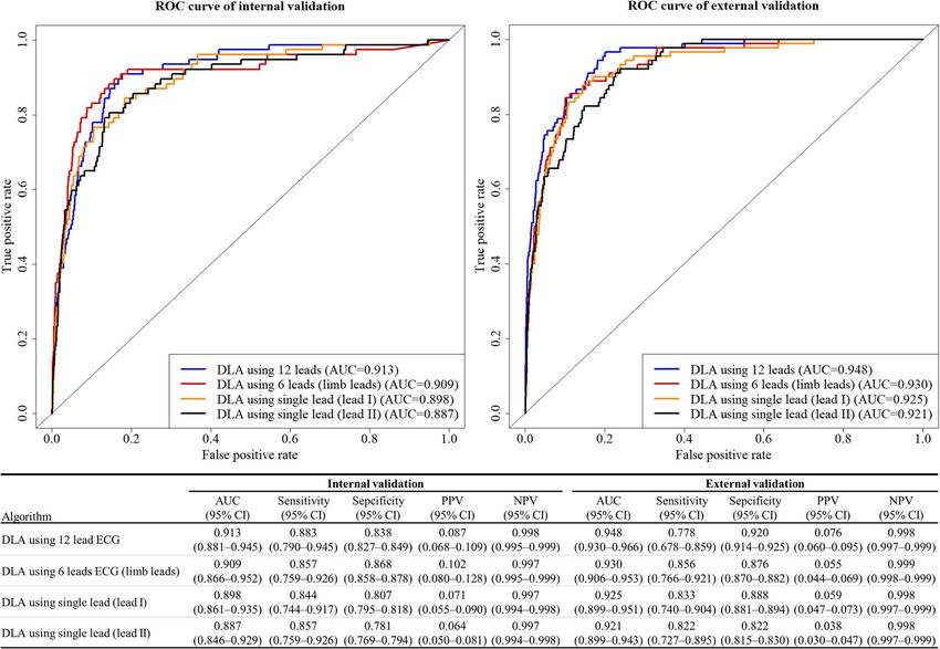
Kwon et al. Scandinavian Journal of Trauma, Resuscitation and Emergency Medicine    (2020) 28:98                         Page 2 of 10

   Several track and trigger systems (TTSs) using discrete              and validated an algorithm using 12-lead ECGs, we used
numeric values such as vital signs and laboratory results               the dataset that was 2D data of 12 × 4000 numbers. Simi-
are used in RRSs [9, 10]. As conventional TTSs have                     larly, for 6-lead and single lead ECGs, we used datasets
limitations in detecting deterioration in patients, several             comprising 6 × 4000 and 1 × 4000 numbers, respectively.
researchers have adopted deep learning based algorithms                 We rearranged the input 2D ECG data in the order V1,
to deal with these numeric values, which performed                      V2, V3, V4, V6, aVL, I, −aVR, II, aVF, and III. Convolu-
better than conventional tools [11–15]. However, the                    tional neural network (CNN) is a well-known deep learn-
performances of these novel TTSs were also not satisfac-                ing architecture for learning 2D image data [20].
tory, and further improvement is needed to use the algo-
rithms with electrical health records. A paradigm shift is              Development of deep learning based artificial intelligence
needed to use a new type of variable to improve the per-                algorithm
formance of predicting cardiac arrest.                                  The DLA was made using many hidden layers of neurons
   Previous studies found QT prolongation, QRS pro-                     to learn complex hierarchical nonlinear representations
longation, fragmented QRS complexes, and early repo-                    from the data [20]. As a block with six stages, it had two
larization to be associated with cardiac arrest [16–19].                convolutional layers, two batch normalization layers, one
However, it is not easy to detect such delicate changes                 max pooling layer, and one dropout layer. This block was
in ECGs, and conventional statistical methods fail to                   fully connected to the one-dimensional (1D) layer com-
build a criteria to predict cardiac arrest using the com-               posed of 128 nodes (Fig. 1). The input layer of epidemi-
plex information of ECGs. The most important aspect of                  ology (age, sex) was concatenated with the 1D layer. There
deep learning is its ability to extract features from high              were two fully connected 1D layers after the flattened layer,
dimensional complex data and formulate algorithms                       and the second layer was connected to the output node,
from various types of data, such as images, two-                        which was composed of one node. The values of the out-
dimensional (2D) data, and waveforms [20]. Recently,                    put node represent the possibility of developing cardiac ar-
deep learning has been used to analyze ECGs for diag-                   rest, and the output node uses a sigmoid function as an
nosing left ventricular hypertrophy, aortic stenosis, atrial            activation function, as the output of the sigmoid function
fibrillation, heart failure, and even determining age and               is between 0 and 1. We used TensorFlow’s open-source
sex [21–24]. We hypothesized that DLAs could effect-                    software library (Google LLC, Mountain View, CA USA)
ively predict cardiac arrests. To test this hypothesis, we              as the backend, and conducted our experiment with Py-
developed and validated a DLA for predicting cardiac                    thon (version 3.5.2; Python Software Foundation, Beaver-
arrest using ECGs.                                                      ton, OR, USA). We conducted additional experiments for
                                                                        the DLA using limb 6-lead and each single-lead (lead I,
Methods                                                                 lead II, lead III, aVR, aVL, and V1–6) ECGs. To develop
This study was approved by the institutional review                     and validate the DLA for these ECGs, we changed the sizes
boards (IRB) of Sejong general hospital (2018–0689) and                 of the filters and convolutional layers, thus adjusting the
Mediplex Sejong hospital (2018–054). Clinical data, in-                 shape of the input datasets. The number of filters, max
cluding digitally stored ECGs, age, sex, and endpoints of               pooling, and fully connected layers were the same as that
admitted patients, were extracted from both hospitals.                  of the 12-lead ECG architecture.
Both IRBs waived the need for informed consent be-
cause of the retrospective nature of the study, using fully             Development and validation datasets
anonymized ECG and health data, and minimal harm.                       Data from hospital A were used for development and in-
                                                                        ternal validation. We identified patients who were ad-
ECG data                                                                mitted to hospital A in the study period (October 2016–
The predictor variables are ECG, age, and sex. Digitally                September 2019), and who had at least one standard
stored 12-lead ECG data were recorded at 500 data points                digital, 10 s, 12-lead ECG acquired in the supine position
per second (500 Hz) at each lead for 10 s. We removed 1 s               during the admission period. We excluded subjects with
each at the beginning and end of the ECG, because these                 missing demographic or electrocardiographic informa-
areas have more artifacts than other parts. Because of this,            tion. As shown in Fig. 2, patients treated at hospital A
the length of each ECG was reduced to 8 s (4000). We                    were randomly and exclusively split into algorithm de-
made a dataset using the entire 12-lead ECG data. We also               velopment (70%) and internal validation (30%) datasets.
used partial datasets from the 12-lead ECG data, such as                Data from hospital B were only used for external valid-
limb 6-lead (aVL, I, −aVR, II, aVF, and III) and single lead            ation, which confirmed that the developed DLA was ro-
(I or II). We selected these leads as they can easily be re-            bust across diverse datasets. The characteristics of the 2
corded by wearable and pad devices in contact with the                  hospitals are different (hospital A is a cardiovascular
patient’s limbs [25]. Consequently, when we developed                   teaching hospital, and hospital B is a community general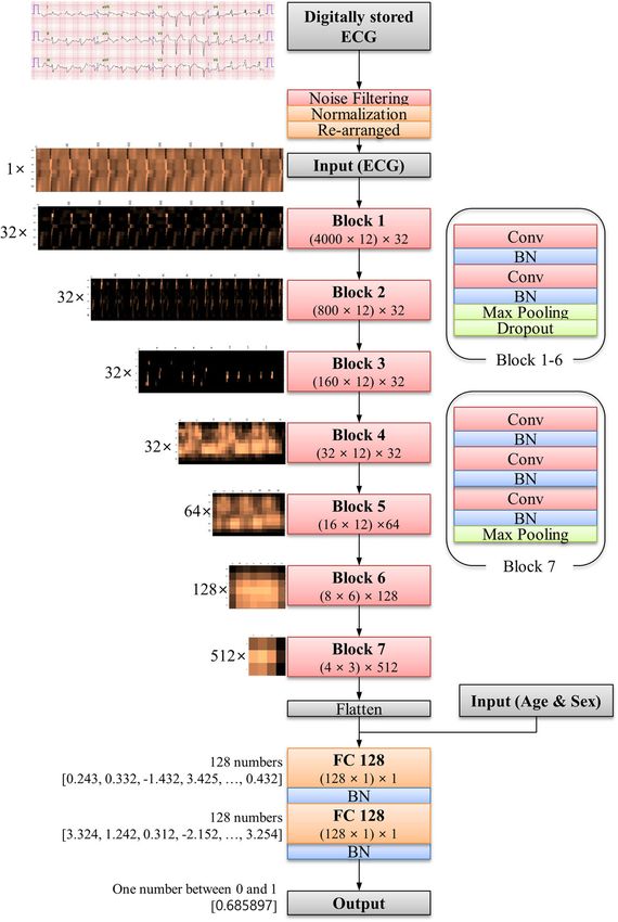
Kwon et al. Scandinavian Journal of Trauma, Resuscitation and Emergency Medicine          (2020) 28:98                                Page 3 of 10

 Fig. 1 Architecture of deep-learning-based algorithm for predicting cardiac arrest. BN denotes batch normalization, Conv convolutional layer, ECG
 electrocardiography, and FC fully connected layer

hospital). We also identified patients who were admitted                   identify the exact time of each endpoint. The objective
to hospital B in the study period (March 2017–Septem-                      of the DLA was to predict whether an ECG was within
ber 2019) and had at least one ECG during it. We also                      the prediction time window of cardiac arrest, which is
excluded subjects in hospital B with missing values. Be-                   the 24 h interval before cardiac arrest. For a patient with
cause the purpose of the validation data was to assess                     cardiac arrest, the ECGs belonging to the prediction
the accuracy of the algorithm, we used only one ECG                        window were labeled as cardiac arrest and other ECGs
from each patient for the internal and external validation                 were labeled as a nonevent. For a patient without cardiac
dataset—the most recent ECG to the endpoints (cardiac                      arrest, all ECGs were labeled as a nonevent. In other
arrest or survival and subsequent discharge).                              words, the aim of the developed DLA was to accurately
                                                                           classify an ECG as cardiac arrest or nonevent.
Endpoint
The endpoint of this research was cardiac arrest, defined                  Statistical analysis
as a lack of palpable pulse, with or without attempted re-                 At each input (ECG, age, and sex) of the validation data,
suscitation. We reviewed electronic health records to                      the DLA calculated the possibility of cardiac arrest in
Kwon et al. Scandinavian Journal of Trauma, Resuscitation and Emergency Medicine   (2020) 28:98                          Page 4 of 10

 Fig. 2 Study flowchart

the range from 0 (nonevent) to 1 (cardiac arrest). To                   could have occurred in nonevent ECGs as well. In other
confirm the performance of the DLA, we compared the                     words, we hypothesized that our DLA would classify
possibility calculated by the DLA with the occurrence of                ECGs with characteristics of deterioration as cardiac ar-
cardiac arrest within 24 h after the time of ECG in the                 rest, giving the initial appearance of a false positive test
validation data. For this, we used the area under the re-               (that is, an ECG classified as cardiac arrest, but not lead-
ceiver operating characteristics curve (AUROC) to meas-                 ing to cardiac arrest within 24 h). To test this hypothesis,
ure the performance of the model. As the purpose of the                 we designed two subgroup analyses with nonevent ECGs
DLA was screening, we evaluated the specificity, the                    in the external validation dataset. We divided the non-
positive predictive value, and the negative predictive                  event ECGs as low and high risk groups defined by the
value at a cut-off point selected for high (90%) sensitivity            DLA. In the analysis of the first subgroup, we confirmed
in development data. Exact 95% confidence intervals                     the occurrence of cardiac arrest over 2 weeks in each
(CIs) were used for all measures of diagnostic perform-                 ECG. We also confirmed the performance of the DLA in
ance except for AUROC. The CI for AUROC was deter-                      predicting deterioration events. The deterioration events
mined based on Sun and Su optimization of the De-long                   were defined as unexpected intensive care unit transfer
method, using the pROC package in R (The R Founda-                      over 2 weeks in each ECG. In the nonevent ECGs of the
tion, Vienna, Austria; www.r-project.org). Statistical sig-             external validation data, we included ECGs which were
nificance for the differences in patient characteristics                acquired in general wards for second subgroup analysis.
was defined as a 2-sided P value of less than 0.001. Mea-               Kaplan-Meier analysis was used to depict the occurrence
sures of the diagnostic performance were summarized                     of delayed cardiac arrest and deterioration events for the
using 2-sided 95% CIs. Analyses were computed using R                   true negative (low risk) versus the false positive (high
software, version 3.4.2.                                                risk) groups over time. Subsequently, Cox proportional
                                                                        hazards regression was used to estimate the hazard for
Subgroup analysis                                                       the delayed cardiac arrest and the deterioration events.
We hypothesized that early in the course of any deteri-
oration, ECG signals would show subtle abnormal pat-                    Visualizing using sensitivity map
terns due to metabolic and structural changes. Although                 To understand the developed DLA and make a compari-
cardiac arrest did not happened within 24 h in nonevent                 son with existing medical knowledge, it was important
ECGs, delayed cardiac arrest and events of deterioration                to identify which regions had significant effects on the
Kwon et al. Scandinavian Journal of Trauma, Resuscitation and Emergency Medicine (2020) 28:98 Page 5 of 10
decision of the DLA. We employed a sensitivity map For endpoint (cardiac arrest within 24 h of ECG), the
using the saliency method, and used it to visualize the AUROC of the 12-lead DLA were 0.913 (95% confidence
ECG regions used by the DLA to predict cardiac arrest. interval: 0.881–0.945) and 0.948 (0.930–0.966) during in-
The map was computed using the first-order gradients ternal and external validation, respectively. As shown in
of the classifier probabilities with respect to the input Fig. 3, the AUROC was 0.004 to 0.027 lower when using 6-
signals. If the probability of a classifier was sensitive to a lead or single-lead ECG than when using 12-lead ECG. As
specific region of the signal, the region would be consid- shown in the Supplemental material 1, when we confirmed
ered as significant in the model. We used a gradient- the performance of DLA using a single lead, a DLA using
weighted class activation map (Grad-CAM) for lead I outperformed other DLAs using other leads.
visualization [26]. Grad-CAM uses the gradient informa- In the external validation data, there were 10,638
tion of the algorithm, and could be used with any activa- ECGs from 10,638 patients labeled as nonevent. We con-
tion function and any architecture of CNNs. ducted first subgroup analysis of the cardiac arrest over
2 weeks with these 10,638 ECGs. Of them, cardiac arrest
Results occurred within 2 weeks in 81 ECGs. The high risk
The study population included 47,505 ECGs of 25,672 group of the DLA showed a significantly higher hazard
patients, in which cardiac arrest occurred in 1054 pa- (Fig. 4) and higher occurrence rate of cardiac arrest than
tients, as shown in Fig. 2. The number of ECGs from the low risk group (5.74% vs. 0.33%, P < 0.001). The sec-
1054 cardiac arrest patients was 2298. Of those 2298 ond subgroup analysis was performed with 10,441 ECGs
ECGs, the number of ECGs labeled as cardiac arrest was acquired in the general ward. Of them, unexpected in-
504. The development dataset from hospital A included tensive care unit transfer within 2 weeks occurred in 112
32,294 ECGs of 10,461 patients. The performance of the ECGs. The high risk group of the DLA showed a signifi-
DLA was then confirmed using 4483 ECGs from the cantly higher hazard (Fig. 4) and higher occurrence rate
4483 patients in the internal validation data from hos- of unexpected intensive care unit transfers than the low
pital A, and 10,728 ECGs from the 10,728 patients in the risk group (4.23% vs. 0.82%, P < 0.001).
external validation data from hospital B. Baseline charac- As shown in Fig. 5, the sensitivity map shows that the
teristics of study population were shown in Table 1. DLA focused mostly on the QRS complex to predict
Table 1 Baseline characteristics
Variables Non-event patients Cardiac arrest patients P-value
n = 24,618 n = 1054
Male, n (%) 13,072 (53.1) 558 (52.9) 0.888
Age, year (mean (sd)) 60.67 (16.69) 72.37 (13.24) < 0.001
Admission to ICU, n (%) 279 (1.1) 638 (60.5) < 0.001
Emergent admission, n (%) 10,491 (42.6) 800 (75.9) < 0.001
Admission division, n (%) < 0.001
Cardiovascular 13,836 (56.2) 760 (72.1)
Cerebrovasclar 2474 (10.0) 142 (13.5)
Respiratory disease 810 (3.3) 63 (6.0)
Other internal medicines 2934 (11.9) 64 (6.1)
Major surgery 4306 (17.5) 18 (1.7)
Others 258 (1.0) 7 (0.7)
Length of stay, day (mean (sd)) 9.34 (15.20) 54.35 (114.36) < 0.001
Heart rate, bpm (mean (sd)) 74.57 (17.76) 93.43 (27.91) < 0.001
PR interval, msec (mean (sd)) 172.03 (31.72) 170.65 (41.28) 0.262
QT interval, msec (mean (sd)) 406.41 (47.17) 393.70 (71.65) < 0.001
QTc (mean (sd)) 445.79 (38.02) 474.27 (46.81) < 0.001
QRS duration, msec (mean (sd)) 97.25 (19.25) 107.62 (28.58) < 0.001
P wave axis (mean (sd)) 43.20 (31.26) 44.89 (47.43) 0.190
R wave axis (mean (sd)) 36.14 (46.52) 39.15 (75.26) 0.046
T wave axis (mean (sd)) 51.17 (59.34) 86.39 (92.21) < 0.001Kwon et al. Scandinavian Journal of Trauma, Resuscitation and Emergency Medicine (2020) 28:98 Page 6 of 10
Fig. 3 Performances of artificial intelligence algorithms for predicting cardiac arrest. AUC denotes area under the receiver operating characteristic
curve, CI confidence interval, DLA deep-learning based artificial intelligence algorithm, NPV negative predictive value, PPV positive predictive
value, and ROC receiver operating characteristic curve
cardiac arrest. The DLA also focused on T-wave, but the model showed good performance using data from an-
significances of these regions were lower than that of the other hospital that was not used for algorithm develop-
QRS complex. ment, and had different patient characteristics and data
shape. At a high-sensitivity (90%) operating point in de-
Discussion velopment data, the DLA performed well as a potential
This is the first study that developed and validated a TTS to predict cardiac arrest, and screen risk in patients
DLA for predicting cardiac arrest using ECGs. This with a negative predictive value greater than 99.8%. The
study reveals that a deep-learning algorithm, one of the model’s performance was better than conventional TTSs,
powerful tools of artificial intelligence, can figure out and similar to recent novel TTSs based on deep learn-
very delicate ECG changes in predicting cardiac arrest. ing. Also, the model’s performance was better than other
The performance for predicting cardiac arrest was pre- commonly used screening tests such as mammography
served with 6-lead or single-lead DLAs. In recent years, for breast cancer (AUROC, 0.78, positive predictive
there many wearable devices for monitoring ECGs have value, 3–12%), and fecal occult blood testing for detect-
been developed [27]. If cardiac arrest could be predicted ing colorectal neoplasia (AUROC 0.71, overall sensitivity,
using ECGs, one can in principle capture the risk of pa- 29%) [28, 29].
tients in general wards or at home through wearable de- The most important aspect of deep learning is its abil-
vices. Additionally, ECGs as raw bio-signal data can be ity to extract features and make an algorithm from vari-
used, which can enhance the performance of recent ous types of data, such as images, 2D data, and
TTSs based on deep learning. waveforms. Here, we used raw ECG data (2D numerical
In development and validation using retrospective data data, 12 × 4000) and interpreted ECG patterns for pre-
of more than 46,000 ECGs, the DLA had a high AUROC dicting cardiac arrest. Attia et al. developed deep-
of 0.913 to 0.948 for predicting cardiac arrest. The learning algorithms for screening cardiac contractileKwon et al. Scandinavian Journal of Trauma, Resuscitation and Emergency Medicine (2020) 28:98 Page 7 of 10 Fig. 4 Cumulative hazard of deterioration event in patients who had no cardiac arrest within 24 h. DLA denotes deep-learning based artificial intelligence algorithm, ECG electrocardiography, and ICU intensive care unit. The cutoff point used for dividing the risk groups was selected when the overall sensitivity was 90% in the development dataset. Cox proportional hazards regression was used to estimate the hazard for the delayed cardiac arrest and the deterioration events dysfunction, predicting the occurrence of atrial fibrilla- prolonged QRS durations and QTs corrected. The heart tion during sinus rhythm, approximating age and sex, rates of cardiac arrest ECGs were greater than that of and detecting hyperkalemia using raw ECG data and nonevent ECGs, and the T wave axes of cardiac arrest demonstrated its feasibility.11,12 Our study group showed ECGs were more rightward than that of nonevent ECGs. that a deep-learning-based algorithm using ECG could As shown in the Supplemental material 2, we described outperform cardiologists in diagnosing left ventricular the features of the high- and low-risk ECG of the DLA. hypertrophy and diagnosis aortic stenosis.13 However, The high-risk ECGs had tachycardia, wide QRS deep learning is often criticized for the unreliability of duration. However, because of the limitations of deep its outcomes because of the unpredictability of the learning, we could not determine the exact process of process. Because of this, we used a sensitivity map to calculation of the risk score by the DLA. Conducting visualize the regions of the ECGs that were used for research of explainable deep learning technologies for decision-making by the DLA. ECG will be the focus of our next study. The map shows that the DLA focused more on the We described the features of cardiac arrest patients in QRS complex to decide and predict cardiac arrest. The the validation dataset in a supplemental material. As DLA also partially focused on the T-wave for predicting shown in the Supplemental material 3, the DLA could cardiac arrest. QRS prolongation has been considered a predict cardiac arrest in several young patients. Al- prognostic marker for mortality among patients with a though a prospective study is needed to prove the clin- variety of cardiovascular diseases [16–18]. Moreover, ical improvement, it is possible to improve clinical QRS fragmentation has been reported to be associated outcomes using the DLA in the general ward. with increased mortality in patients with structural heart Although the developed DLA could detect the deteri- disease [19]. In this study, the cardiac arrest ECGs had oration in a patient, several factors impede its
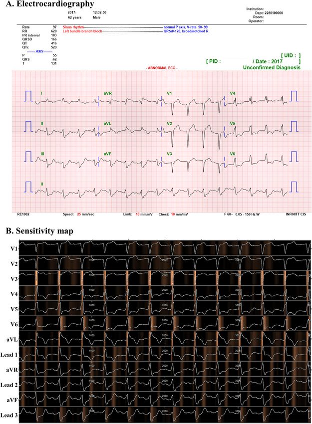
Kwon et al. Scandinavian Journal of Trauma, Resuscitation and Emergency Medicine            (2020) 28:98                                  Page 8 of 10

 Fig. 5 Electrocardiography and sensitivity map of patient with cardiac arrest. This is electrocardiography of patient who was 62 years old and was
 occurred cardiac arrest in external validation hospital. The cardiac arrest occurred 18 min after acquiring electrocardiography. The deep learning
 based artificial intelligence algorithm predicted cardiac arrest in this patient with a value of 0.685897, which was 32.7 times the cut-off value of
 sensitivity 90% in development dataset

application in real clinical practice. Owing to the lack of                  Therefore, previous studies on alarm detection systems
required resources in the ICU, we could not transfer all                     for deterioration in patients have focused on the transfer
high-risk patients to the ICU. However, we could use                         of patients to the ICU as the endpoint.
tele-monitoring devices to monitor high-risk patients,                         Our study has several limitations to be resolved in the
enabling the physicians of the rapid response team to re-                    future. First, this was a retrospective study using conven-
evaluate these patients. Moreover, it is important to note                   tional 12-lead ECGs. A prospective study is warranted to
that it is difficult to prove clinical improvement; if car-                  determine the association of the DLA, and enhancement
diac arrests were prevented due to increased monitoring                      in detecting cardiac arrest and improving clinical out-
of patients, this outcome would not be measurable.                           comes. In such future studies, it is important that we
Kwon et al. Scandinavian Journal of Trauma, Resuscitation and Emergency Medicine              (2020) 28:98                                       Page 9 of 10

exclude patients who experienced cardiac arrest without                       Grad-CAM: Gradient-weighted class activation map; IRB: Institutional review
any resuscitation attempts because such supposed clin-                        board; RRS: Rapid response system; TTS: Track and trigger system; 1D: One-
                                                                              dimensional; 2D: Two-dimensional
ical improvements possess little perceived value, espe-
cially if the patients are not resuscitated. A study for                      Acknowledgements
confirming the accuracy of data from various wearable                         This research was results of a study on the “High Performance Computing
                                                                              Support” Project, supported by the ‘Ministry of Science and ICT and National
or portable ECG devices is warranted to apply the DLA                         IT Industry Promotion Agency of South Korea.
to those devices. If we adopt DLAs in daily living, a
study is also needed to confirm the performance at                            Authors’ contributions
home and general environments. Second, the perform-                           JK was principal investigator, designed the study, prepared the study data,
                                                                              analyzed the data, prepared the figures, and drafted the manuscript. KHK
ance of the DLA needs to be enhanced in order to use it                       and KHJ contributed to the design of the study, prepared data, and revised
as a reliable cardiac arrest detecting tool. Although the                     the manuscript. SYL and BHO contributed to the design of the study,
NPV was over 99%, the PPV was only 8% at the point of                         interpreted the findings, and revised the manuscript. JP supervised the study,
                                                                              interpreted the findings, and contributed to the manuscript. All authors read
high sensitivity. As there are several methodologies that                     and approved the final manuscript.
have been developed in deep learning and computer sci-
ence, we could develop higher performance DLAs in the                         Funding
near future. We also developed a high performance TTS,                        This work was supported by the National Research Foundation of Korea
                                                                              (NRF) grant funded by the Korea government (MSIT) (No.
by combining the DLA with discrete numeric variables                          2020R1F1A1073791).
such as the vital sign. Thirdly, we need to explore the
decision-making process of the DLA further. For ex-                           Availability of data and materials
                                                                              The datasets used and/or analyzed during the current study are available
ample, additional experiments are required to under-                          from the corresponding author on reasonable request.
stand the deep learning process better, and thereby
understand which exact characteristics of the QRS com-                        Ethics approval and consent to participate
plex and the T wave influence the algorithm’s decision.                       The study was performed in accordance with the Declaration of Helsinki and
                                                                              approved by the institutional review boards (IRB) of Sejong general hospital
Explainable artificial intelligence has been studied and                      (file number: 2018–0689) and Mediplex Sejong hospital (file number: 2018–
reported on recently, so the “black box” limitation could                     054), South Korea. Both IRBs waived the need for informed consent because
be solved in the near future [30]. This subject will be                       of the retrospective nature of the study, using fully anonymized ECG and
                                                                              health data, and minimal harm.
our next area of study, and this might turn out to be the
new standard for discovering new medical knowledge                            Consent for publication
about diseases and ECGs.                                                      Not applicable.

                                                                              Competing interests
Conclusion                                                                    KHK, KHJ, SYL, and BHO declare that they have no competing interests. JK
The newly developed deep learning-based artificial                            and JP are co-founder and stakeholder in Medical AI Co Ltd., a medical artifi-
intelligence algorithm demonstrated a high performance                        cial intelligence company. JK is researcher of Body friend co. There are no
                                                                              products in development or marketed products to declare. This dose not
in predicting cardiac arrest using not only a 12-lead, but                    alter our adherence to Scandinavian Journal of Trauma, Resuscitation and
also a single-lead ECG. The results indicate that cardiac                     Emergency Medicine.
arrest could be screened and predicted not only with a
                                                                              Author details
conventional 12-lead ECG, but also with a single-lead                         1
                                                                               Department of Critical Care and Emergency Medicine, Mediplex Sejong
ECG using a wearable device that employs the artificial                       Hospital, 20, Gyeyangmunhwa-ro, Gyeyang-gu, Incheon, Republic of Korea.
                                                                              2
intelligence algorithm.                                                        Artificial Intelligence and Big Data Research Center, Sejong Medical
                                                                              Research Institute, Bucheon, South Korea. 3Medical research team, Medical AI,
                                                                              co., Seoul, South Korea. 4Medical R&D Team, Body Friend, co., Seoul, South
Supplementary information                                                     Korea. 5Division of Cardiology, Cardiovascular Center, Mediplex Sejong
Supplementary information accompanies this paper at https://doi.org/10.       Hospital, Incheon, South Korea.
1186/s13049-020-00791-0.
                                                                              Received: 29 August 2020 Accepted: 22 September 2020

 Additional file 1: Supplemental material 1. Performance of deep
 learning algorithm based on single lead electrocardiography.
                                                                              References
 Additional file 2: Supplemental material 2. Electrocardiographic             1. Andersen LW, Holmberg MJ, Berg KM, Donnino MW, Granfeldt A. In-
 features of high and low risk group defined by deep learning based               Hospital Cardiac Arrest. JAMA [Internet]. 2019;321:1200.
 algorithm.                                                                   2. Holmberg MJ, Ross CE, Fitzmaurice GM, Chan PS, Duval-Arnould J,
 Additional file 3: Supplemental material 3. Features of cardiac arrest           Grossestreuer AV, et al. Annual incidence of adult and pediatric in-hospital
 patients in validation datasets.                                                 cardiac arrest in the United States. Circ Cardiovasc Qual Outcomes. 2019;
                                                                                  12(7):e005580.
                                                                              3. Hayashi M, Shimizu W, Albert CM. The Spectrum of epidemiology
Abbreviations                                                                     underlying sudden cardiac death. Circ Res. 2015;116:1887–906.
AUROC: Area under the receiver operating characteristics curve;               4. Merchant RM, Yang L, Becker LB, Berg RA, Nadkarni V, Nichol G, et al.
CI: Confidence interval; CNN: Convolutional neural network; DLA: Deep-            Incidence of treated cardiac arrest in hospitalized patients in the United
learning-based artificial intelligence algorithm; ECG: Electrocardiography;       States. Crit Care Med. 2012;39:2401–6.
Kwon et al. Scandinavian Journal of Trauma, Resuscitation and Emergency Medicine (2020) 28:98 Page 10 of 10
5. Benjamin EJ, Virani SS, Callaway CW, Chang AR, Cheng S, Chiuve SE, et al. 26. Selvaraju RR, Cogswell M, Das A, Vedantam R, Parikh D, Batra D. Grad-CAM:
Heart disease and stroke statistics—2018 update: a report from the visual explanations from deep networks via gradient-based localization. Proc
American Heart Association. Circulation. 2018;137:67–492. IEEE Int Conf Comput Vis. 2017;1:618–26.
6. Nadkarni VM. First documented rhythm and clinical outcome from in- 27. Steinhubl SR, Waalen J, Edwards AM, Ariniello LM, Mehta RR, Ebner GS, et al.
hospital cardiac arrest among children and adults. JAMA. 2006;295:50. Effect of a home-based wearable continuous ECG monitoring patch on
7. Lyons PG, Edelson DP, Churpek MM. Rapid response systems. Resuscitation. detection of undiagnosed atrial fibrillation. JAMA. 2018;320:146.
2018;128:191–7. 28. Pisano ED, Gatsonis C, Hendrick E, Yaffe M, Baum JK, Acharyya S, et al.
8. So RKL, Bannard-Smith J, Subbe CP, Jones DA, van Rosmalen J, Lighthall GK. Diagnostic performance of digital versus film mammography for breast-
The association of clinical frailty with outcomes of patients reviewed by cancer screening. N Engl J Med. 2005;353:1773–83.
rapid response teams: an international prospective observational cohort 29. Haug U, Kuntz KM, Knudsen AB, Hundt S, Brenner H. Sensitivity of
study. Crit Care. 2018;22:227. immunochemical faecal occult blood testing for detecting left- vs right-
9. Romero-Brufau S, Huddleston JM, Naessens JM, Johnson MG, Hickman J, sided colorectal neoplasia. Br J Cancer. 2011;104:1779–85.
Morlan BW, et al. Widely used track and trigger scores: Are they ready for 30. Mittelstadt B, Russell C, Wachter S. Explaining explanations in AI. FAT* 2019 -
automation in practice? Resuscitation. 2014;85:549–52 European Proc 2019 Conf fairness, accountability. Transpar. 2019;1:279–88.
Resuscitation Council, American Heart Association, Inc., and International
Liaison Committee on Resuscitation.~Published by Elsevier Ireland Ltd.
10. Dziadzko MA, Novotny PJ, Sloan J, Gajic O, Herasevich V, Mirhaji P, et al.
Publisher’s Note
Springer Nature remains neutral with regard to jurisdictional claims in
Multicenter derivation and validation of an early warning score for acute
published maps and institutional affiliations.
respiratory failure or death in the hospital. Crit Care. 2018;22:286.
11. Parshuram CS, Dryden-Palmer K, Farrell C, Gottesman R, Gray M, Hutchison
JS, Helfaer M, Hunt EA, Joffe AR, Lacroix J, Moga MA, Nadkarni V, Ninis N,
Parkin PC, Wensley D, Willan AR, Tomlinson GA; Canadian Critical Care Trials
Group and the EPOCH Investigators. Effect of a PediatricEarly Warning
System on All-Cause Mortality in Hospitalized Pediatric Patients: The EPOCH
Randomized Clinical Trial. JAMA. 2018;319(10):1002–12. https://doi.org/10.
1001/jama.2018.0948.
12. Churpek MM, Yuen TC, Winslow C, Meltzer DO, Kattan MW, Edelson DP.
Multicenter comparison of machine learning methods and conventional
regression for predicting clinical deterioration on the wards. Crit Care Med.
2016;44:368–74.
13. Cho KJ, Kwon O, Kwon JM, Lee Y, Park H, Jeon KH, Kim KH, Park J, Oh BH.
Detecting Patient Deterioration Using Artificial Intelligence in a Rapid
Response System. Crit Care Med. 2020;48(4):e285–9. https://doi.org/10.1097/
CCM.0000000000004236.
14. Kwon J-M, Lee Y, Lee Y, Lee S, Park J. An algorithm based on deep learning
for predicting in-hospital cardiac arrest. J Am Heart Assoc. 2018;7:e008678.
15. Shillan D, Sterne JAC, Champneys A, Gibbison B. Use of machine learning to
analyse routinely collected intensive care unit data: a systematic review. Crit
Care. 2019;23:284.
16. Kashani A, Barold SS. Significance of QRS complex duration in patients with
heart failure. J Am Coll Cardiol. 2005;46:2183–92.
17. Teodorescu C, Reinier K, Uy-Evanado A, Navarro J, Mariani R, Gunson K, et al.
Prolonged QRS duration on the resting ECG is associated with sudden
death risk in coronary disease, independent of prolonged ventricular
repolarization. Hear Rhythm. 2011;8:1562–7.
18. Baslaib F, Alkaabi S, Yan AT, Yan RT, Dorian P, Nanthakumar K, et al. QRS
prolongation in patients with acute coronary syndromes. Am Heart J. 2010;
159:593–8.
19. Das MK, El Masry H. Fragmented QRS and other depolarization
abnormalities as a predictor of mortality and sudden cardiac death. Curr
Opin Cardiol. 2010;25:59–64.
20. LeCun Y, Bengio Y, Hinton G. Deep learning. Nature. 2015;521:436–44.
21. Attia ZI, Kapa S, Lopez-Jimenez F, McKie PM, Ladewig DJ, Satam G, et al.
Screening for cardiac contractile dysfunction using an artificial intelligence–
enabled electrocardiogram. Nat Med. 2019;25(1):70–4.
22. Attia ZI, Noseworthy PA, Lopez-Jimenez F, Asirvatham SJ, Deshmukh AJ,
Gersh BJ, et al. An artificial intelligence-enabled ECG algorithm for the
identification of patients with atrial fibrillation during sinus rhythm: a
retrospective analysis of outcome prediction. Lancet. 2019;394:861–7.
23. Attia ZI, Friedman PA, Noseworthy PA, Lopez-Jimenez F, Ladewig DJ, Satam
G, et al. Age and sex estimation using artificial intelligence from standard
12-Lead ECGs. Circ Arrhythmia Electrophysiol. 2019;12(9):e007284.
24. Kwon J-M, Jeon K-H, Kim HM, Kim MJ, Lim SM, Kim K-H, et al. Comparing
the performance of artificial intelligence and conventional diagnosis criteria
for detecting left ventricular hypertrophy using electrocardiography.
Europace. 2020;22(3):412–9.
25. Halcox JPJ, Wareham K, Cardew A, Gilmore M, Barry JP, Phillips C, et al.
Assessment of remote heart rhythm sampling using the AliveCor heart
monitor to screen for atrial fibrillation: the REHEARSE-AF study. Circulation.
2017;136:1784–94.You can also read