Defining a therapeutic range for regeneration of ischemic myocardium via shock waves - Nature
←
→
Page content transcription
If your browser does not render page correctly, please read the page content below
www.nature.com/scientificreports
OPEN Defining a therapeutic range
for regeneration of ischemic
myocardium via shock waves
Leo Pölzl1,2,7, Felix Nägele1,7, Jakob Hirsch1, Michael Graber1, Daniela Lobenwein1,2,
Elke Kirchmair1, Rosalie Huber1, Christian Dorfmüller3, Sophia Lechner1, Georg Schäfer4,
Martin Hermann5, Helga Fritsch2, Ivan Tancevski6, Michael Grimm1, Johannes Holfeld1 &
Can Gollmann‑Tepeköylü1*
Shockwave therapy (SWT) represents a promising regenerative treatment option for patients with
ischemic cardiomyopathy. Although no side-effects have been described upon SWT, potential cellular
damage at therapeutic energies has not been addressed so far. In this work, we aimed to define a
therapeutic range for shock wave application for myocardial regeneration. We could demonstrate
that SWT does not induce cellular damage beneath energy levels of 0.27 mJ/mm2 total flux density.
Endothelial cell proliferation, angiogenic gene expression and phosphorylation of AKT and ERK
are enhanced in a dose dependent manner until 0.15 mJ/mm2 energy flux density. SWT induces
regeneration of ischemic muscle in vivo via expression of angiogenic gene expression, enhanced
neovascularization and improved limb perfusion in a dose-dependent manner. Therefore, we provide
evidence for a dose-dependent induction of angiogenesis after SWT, as well as the absence of cellular
damage upon SWT within the therapeutic range. These data define for the first time a therapeutic
range of SWT, a promising regenerative treatment option for ischemic cardiomyopathy.
Abbreviations
HUVEC Human umbilical vein endothelial cells
qPCR Real time polymerase chain reaction
LDPI Laser Doppler perfusion imaging
LV Left ventricle
SW Shock wave
SWT Shock wave therapy
VEGF Vascular endothelial growth factor
VEGFR2 Vascular endothelial growth factor receptor 2
IHD Ischemic heart disease
Ischemic heart disease (IHD) is the leading cause of death in the European Union and the Western world1,2.
Prevalence is rising constantly due to an aging population. Ischemia results in replacement of functional cardio-
myocytes with non-contractile fibrotic scar tissue. Decreased cardiac output due to a remodeled left ventricle
causes the deadly syndrome of heart failure3. Affected patients suffer from poor quality of live with obscure
prognosis4. Repeated hospitalizations and incapacity to work contribute to the severe socio-economic burden
of heart f ailure4–6.
Despite intensive research in the field, modern pharmacotherapy mainly aims at symptom control rather than
regeneration of scar tissue7. As novel therapeutic approaches including gene and stem cell therapy remain purely
experimental, there is a major need for innovative approaches for the regeneration of ischemic myocardium8–10.
Shock waves are specific pressure-waves which have been used for kidney stone disintegration for more than
three decades in the medical fi eld11. In lower energies, they exhibit potent regenerative properties. Beneficial
1
Department of Cardiac Surgery, Medical University of Innsbruck, Innsbruck, Austria. 2Institute of Clinical and
Functional Anatomy, Medical University of Innsbruck, Innsbruck, Austria. 3Heart Regeneration Technologies GmbH,
Innsbruck, Austria. 4Department of Pathology, Medical University of Innsbruck, Innsbruck, Austria. 5Department
of Anesthesiology, Medical University of Innsbruck, Innsbruck, Austria. 6Department of Internal Medicine II,
Infectious Diseases, Pneumology, Rheumatology, Medical University of Innsbruck, Innsbruck, Austria. 7These
authors contributed equally: Leo Pölzl and Felix Nägele. *email: can.tepekoeylue@i‑med.ac.at
Scientific Reports | (2021) 11:409 | https://doi.org/10.1038/s41598-020-79776-z 1
Vol.:(0123456789)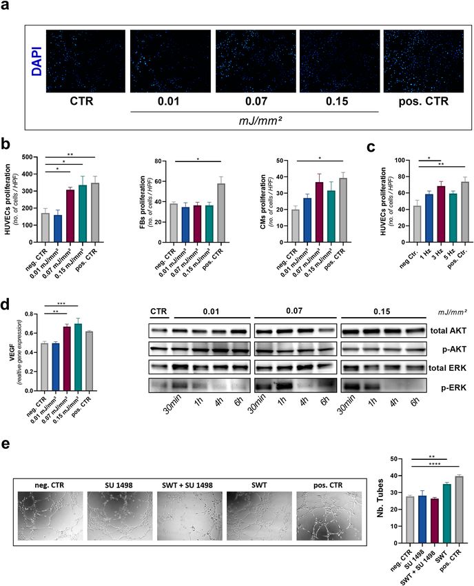
www.nature.com/scientificreports/
effects on bone non-unions, chronic tendonitis (e.g. tennis-elbow) and diabetic wound healing disorders are
well described and established in clinical routine12,13. The observed effects are mainly attributed to the induc-
tion of angiogenesis14–16. Our group could show recently that the mechanical stimulus of SWs cause release of
(a) angiogenic growth factors from the extracellular m atrix17 and (b) specific extracellular vesicles containing
angiogenic cargo . Concomitant stimulation of inflammation enhances the angiogenic response19 .
18
Over the past years, we showed a strong angiogenic effect of SWT improving left ventricular function in
ischemic cardiomyopathy in small and large animal models. Treatment caused neovascularization and resulted
in reduction of myocardial s car17,20. Thus, SWT emerged as promising treatment strategy for ischemic cardio-
myopathy. As a consequence, we initiated a prospective-randomized clinical trial, the CAST (safety and efficacy
of direct Cardiac Shockwave Therapy in patients with ischemic cardiomyopathy undergoing coronary artery
bypass grafting) trial (ClinicalTrials.gov Identifier: NCT03859466). The trial is currently in the recruitment phase.
Despite the promising effects of SWT and their impact on the treatment of IHD, the biophysical principles
behind the therapy are not well understood. Although no side-effects have been described upon SWT, potential
cellular damage at angiogenic acting energy levels was never determined in studies. Dose–response studies
regarding SWT are missing so far. Thus, it remains uncertain whether the underlying mechanism of angiogen-
esis upon shock-wave therapy is based on a “damage-repair” principle. For this purpose, we aimed to define a
therapeutic range for SW application in vitro and in vivo.
Results
Shock wave therapy inflicts no cellular damage at therapeutic energies. Shock waves destroy
kidney stones at high energy levels, but regenerate tissue at low energy levels11–13.To evaluate whether SWT
induces cellular necrosis at commonly used energy levels, we subjected human endothelial cells to different
energy levels of SWT in T25 flasks. Release of LDH was measured 1 h after treatment to evaluate necrosis.
No signs of necrosis were detected upon SWT at energy flux density levels of 0.01 mJ/mm2, 0.07 mJ/mm2 and
0.15 mJ/mm2 (Fig. 1a). However, we found evidence of necrosis at 0.27 mJ/mm2.
Shock waves are physical pressure waves, their reflection can result either in positive interference amplify-
ing the energy of the primary wave, or cause negative interference, extinguishing the wave. Like ultrasound
waves, shock waves are reflected whenever the wave encounters a material with a different density (acoustical
impedance)21.
Thus, reflection and potential consecutive interference depends on the size and form of the cell culture flask.
To evaluate whether the occurrence of necrosis might depend on the size and form of the cell culture flask in
which the cells undergo SWT, we repeated the experiment using different common cell culture flasks and well
plates (Fig. 1b). In contrast to the first experiment, we could observe necrosis already at a level as low as 0.15 mJ/
mm2 in plates with a surface smaller than 9.5 cm2 (12, 24 and 96 well plates) (Fig. 1c). In contrast, in 6 well plates
and T25 flasks necrosis only occurred at 0.27 mJ/mm2. These experiments suggest phenomena of constructive
interference after SWT in smaller well plates. Cardiomyocytes and fibroblast inevitable receive SWT during direct
epicardial application to the heart. To evaluate whether SWT induces necrosis in other cardiac domestic cells,
we subjected cardio myocytes and cardiac fibroblasts to SWT. However, we could not observe any differences
compared to human endothelial cells using therapeutic energy levels (Fig. 1d).
Positive pressure induces cellular damage. Within the targeted tissue, shock waves induce alternating
positive and negative pressure due to their wave p rofile22. However, whether the positive squeezing or the nega-
tive pulling acting pressure induces the described effects remains unknown.
Therefore, we evaluated pressure levels at different energy levels utilizing a hydrophone (Fig. 1e). The posi-
tive pressure increased concomitant with the total energy flux density and necrosis (Fig. 1f). The measured
necrosis was thereby induced by the positive pressure of the SW, since negative peak pressure of SW treatment
increased in a linear matter with corresponding energy levels, until reaching a plateau at 3.8 MPa at an energy
level of 0.11 mJ/mm2 (Fig. 1g). For all further experiments energy levels within the therapeutiv range were used.
SWT induces dose‑dependent angiogenesis in vitro. In a next set of experiments, we aimed to evalu-
ate whether angiogenesis upon SWT was dose dependent. Proliferation of endothelial cells is crucial for the
sprouting of newly formed v essels23. Therefore, human endothelial cells were subjected to SWT and analyzed
for proliferation. We could demonstrate that beginning at an energy level of 0.07 mJ/mm2 SWT induced dose-
dependent proliferation compared to untreated controls in human endothelial cells (Fig. 2a,b). In contrast,
human fibroblasts showed no proliferation upon therapy. However, proliferation was enhanced after SWT in a
human cardiomyocyte cell line at an energy flux density of 0.07 mJ/mm2 (Fig. 2b). Moreover, effects depended on
frequency, as SW treatment with 3 Hz induced proliferation of endothelial cells, whereas the effect was markedly
reduced with 1 or 5 Hz (Fig. 2c). In line with these findings, we found a dose dependent increase of angiogenic
Vascular Endothelial Growth Factor (VEGF) expression after SW treatment (Fig. 2d). VEGF activates its recep-
tor VEGFR2, followed by phosphorylation of the kinases AKT and ERK both of which induce angiogenesis24,25.
To evaluate angiogenic signaling upon SWT, we analyzed phosphorylation of both AKT and ERK after SWT.
SWT induced phosphorylation of AKT and ERK 30 min and 1 h after SW treatment. Interestingly, although
phosphorylation of AKT was independent of SW dose, activation of ERK was dose-dependent (Fig. 2d). These
findings indicate a dose dependent angiogenic and proliferative effect of SWT. To assess whether SW effects
depended on VEGF signaling, we performed a tube formation assay in the presence of the VEGFR2 inhibitor
SU1498. Angiogenesis upon SWT was abolished upon pretreatment with VEGFR2 indicating a crucial role of
the VEGF axis in the angiogenic response upon SWT (Fig. 2e).
Scientific Reports | (2021) 11:409 | https://doi.org/10.1038/s41598-020-79776-z 2
Vol:.(1234567890)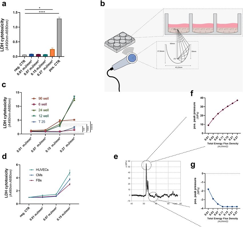
www.nature.com/scientificreports/
Figure 1. Shock wave therapy induces necrosis only at very high energy levels. (a) To evaluate at which
energy levels SWT would induce cellular damage, we performed an LDH assay upon therapy. SWT caused no
necrosis induced until energy levels of 0.27 mJ/mm2 total flux density in T25 flasks. Means ± SEM. *p < 0.05,
****p < 0.0001. n = 3. (b) Scheme for the mechanical setup of SWT in vitro: To evaluate whether the occurrence
of necrosis might depend on the size and form of the cell culture flask in which the cells undergo SWT,
HUVECs were seeded in 6, 12, 24 and 96 well plates and subjected to SWT. (c) In microwell plates with a
surface smaller than 9,5cm2 (12, 24 and 96 well plates) necrosis could be measured at a level as low as 0.15 mJ/
mm. This might be attributed to phenomena of constructive interference. Means ± SEM. **p < 0.01; ***p < 0.001,
****p < 0.0001. n = 3–4. (d) Comparable levels of necrosis could be discovered in fibroblasts and cardiomyocytes
upon shockwave therapy. (e) Within the targeted tissue, shock waves induce alternating positive and negative
pressure due to their specific wave profile. Positive and negative pressure induced by SWs were measured by
a hydrophone showing an increase of pressure upon release of the wave with subsequent decrease to negative
pressures. (f) Positive pressure increases concomitant with the total energy flux density and thus necrosis. (g)
Initially, negative pressure increases concomitant with the total flux density until plateauing at a level of 3,8 MPa.
Statistical comparisons between multiple groups: one-way ANOVA with Tukey post hoc analysis.
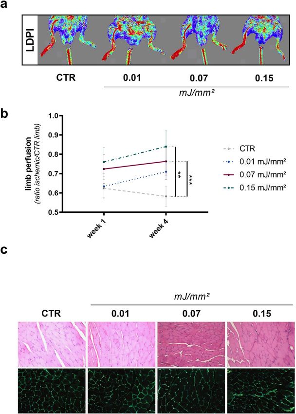
SWT improves limb perfusion. To assess dose-dependency in vivo, we performed SWT in a murine
model of hind limb ischemia. We found improved limb perfusion compared to untreated mice 4 weeks after
SW treatment (Fig. 3a). As anticipated from our in vitro findings, energy levels of 0.07 and 0.15 mJ/mm2 had
the most beneficial effects on hind limb regeneration (Fig. 3b). To evaluate possible cytotoxic effects in vivo, SW
treated muscle samples underwent pathological examination. There was no evidence of necrosis or cellular dam-
age, independent of dose (Fig. 3c).
Scientific Reports | (2021) 11:409 | https://doi.org/10.1038/s41598-020-79776-z 3
Vol.:(0123456789)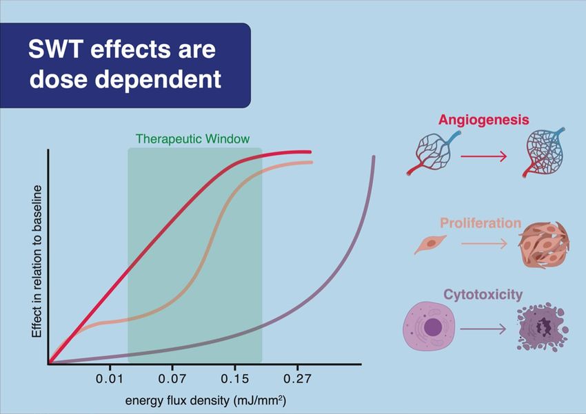
www.nature.com/scientificreports/

                                           Figure 2.  SWT induces angiogenesis in a dose-dependent manner in vitro. (a) To evaluate proliferation, HUVECs were subjected
                                           to SWT at different energy levels and analyzed via DAPI staining after 24 h. (b) SWT induced proliferation of HUVECs in a dose-
                                           dependent manner, beginning at an energy level of 0.07 mJ/mm2. Proliferation of cardio myocytes was enhanced upon shockwave
                                           therapy. In contrast no proliferative effect could be observed in fibroblasts. Means ± SEM. *p < 0.05, **p < 0.01. n = 6. (c) The highest
                                           proliferative effect of SWT was observed upon therapy at 3 Hz. (d) SWT enhanced the mRNA expression of VEGF in a dose
                                           dependent-manner. Means ± SEM. SWT resulted in phosphorylation of angiogenic AKT and ERK 30 min and 1 h after SW treatment.
                                           **p < 0.01; ***p < 0.001. n = 6. (e) To assess whether SW effects depended on VEGF signaling, a functional angiogenesis assay, the tube
                                           formation assay was performed. Angiogenic effect of SWT abolished upon treatment with VEGFR2 inhibitor SU 1498. **p < 0.01;
                                           ****p < 0.0001. n = 6. Statistical comparisons between multiple groups: one-way ANOVA with Tukey post hoc analysis. Blots are
                                           displayed in cropped format.

          Scientific Reports |   (2021) 11:409 |                    https://doi.org/10.1038/s41598-020-79776-z                                                                     4

Vol:.(1234567890)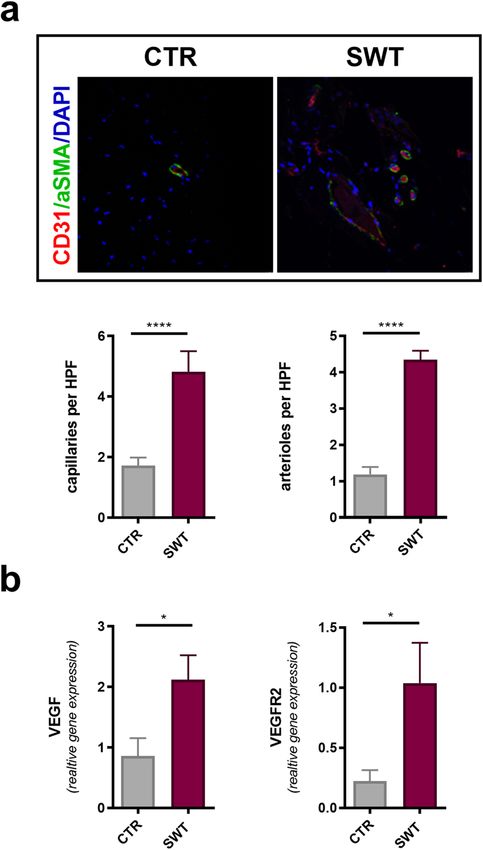
www.nature.com/scientificreports/

                                 Figure 3.  SWT improves limb perfusion dose-dependently. (a, b) To evaluate dose–response relationship
                                 in vivo, mice were subjected to hind limb ischemia and treated with different SW doses thereafter. Treatment
                                 with 0.07 and 0.15 mJ/mm2 improved limb perfusion 4 weeks after treatment compared to untreated controls,
                                 whereas treatment with 0.01 mJ/mm2 showed no beneficial effect . Means ± SEM. **p < 0.01; ***p < 0.001.
                                 n = 5–10. (c) To asses necrosis upon SWT in vivo, limbs of mice were treated with SWT. Upon treatment with
                                 different levels of energy flux density, no signs of necrosis could be observed in pathological examination.
                                 Statistical comparisons between multiple groups: one-way ANOVA with Tukey post hoc analysis.

                                 SWT induces angiogenesis in vivo.            To assess whether improved limb perfusion was due to angiogenesis
                                 we stained ischemic muscles for newly formed vessels. We found an increase of capillaries as well as arterioles
                                 in mice treated at an energy level of 0.07 mJ/mm2 in comparison to untreated animals (Fig. 4a). In parallel, gene
                                 expression levels of VEGF and its receptor VEGFR2 were increased after SWT (Fig. 4b).

Scientific Reports |   (2021) 11:409 |               https://doi.org/10.1038/s41598-020-79776-z                                                 5

                                                                                                                                           Vol.:(0123456789)
www.nature.com/scientificreports/
Figure 4. SWT induces angiogenesis in vivo. (a) To evaluate angiogenesis in ischemic muscles, we performed
immunostaining of endothelial cells and vascular smooth muscle cells. Quantification showed increased
numbers of capillaries and arterioles in ischemic limbs subjected to SWT. Means ± SEM. ****p < 0.0001. n = 5–10.
(b) qPCR revealed increased gene expression of VEGF and VEGFR2 in ischemic muscle 3 days after SW
treatment. Means ± SEM. *p < 0.05. N = 5–10. Statistical comparisons between two groups: Student’s t test.
Discussion
The prevalence of ischemic heart failure rises due to constantly aging population. Frequent rehospitalizations
and incapacity to work cause a severe socio-economic burden for Western health care systems4–6. Despite mod-
ern pharmacotherapy, ischemic heart failure is still associated with poor outcomes. Current treatments aim at
symptom control rather than myocardial regeneration. Novel therapies as gene or stem cell therapy showed
promising results in pre-clinical experiments. However, they have failed to gain routine clinical application
due to unfavorable side effect profiles and ethical concerns8–10. Therefore, there is major need in novel and safe
therapy for patients suffering from ischemic cardiomyopathy.
Shockwave therapy has been used in clinical routine in various indications for more than 30 years without
showing any form of side effects11. Beneficial effects of SWT in ischemic heart failure via induction of angio-
genesis were shown b efore18,20,26–28. On the other hand, SWT has been used at much higher energy levels for the
destruction of kidney stones. Hence, depending on the used energy level, SW can both induce tissue regeneration
as well as stone disintegration. However, the dose–response profile of SWT has not been defined so far.
In this work, we could define for the first time a therapeutic range of SWT. Within the therapeutic range,
SWT did not cause cellular damage. Furthermore, we could show that necrosis beyond energy levels of 0.27 mJ/
mm2 is attributed to positive pressure, since negative pressure released by SW plateaus already at an energy level
of 0.11 mJ/mm2.
Other cellular responses to increased pressure upon SWT, as the formation of caveolae, have been described
previously29. However, earlier works performed experiments only at one single level of energy flux density.
Since a therapeutic range for SWT has not been defined yet, we subject endothelial cells to SWT at differ-
ent energy levels. We could demonstrate that the proliferative effect of SWT is dose dependent and plateaus at
an energy level of 0.07 mJ/mm2. Concomitant with these findings we could observe a dose depended increase
of angiogenic gene expression with subsequent phosphorylation of protein kinases AKT and ERK upon SWT.
Therefore we could demonstrate in vitro an extended therapeutic range of the SWT, since experiments in earlier
works were only performed at an energy flux density of 0.08 mJ/mm219,26.
Scientific Reports | (2021) 11:409 | https://doi.org/10.1038/s41598-020-79776-z 6
Vol:.(1234567890)www.nature.com/scientificreports/
Figure 5. Central picture. These data define for the first time a therapeutic range of SWT. We provide evidence
for a dose-dependent induction of angiogenesis after SWT, as well as the absence of cellular damage upon SWT
within the therapeutic range.
In a murine model of hind limb ischemia we investigated the effects of SW treatment on ischemic tissue. As
anticipated from earlier in vitro findings, we could show again a dose dependent increase in limb perfusion in
animals subjected to SWT. Immunofluorescence staining revealed an increased amount of capillaries and arte-
rioles in ischemic muscles after SWT. Combined with increased angiogenic gene expression in vivo after SWT,
these findings underline the angiogenic effect of SWT. However, these experiments show a dose–response profile
in soft tissue only. Whether the beneficial effects of SWT on bone non-unions is in the same therapeutic range
has to be demonstrated in future w orks12,13. We found no damage after SWT in muscle tissue, previous studies
found no evidence of cellular damage after SWT of ischemic m yocardium17,20.
Overall, we provide evidence for a dose-dependent induction of angiogenesis after SWT (Fig. 5). VEGF signal-
ing resulted in endothelial proliferation and neovascularization improving limb perfusion of ischemic muscle. We
were able to define a therapeutic range of SWT from 0.07 to 0.15 mJ/mm2. Moreover, we could show the absence
of necrosis upon SWT within the therapeutic range. Hence, the angiogenic properties are not attributed to a
“damage-repair” mechanism, but rather due to a specific induction of angiogenic signaling pathways. Combined
the results of this study and earlier works, suggest the SWT as a novel and side effect free treatment option for
ischemic heart failure17,18,20. Translating our findings to a clinical setting, the currently recruiting CAST trial
(ClinicalTrials.gov Identifier: NCT03859466) investigates the safety and efficacy of cardiac SWT during CABG
surgery for myocardial regeneration.
Materials and methods
Cell culture. Human umbilical vein endothelial cells (HUVECs) were isolated from umbilical cords
obtained by Caeseream sections. Informed consent was obtained from all participants and/or their legal guard-
ians. Approval of this study protocol was given from the ethics committee of Innsbruck Medical University (no.
UN4435) and complied to the Declaration of Helsinki. Isolation of endothelial cells was performed as described
in detail before19. HUVECs were grown in endothelial growth medium (EGM) (Lonza, Basel, Switzerland) and
cultured as described previously. Fibroblasts and cardio myocytes were purchased from Promo Cell (Heidelberg,
Germany) and cultured in DMEM containing 10% FCS (Lonza, Basel, Switzerland) and Myocyte basal medium
(Pan Bio-Tech, Aidenbach, Germany) respectively. Cells were used until passage 5.
Shock wave therapy. Electrohydraulic generated shockwave treatment was applied as described
reviously15. The Orthogold 180 device with applicator CE50 (MTS Europe GmbH, Konstanz, Germany) was
p
used for all treatments. Respectively cells or animals were treated with 300 impulses with an energy flux density
of 0.01, 0.07, 0.15 and 0.27 mJ/mm2 at frequencies ranging from 1 to 5 Hz. Common ultrasonic gel Skintact
(Leonhard Lang, Innsbruck, Austria) was used for coupling.
Necrosis assay. Thermo Scientific Pierce LDH Cytotoxicity Assay Kit (Thermo Scientific Waltham, MA)
was used for quantitatively measure for cellular cytotoxicity and cytolysis. HUVECs, fibroblasts and cardio myo-
cytes were seeded in T25 flasks, 96, 24, 12 and 6 well plates and cultivated in cell specific media (EGM (Lonza,
Scientific Reports | (2021) 11:409 | https://doi.org/10.1038/s41598-020-79776-z 7
Vol.:(0123456789)www.nature.com/scientificreports/
Basel, Switzerland), DMEM (Pan Bio-Tech, Aidenbach, Germany), Myocyte basal medium (Promo Cell, Hei-
delberg, Germany)). 1 h after shockwave treatment LDH levels of supernatant were measured via Elisa reader,
according to manufacture.
Western blotting. Western blot for protein expression was performed as described previously19. HUVECs
seeded in 6 well plates were processed 30 min, 1 h, 4 h and 6 h after shockwave treatment. The blots were probed
with monoclonal rabbit anti-pAKT, monoclonal rabbit anti-AKT, monoclonal mouse anti-pERK (all Cell Signal-
ing Technology, Cambridge, UK), polyclonal rabbit anti-ERK (Santa Cruz Biotechnology, Dallas, US, MA) and
mouse β-actin (Sigma Aldrich, St. Louis, MO) antibody.
Proliferation. 24 h after shockwave treatment HUVECs, fibroblasts and cardio myocytes were fixed in 4%
paraformaldehyde. Subsequently, DAPI (Abcam, Cambridge, UK) was used for nuclear counterstaining. Cells
were examined using AxioVision Rel.4.8 software (Carl Zeiss, Oberkochen, Germany) and quantified with
Image J (NIH, Bethesda, MD, USA)30.
Tube formation assay. 96 well plates were coated with matrigel (Corning, Munich, Germany) and incu-
bated at 37 °C for 1 h. HUVECs were seeded and SWT was performed subsequently. Cells were treated with
10 mM SU1498 (Calbiochem, Santiago, Ca) for VEGF R2 inhibition. Pictures of tubes were acquired 5 h after
seeding using AxioVision Rel.4.8 software (Carl Zeiss, Oberkochen, Germany) and quantified with Image J
(NIH, Bethesda, MD, USA)30.
qPCR. As reported previously total RNA was extracted from cells using the Monarch Total RNA Miniprep
Kit (New England Bio Labs, Ipswich, MA) according to manufacturer`s instructions19. Briefly, real-time reverse
transcription-polymerase chain reaction (qPCR) for gene expression analysis was performed with the ABI
PRISM 7500 Sequence Detection System (Applied Biosystems, Life Technologies, Carlsbad, CA, USA). For per-
forming the qPCR reaction a final volume of 14 µl containing 4 µl cDNA, 6 µl Luna Master Mix (New England
Bio Labs, Ipswich, MA), 10 µm of Primer and 1,8 µl nuclease free water was used. Primers were designed using
Primer Express Software (Applied Biosystems, Foster City, CA) and are listed below. The amplification consisted
of a two-step PCR (40 cycles; 1 min denaturation step 1 at 95 °C for 15 sec annealing/extension step at 60 °C
for 30 sec). Specific gene expression was normalized to the housekeeping gene GAPDH given by the formula
2 − ΔCt. The result for the relative gene expression was calculated by the 2-DDCt method. The mean Ct values
were calculated from double determinations and samples were considered negative if the Ct values exceeded 40.
forward reverse
caccacttcgtgat-
VEGF human gcctccgaaaccatgaactttc
gattctgc
tcgctggtagacatc-
VEGF mouse accctggctttactgctgtac
catgaac
VEGFR2 (KDR) mouse tgatactggagcctacaagtgc tgatgtacacgatgccatgc
Animal experiments. Experiments were approved by the Austria animal care and use committee and
were conform to the “Guide for the Care and Use of Laboratory Animals” published by the US National Insti-
tutes of Health (NIH Publication No. 85–23, 1996, revised 2011; available from: www.nap.edu/catalog/5410.
html. Hind limb ischemia was induced as described p reviously18. Briefly, 12–15 weeks old male C57/BL6 mice
(Charles River, Sulzfeld, Germany) were administered to anesthesia via an intraperitoneal injection of ketamine
hydrochloride (Ketanest, Graeub, Switzerland, 80 mg/kg body weight) and xylazine hydrochloride (Xylasol, ani-
Medica, Germany, 5 mg/kg body weight). Popliteal artery and femoral artery proximal to the branching into
saphenous were ligated with 7–0 polypropylene sutures (Ethicon, USA) and femoral artery was excited sub-
sequently. Limb perfusion was measured using a laser Doppler perfusion image analyser (Moor Instruments,
USA). Therefore, animals were kept on a 37 °C tempered heating plate. Limb perfusion was calculated as ration
of left (operated, ischemic limb) to right (not operated, non-ischemic limb). For elevation of necrosis upon SWT
in vivo, mice were administered to anesthesia as described above. Limbs were treated with SWT subsequently.
15 min after SW application skeletal muscle was harvested for histological processing.
Immunofluorescence staining. Immunofluorescence staining was performed as described previ-
ously to analyze number of vessels19. Histological sections of the heart were incubated with monoclonal rat
anti-CD31 (Dianova, Hamburg, Germany) or rabbit polyclonal anti-alpha smooth muscle actin antibodies
(Abcam,Cambridge, UK) over night at 4 °C. For WGA staining histological sections were incubated at room
temperature with WGA (Invitrogen, Carlsbad, CA) for 15 min. Five areas per sample were analyzed. Sections
were examined with a Zeiss Axioplan 2 (Zeiss, Oberkochen, Germany) and a Leica SP5 confocal microscope
(Leica, Wetzlar, Germany). Images were analyzed using ImageJ software (NIH, Bethesda, MD, USA) and pro-
cessed with Adobe Photoshop CS5.1 for Mac (Adobe Systems Inc., San Jose, CA, USA)30.
Statistic. Graphs are presented as dot plots. Experiments illustrating a time course or dose response are repre-
sented as line graphs. All results are expressed as mean + SEM. Statistical comparisons between two groups were
Scientific Reports | (2021) 11:409 | https://doi.org/10.1038/s41598-020-79776-z 8
Vol:.(1234567890)www.nature.com/scientificreports/
performed by Student’s t. Multiple groups were analyzed by one-way ANOVA with Tukey post hoc analysis to
determine statistical significance. Probability values < 0.05 were considered statistically significant.
Data availability
The datasets generated during and/or analysed during the current study are available from the corresponding
author on reasonable request.
Received: 13 December 2019; Accepted: 14 December 2020
References
1. Lim, S. S. et al. A comparative risk assessment of burden of disease and injury attributable to 67 risk factors and risk factor clusters
in 21 regions, 1990–2010: a systematic analysis for the Global Burden of Disease Study 2010. Lancet 380, 2224–2260 (2012).
2. Pagidipati, N. J. & Gaziano, T. A. Estimating deaths from cardiovascular disease: a review of global methodologies of mortality
measurement. Circulation 127, 749–756 (2013).
3. Jessup, M. & Brozena, S. Heart failure. N. Engl. J. Med. 348, 2007–2018 (2003).
4. Marwick, T. H. et al. Functional status and quality of life in patients with heart failure undergoing coronary bypass surgery after
assessment of myocardial viability. J. Am. Coll. Cardiol. 33, 750 (1999).
5. Roger, V. L. et al. Heart disease and stroke statistics—2011 update: a report from the American Heart Association RD on behalf
of the American Heart Association Statistics Committee and Stroke Statistics Subcommittee. Circulation 123, 18–209 (2011).
6. Hiat, W. R. Medical treatment of peripheral arterial disease and claudication. N. Engl. J. Med. 344, 1608–1621 (2001).
7. Ponikowski, P. et al. 2016 ESC Guidelines for the diagnosis and treatment of acute and chronic heart failure: the Task Force for the
diagnosis and treatment of acute and chronic heart failure of the European Society of Cardiology (ESC). Eur. Heart J. 37, 2129–2220
(2016).
8. Gupta, R., Tongers, J. & Losordo, D. Human studies of angiogenic gene therapy. Circ. Res. 105, 724–736 (2009).
9. Sadahiro, T., Yamanaka, S. & Ieda, M. Direct cardiac reprogramming: progress and challenges in basic biology and clinical applica-
tions. Circ. Res. 116, 1378–1391 (2015).
10. Sanganalmath, S. K. & Bolli, R. Cell therapy for heart failure: a comprehensive overview of experimental and clinical studies, cur-
rent challenges, and future directions. Circ. Res. 113, 810–834 (2013).
11. Pearle, M. S. Shock-wave lithotripsy for renal calculi. N. Engl. J. Med. 367, 50–57 (2012).
12. Delius, M. Biological effects of extracorporeal shock waves. Ultrason. Symp. Proc. 2, 983–990 (1989).
13. Romeo, P., Lavanga, V., Pagani, D. & Sansone, V. Extracorporeal shock wave therapy in musculoskeletal disorders: a review. Med.
Princ. Pract. 23, 7–13 (2013).
14. Stojadinovic, A. et al. Angiogenic response to extracorporeal shock wave treatment in murine skin isografts. Angiogenesis 11,
369–380 (2008).
15. Holfeld, J. et al. Low energy shock wave therapy induces angiogenesis in acute hind-limb ischemia via VEGF receptor 2 phospho-
rylation. PLoS ONE 9, 1–7 (2014).
16. Mittermayr, R. et al. Extracorporeal shock wave therapy (ESWT) minimizes ischemic tissue necrosis irrespective of application
time and promotes tissue revascularization by stimulating angiogenesis. Ann.. Surg. 253, 1024–1032 (2011).
17. Gollmann-Tepeköylü, C. et al. Shock wave therapy improves cardiac function in a model of chronic ischemic heart failure: evi-
dence for a mechanism involving VEGF signaling and the extracellular matrix. J. Am. Heart Assoc. 7, 20. https://doi.org/10.1161/
JAHA.118.010025 (2018).
18. Gollmann-Tepeköylü, C. et al. miR-19a-3p containing exosomes improve function of ischemic myocardium upon shock wave
therapy. Cardiovasc. Res. 116, 1226–1236 (2019).
19. Holfeld, J. et al. Toll-like receptor 3 signaling mediates angiogenic response upon shock wave treatment of ischemic muscle. Car-
diovasc.. Res. 109, 331–343 (2016).
20. Holfeld, J. et al. Epicardial shock-wave therapy improves ventricular function in a porcine model of ischaemic heart disease. J.
Tissue Eng. Regen. Med. 10, 1057–1064 (2016).
21. Dietz-Laursonn, K., Beckmann, R., Ginter, S., Radermacher, K. & de la Fuente, M. In-vitro cell treatment with focused shockwaves-
influence of the experimental setup on the sound field and biological reaction. J. Ther. Ultrasound. 4, 10 (2016).
22. Holfeld, J. et al. Shock wave application to cell cultures. J. Vis. Exp. 86, 51076 (2014).
23. Otrock, Z. K., Mahfouz, R. A. R., Makarem, J. A. & Shamseddine, A. I. Understanding the biology of angiogenesis: review of the
most important molecular mechanisms. Blood Cells, Mol Dis. 39, 212–220 (2007).
24. Fallah, A. et al. Therapeutic targeting of angiogenesis molecular pathways in angiogenesis-dependent diseases. Biomed. Pharma-
cotherapy. 110, 775–785 (2019).
25. Hausenloy, D. J. & Yellon, D. M. Myocardial ischemia-reperfusion injury: a neglected thera- peutic target. J. Clin. Invest. 123,
92–100 (2013).
26. Tepeköylü, C. et al. Shockwaves prevent from heart failure after acute myocardial ischaemia via RNA/protein complexes. J. Cell.
Mol. Med. 21, 791–801 (2017).
27. Nishida, T. et al. Ameliorates Ischemia-Induced Myocardial dysfunction in pigs in vivo. Circulation 110, 3055–3061 (2004).
28. Shindo, T. & Shimokawa, H. Therapeutic angiogenesis with sound waves. Ann. Vasc. Dis. 13, 116–125 (2020).
29. Hatanaka, K. et al. Molecular mechanisms of the angiogenic effects of low-energy shock wave therapy: roles of mechanotransduc-
tion. Am. J. Physiol. Cell Physiol. 311, C378–C385 (2016).
30. Schindelin, J. et al. Fiji: an open-source platform for biological-image analysis Nat. Methods 9(676), 682 (2012).
Acknowledgements
Images partly made with biorender.com .
Author contributions
Conception and design of the research: L.P., C.G.T., J.S.H.; Acquisition of data: L.P., M.G., J.H., R.H., F.N., C.D.,
D.L., E.K., M.H., S.L., I.T., G.S.: Analysis and interpretation of the data: C.G.T., L.P., M.G., J.S.H., J.H., H.F., C.D.,
S.L., I.T.; Statistical analysis: L.P., C.G.T., J.S.H.; Obtaining funding and supervising the work: C.G.T., J.S.H., L.P.,
I.T.; Drafting the manuscript: L.P., C.G.T., J.S.H., J.H.
Funding
This study was supported by an unrestricted AUVA research grant to JSH and CGT.
Scientific Reports | (2021) 11:409 | https://doi.org/10.1038/s41598-020-79776-z 9
Vol.:(0123456789)www.nature.com/scientificreports/
Competing interests
JSH, CD and MG are shareholders of Heart Regeneration Technologies GmbH, an Innsbruck Medical University
spin-off aiming to promote cardiac shockwave therapy (www.heart-regernation.com). All other authors have
nothing to disclose.
Additional information
Supplementary Information The online version contains supplementary material available at https://doi.
org/10.1038/s41598-020-79776-z.
Correspondence and requests for materials should be addressed to C.G.-T.
Reprints and permissions information is available at www.nature.com/reprints.
Publisher’s note Springer Nature remains neutral with regard to jurisdictional claims in published maps and
institutional affiliations.
Open Access This article is licensed under a Creative Commons Attribution 4.0 International
License, which permits use, sharing, adaptation, distribution and reproduction in any medium or
format, as long as you give appropriate credit to the original author(s) and the source, provide a link to the
Creative Commons licence, and indicate if changes were made. The images or other third party material in this
article are included in the article’s Creative Commons licence, unless indicated otherwise in a credit line to the
material. If material is not included in the article’s Creative Commons licence and your intended use is not
permitted by statutory regulation or exceeds the permitted use, you will need to obtain permission directly from
the copyright holder. To view a copy of this licence, visit http://creativecommons.org/licenses/by/4.0/.
© The Author(s) 2021
Scientific Reports | (2021) 11:409 | https://doi.org/10.1038/s41598-020-79776-z 10
Vol:.(1234567890)You can also read


