INFECTION OF DOGS WITH SARS-COV-2 - NATURE
←
→
Page content transcription
If your browser does not render page correctly, please read the page content below
Article

Infection of dogs with SARS-CoV-2
https://doi.org/10.1038/s41586-020-2334-5                      Thomas H. C. Sit1, Christopher J. Brackman1, Sin Ming Ip1, Karina W. S. Tam1, Pierra Y. T. Law1,
                                                               Esther M. W. To1, Veronica Y. T. Yu1, Leslie D. Sims2, Dominic N. C. Tsang3, Daniel K. W. Chu4,
Received: 12 March 2020
                                                               Ranawaka A. P. M. Perera4, Leo L. M. Poon4 & Malik Peiris4,5 ✉
Accepted: 6 May 2020

Published online: 14 May 2020
                                                               Severe acute respiratory syndrome coronavirus 2 (SARS-CoV-2) was first detected in
    Check for updates                                          Wuhan in December 2019 and caused coronavirus disease 2019 (COVID-19)1,2. In 2003,
                                                               the closely related SARS-CoV had been detected in domestic cats and a dog3. However,
                                                               little is known about the susceptibility of domestic pet mammals to SARS-CoV-2. Here,
                                                               using PCR with reverse transcription, serology, sequencing the viral genome and virus
                                                               isolation, we show that 2 out of 15 dogs from households with confirmed human cases
                                                               of COVID-19 in Hong Kong were found to be infected with SARS-CoV-2. SARS-CoV-2
                                                               RNA was detected in five nasal swabs collected over a 13-day period from a 17-year-old
                                                               neutered male Pomeranian. A 2.5-year-old male German shepherd was positive for
                                                               SARS-CoV-2 RNA on two occasions and virus was isolated from nasal and oral
                                                               swabs. Antibody responses were detected in both dogs using plaque-reduction-
                                                               neutralization assays. Viral genetic sequences of viruses from the two dogs were
                                                               identical to the virus detected in the respective human cases. The dogs remained
                                                               asymptomatic during quarantine. The evidence suggests that these are instances of
                                                               human-to-animal transmission of SARS-CoV-2. It is unclear whether infected dogs can
                                                               transmit the virus to other animals or back to humans.

In Hong Kong, when a person is diagnosed with COVID-19, they are                              swabs and a faecal sample were collected. Additional specimens for
hospitalized and household contacts regarded as ‘close contacts’ are                          virus detection were collected from the dog on six further occasions.
quarantined in designated centres. Affected pet owners are given                              A blood sample was collected on 3 March 2020 for serological testing
the option of having their dogs and cats looked after and isolated by                         (see Fig. 1). Throughout the period in quarantine, the dog remained
the Hong Kong Agriculture, Fisheries and Conservation Department                              bright and alert with no obvious change in clinical condition.
(AFCD). Specimens are collected from these animals to assess whether                             SARS-CoV-2 RNA was detected from nasal swabs collected from dog
they are infected with SARS-CoV-2 and to assist in determining the                            1 by quantitative PCR with reverse transcription (RT–qPCR)4,5 in five
best methods for managing animals in quarantine, including timing of                          consecutive specimens collected on and between 26 February and 9
release back to the owner. Fifteen dogs and seven cats from households                        March 2020 (Table 1). Rectal and faecal specimens tested negative.
with known COVID-19 cases had been quarantined and tested as of 27                            Attempts to culture the virus from the dog were unsuccessful, prob-
March 2020. During this period, two dogs returned virological test                            ably owing to the low viral load (range 7.5 × 10² to 2.6 × 10⁴ RNA copies
results demonstrating that they were infected.                                                per ml of specimen); in human patients with COVID-19, virus isolation
                                                                                              had a low probability of success when viral load in the specimen was
                                                                                              less than 106 per ml (ref. 6).
Results                                                                                          Dog 2 was a 2.5-year-old male German shepherd in good health from a
Dog 1 is a 17-year-old neutered male Pomeranian that had a number of                          household in which the owner developed symptoms on 10 March 2020
pre-existing diseases, including a grade II heart murmur, systemic and                        and was diagnosed with COVID-19 on 17 March 2020. Specimens from
pulmonary hypertension, chronic renal disease, hypothyroidism and a                           this dog were collected six times between 18 and 30 March 2020. Oral
previous history of hyperadrenocorticism (F. Chan, personal commu-                            and nasal swabs tested positive for SARS-CoV-2 RNA on the first two
nication). The owner of dog 1 was a 60-year-old woman who developed                           occasions (Table 1). Rectal swabs collected on 18 March 2020 tested
symptoms on 12 February 2020 and was diagnosed with COVID-19 on                               positive in four of the six assays, all with higher Ct values (lower viral
24 February 2020. A female domestic helper in the household devel-                            load) than those obtained from oral and nasal swabs. A second dog
oped a fever on 16 February 2020 and was subsequently confirmed                               kept in the household was sampled on four occasions between 18 and
to be infected with SARS-CoV-2 (secondary case A). The remaining                              30 March and tested negative for SARS-CoV-2 RNA in all tests.
three members of the household were sent to a quarantine centre on                               Serum samples collected from dog 1 on 3 March 2020, and from dog
26 February 2020, and one of them was confirmed to be infected on                             2 on 19, 23 and 30 March 2020 were tested for SARS-CoV-2 antibody
7 March 2020 (secondary case B). Dog 1 was transferred to a holding                           using 90% plaque-reduction neutralization tests (PRNT90)7. Serum from
facility managed by AFCD on 26 February 2020 and nasal, oral and rectal                       dog 1 had a PRNT90 titre of 1:80; serum from dog 2 had PRNT90 titres of
1
 Agriculture, Fisheries and Conservation Department, Government of the Hong Kong SAR, Hong Kong Special Administrative Region, Hong Kong, China. 2Asia Pacific Veterinary Information
Services, Melbourne, Victoria, Australia. 3Public Health Laboratory Centre, Centre for Health Protection, Department of Health, Government of the Hong Kong SAR, Hong Kong Special
Administrative Region, Hong Kong, China. 4School of Public Health, The University of Hong Kong, Hong Kong Special Administrative Region, Hong Kong, China. 5HKU-Pasteur Research Pole,
The University of Hong Kong, Hong Kong Special Administrative Region, Hong Kong, China. ✉e-mail: malik@hku.hk

776 | Nature | Vol 586 | 29 October 2020
Owner 1 Owner 1 Owner 2 Owner 2
First clinical signs Hospitalized First clinical signs Hospitalized
Cough Positive test Headache, back pain Positive test
Humans
Secondary case A Secondary case A Secondary case B
First clinical signs Hospitalized Positive test
Fever Positive test Asymptomatic
Family quarantined
12 Feb 14 Feb 16 Feb 24 Feb 26 Feb 28 Feb 1 Mar 3 Mar 5 Mar 7 Mar 9 Mar 11 Mar 13 Mar 15 Mar 17 Mar 19 Mar 21 Mar 23 Mar 25 Mar 30 Mar
Dog 1 Dog 1
Isolated Died
Dog 1 Dog 1 Dog 2
Blood for Returned Blood for
Dogs
serology to owner serology
Dog 1 Dog 1 Dog 1 Dog 2 Dog 2
Positive Positive Negative Positive Negative
Nasal and Nasal swabs Nasal and Nasal and Nasal and
oral swabs oral swabs oral swabs oral swabs
Fig. 1 | Timeline. A timeline of clinical events in the human and dog SARS-CoV-2 infection cases that were analysed in this study.
1:10 (19 March), 1:40 (23 March) and 1:160 (30 March). The second dog respectively. The viral sequences from the index case and two second-
in the household of dog 2 remained antibody-negative on 30 March ary cases were identical across the full genome. Viral RNA from the
2020. Twenty control dog sera tested negative for PRNT90-neutralizing nasal swabs of dog 2 collected on 18 and 19 March 2020 and the human
antibody. index case from the same household were sequenced and found to be
Viral RNA from the nasal swab specimen collected from dog 1 on 26 identical across the full genome (29,764 nucleotides). The viruses from
and 28 February 2020 was sequenced directly from the clinical speci- the two households, however, were clearly distinguishable (Fig. 2).
men and compared with the virus found in clinical specimens from the
owner and secondary cases A and B. The full virus genome sequence
(29,764 nucleotides) was obtained from the index case and from sec- Discussion
ondary cases A and B. Viral sequences of length 27,871 nucleotides (nt) Our results demonstrate infection of two dogs by SARS-CoV-2.
(94% of the genome) and 26,025 nt (93% of the genome) were obtained Angiotensin-converting enzyme 2 (ACE2) is known to be the human
from the nasal swabs of dog 1 collected on 26 and 28 February 2020, receptor for SARS-CoV-2, and canine ACE2 is similar to that of humans
Table 1 | RT–qPCR testing results on nasal and oral swabs of the dogs and serology
TLVL laboratory HKU laboratory
Ct (E) Ct (RdRp) Ct (nsp14) Ct (N) Ct (nsp16) Ct (M) Serum
PRNT90 titre
Date of Nasal Oral Nasal Oral Nasal Oral Nasal N gene copies Oral Nasal Oral Nasal Oral
collection per ml (nasal)
Dog 1 26 Feb 33.90 34.52 38.97 Neg. 36.76 37.96 34.71 11,741 36.48 37.94 39.25 36.91 37.95
(potential
28 Feb 31.98 Neg. 37.44 Neg. 38.96 39.01 34.58 10,145 Neg. 38.64 Neg. 38.97 Neg.
exposure
12–26 2 Mar 31.69 Neg. Neg. Neg. 32.49 Neg. 33.2 25,788 Neg. 32.71 Neg. 32.41 Neg.
Feb)
3 Mar 1:80
5 Mar 33.58 Neg. 38.53 Neg. 39.14 Neg. 38.43 751 Neg. 37.72 Neg. Neg. Neg.
9 Mar 30.07 Neg. Neg. Neg. 35.86 Neg. 34.97 7,777 Neg. 36.96 Neg. 36.24 Neg.
12 Mar Neg. Neg. Neg. Neg. Neg. Neg. Neg. Neg. Neg. Neg. Neg. Neg. Neg.
13 Mar Neg. Neg. Neg. Neg. Neg. Neg. Neg. Neg. Neg. Neg. Neg. Neg. Neg.
Dog 2 18 Mar 24.85 26.60 31.19 32.63 26.74 28.72 27.31 724,500 29.33 28.26 30.29 27.73 29.49
(potential
19 Mar 28.11 31.23 36.12 38.45 32.98 36.09 32.66 62,933 36.98 33.65 36.95 32.17 35.97Article
rectal swabs two days after challenge, and one dog had viral RNA in a
Belgium EPI_ISL_407976 rectal swab six days after challenge. No virus was detected in oropharyn-
Taiwan EPI_ISL_411926
geal swabs, but nasal swabs were not collected10. Our results suggest
Finland EPI_ISL_407079 higher viral load and increased duration of viral shedding in nasal swabs
Guangdong EPI_ISL_406538 compared with oral swabs. The experimental challenge study reported
Shenzhen EPI_ISL_406595 that cats had large quantities of virus in nasal mucosa and other tissues,
USA EPI_ISL_410044
France EPI_ISL_410486 and that they shed sufficient virus to allow cat-to-cat transmission10.
Wuhan EPI_ISL_408514 A cat that was in contact with a human patient with COVID-19 tested
Germany EPI_ISL_406862
Brazil EPI_ISL_412964
positive for SARS-CoV-2 in Belgium11. SARS-CoV-2 RNA was detected in
Finland EPI_ISL_412971 a cat in Hong Kong after the cut-off date for the present study; the cat
Italy EPI_ISL_412973 was from a household with a confirmed case of COVID-19.
Canine case 2 (18, 19 March)
Index case 2 These findings and the results from animal testing during the SARS
Germany EPI_ISL_412912 outbreak in 20033 have potential implications for the management of
Mexico EPI_ISL_412972
France EPI_ISL_410984 mammalian pets owned by people who develop SARS-CoV-2 infection.
Japan EPI_ISL_412968 There is no evidence that domestic animals had any role in onward trans-
HK_case14
HK_case78 mission of the SARS outbreak3. However, from a precautionary point of
view, pets belonging to patients with COVID-19 could be isolated and
Italy EPI_ISL_410546 tested for SARS-CoV-2, as is being done in Hong Kong.
Australia EPI_ISL_406844 The findings also have implications for future zoonotic transmis-
South_Korea EPI_ISL_411929 sion events by the precursor virus of SARS-CoV-2. Rhinolophid bats
Sweden EPI_ISL_411951
HK_case49 are considered a probable reservoir of the precursor of SARS-CoV-212.
HK_case52 However, on the basis of experiences with SARS virus, intermediate
HK_case53
HK_case12 hosts probably serve to bridge transmission from bats to humans.
HK_case42 Dogs, other canids and felids can be sold in or present in the vicinity of
Canine case 1
Index case 1
wild-game animal markets, the presumed source for the initial zoonotic
Secondary case A spillover of SARS-CoV-2. Studies into the origin of SARS-CoV-2 should
Secondary case B investigate these species to determine whether they have any role in
5.0 × 10⁻⁵
spillover events.
Fig. 2 | A phylogenetic tree of SARS-CoV-2 showing viruses from infected
dogs and humans in Hong Kong. Virus sequences from humans and dogs
from the two affected households are shown in red. Other virus sequences Online content
from human patients in Hong Kong are shown in blue. Selected full and partial Any methods, additional references, Nature Research reporting sum-
(longer that 23,000 nt) virus genomes from the GISAID database are included maries, source data, extended data, supplementary information,
in this analysis. The tree is unrooted and was constructed using the acknowledgements, peer review information; details of author con-
maximum-likelihood method with PhyML. tributions and competing interests; and statements of data and code
availability are available at https://doi.org/10.1038/s41586-020-2334-5.
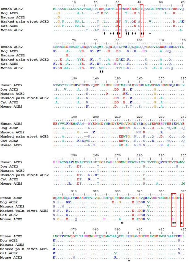
(Extended Data Fig. 1). Of the 18 amino acids known to be involved in 1. Zhu, N. et al. A novel coronavirus from patients with pneumonia in China, 2019. N. Engl. J.
the interaction between ACE2 and the spike receptor binding domain Med. 382, 727–733 (2020).
(RBD) of SARS-CoV-2, five differ between humans and dogs, but none 2. World Health Organization. Coronavirus Disease 2019 (COVID-19) Situation Report 50
(2020); https://www.who.int/docs/default-source/coronaviruse/situation-reports/202003
of these are in regions known to disrupt the interaction between the 10-sitrep-50-covid-19.pdf?sfvrsn=55e904fb_2
RBD of SARS-CoV and ACE28. 3. World Health Organization. Consensus Document on the Epidemiology of Severe Acute
Respiratory Syndrome (SARS) (2003); https://apps.who.int/iris/bitstream/
Our evidence suggests that human-to-animal transmission of
handle/10665/70863/WHO_CDS_CSR_GAR_2003.11_eng.pdf
SARS-CoV-2 is possible. We do not have information on whether the 4. Chu, D. K. W. et al. Molecular diagnosis of a novel coronavirus (2019-nCoV) causing an
virus can cause illness in dogs, but there were no specific symptoms outbreak of pneumonia. Clin. Chem. 66, 549–555 (2020).
5. Corman, V. M. et al. Detection of 2019 novel coronavirus (2019-nCoV) by real-time
in either of the infected dogs while they were shedding virus. The
RT–PCR. Euro Surveill. 25 (2020).
Pomeranian died two days after release from isolation, probably 6. Wölfel R, et al. Virological assessment of hospitalized patients with COVID-2019. Nature
owing to the pre-existing underlying diseases; we were unable to per- 581, 465–469 (2020).
7. Choe, P. G. et al. MERS-CoV antibody responses 1 year after symptom onset, South Korea,
form a post mortem examination. Whether infected dogs could trans-
2015. Emerg. Infect. Dis. 23, 1079–1084 (2017).
mit the virus to other animals or back to humans remains unknown. 8. Lan J et al. Structure of the SARS-CoV-2 spike receptor-binding domain bound to the
The owner of dog 2 had a second, crossbreed dog in which neither ACE2 receptor. Nature 581, 215–220 (2020).
9. IDEXX. IDEXX SARS-CoV-2 (COVID-19) RealPCR Test (2020); https://www.idexx.com/en/
viral RNA nor antibody responses were detected, suggesting that
veterinary/reference-laboratories/idexx-sars-cov-2-covid-19-realpcr-test/
transmission had not occurred between the two dogs sharing the 10. Shi, J. et al. Susceptibility of ferrets, cats, dogs, and other domesticated animals to
household. SARS-coronavirus 2. Science 368, 1016–1020 (2020).
11. SciCom. Risque Zoonotique du SARS-CoV2 (Covid-19) Associé aux Animaux de
These two cases in Hong Kong demonstrate that dogs can acquire
Compagnie: Infection de l’Animal vers l’Homme et de l’Homme vers l’Animal (2020);
infection in households with SARS-CoV-2-infected humans. A survey http://www.afsca.be/comitescientifique/avis/2020/_documents/
of 4,000 specimens from dogs, cats and horses from places where Conseilurgentprovisoire04-2020_SciCom2020-07_Covid-19petitsanimauxdomestiqu
es_27-03-20_001.pdf
community transmission of SARS-CoV-2 was occurring in humans did 12. Zhou, P. et al. A pneumonia outbreak associated with a new coronavirus of probable bat
not detect any positive results, suggesting that the virus is not widely origin. Nature 579, 270–273 (2020).
circulating in pet animals9. Unlike our study, this previous study did not
Publisher’s note Springer Nature remains neutral with regard to jurisdictional claims in
specifically investigate dogs from households of patients with COVID-19.
published maps and institutional affiliations.
A challenge study in five six-week-old beagles demonstrated serocon-
version in two dogs and detection of viral RNA (up to 106.5 copies) in © The Author(s), under exclusive licence to Springer Nature Limited 2020
778 | Nature | Vol 586 | 29 October 2020Methods with multiple gene specific primers targeting different regions of the
viral genome (Supplementary Table). The synthesized cDNA was then
Data reporting subjected to multiple overlapping PCRs using Platimum Taq DNA poly-
No statistical methods were used to predetermine sample size. The merase (Thermo Fisher Scientific) using the protocol provided by the
experiments were not randomized. The investigators were not blinded manufacturer. The PCRs performed were in sizes of around 2,000 bp
to allocation during experiments and outcome assessment. designed to cover the whole virus genome. PCR amplicons were visual-
ized by agarose gel electrophoresis. Nested PCRs were performed when
Specimen collection necessary for genome amplification. Aliquots of 5 μl PCR products and
Specimens from dogs and cats were collected by veterinarians from ani- DNA ladder were loaded into wells in 2% agarose gel. Electrophoresis
mals sent to the AFCD isolation centre and included deep oropharyngeal was run at 120 V for 20 min in TAE buffer and the DNA band was visual-
and nasal swabs and a sample of fresh faeces and/or a rectal swab, placed ized with SYBR safe DNA gel stain.
in virus transport medium and kept on cool-packs until arrival in the labo- PCR amplicons obtained from the same specimens were pooled and
ratory. Virus transport medium comprised Medium 199 (Sigma M0393) sequenced using MiSeq sequencing platform (Illumina). Sequencing
as basal medium, 0.5% bovine serum albumin, antibiotics (penicillin g, library was prepared by Nextera XT DNA library prep Kit (Illumina) fol-
streptomycin sulfate, polymyxin B sulfate, sulfamethoxazole, nystatin, lowing standard protocols. Generated sequencing reads were mapped
gentamicin sulfate, ofloxacin). Specimens were collected on at least 3 to a reference virus genome by BWA14 and genome consensus was gen-
occasions (on arrival in the isolation centre and in the two days before erated by Geneious version 11.1.4 (https://www.geneious.com) with a
release). Any animal that had a positive test was retested until no positive minimal coverage depth of 20. Percentage of nucleotides at each posi-
results were obtained. Owners provided written consent at the time their tion of the genome was calculated by bam-readcount (https://github.
pets were moved to isolation to allow specimens to be collected and tested. com/genome/bam-readcount) with minimal base quality score of 20
Control specimens including nasal, oral, rectal swabs and faeces were and minimum mapping quality score of 20.
collected from 21 stray dogs soon after euthanasia. Stored residual sera
from 20 dogs collected for diagnostic purposes from veterinary clinics Plaque reduction neutralization tests
during 2017–2018 were used as controls for serology. BetaCoV/Hong Kong/VM20001061/2020 isolated from the nasophar-
Specimens from humans were collected and tested by RT–qPCR as ynx aspirate and throat swab of a COVID-19 patient in Hong Kong was
part of routine clinical care and the viruses genetically sequenced as grown in Vero E6 cells (ATCC CRL-1586). Cells were regularly tested to
part of the routine public health response (Institutional Review Board exclude mycoplasma contamination. Stock virus was prepared and
approval UW20-168). aliquoted and stored at −80 °C until use. The virus stock was titrated
in quadruplicate in Vero-E6 cells in 24-well tissue culture plates (TPP
Quantitative RT–PCR Techno Plastic Products) in a biosafety level 3 facility. After one hour
At the AFCD laboratory, RNA from 200 μl specimen in virus trans- incubation in 5% CO2 incubator, the plates were overlaid with 1% aga-
port medium was extracted using NucliSENS easyMag extraction kit rose in cell culture medium and incubated for 3 days when the plates
(BioMerieux) following instructions provided by the manufacturer were fixed and stained and plaque forming units per ml of the virus
and eluted into 60 μl. The RNA was tested for SARS-CoV-2 RNA in a stock was determined. Serial dilutions of serum samples were then
commercial assay RT–qPCR assay for the E and RdRp gene sequences incubated with 30–40 plaque-forming units of virus for 1 h at 37 °C.
(TIB Molbiol Lightmix Modular Assays) based on published RT–qPCR The virus–serum mixtures were added on to Vero cell monolayers,
assay for SARS-CoV-25. Positive, negative and inhibitor controls were incubated, overlaid and stained as above. Antibody titres were defined
included in each RT–qPCR run and work-flow precautions were in as the highest serum dilution that resulted in >90% (PRNT90) reduction
place to minimise PCR contamination. Positive samples were sent to in the number of plaques7.
the HKU as an independent reference laboratory for confirmation.
Viral RNA from the original swabs referred by the AFCD laboratory were Virus isolation
independently extracted at the HKU using the QIAamp viral RNA minikit Fresh nasal and oral swab fluid collected from SARS-CoV-2 PCR con-
(Qiagen, Hilden, Germany) following the manufacturer’s instructions. firmed dogs in viral transport media were used as the inoculum for
Swab supernatant (160 μl) was used for RNA extraction with the final elu- virus isolation. In brief, Vero E6 (ATCC CRL-1586) cells were cul-
tion volume being 60 μl. One-step RT–qPCR assays were run for previ- tured for 24 h in a 24-well plate format (TPP Techno Plastic Products)
ously published nsp14 and N genes, which detect SARS-CoV, SARS-CoV-2 before inoculation. Culture medium was minimal essential medium
and bat SARS-CoV4. In addition, RT–qPCR assays for nsp16 and M that containing 2% fetal bovine serum, 100 units ml−1 penicillin and 100 μg
are specific for SARS-CoV-2 with no cross-reaction with SARS-CoV were ml−1 streptomycin. The swab fluids were centrifuged at 5,000 rpm
also used. The forward primer (5′-GGWCAAATCAATGATATGATTTT), for 10 min at 4 °C in a benchtop centrifuge and the supernatant was
reverse prime (5′-GTTGTTAACAAGAACATCACTAGA) and probe separated and inoculated on to Vero E6 cells in alternative wells
(5′-FAM-AAGTCTRCCTTTACTAAGAAGAGA-TAMRA-3′) were used for the of the 24-well plate. After two hours incubation for adsorption in
ORF1b-nsp16 assay and forward primer (5′-GGYTCTAARTCACCCA a 37 °C incubator containing 5% CO 2, fresh virus growth medium
TTCA-3′), reverse prime (5′-TGATACTCTARAAAGTCTTCATA-3′) and was added to a final volume of 1 ml and then incubated in a 37 °C
probe (5′-FAM-AATTTAGGTTCCTGGCAATTAATT-TAMRA-3′) were used incubator containing 5% CO2 for six days. The presence of cyto-
for the M gene assay. The thermal cycling conditions were identical to pathic effect (CPE) was looked for daily. Additionally, the aliquots
those published for the nsp14 and N gene assays4. Positive, negative and of culture supernatant samples was collected into AVL buffer at 0 h,
inhibitor controls were included in each RT–qPCR run and work-flow 24 h, 48 h and 72 h post inoculation for PCR. The culture medium
precautions were in place to minimise PCR contamination13. was replaced as required with fresh culture medium. Cell cultures
Nasal, oral, rectal swabs and faecal samples from 21 control dogs that were negative for virus growth were blind-passaged again after
were run by all six RT–qPCR assays with negative results. No evidence six days. The cultures that were positive for virus growth as judged
of PCR inhibition was seen in any of these RNA extracts. by cytopathic effect and increasing viral load by RT–qPCR were col-
lected and passed on to new cull culture wells in 24-well plates and
Sequencing the viral genomes then progressively onto cells in T25 culture flasks (Greiner Bio-one).
To amplify the virus genome, reverse transcription reactions were set Mock inoculated Vero E6 cells were used as negative control for each
up using superscript IV reverse transcriptase (Thermo Fisher Scientific) isolation experiment.Article
published. We thank E. Tai for information on infection in pet animals during the SARS
outbreak; E. Yiu for design and preparation of the timeline; H.-L. Yen for providing
Reporting summary
control canine sera; P. Krishnan and D. Yuet Mei Ng for technical assistance; the core
Further information on research design is available in the Nature facility at the Centre for PanorOmic Sciences at the University of Hong Kong for the
Research Reporting Summary linked to this paper. Illumina MiSeq sequencing of viral genomes; and the originating and submitting
laboratories for sharing genetic sequences and other associated data through the
GISAID Initiative for SARS-CoV-2 genome sequences. We acknowledge research funding
for M.P. from the US National Institute of Allergy and Infectious Diseases (NIAID) under
Data availability Centers of Excellence for Influenza Research and Surveillance (CEIRS) contract no.
HHSN272201400006C.
Data that support the findings of this study have been deposited at
GenBank with accession numbers MT215193, MT215194, MT215195, Author contributions T.H.C.S., E.M.W.T., M.P., L.D.S. and C.J.B. were responsible for design of
MT270814, MT270815 and MT276600. The sequencing primers used the study. Monitoring and collection of samples from dogs was undertaken by E.M.W.T. and
V.Y.T.Y., and data and samples from humans was curated by D.N.C.T. Molecular diagnostics was
for full genome sequencing of SARS-CoV-2 are available in the Sup- undertaken and overseen by P.Y.T.L., S.M.I., K.W.S.T. and D.K.W.C. Virus genetic sequencing was
plementary Table. undertaken by D.K.W.C., L.L.M.P. and M.P. L.D.S. and T.H.C.S. drafted the manuscript. Data
analysis and critical review of the manuscript was undertaken by all authors.
13. Bustin S.A. et al. MIQE précis: practical implementation of minimum standard guidelines
for fluorescence-based quantitative real-time PCR experiments. BMC Mol. Biol. 11, 74 Competing interests The authors declare no competing interests.
(2010).
14. Li, H. & Durbin, R. Fast and accurate short read alignment with Burrows–Wheeler Additional information
transform. Bioinformatics 25, 1754–1760 (2009). Supplementary information is available for this paper at https://doi.org/10.1038/s41586-020-
2334-5.
Correspondence and requests for materials should be addressed to M.P.
Acknowledgements We acknowledge the work by staff of the Hong Kong SAR Centre for Peer review information Nature thanks Linda Saif and the other, anonymous, reviewer(s) for
Health Protection who undertook investigations of the human cases in the affected their contribution to the peer review of this work.
household. We thank the dog owners for allowing the material on these cases to be Reprints and permissions information is available at http://www.nature.com/reprints.Extended Data Fig. 1 | Sequence alignment of ACE2 proteins from human, interaction between human ACE2 and RBD of SARS-CoV are highlighted in red dog, macaque, masked palm civet, cat and mouse. Amino acid residues of boxes and these amino acid residues are all conserved between human and dog human ACE2 that are experimentally shown to interact with the RBD of ACE2 proteins. SARS-CoV-28 are denoted by asterisks. Mutations known to disrupt the
nature research | reporting summary
                                                                                                      Corresponding author(s): Malik Peiris
                                                                                                      Last updated by author(s): 22 April 2020

Reporting Summary
Nature Research wishes to improve the reproducibility of the work that we publish. This form provides structure for consistency and transparency
in reporting. For further information on Nature Research policies, seeAuthors & Referees and theEditorial Policy Checklist .

Statistics
For all statistical analyses, confirm that the following items are present in the figure legend, table legend, main text, or Methods section.

n/a Confirmed
           The exact sample size (n) for each experimental group/condition, given as a discrete number and unit of measurement
           A statement on whether measurements were taken from distinct samples or whether the same sample was measured repeatedly
           The statistical test(s) used AND whether they are one- or two-sided
           Only common tests should be described solely by name; describe more complex techniques in the Methods section.

           A description of all covariates tested
           A description of any assumptions or corrections, such as tests of normality and adjustment for multiple comparisons
           A full description of the statistical parameters including central tendency (e.g. means) or other basic estimates (e.g. regression coefficient)
           AND variation (e.g. standard deviation) or associated estimates of uncertainty (e.g. confidence intervals)

           For null hypothesis testing, the test statistic (e.g. F, t, r) with confidence intervals, effect sizes, degrees of freedom and P value noted
           Give P values as exact values whenever suitable.

           For Bayesian analysis, information on the choice of priors and Markov chain Monte Carlo settings
           For hierarchical and complex designs, identification of the appropriate level for tests and full reporting of outcomes
           Estimates of effect sizes (e.g. Cohen's d, Pearson's r), indicating how they were calculated
                                                 Our web collection on statistics for biologists contains articles on many of the points above.

Software and code
Policy information about availability of computer code
  Data collection              No commercial or open source databases used.

  Data analysis                Short sequence read alignment using Burrows-Wheeler transform, Li & Durbin. Bioinformatics 2009; 25: 1754; Genome consensus
                               generated using Geneious ver 11.1.4; Percentages of nucleotides at each position of the genome using bam-readcount (https://
                               github.com/genome/bam-readcount)
For manuscripts utilizing custom algorithms or software that are central to the research but not yet described in published literature, software must be made available to editors/reviewers.
We strongly encourage code deposition in a community repository (e.g. GitHub). See the Nature Research guidelines for submitting code & software for further information.

Data
Policy information about availability of data
 All manuscripts must include a data availability statement. This statement should provide the following information, where applicable:
     - Accession codes, unique identifiers, or web links for publicly available datasets
     - A list of figures that have associated raw data
     - A description of any restrictions on data availability
                                                                                                                                                                                                    October 2018

GenBank accession numbers MT215193, MT215194, MT215195, MT270814, MT270815 and MT276600. Data will be released 1st May 2020

                                                                                                                                                                                                          1
Field-specific reporting
Please select the one below that is the best fit for your research. If you are not sure, read the appropriate sections before making your selection.
nature research | reporting summary
Life sciences study design
All studies must disclose on these points even when the disclosure is negative.
Sample size 15 dogs, 7 cats and four human patients. All eligible subjects during study period 10th Feb - 27th March 2020 were included. Sample size
calculation is not relevant.
Data exclusions No data exclusions
Replication RT-PCR assays have been done independently in two different laboratories with up to 6 different gene targets. All positive results were
confirmed by re-extraction and repeat PCR on the original specimen.
Randomization Not relevant. An observational study. No intervention investigated
Blinding Not relevant. An observational study. No intervention investigated
Reporting for specific materials, systems and methods
We require information from authors about some types of materials, experimental systems and methods used in many studies. Here, indicate whether each material,
system or method listed is relevant to your study. If you are not sure if a list item applies to your research, read the appropriate section before selecting a response.
Materials & experimental systems Methods
n/a Involved in the study n/a Involved in the study
Antibodies ChIP-seq
Eukaryotic cell lines Flow cytometry
Palaeontology MRI-based neuroimaging
Animals and other organisms
Human research participants
Clinical data
Eukaryotic cell lines
Policy information about cell lines
Cell line source(s) Vero-E6 cells (ATCC CRL-1586)
Authentication Cell lines obtained from ATCC. Original cell stocks maintained in liquid N2 storage and each thawed aliquot discarded after 20
cell passages.
Mycoplasma contamination Confirmed to be free of mycoplasma using two methods. A cell culture based kit from Invivogen. PlasmoTest™ - Mycoplasma
Detection Kit and a PCR assay from ABM. https://www.abmgood.com/pcr-mycoplasma-detection-kit-g238.html
Commonly misidentified lines No commonly misidentified cell lines used.
(See ICLAC register)
Animals and other organisms
Policy information about studies involving animals; ARRIVE guidelines recommended for reporting animal research
Laboratory animals None
Wild animals None
Field-collected samples Samples collected by veterinarians of the Department of Agriculture, Fisheries and Conservations as part of routine management
of the animals
October 2018
Ethics oversight Agriculture, Fisheries and Conservation Department
Note that full information on the approval of the study protocol must also be provided in the manuscript.
2You can also read