Case Report: Changes in Cytokine Kinetics During the Course of Disease in a Japanese Patient With Multisystem Inflammatory Syndrome in Children
←
→
Page content transcription
If your browser does not render page correctly, please read the page content below
CASE REPORT
                                                                                                                                                    published: 21 July 2021
                                                                                                                                           doi: 10.3389/fped.2021.702318

                                                Case Report: Changes in Cytokine
                                                Kinetics During the Course of
                                                Disease in a Japanese Patient With
                                                Multisystem Inflammatory Syndrome
                                                in Children
                                                Satoshi Takasago 1 , Aiko Sakai 2 , Masaya Sugiyama 2 , Masashi Mizokami 2 ,
                                                Hiromichi Hamada 3 , Yukihito Ishizaka 4 , Tohru Miyoshi-Akiyama 5 , Akihiro Matsunaga 4 ,
                                                Mikako Ueno 4 , Hiroyuki Shichino 1 and Ayumi Mizukami 1*
                                                1
                                                  Department of Pediatrics, Center Hospital of the National Center for Global Health and Medicine, Tokyo, Japan, 2 Genome
                                                Medical Sciences Project, Research Institute, National Center for Global Health and Medicine, Tokyo, Japan, 3 Department of
                                                Pediatrics, Graduate School of Medicine, Chiba University, Chiba, Japan, 4 Department of Intractable Diseases, Research
                                                Institute, National Center for Global Health and Medicine, Tokyo, Japan, 5 Department of Infectious Diseases, Research
                                                Institute, National Center for Global Health and Medicine, Tokyo, Japan

                                                Multisystem inflammatory syndrome in children (MIS-C) is a severe disease that
                       Edited by:
                 Kuender D. Yang,               is reportedly linked to coronavirus disease 2019. Affected patients present with
   Mackay Memorial Hospital, Taiwan             gastrointestinal symptoms and cardiovascular dysfunction, in addition to Kawasaki
                         Reviewed by:           disease-like features, suggesting the potential for overlapping disease mechanisms.
                         Ilaria Maccora,
            University of Florence, Italy
                                                Kawasaki disease has been reported among individuals of East Asian ethnicities, whereas
                    Mamoru Ayusawa,             there is minimal clinical literature regarding the occurrence of MIS-C among individuals
               Nihon University, Japan
                                                of Asian ethnicities. A few reports thus far have described changes in cytokine kinetics
                 *Correspondence:
                                                during the course of disease in patients with MIS-C. We followed the temporal cytokine
                    Ayumi Mizukami
        aymizukami@hosp.ncgm.go.jp              kinetics in a 9-year-old Japanese girl who exhibited a classical trajectory of MIS-C.
                                                The patient exhibited right cervical swelling and pain, abdominal pain, vomiting, and
                    Specialty section:          lip reddening, which developed 31 days after she was diagnosed with severe acute
          This article was submitted to
                 Pediatric Immunology,          respiratory syndrome coronavirus-2 infection. The patient was diagnosed with Kawasaki
                a section of the journal        disease on her fifth day of illness; because she fulfilled the criteria for MIS-C, she was
                  Frontiers in Pediatrics
                                                also diagnosed with this disease on her fifth day of illness. Her fever rapidly resolved upon
              Received: 29 April 2021
                                                administration of intravenous immunoglobulin, aspirin, and prednisolone. On the patient’s
             Accepted: 21 June 2021
             Published: 21 July 2021            sixth day of illness, she developed acute myocarditis, which was treated with two
                               Citation:        diuretics and one vasodilator; the myocarditis ameliorated within a few days. Analyses of
    Takasago S, Sakai A, Sugiyama M,            temporal kinetics for 71 serum cytokines revealed several patterns of cytokine changes
   Mizokami M, Hamada H, Ishizaka Y,
     Miyoshi-Akiyama T, Matsunaga A,            that were consistent with the patient’s clinical course of disease. Importantly, there was
  Ueno M, Shichino H and Mizukami A             a clear distinction between cytokines that did and did not decrease rapidly following
      (2021) Case Report: Changes in
                                                post-treatment fever resolution. These findings may be useful for the assessment of
Cytokine Kinetics During the Course of
   Disease in a Japanese Patient With           disease status and selection of therapy in patients with similar symptoms; they may also
  Multisystem Inflammatory Syndrome             provide insights for basic and clinical research regarding MIS-C.
 in Children. Front. Pediatr. 9:702318.
      doi: 10.3389/fped.2021.702318             Keywords: MIS-C, PIMS-TS, COVID-19, SARS-CoV-2, Kawasaki disease, cytokine, case report

Frontiers in Pediatrics | www.frontiersin.org                                         1                                               July 2021 | Volume 9 | Article 702318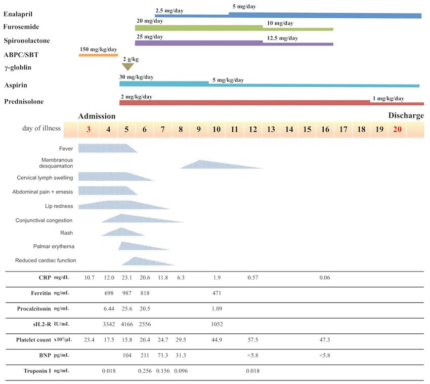
Takasago et al.                                                                                                 Temporal Cytokine Kinetics in MIS-C

INTRODUCTION                                                               The patient’s blood pressure was 113/70 mmHg (i.e., within the
                                                                           reference range). She had a positive test result for SARS-CoV-2,
Multisystem inflammatory syndrome in children (MIS-C; also                 according to FilmArray (bioMérieux SA, Marcy-l’Étoile, France)
known as pediatric inflammatory multisystem syndrome toxic                 analysis of a nasopharyngeal swab. Elevated leukocyte count
shock) is a condition that has been linked with coronavirus                (9.39 × 10³ /µL), elevated neutrophil count (8.45 × 10³ /µL),
disease 2019 (COVID-19) mainly in Europe and North America                 reduced lymphocyte count (0.47 × 10³ /µL), elevated C-reactive
since April 2020; it reportedly involves serious complications             protein level (10.77 mg/dL), and reduced serum sodium level
including Kawasaki disease-like features (1, 2). MIS-C occurs              (131 mEq/L) were noted. Platelet count (234 × 103 /µL) was
within 2–6 weeks after an acute infection with severe acute                within the reference range. Chest and abdominal X-ray findings
respiratory syndrome coronavirus-2 (SARS-CoV-2). Distinct                  were normal.
features include an older peak age of onset, compared with                     The laboratory measures from hospitalization to diagnosis
patients with classical Kawasaki disease (3); they also include            are shown in Table 1; the patient’s course of disease following
frequent gastrointestinal symptoms and greater incidences of               hospitalization is shown in Figure 1. After admission, the
both cardiovascular dysfunction and intensive care management              patient was diagnosed with purulent cervical lymphadenitis;
than in patients with classical Kawasaki disease (4). While                she then began broad-spectrum antimicrobial treatment
Kawasaki disease is more prominent among individuals of East               with ampicillin/sulbactam (150 mg/kg/day). However, the
Asian ethnicities, MIS-C reportedly occurs more often among                fever persisted and the patient showed increasing levels of
individuals of Afro-Caribbean ethnicities (5); to the best of our          inflammatory reaction markers (e.g., C-reactive protein,
knowledge, there is minimal clinical literature regarding the              erythrocyte sedimentation rate, and procalcitonin). On the
occurrence of MIS-C among individuals of Asian ethnicities.                patient’s fifth day of illness, she developed bilateral bulbar
Furthermore, a few reports thus far have described changes in              conjunctival congestion, anterior chest erythema, and right
cytokine kinetics during the course of MIS-C with sufficient detail        palm erythema, in addition to exacerbation of lip reddening;
to identify cytokine changes in relation to clinical manifestations.       the patient thus fulfilled all six Japanese criteria for Kawasaki
We presume that an understanding of these changes may help                 disease (6) (Supplementary Figure 1). The patient did not
to elucidate the underlying etiology of MIS-C. Here, we describe           exhibit bacillus Calmette–Guérin inoculation site redness.
a patient whose course of MIS-C followed a trajectory similar              Furthermore, she exhibited elevated leukocyte count (14.07 ×
to that observed in previously reported patients; additionally,            103 /µL), elevated neutrophil count (12.38 × 10³ /µL), elevated
we describe the temporal kinetics of 71 cytokines, which were              C-reactive protein level (23.05 mg/dL), elevated procalcitonin
analyzed using serum from our patient.                                     level (25.56 ng/mL), elevated aspartate aminotransferase level (62
                                                                           U/L), elevated alanine aminotransferase level (52 U/L), reduced
CASE DESCRIPTION                                                           platelet count (158 × 103 /µL), prolonged prothrombin time
                                                                           (14.3 s; international normalized ratio: 1.17), elevated fibrinogen
A 9-year-old Japanese girl with no notable history of medical              level (691 mg/dL), and elevated D-dimer level (4.9 µg/mL)
problems was diagnosed with SARS-CoV-2 infection on the basis              (Table 1). Because the patient fulfilled the WHO preliminary
of polymerase chain reaction (PCR) analysis of nasopharyngeal              criteria (7), she was diagnosed with MIS-C.
swab sample. Her parents are both Japanese. The grandmother                    Following diagnosis of MIS-C, the patient exhibited normal
was the first in the family to be infected with SARS-CoV-2.                cardiac function according to echocardiography performed prior
Subsequently, the patient, both parents, and one sibling were              to treatment start, with no indications of pericardial effusion.
positive for SARS-CoV-2 on PCR test. There was no contact                  She did not exhibit coronary artery dilation, but demonstrated
with SARS-CoV-2 positive patients at school or other places                enhanced coronary artery wall brightness. Furthermore, she had
during the same period. The patient exhibited fever and olfactory          no unusual electrocardiographic findings. We expected that the
dysfunction for 2 days, although her symptoms were mild. She               patient was at considerable risk of coronary aneurysm, on the
did not require hospitalization and spontaneously recovered                basis of previous findings by Kobayashi et al. (8, 9). Accordingly,
during in-home quarantine. At 31 days after the patient had                we administered intravenous immunoglobulin (IVIG; 2 g/kg)
been diagnosed with SARS-CoV-2 infection, she developed                    and aspirin (ASA; 30 mg/kg/day), which constitute standard
fever, swelling and pain in the right cervical region, as well as          treatment for Kawasaki disease, beginning on the 5th day of
abdominal pain and vomiting. One day later, she presented to               illness; we also began administration of prednisolone (PSL;
our hospital for outpatient treatment. Although the patient was            2 mg/kg/day) at that time. The patient’s treatment response
able to return home for approximately 24 hours, her symptoms               was favorable, such that the fever resolved within 12 hours
subsequently worsened; she required emergency admission on                 of treatment initiation; the patient’s other symptoms showed
the following day (third day of illness).                                  progressive amelioration. Furthermore, the serum levels of
   Upon admission, the patient exhibited a fever of 40.2◦ C,               inflammation markers (e.g., C-reactive protein, procalcitonin,
spontaneous pain throughout the abdomen, vomiting, and right               ferritin, and soluble interleukin-2 receptor; Figure 1) also
cervical pain. Physical examination revealed multilocular cervical         decreased relatively rapidly from a peak on the 5th day of
lymphadenopathy with a maximum diameter of 30 mm, mild                     illness. On the 6th day of illness, however, she exhibited reduced
redness of the lips and oral cavity. There were no indications of          left ventricular systolic function (ejection fraction 50.2%) and
bulbar conjunctival congestion, rash, or distal extremity changes.         enhanced left ventricular end-diastolic diameter (Figure 2A),

Frontiers in Pediatrics | www.frontiersin.org                          2                                       July 2021 | Volume 9 | Article 702318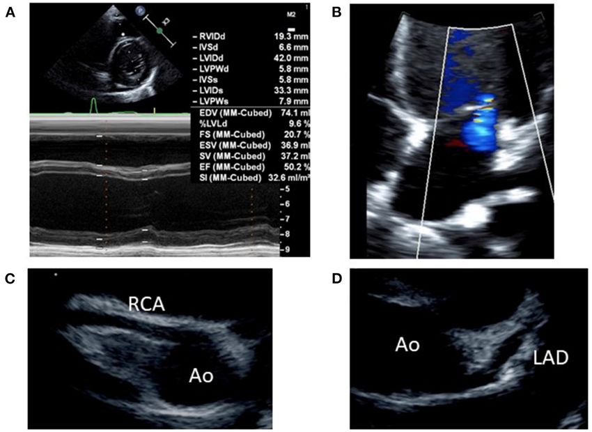
Takasago et al.                                                                                                               Temporal Cytokine Kinetics in MIS-C

TABLE 1 | Blood test results from admission (3rd day of illness) to MIS-C diagnosis (5th day of illness).

                                                               Day 3                         Day 4                   Day 5                     Reference range

White blood cell (×103 /µL)                                     9.39                          7.63                   14.07                          4.5–13.5
Neutrophil (×103 /µL)                                           8.45                          6.71                   12.38                           1.8–8.0
Lymphocyte (×103 /µL)                                           0.47                          0.53                   1.41                            1.5–6.5
Hemoglobin (g/dL)                                               14.0                          11.9                   12.3                          11.5–14.8
Platelets (×104 /µL)                                            23.4                          18.4                   15.8                          15.0–40.0
Albumin (g/dL)                                                  3.9                            3.2                    3.2                            3.5–5.0
Total bilirubin (mg/dL)                                         0.6                            0.5                    0.5                            0.4–1.5
Aspartate aminotransferase (IU/L)                                41                            60                     62                             16–38
Alanine aminotransferase (IU/L)                                  25                            39                     52                            4.0–25.0
Lactate dehydrogenase (IU/L)                                    276                           288                     322                           286–606
Creatinine kinase (IU/L)                                        140                           372                     198                            41–212
Blood urea nitrogen (mg/dL)                                     13.4                           8.0                    8.3                           8.0–20.0
Creatinine (mg/dL)                                              0.36                          0.35                   0.46                          0.34–0.51
Sodium (mEq/L)                                                  131                           126                     130                           138–145
Potassium (mEq/L)                                               3.9                            3.2                    3.6                            3.4–4.7
Chloride (mEq/L)                                                 95                            94                     94                             98–106
C-reactive protein (mg/dL)                                     10.77                          9.57                   23.05
Takasago et al. Temporal Cytokine Kinetics in MIS-C
FIGURE 1 | Course of disease after admission. ABPC/SBT, ampicillin/sulbactam; CRP, C-reactive protein; sIL2-R, soluble interleukin-2 receptor; BNP, B-type
natriuretic peptide.
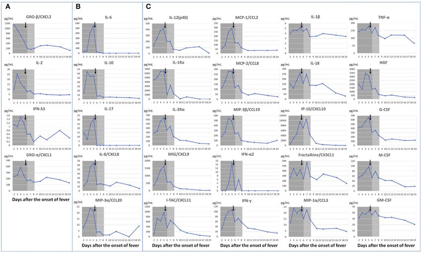
screening panel (Bio-Rad Laboratories Inc.). We also performed reduction in titers after resolution of myocarditis (Figure 3C:
chemiluminescence enzyme immunoassay analyses using an IL-1 receptor antagonist (Ra), CXCL9, CXCL11, CCL2,
HISCL-5000 (Sysmex Asia Pacific Pte Ltd., Singapore) and interferon-γ, IL-1β, IL-18, CXCL10, and tumor necrosis
ELISA assay (R&D systems, Inc.). We investigated changes factor-α et al.); (4) low titers in the acute phase, followed by
in these 71 markers during the patient’s course of disease elevation (CCL17 and SCGF-β); and (5) no change [CCL11,
(Supplementary Figure 3). CCL15, CCL23, IL-3, IL-4, IL-5, IL-12(p70)]. Elevations of
Cytokine changes were striking, such that temporal cytokine the proliferative/hematopoietic factors (hepatocyte growth
kinetics exhibited five patterns: (1) a peak before treatment factor, granulocyte colony-stimulating factor, macrophage
initiation on the 5th day of illness (Figure 3A: interleukin colony-stimulating factor, and granulocyte-macrophage
(IL)-2, interferon-λ3 et al.); (2) elevation until the 5th day of colony-stimulating factor) were also observed in the presence
illness, followed by rapid reduction on the 6th day of illness of inflammation.
after treatment initiation and concomitant fever resolution The patient’s parents provided informed consent for
(Figure 3B: IL-6, IL-10, IL-17, IL-8, CCL20); (3) persistence of publication of this report, including the publication of
high titers for several days after fever resolution, followed by a photographs of the patient’s face and body. The patient also
Frontiers in Pediatrics | www.frontiersin.org 4 July 2021 | Volume 9 | Article 702318Takasago et al. Temporal Cytokine Kinetics in MIS-C
FIGURE 2 | Echocardiography findings on the 6th day of illness: (A) M-mode (left ventricular ejection fraction, 50.2%), (B) mild mitral regurgitation by 4-chamber view,
(C) right coronary artery (seg1: 3.0 mm, Z score+1.6), Z score was calculated by “Z score calculator of coronary arterial diameter” (http://raise.umin.jp/zsp/
calculator/), (D) left coronary artery (seg5: 3.0 mm, Z score+0.9, seg6: 2.7 mm, Z score+1.3, seg11: 2.6 mm, Z score+1.5). Ao, aorta; RCA, right coronary artery;
LAD, left anterior descending artery.
provided informed assent. This clinical analysis was performed occur after the initiation of treatment for MIS-C. In a previous
in accordance with the Declaration of Helsinki and the Ethical report regarding Kawasaki disease shock syndrome (10), which
Guidelines of the Ministry of Health, Labor and Welfare of comprises vasculitis due to hypercytokinemia (similar to MIS-C),
Japan; it was approved by the Research Ethics Committees of the hypotension occurred either during or after IVIG administration
National Center for Global Health and Medicine. in three of six patients.
Feldstein et al. (1) reported the findings of a series
of 186 patients diagnosed with MIS-C in 26 states within
DISCUSSION the United States; importantly, Kawasaki disease-like features
(e.g., conjunctival congestion, rash, and coronary aneurysm
This patient exhibited MIS-C involving a spectrum of symptoms formation) were observed in 74 patients (40%). Furthermore,
along with elevated inflammatory responses within a few days of Toubiana et al. (4) reported that 21 patients with MIS-C
fever onset. The response to treatment comprising IVIG, ASA, had presented with Kawasaki disease-like symptoms and were
and PSL was good; her inflammatory responses rapidly reduced hospitalized; the median onset age was 7.9 years (3.7–16.6 years),
with fever resolution. The patient’s current medical history, which is higher than the peak age for Kawasaki disease (3).
symptoms (fever, abdominal pain, nausea, and manifestations Moreover, all 21 patients had gastrointestinal symptoms such as
of Kawasaki disease), and responses to treatment were also vomiting and abdominal pain, 16 (76%) developed myocarditis,
consistent with reports from Europe and the United States 17 (81%) were admitted to an intensive care unit, and 11
(1, 2, 4). Although it is difficult to clearly differentiate MIS- (52%) required mechanical ventilation; these findings suggest
C from Kawasaki shock syndrome, the clinical picture of a pathology that is distinct from Kawasaki disease. Moreover,
this case is closer to that of MIS-C because of the obvious there have been many reports of MIS-C from Europe and
preceding SARS-CoV-2 infection, the older age, the strong the United States (11), as well as reports of disease among
abdominal pain and vomiting, and the thrombocytopenia and individuals of Afro-Caribbean and Hispanic ethnicities (4, 5).
lymphopenia (10). This patient’s cardiac function was reduced To the best of our knowledge, reports from Asia are rare,
after fever resolution and required cardiovascular intervention. with only one report from India (12) and three reports from
Based on our clinical experience, we assumed that the effects Korea (13).
of vasculitis inflammation on myocardial tissue were delayed. MIS-C and Kawasaki disease are both vasculitic syndromes
Because cardiac function, vascular permeability, and volume that are presumed to arise from cytokine storms; analyses of
overload due to treatment would all contribute to the patient’s their cytokine characteristics may aid in understanding the
disease state, we presumed that reduced cardiac function would underlying mechanisms of disease. Thus far, some studies
Frontiers in Pediatrics | www.frontiersin.org 5 July 2021 | Volume 9 | Article 702318Takasago et al. Temporal Cytokine Kinetics in MIS-C FIGURE 3 | Dynamic changes in cytokines according to changes in clinical symptoms. Febrile period shown in dark gray; duration of myocarditis shown in light gray. ↓ indicates IVIG+PSL+ASA start timing. Dots represent blood draw timings. (A) Cytokines with peak titers before treatment initiation. (B) Cytokines with rapid reductions following fever resolution. (C) Cytokines that persisted at high titers for an extended period following fever resolution, which then decreased with amelioration of myocarditis. ASA, aspirin; IVIG, intravenous immunoglobulin; PSL, prednisolone; IL, interleukin; MIP, macrophage inflammatory protein; IFN, interferon; MCP, monocyte chemoattractant protein; MIG, monokine induced by interferon-γ; IP, interferon-γ-induced protein; TNF, tumor necrosis factor; HGF, hepatocyte growth factor; G-CSF, granulocyte colony-stimulating factor; M-CSF, macrophage colony-stimulating factor; GM-CSF, granulocyte-macrophage colony-stimulating factor. have described acute phase cytokine assay findings of MIS- peaked prior to the fifth day of illness (i.e., when the treatment C (14–21), as well as differences in acute phase cytokine was started; Figure 3) may be involved in the pathogenesis findings in patients with Kawasaki disease, macrophage of MIS-C. activating syndrome, severe COVID-19, and MIS-C (15– In particular, those shown in Figure 3A peaked out 17, 20, 21); however, we have found only a few reports before the start of treatment, and we could observe that describe temporal changes in cytokines in the context them in daily samples obtained from the 2nd to 5th day of clinical symptomology for an individual patient with of illness. Notably, IL-2 is associated with the inositol- MIS-C. In the present report, we closely evaluated the trisphosphate 3-kinase C (ITPKC) gene, which was associated kinetics of multiple cytokines from our patient’s sera, which with susceptibility to Kawasaki disease in a previous had been obtained prior to diagnosis and over the entire genome-wide association study (23). It has been reported treatment trajectory. that patients with single-nucleotide polymorphisms that We observed kinetics of various cytokines that are reportedly convey susceptibility to Kawasaki disease have reduced increased in patients with MIS-C, including IL-1Ra (14, 21), expression of ITPKC, which leads to T-cell activation IL-1β (19), IL-6 (15–20, 22), IL-8 (18, 19), IL-10 (16, 20), IL- and IL-2 elevation (23). We also observed the early peak 17 (17), IL-18 (20), CXCL10 (15, 17, 21, 22), tumor necrosis of CXCL1 and CXCL2 (Figure 3A), which may suggest factor-α (18, 19), interferon-γ (20, 21), CCL3 (17), CCL20 (17), “neutrophil activation” involvement in the pathogenesis CCL19 (17), CCL2 (21, 22), CXCL1 (17), and CXCL11 (17). of MIS-C. We found five patterns in the cytokine kinetics in contrast The cytokines with titers that peaked on the 5th day of with patient’s symptoms. Although it is not possible to clearly illness and rapidly decreased after treatment as symptoms divide all 71 cytokines into five patterns with only a single case improved on the next day of treatment are shown in Figure 3B; study, here, we demonstrate the 29 cytokines that show the those cytokines sensitively reflected the patient’s clinical course, most characteristic features (Figure 3). Cytokines with titers that and thus, may be directly related to MIS-C disease activity. Frontiers in Pediatrics | www.frontiersin.org 6 July 2021 | Volume 9 | Article 702318
Takasago et al.                                                                                                Temporal Cytokine Kinetics in MIS-C

IL-6 is a major pro-inflammatory cytokine that was reportedly             findings may aid in elucidating cytokine kinetics that influence
elevated in patients with MIS-C in previous studies (15–                  treatment responsiveness; they may also facilitate the selection of
20, 22). In our patient, IL-6 titer also rapidly increased                agents for add-on therapy in patients who are unresponsive to
(21.1-fold) with the onset of symptoms. However, another                  initial treatment.
report indicated that IL-6 elevation occurs less frequently
compared with IL-1 receptor antagonist elevation (14); therefore,
further data are needed regarding patients with MIS-C. The                CONCLUSION
importance of IL-17 in MIS-C is controversial. Consiglio et al.
report that IL-17A drives Kawasaki disease but not MIS-C                  We conducted comprehensive temporal analyses of serum
hyperinflammation (15), while reports by Esteve-Sole et al.               cytokine kinetics for the entire course of disease, along with an
(21) and Gruber et al. (17) suggest that IL-17 is elevated in             investigation of clinical symptoms, in a Japanese patient with
MIS-C patients. In our case, IL-17 was rapidly elevated (5.2-             MIS-C who exhibited a classical disease trajectory. Our findings
fold) by the time of diagnosis and decreased to less than the             regarding cytokines that changed during the course of disease
measurement sensitivity with fever resolution on the day after            may provide useful information for elucidating disease status and
treatment initiation.                                                     selecting therapy; the findings will be strengthened by additional
    The clinical significance of cytokines with titers that did not       analyses of cytokine kinetics in patients with MIS-C.
immediately decline to baseline after the fifth day of illness (but
showed a gradual decline; Figure 3C) is difficult to determine
on the basis of data from our single patient; however, Caldarale          DATA AVAILABILITY STATEMENT
et al. showed that IL-6, CXCL8, CCL2, CXCL9, and CXCL10 were
higher in MIS-C patients than in pediatric COVID-19 patients,             The original contributions presented in the study are included
and that treatment with IVIG and methylprednisolone resulted              in the article/Supplementary Material, further inquiries can be
in a rapid decrease in IL-6, but the decrease of CCL 2, CXCL9,            directed to the corresponding author.
and CXCL10 were delayed (22). The same changes in these
cytokines/chemokines were observed in our study. In our case,
IL-6 decreased on the day after treatment; in contrast, CXCL9,
                                                                          ETHICS STATEMENT
CXCL10, MCP-1 and some others took about 5 days. Caldarale                The studies involving human participants were reviewed and
et al. discussed that these interferon-γ induced chemokines               approved by Research Ethics Committees of the National Center
(CXCL9 and CXCL10) showed significant changes in MIS-C                    for Global Health and Medicine. Written informed consent to
patients and are markers of Th1 type immune response (22).                participate in this study was provided by the participants’ legal
In our study, in addition to CXCL9 and CXCL10, interferon-                guardian/next of kin.
γ also showed the same pattern. The increase in IL-1 receptor
antagonist titer was consistent with the findings in a previous
report (14). IL-1β and IL-18 titers were only mildly elevated, and        AUTHOR CONTRIBUTIONS
the mild elevation of the IL-18 titer was also consistent with the
previously reported findings (20). These IL-1 family cytokines are        ST and AS contributed equally to the study design and writing of
reportedly associated with Kawasaki disease-related myocarditis           the manuscript. ST, AS, and AMi designed the study and wrote
(24), but further studies are needed to determine their clinical          the initial draft of the manuscript. AS, MS, and MM contributed
significance in patients with MIS-C.                                      to the analysis and interpretation of cytokines, and assisted in
    Previous reports have indicated that the cytokine profiles of         the preparation of the manuscript. YI, TM-A, AMa, and MU
patients with MIS-C had some overlap and some differences                 contributed to the analysis and interpretation of viral PCR and
from those of patients with COVID-19, macrophage                          serum antibodies to SARS-CoV-2, and assisted in the preparation
activation syndrome, and Kawasaki disease (15, 16, 20, 21).               of the manuscript. HH and HS contributed to critically review
Immunoglobulins and steroids have been used as treatments                 of the manuscript. All authors approved the final version of
for these conditions and, within the past 10 years, molecular             the manuscript, and agree to be accountable for all aspects of
targeted therapies associated with specific cytokines (e.g.,              the work in ensuring that questions related to the accuracy or
tocilizumab, infliximab, and anakinra) have become available              integrity of any part of the work are appropriately investigated
as a treatment option. However, there remains a lack of clear             and resolved.
evidence regarding which drugs to use for specific conditions.
The identification of cytokines associated with clinical features
and outcomes is necessary in understanding pathogenesis and               FUNDING
guiding treatment decisions.
    The primary limitation of this report is its focus on a               This work was supported by Japan Agency for Medicine
single patient. Thus, similar assessments of cytokine kinetics            Research and Development (AMED), Research Program on
are needed in patients with MIS-C; in particular, such reports            Emerging and Re-emerging Infectious Disease (JP19fk0108164,
should focus on patients who are unresponsive to treatment                JP20f0108164, JP19fk0108104, JP20fk0108104, JP20fk0108416,
and on patients who receive other therapeutic agents. The                 JP20fk0108262h0001, and JP19fk0108163).

Frontiers in Pediatrics | www.frontiersin.org                         7                                       July 2021 | Volume 9 | Article 702318
Takasago et al. Temporal Cytokine Kinetics in MIS-C
ACKNOWLEDGMENTS SUPPLEMENTARY MATERIAL
We thank Ryan Chastain-Gross, Ph.D., from Edanz Group The Supplementary Material for this article can be found
(https://en-author-services.edanz.com/ac) for editing a draft of online at: https://www.frontiersin.org/articles/10.3389/fped.
this manuscript. 2021.702318/full#supplementary-material
REFERENCES syndrome following SARS-CoV-2 infection in Switzerland. Front Pediatr.
(2021) 8:594127. doi: 10.3389/fped.2020.594127
1. Feldstein LR, Rose EB, Horwitz SM, Collins JP, Newhams MM, Son 15. Consiglio CR, Cotugno N, Sardh F, Pou C, Amodio D, Rodriguez L, et al.
MB, et al. Multisystem inflammatory syndrome in U.S. children and The immunology of multisystem inflammatory syndrome in children with
adolescents. N Engl J Med. (2020) 383:334–46. doi: 10.1056/NEJMoa20 COVID-19. Cell. (2020) 183:968–81. doi: 10.1016/j.cell.2020.09.016
21680 16. Lee PY, Day-Lewis M, Henderson LA, Friedman KG, Lo J, Roberts JE,
2. Belot A, Antona D, Renolleau S, Javouhey E, Hentgen V, Angoulvant F, et al. Distinct clinical and immunological features of SARS-CoV-2-induced
et al. SARS-CoV-2-related paediatric inflammatory multisystem syndrome, an multisystem inflammatory syndrome in children. J Clin Invest. (2020)
epidemiological study, France, 1 March to 17 May 2020. Euro Surveill. (2020) 130:5942–50. doi: 10.1172/JCI141113
25:2001010. doi: 10.2807/1560-7917.ES.2020.25.22.2001010 17. Gruber CN, Patel RS, Trachtman R, Lepow L, Amanat F, Krammer
3. Ae R, Makino N, Kosami K, Kuwabara M, Matsubara Y, Nakamura F, et al. Mapping systemic inflammation and antibody responses in
Y. Epidemiology, treatments, and cardiac complications in patients with multisystem inflammatory syndrome in children (MIS-C). Cell. (2020)
Kawasaki disease: the nationwide survey in Japan, 2017–2018. J Pediatr. (2020) 183:982–95. doi: 10.1016/j.cell.2020.09.034
225:23–9.e2. doi: 10.1016/j.jpeds.2020.05.034 18. Kwak JH, Lee SY, Choi JW, Korean Society of Kawasaki Disease. Clinical
4. Toubiana J, Poirault C, Corsia A, Bajolle F, Fourgeaud J, Angoulvant F, et al. features, diagnosis and outcomes of multisystem inflammatory syndrome in
Kawasaki-like multisystem inflammatory syndrome in children during the children associated with coronavirus disease 2019. Clin Exp Pediatr. (2021)
covid-19 pandemic in Paris, France: prospective observational study. BMJ. 64:68–75. doi: 10.3345/cep.2020.01900
(2020) 369:m2094. doi: 10.1136/bmj.m2094 19. Calò Carducci FI, De Ioris MA, Agrati C, Carsetti R, Perrotta D,
5. Riphagen S, Gomez X, Gonzalez-Martinez C, Wilkinson N, Theocharis P. D’Argenio P, et al. Hyper inflammation in two severe acute respiratory
Hyperinflammatory shock in children during COVID-19 pandemic. Lancet. syndrome coronavirus 2-infected adolescents successfully treated with the
(2020) 395:1607–8. doi: 10.1016/S0140-6736(20)31094-1 interleukin-1 inhibitor anakinra and glucocorticoids. Front Pediatr. (2020)
6. Kobayashi T, Ayusawa M, Suzuki H, Abe J, Ito S, Kato T, et al. Revision of 8:576912. doi: 10.3389/fped.2020.576912
diagnostic guidelines for Kawasaki disease (6th revised edition). Pediatr Int. 20. Gkoutzourelas A, Bogdanos DP, Sakkas LI. Kawasaki disease and COVID19.
(2020) 62:1135–8. doi: 10.1111/ped.14326 Mediterr J Rheumatol. (2020) 31(Suppl 2):268–74. doi: 10.31138/mjr.31.3.268
7. World Health Organization. Multisystem Inflammatory Syndrome in Children 21. Esteve-Sole A, Anton J, Pino-Ramirez RM, Sanchez-Manubens J,
and Adolescents With COVID-19: Scientific Brief, 15 May 2020. Geneva: World Fumadó V, Fortuny C, et al. Similarities and differences between the
Health Organization (2020). immunopathogenesis of COVID-19-related pediatric inflammatory
8. Kobayashi T, Inoue Y, Takeuchi K, Okada Y, Tamura K, multisystem syndrome and Kawasaki disease. J Clin Invest. (2021)
Tomomasa T, et al. Prediction of intravenous immunoglobulin 131:e144554. doi: 10.1172/JCI144554
unresponsiveness in patients with Kawasaki disease. Circulation. (2006) 22. Caldarale F, Giacomelli M, Garrafa E, Tamassia N, Morreale A,
113:2606–12. doi: 10.1161/CIRCULATIONAHA.105.592865 Poli P, et al. Plasmacytoid dendritic cells depletion and elevation
9. Kobayashi T, Saji T, Otani T, Takeuchi K, Nakamura T, Arakawa H, of IFN-g dependent chemokines CXCL9 and CXCL10 in children
et al. Efficacy of immunoglobulin plus prednisolone for prevention of with multisystem inflammatory syndrome. Front immunol. (2021)
coronary artery abnormalities in severe Kawasaki disease (RAISE study): a 12:654587. doi: 10.3389/fimmu.2021.654587
randomized, open-label, blinded-endpoints trial. Lancet. (2012) 379:1613– 23. Onouchi Y, Gunji T, Burns JC, Shimizu C, Newburger JW, Yashiro M,
20. doi: 10.1016/S0140-6736(11)61930-2 et al. ITPKC functional polymorphism associated with Kawasaki disease
10. Suzuki J, Abe K, Matsui T, Honda T, Yasukawa K, Takanashi J, et al. susceptibility and formation of coronary artery aneurysms. Nat Genet. (2008)
Kawasaki disease shock syndrome in Japan and comparison with multisystem 40:35–42. doi: 10.1038/ng.2007.59
inflammatory syndrome in children in European countries. Front Pediatr. 24. Dusser P, Kone-Paut I. IL-1 inhibition may have an important role
(2021) 9:625456. doi: 10.3389/fped.2021.625456 in treating refractory Kawasaki disease. Front Pharmacol. (2017)
11. Tang Y, Li W, Baskota M, Zhou Q, Fu Z, Luo Z, et al. Multisystem 8:163. doi: 10.3389/fphar.2017.00163
inflammatory syndrome in children during the coronavirus disease 2019
(COVID-19) pandemic: a systematic review of published case studies. Transl Conflict of Interest: The authors declare that the research was conducted in the
Pediatr. (2021) 10:121–35. doi: 10.21037/tp-20-188 absence of any commercial or financial relationships that could be construed as a
12. Rauf A, Vijayan A, John ST, Krishnan R, Latheef A. Multisystem potential conflict of interest.
inflammatory syndrome with features of atypical Kawasaki
disease during COVID-19 pandemic. Indian J Pediatr. (2020) Copyright © 2021 Takasago, Sakai, Sugiyama, Mizokami, Hamada, Ishizaka,
87:745–7. doi: 10.1007/s12098-020-03357-1 Miyoshi-Akiyama, Matsunaga, Ueno, Shichino and Mizukami. This is an open-
13. Choe YJ, Choi EH, Choi JW, Eun BW, Eun LY, Kim Y, et al. access article distributed under the terms of the Creative Commons Attribution
Surveillance of COVID-19–associated multisystem inflammatory License (CC BY). The use, distribution or reproduction in other forums is permitted,
syndrome in children, South Korea. Emerg Infect Dis. (2021) provided the original author(s) and the copyright owner(s) are credited and that the
27:1196–200. doi: 10.3201/eid2704.210026 original publication in this journal is cited, in accordance with accepted academic
14. Fouriki A, Fougère Y, De Camaret C, Blanchard Rohner G, Grazioli S, Wagner practice. No use, distribution or reproduction is permitted which does not comply
N, et al. Case report: case series of children with multisystem inflammatory with these terms.
Frontiers in Pediatrics | www.frontiersin.org 8 July 2021 | Volume 9 | Article 702318You can also read