Proteomic Analysis of Hepatocellular Carcinoma Tissues With Aminopeptidase 3 and Phosphoenolpyruvate Carboxykinase 2 Encapsulation Shows ...
←
→
Page content transcription
If your browser does not render page correctly, please read the page content below
CANCER GENOMICS & PROTEOMICS 18: 307-316 (2021)
doi:10.21873/cgp.20261

  Proteomic Analysis of Hepatocellular Carcinoma Tissues With
         Encapsulation Shows Up-regulation of Leucine
  Aminopeptidase 3 and Phosphoenolpyruvate Carboxykinase 2
                KEISUKE KUHARA1,2, TAKAO KITAGAWA1, BYRON BARON3, KAZUHIRO TOKUDA4,
                    KAZUHIKO SAKAMOTO5, HIROAKI NAGANO5, KAZUYUKI NAKAMURA6,
                  MASANOBU KOBAYASHI1, HIROKI NAGAYASU2 and YASUHIRO KURAMITSU1

       1Advanced   Research Promotion Centre, Health Sciences University of Hokkaido, Ishikari-Tobetsu, Japan;
           2Division of Oral and Maxillofacial Surgery, Department of Human Biology and Pathophysiology,
                 School of Dentistry, Health Sciences University of Hokkaido, Ishikari-Tobetsu, Japan;
                  3Centre for Molecular Medicine and Biobanking, University of Malta, Msida, Malta;
             4Graduate School of Health and Welfare, Yamaguchi Prefectural University, Yamaguchi, Japan;
                           5Department of Gastroenterological, Breast and Endocrine Surgery,

                            Yamaguchi University Graduate School of Medicine, Ube, Japan;
               6Centre of Clinical Laboratories in Tokuyama Medical Association Hospital, Shunan, Japan

Abstract. Background/Aim: Cancer is the most fatal disease          Results: Two-DE analysis showed 14 spots whose expression
worldwide whose most lethal characteristics are invasion and        was different between encapsulated and non-encapsulated
metastasis. Hepatocellular carcinoma (HCC) is one of the            HCC tissues. Of these, 9 were up-regulated and 5 were down-
most fatal cancers worldwide. HCC often shows                       regulated in HCC tissues without encapsulation. The
encapsulation, which is related to better prognosis. In this        validation by Western blot confirmed that leucine
study, proteomic analysis of HCC tissues with and without           aminopeptidase 3 (LAP3) and phosphoenolpyruvate
encapsulation was performed, in order to elucidate the factors      carboxykinase mitochondrial (PCK2) were up-regulated
which play important roles in encapsulation. Materials and          significantly in HCC tissues with a capsule, compared to HCC
Methods: Five HCC tissues surrounded by a capsule and five          tissues that broke the capsule. Conclusion: These findings
HCC tissues which broke the capsule were obtained from              suggest that LAP3 and PCK2 could be factors responsible for
patients diagnosed with HCC who underwent surgical liver            the maintenance of encapsulation in HCC tissues.
resection. Protein samples from these tissues were separated
by two-dimensional gel electrophoresis (2-DE), and the              Although multidisciplinary therapies such as surgery,
protein spots whose expression was different between                radiation and chemotherapy have made progress in the
encapsulated and non-encapsulated HCC tissues were                  treatment of cancer, the latter is still one of the leading
identified through gel imaging analysis software. The selected      causes of death in many countries. Many types of cancer
protein spots were analyzed and identified by liquid                show strong invasive and metastatic capability. Cancer
chromatography-tandem mass spectrometry (LC-MS/MS).                 metastasis is composed of a number of steps. In principle,
                                                                    cancer cells show epithelial-to-mesenchymal transition
                                                                    (EMT) and migrate into surrounding tissues in the course of
                                                                    degrading extracellular matrix from cancer primary tissues.
This article is freely accessible online.                           After intravasation, the cancer cells move to distant organs.
                                                                    Then, cancer cells extravasate from blood vessels, and
Correspondence to: Yasuhiro Kuramitsu, MD, Ph.D., Advanced          invade the parenchyma and infiltrate adjacent tissues. Lastly,
Research Promotion Centre, Health Sciences University of            the cancer cells establish a metastatic tumor tissue with a
Hokkaido, 1757 Kanazawa, Ishikari-Tobetsu, Hokkaido 061-0293,
                                                                    wealth of tumor vasculature (1).
Japan. Tel: +81 133231630, Fax: +81 133231782, e-mail:
climates@hoku-iryo-u.ac.jp
                                                                       So far, many types of anti-metastatic drugs, targeting
                                                                    multiple steps in the process of metastasis have been
Key Words: HCC, proteomics, encapsulation, leucine aminopeptidase   developed. Anti-matrix metalloproteinase (anti-MMP) drugs
3, phosphoenolpyruvate carboxykinase mitochondrial.                 marimastat (2), prinomastat (3), BMS-275291, metastat (4)

                                                                                                                              307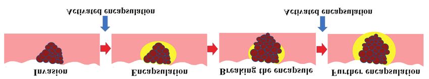
CANCER GENOMICS & PROTEOMICS 18: 307-316 (2021)

Figure 1. Cancer cells invade the surrounding tissue. Encapsulation against invading cancer cells is induced by the activated molecules which
maintain the encapsulation. However, cancer cells can reinvade the surrounding tissue. The identified molecules are activated and induce the
encapsulation for invading cancer cells repeatedly.

and neovastat (5) have been developed, while SU5416 and                 identify molecules which may be responsible for the
SU6668 are anti-angiogenesis drugs (6), and have been t used            maintenance of encapsulation of HCC tissues.
in the phase II studies for several types of cancer. However,
unfortunately all of these never yielded sufficient results,            Materials and Methods
because any doctor does not choose them for the priority
routine therapy for patients with advanced cancer (7).                  Tumor samples. Five HCC tissues surrounded by a capsule and five
   In hepatocellular carcinoma (HCC), tissues are sometimes             HCC tissues with a broken capsule were obtained from patients
                                                                        diagnosed with HCC who underwent surgical liver resection at the
encapsulated with fibrous tissue (8). Ng et al. reported a
                                                                        Department of Surgery II, Yamaguchi University Hospital (Table I).
pathologic study of 189 cases in which encapsulated tumors              None of the patients received any preoperative therapy. The study
showed a much lower incidence of direct liver invasion                  protocol was approved by the Institutional Review Board for
(p
Kuhara et al: Proteomic Analysis of HCC Τissues With or Without Encapsulation software (Nonlinear Dynamics Ltd, Newcastle upon Tyne, UK), and Results the differences in expression between fully capsulated HCC tissues and HCC tissues with capsular invasion were analyzed statistically Figures 2A and B show the 2-DE patterns of hepatocellular by ANOVA; p39 indicate identity or extensive homology (p
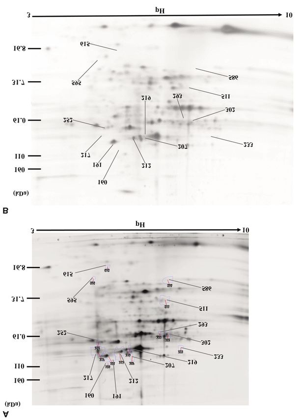
CANCER GENOMICS & PROTEOMICS 18: 307-316 (2021)

Figure 2. 2-DE gel pattern of (A) HCC tissue without encapsulation, and (B) HCC tissue with encapsulation. Proteins (80 μg) were separated on
precast polyacrylamide gel with a linear concentration gradient of 5-20%. After fixing, the gels were stained with Flamingo Gel Stain™.

310
Kuhara et al: Proteomic Analysis of HCC Τissues With or Without Encapsulation

Table I. Summary of all cases of hepatocellular carcinoma.           Table II. Protein spots that decreased or increased in HCC tissues
                                                                     without capsule.
No.   Tumor size    Differentiation   Capsular    Vascular   Stage
        (cm)                          invasion    invasion           #      Anova     Fold    Average normalised   Average normalised
                                                                             (p)                   volumes              volumes
1         2.5          Moderate           –           –       2
2         2.5           Well              –           –       2                                With capsulation    Without capsulation
3         1.2          Moderate           –           –       1
4         2.2          Moderate           –           –       2      160     0.017     4.3       8.930e+004            3.884e+005
5         2.5          Moderate           –           –       2      207     0.019     2.0       2.396e+006            4.825e+006
6        13.0           Poor              +           +       3      217     0.021     2.6       4.910e+006            1.276e+007
7         5.0          Moderate           +           +       3      191     0.022     1.5       1.048e+007            1.609e+007
8         5.9          Moderate           +           +       3      302     0.024     1.5       4.063e+006            2.665e+006
9        13.0          Moderate           +           +       3      293     0.026     2.1       1.297e+006            6.285e+005
10       14.0           Poor              +           +       3      615     0.027     3.3       9.248e+004            3.055e+005
                                                                     252     0.031     1.7       2.260e+006            3.832e+006
                                                                     511     0.031     1.4       4.434e+006            3.248e+006
                                                                     233     0.033     1.3       5.065e+005            4.050e+005
                                                                     212     0.039     1.4       1.492e+007            2.119e+007
                                                                     586     0.040     2.1       2.738e+006            1.281e+006
   HSPs are proteins which mount responses to cellular stress        219     0.041     1.7       1.377e+006            2.340e+006
                                                                     595     0.048     2.4       1.082e+005            2.572e+005
and play roles as molecular chaperones in cells (17). HSPs
are classified into the HSP27, HSP40, HSP47, HSP60,
HSP70, HSP110 and HSP150 families according to their
molecular weight. The role of HSPD1 on the growth of
cancer cells is highly controversial (18). HSPA1A and
HSPA8 are molecular chaperone which are related to various           is essential for cell proliferation and survival, the up-
intracellular process (19). HSPA5 is a molecular chaperone           regulation of eIF5A1 in HCC tissues without encapsulation
which is also known as binding immunoglobulin protein                seems to be consistent with the characteristics of progressing
(BiP) (20). HSPA9, also called GRP75 or mortalin, is located         HCC cells.
in the mitochondria and inhibits cell division and is                   PSMB9 is a component of the proteasome which contributes
implicated in the control of cell proliferation and cellular         to the complete assembly of the 20S proteasome complex (30).
aging (21). HSP 70 kDa family members have been reported             Rouette et al. showed that expression of the immunoproteasome
to be related to malignancies. HSPA1A, HSPA8, HSPD1 and              including PSMB9 is elevated in most cancer types (31). Even
HSPD1 are known to function in various intracellular                 though it is still not clear why the immunoproteasome is up-
reactions and are up-regulated in cancer cells (22). Some of         regulated in cancer cells, its function may be increased in highly
the HSP 70 kDa members are used as prognostic markers                proliferating cancer cells.
and therapeutic target molecules for cancer treatment (23).             RRBP1 is an endoplasmic reticulum membrane protein
Takashima et al. reported that HSP70 kDa family members              (32) which has essential roles for ribosomal function. One
were up-regulated in HCV-associated HCC tissues compared             function is binding and the other is translocation of nascent
to adjacent non-tumoral tissues (24). Jubran et al. reported         proteins across the membrane of the rough endoplasmic
that determination of HSPA9 and Hsp70 in blood could be a            reticulum (33). The up-regulation of RRBP1 in many types
useful additive prognostic tool in guiding clinical                  of cancer cells and tissues has been reported by many
management of patients with colorectal cancer (25). Our              research groups (34-36). In line with these, cancer cells may
present study showed up-regulation of HSP70 kDa family               utilize RRBP1 to break encapsulation and proliferate.
proteins in HCC tissues without encapsulation compared to               In addition, PCK2, LAP3, PEBP1 and PBLD were
encapsulated HCC tissues. These HSPs appear to be related            identified as downregulated proteins in HCC tissue without
to the HCC progression process.                                      encapsulation. By means of Western blotting, the significant
   EIF5A is a translation factor. eIF5A has two isoforms,            down-regulation of PCK2 and LAP3 was confirmed.
eIF5A1 and eIF5A2. eIF5A1 is ubiquitously expressed in                  Wu et al. reported down-regulation of PBLD in HCC
most cells. It is essential for cell proliferation and survival      tissues while PBLD inhibited HCC progression via Ras and
(26). In recent years, the role of eIF5A on cancers, diabetes        Rap1 (37). Li et al. found PBLD inhibited HCC cell growth
mellitus, HIV-infection etc. have been identified and it has         and invasion in vitro and tumor growth in vivo, and they
been exploited as a therapeutic target molecule (27).                concluded that elevated PBLD expression might reduce HCC
Upregulation of eIF5A1 in various cancer tissues and cells           cell growth and invasion via inactivation of several
has been reported in multiple studies (28, 29). Since eIF5A1         tumorigenesis-related signaling pathways (38).

                                                                                                                                    311
CANCER GENOMICS & PROTEOMICS 18: 307-316 (2021)
Figure 3. Western blot analysis of PEBP, LAP3, PCK2 and actin in HCC tissues with or without encapsulation. The protein expression of PEBP,
LAP3 and PCK2 (bands of 21, 56 and 70 kDa, respectively) was reduced in HCC tissues with encapsulation compared to HCC tissues without
encapsulation.
Table III. Identification of proteins up-regulated in HCC tissues without capsulation.
Spot Protein name Accession Theoretical Theoretical Distinct Sequence MS/MS Change in
No. No. pI Mr peptides coverage (%) search score spots (fold)
207 Stress-70 protein, mitochondrial P38646 5.87 73,680 17 27 354 +2.0
219 Heat shock 70 kDa protein 1A P0DMV8 5.47 70,052 11 20 172 +1.7
212 Heat shock cognate 71 kDa protein P11142 5.37 70,898 45 35 612 +1.4
615 Eukaryotic translation factor 5A-1 P63241 5.07 16,832 7 30 111 +3.3
595 Proteasome subunit beta type-9 P28065 4.89 23,264 18 30 232 +2.4
252 60 kDa heat shock protein, mitochondrial P10809 5.70 61,055 28 56 479 +1.7
217 Heat shock cognate 71 kDa protein P11142 5.37 70,898 3 6 63 +2.6
191 Endoplasmic reticulum chaperone BiP P11021 5.07 72,333 30 34 522 +1.5
160 Ribosome-binding protein 1 Q9P2E9 5.45 108,632 11 13 260 +4.3
Table IV. Identification of proteins down-regulated in HCC tissues without capsulation.
Spot Protein name Accession Theoretical Theoretical Distinct Sequence MS/MS Change in
No. No. pI Mr peptides coverage (%) search score spots (fold)
233 Phosphoenolpyruvate carboxykinase Q16822 6.63 70,699 19 33 251 –1.3
mitochondrial
302 Leucine aminopeptidase 3 P28838 8.03 56,166 37 44 440 –1.5
293 Leucine aminopeptidase 3 P28838 8.03 56,166 44 57 745 –2.1
511 Phenazine biosynthesis-like P30039 7.10 31,785 40 76 381 –1.4
domain-containing protein
586 Phosphatidylethanolamine-binding protein 1 P30086 7.01 21,057 10 47 176 –2.1
312Kuhara et al: Proteomic Analysis of HCC Τissues With or Without Encapsulation Figure 4. The intensity of (A) PEBP (B) LAP3 bands and (C) PCK2 bands, normalized by actin, in HCC tissues with or without encapsulation. The intensity of LAP3 and PCK2 bands were normalized by actin and were significantly reduced in HCC tissues without encapsulation compared to HCC tissues with encapsulation (p
CANCER GENOMICS & PROTEOMICS 18: 307-316 (2021)

   PEBP1, also known as Raf kinase inhibitory protein                 cells and extruded transformed cells from the epithelium.
(RKIP), is an inhibitor of the MEK phosphorylation by Raf-            They called this process cell competition. It is not clear how
1. PEBP1 inhibits the Raf-1/MEK/ERK pathway by direct                 cell competition starts, but vimentin induced by LAP3 may
inhibition of Raf-1 and MEK (39), and has anti-metastatic and         have an important role in encapsulation of HCC tissues (51).
pro-apoptotic properties in cancer cells (40). Although Western          In conclusion, in HCC tissues with a broken capsule, the
blotting could not show significant down-regulation of PEBP1          protein levels of LAP3 and PCK2 were significantly down-
in encapsulated HCC tissues compared to HCC tissues without           regulated. The role of both molecules on the maintenance of
encapsulation, there is a possibility that PEBP1 has a role in        encapsulation of HCC tissues is not clear at present.
the maintenance of encapsulation in HCC tissues.                      However, further investigation of LAP3 and PCK2 may
   Significant down-regulation of PCK2 in the HCC tissues             elucidate their important function for cancer tissue
without encapsulation was confirmed by Western blotting. PCK2         encapsulation, and this could be used as an anti-metastatic
is a kinase located in the mitochondria. PCK2 phosphorylates          strategy to induce encapsulation for cancer tissues and
oxaloacetic acid and produces phosphoenolpyruvic acid. This           prevent metastasis.
phosphoenolpyruvic acid goes through the mitochondrial
membrane, and produces glucose (glyconeogenesis) (41). In the         Conflicts of Interest
central part of cancer tissues, the supply of glucose is not enough
for proliferating cancer cells. PCK2 plays an important role in       The Authors declared no potential conflicts of interest with respect
the supply of glucose in cancer cells by using gluconeogenesis.       to the research, authorship, and/or publication of this article.
Although some reports showed that the activation of PCK2
increases the proliferative ability of cancer cells (41), the         Author’s Contributions
expression of PCK2 was reported to be down-regulated in
ovarial cancer cells (42), aflatoxin-induced HCC cells (43),          Y.K., K.N., H.N. and M.K. designed the research; Y.K., K.K., T.K.
pancreatic cancer cells (44), renal cell carcinoma tissues (45),      and K.T. performed the experiments; K.S. and H.N. provided the
                                                                      HCC tissues; K.K., Y.K. and B.B. wrote the article.
and osteosarcoma tissues (46). The most interesting report about
PCK2-expression is a quantitative proteomic differential display
by using isobaric tags for relative and absolute quantification       Acknowledgements
(iTRAQ) for HCC tissues and adjacent non-cancerous tissues.
The result showed down-regulation of PCK2 in HCC tissues              This work was supported in part by Grants-in-Aid from the Ministry
                                                                      of Education, Science, Sports, and Culture of Japan (no. 17K07218
compared to adjacent non-cancerous tissues. Although they
                                                                      to Yasuhiro Kuramitsu).
compared PCK2 expression between HCC cancerous and
adjacent non-cancerous tissues, our result, where PCK2 was
down-regulated in HCC tissues without encapsulation, has a
                                                                      References
similar result (47).
                                                                      1 Park HA, Brown SR and Kim Y: Cellular mechanisms of
   Significant down-regulation of LAP3 in the HCC tissues
                                                                        circulating tumor cells during breast cancer metastasis. Int J Mol
without encapsulation was also confirmed by Western                     Sci 21(14): 5040, 2020. PMID: 32708855. DOI: 10.3390/
blotting. LAP3 is a cell surface aminopeptidase. LAP3                   ijms21145040
catalyzes the hydrolysis of leucine residues from the protein         2 Layfield HJ, Williams HF, Ravishankar D, Mehmi A, Sonavane
amino termini (48). It has been reported that LAP3 is related           M, Salim A, Vaiyapuri R, Lakshminarayanan K, Vallance TM,
to invasion and metastasis of cancer cells by controlling               Bicknell AB, Trim SA, Patel K and Vaiyapuri S: Repurposing
signal transduction. Fang et al. reported that LAP3 up-                 cancer drugs batimastat and marimastat to inhibit the activity of
regulates fascin, which phosphorylates the HSP27-NF-κB                  a group I metalloprotease from the venom of the western
                                                                        diamondback rattlesnake, Crotalus atrox. Toxins (Basel) 12(5):
signalling pathway and promotes the metastatic ability of
                                                                        309, 2020. PMID: 32397419. DOI: 10.3390/toxins12050309
cancer cells. Furthermore, they showed that LAP3 up-                  3 Heath EI, Burtness BA, Kleinberg L, Salem RR, Yang SC,
regulated matrix metalloproteinase-2 (MMP-2) and MMP-9,                 Heitmiller RF, Canto MI, Knisely JP, Topazian M, Montgomery
and high LAP3 expression was correlated with the grade of               E, Tsottles N, Pithavala Y, Rohmiller B, Collier M and Forastiere
malignancy (49). Tian et al. clarified that LAP3 promoted               AA: Phase II, parallel-design study of preoperative combined
motility and invasiveness of HCC cells (48). Wang et al.                modality therapy and the matrix metalloprotease (mmp) inhibitor
reported that vimentin, a mesenchymal marker which strongly             prinomastat in patients with esophageal adenocarcinoma. Invest
                                                                        New Drugs 24(2): 135-140, 2006. PMID: 16502351. DOI:
relates to EMT, was up-regulated in breast cancer tissues, and
                                                                        10.1007/s10637-006-5934-5
LAP3 induced expression of vimentin (50). Although the                4 Heath EI and Grochow LB: Clinical potential of matrix
LAP3-vimentin axis seems to be a promoter of invasion and               metalloprotease inhibitors in cancer therapy. Drugs 59(5): 1043-
metastasis, Kajita et al. reported that vimentin accumulated            1055, 2000. PMID: 10852638. DOI: 10.2165/00003495-
in normal cells specifically at the interface with transformed          200059050-00002

314
Kuhara et al: Proteomic Analysis of HCC Τissues With or Without Encapsulation
5 Brown PD: Ongoing trials with matrix metalloproteinase 17 Wu J, Liu T, Rios Z, Mei Q, Lin X and Cao S: Heat shock
inhibitors. Expert Opin Investig Drugs 9(9): 2167-2177, 2000. proteins and cancer. Trends Pharmacol Sci 38(3): 226-256, 2017.
PMID: 11060801. DOI: 10.1517/13543784.9.9.2167 PMID: 28012700. DOI: 10.1016/j.tips.2016.11.009
6 Abdollahi A, Lipson KE, Han X, Krempien R, Trinh T, Weber 18 Hoter A, Rizk S and Naim HY: Heat shock protein 60 in
KJ, Hahnfeldt P, Hlatky L, Debus J, Howlett AR and Huber PE: hepatocellular carcinoma: Insights and perspectives. Front Mol
SU5416 and SU6668 attenuate the angiogenic effects of Biosci 7: 60, 2020. PMID: 32351972. DOI: 10.3389/fmolb.
radiation-induced tumor cell growth factor production and 2020.00060
amplify the direct anti-endothelial action of radiation in vitro. 19 Radons J: The human HSP70 family of chaperones: where do
Cancer Res 63(13): 3755-3763, 2003. PMID: 12839971. we stand? Cell Stress Chaperones 21(3): 379-404, 2016. PMID:
7 Anderson RL, Balasas T, Callaghan J, Coombes RC, Evans J, 26865365. DOI: 10.1007/s12192-016-0676-6
Hall JA, Kinrade S, Jones D, Jones PS, Jones R, Marshall JF, 20 Wang J, Lee J, Liem D and Ping P: HSPA5 Gene encoding Hsp70
Panico MB, Shaw JA, Steeg PS, Sullivan M, Tong W, Westwell chaperone BiP in the endoplasmic reticulum. Gene 618: 14-23,
AD, Ritchie JWA and Cancer Research UK and Cancer 2017. PMID: 28286085. DOI: 10.1016/j.gene.2017.03.005
Therapeutics CRC Australia Metastasis Working Group: A 21 Domanico SZ, DeNagel DC, Dahlseid JN, Green JM and Pierce
framework for the development of effective anti-metastatic SK: Cloning of the gene encoding peptide-binding protein 74
agents. Nat Rev Clin Oncol 16(3): 185-204, 2019. PMID: shows that it is a new member of the heat shock protein 70
30514977. DOI: 10.1038/s41571-018-0134-8 family. Mol Cell Biol 13(6): 3598-3610, 1993. PMID: 7684501.
8 Lim JH, Choi D, Park CK, Lee WJ and Lim HK: Encapsulated DOI: 10.1128/mcb.13.6.3598
hepatocellular carcinoma: CT-pathologic correlations. Eur Radiol 22 Chatterjee S and Burns TF: Targeting heat shock proteins in
16(10): 2326-2333, 2006. PMID: 16547706. DOI: 10.1007/ cancer: A promising therapeutic approach. Int J Mol Sci 18(9):
s00330-006-0203-8 1978, 2017. PMID: 28914774. DOI: 10.3390/ijms18091978
9 Ng IO, Lai EC, Ng MM and Fan ST: Tumor encapsulation in 23 Narayanankutty V, Narayanankutty A and Nair A: Heat Shock
hepatocellular carcinoma. A pathologic study of 189 cases. Proteins (HSPs): A novel target for cancer metastasis prevention.
Cancer 70(1): 45-49, 1992. PMID: 1318778. DOI: 10.1002/ Curr Drug Targets 20(7): 727-737, 2019. PMID: 30526455.
1097-0142(19920701)70:13.0.co;2-7 DOI: 10.2174/1389450120666181211111815
10 Wu TH, Yu MC, Chen TC, Lee CF, Chan KM, Wu TJ, Chou HS, 24 Takashima M, Kuramitsu Y, Yokoyama Y, Iizuka N, Toda T,
Lee WC and Chen MF: Encapsulation is a significant prognostic Sakaida I, Okita K, Oka M and Nakamura K: Proteomic
factor for better outcome in large hepatocellular carcinoma. J profiling of heat shock protein 70 family members as biomarkers
Surg Oncol 105(1): 85-90, 2012. PMID: 22161900. DOI: for hepatitis C virus-related hepatocellular carcinoma.
10.1002/jso.22060 Proteomics 3(12): 2487-2493, 2003. PMID: 14673798. DOI:
11 Ooi LP, Crawford DH, Gotley DC, Clouston AD, Strong RW, 10.1002/pmic.200300621
Gobe GC, Halliday JW, Bridle KR and Ramm GA: Evidence 25 Jubran R, Kocsis J, Garam N, Maláti É, Gombos T, Barabás L,
that “myofibroblast-like” cells are the cellular source of capsular Gráf L, Prohászka Z and Fishelson Z: Circulating mitochondrial
collagen in hepatocellular carcinoma. J Hepatol 26(4): 798-807, stress 70 protein/mortalin and cytosolic Hsp70 in blood: Risk
1997. PMID: 9126792. DOI: 10.1016/s0168-8278(97)80245-0 indicators in colorectal cancer. Int J Cancer 141(11): 2329-2335,
12 Bridle KR, Crawford DH, Powell LW and Ramm GA: Role of 2017. PMID: 28791678. DOI: 10.1002/ijc.30918
myofibroblasts in tumour encapsulation of hepatocellular 26 Mathews MB and Hershey JW: The translation factor eIF5A and
carcinoma in haemochromatosis. Liver 21(2): 96-104, 2001. human cancer. Biochim Biophys Acta 1849(7): 836-844, 2015.
PMID: 11318978. DOI: 10.1034/j.1600-0676.2001.021002096.x PMID: 25979826. DOI: 10.1016/j.bbagrm.2015.05.002
13 Kuramitsu Y, Hayashi E, Okada F, Tanaka T, Zhang X, Ueyama 27 Kaiser A: Translational control of eIF5A in various diseases.
Y and Nakamura K: Proteomic analysis for nuclear proteins Amino Acids 42(2-3): 679-684, 2012. PMID: 21818564. DOI:
related to tumour malignant progression: A comparative proteomic 10.1007/s00726-011-1042-8
study between malignant progressive cells and regressive cells. 28 Mémin E, Hoque M, Jain MR, Heller DS, Li H, Cracchiolo B,
Anticancer Res 30(6): 2093-2099, 2010. PMID: 20651356. Hanauske-Abel HM, Pe’ery T and Mathews MB: Blocking
14 Kuramitsu Y, Miyamoto H, Tanaka T, Zhang X, Fujimoto M, eIF5A modification in cervical cancer cells alters the expression
Ueda K, Tanaka T, Hamano K and Nakamura K: Proteomic of cancer-related genes and suppresses cell proliferation. Cancer
differential display analysis identified upregulated astrocytic Res 74(2): 552-562, 2014. PMID: 24220243. DOI:
phosphoprotein PEA-15 in human malignant pleural 10.1158/0008-5472.CAN-13-0474
mesothelioma cell lines. Proteomics 9(22): 5078-5089, 2009. 29 Lam F, Jankova L, Dent OF, Molloy MP, Kwun SY, Clarke C,
PMID: 19771552. DOI: 10.1002/pmic.200800284 Chapuis P, Robertson G, Beale P, Clarke S, Bokey EL and Chan
15 Kuramitsu Y, Hayashi E, Okada F, Zhang X, Tanaka T, Ueyama C: Identification of distinctive protein expression patterns in
Y and Nakamura K: Staining with highly sensitive coomassie colorectal adenoma. Proteomics Clin Appl 4(1): 60-70, 2010.
brilliant blue SeePico™ stain after Flamingo™ fluorescent gel PMID: 21137016. DOI: 10.1002/prca.200900084
stain is useful for cancer proteomic analysis by means of two- 30 Hayashi T, Kawano M, Sano K, Ichimura T, Gur G, Yaish P,
dimensional gel electrophoresis. Anticancer Res 30(10): 4001- Zharhary D, Kanai Y, Tonegawa S, Shiozawa T, Yaegashi N and
4005, 2010. PMID: 21036714. Konishi I: A novel diagnostic biomarker for human uterine
16 Baron B, Fujioka T, Kitagawa T, Maehara S, Maehara Y, leiomyosarcoma: PSMB9/β1i. Chin Clin Oncol 6(2): 22, 2017.
Nakamura K and Kuramitsu Y: Comparative proteomic analysis PMID: 28482675. DOI: 10.21037/cco.2017.04.03
of two stress-management strategies in pancreatic cancer. Cancer 31 Rouette A, Trofimov A, Haberl D, Boucher G, Lavallée VP,
Genomics Proteomics 12(2): 83-87, 2015. PMID: 25770192. D’Angelo G, Hébert J, Sauvageau G, Lemieux S and Perreault
315CANCER GENOMICS & PROTEOMICS 18: 307-316 (2021)
C: Expression of immunoproteasome genes is regulated by cell- 43 Cai P, Zheng H, She J, Feng N, Zou H, Gu J, Yuan Y, Liu X, Liu
intrinsic and -extrinsic factors in human cancers. Sci Rep 6: Z and Bian J: Molecular mechanism of aflatoxin-induced
34019, 2016. PMID: 27659694. DOI: 10.1038/srep34019 hepatocellular carcinoma derived from a bioinformatics analysis.
32 Benyamini P, Webster P and Meyer DI: Knockdown of p180 Toxins (Basel) 12(3): 203, 2020. PMID: 32210020. DOI:
eliminates the terminal differentiation of a secretory cell line. 10.3390/toxins12030203
Mol Biol Cell 20(2): 732-744, 2009. PMID: 19037105. DOI: 44 Wang L, Liu HL, Li Y and Yuan P: Proteomic analysis of
10.1091/mbc.e08-07-0682 pancreatic intraepithelial neoplasia and pancreatic carcinoma in
33 Savitz AJ and Meyer DI: 180-kD ribosome receptor is essential rat models. World J Gastroenterol 17(11): 1434-1441, 2011.
for both ribosome binding and protein translocation. J Cell Biol PMID: 21472101. DOI: 10.3748/wjg.v17.i11.1434
120(4): 853-863, 1993. PMID: 8381785. DOI: 10.1083/jcb. 45 Xiong Z, Yuan C, Shi J, Xiong W, Huang Y, Xiao W, Yang H,
120.4.853 Chen K and Zhang X: Restoring the epigenetically silenced
34 Liu S, Lin M, Ji H, Ding J, Zhu J, Ma R and Meng F: RRBP1 PCK2 suppresses renal cell carcinoma progression and increases
overexpression is associated with progression and prognosis in sensitivity to sunitinib by promoting endoplasmic reticulum
endometrial endometrioid adenocarcinoma. Diagn Pathol 14(1): stress. Theranostics 10(25): 11444-11461, 2020. PMID:
7, 2019. PMID: 30684972. DOI: 10.1186/s13000-019-0784-6 33052225. DOI: 10.7150/thno.48469
35 Liang X, Sun S, Zhang X, Wu H, Tao W, Liu T, Wei W, Geng J 46 Zhang Y, Zhao H, Xu W, Jiang D, Huang L and Li L: High
and Pang D: Expression of ribosome-binding protein 1 correlates expression of PQBP1 and low expression of PCK2 are
with shorter survival in Her-2 positive breast cancer. Cancer Sci associated with metastasis and recurrence of osteosarcoma and
106(6): 740-746, 2015. PMID: 25845758. DOI: 10.1111/ unfavorable survival outcomes of the patients. J Cancer 10(9):
cas.12666 2091-2101, 2019. PMID: 31205570. DOI: 10.7150/jca.28480
36 Ma J, Ren S, Ding J, Liu S, Zhu J, Ma R and Meng F: 47 Chaerkady R, Harsha HC, Nalli A, Gucek M, Vivekanandan P,
Expression of RRBP1 in epithelial ovarian cancer and its clinical Akhtar J, Cole RN, Simmers J, Schulick RD, Singh S,
significance. Biosci Rep 39(7): 2019. PMID: 31285390. DOI: Torbenson M, Pandey A and Thuluvath PJ: A quantitative
10.1042/BSR20190656 proteomic approach for identification of potential biomarkers in
37 Wu J, Niu Q, Yuan J, Xu X and Cao L: Novel compound hepatocellular carcinoma. J Proteome Res 7(10): 4289-4298,
cedrelone inhibits hepatocellular carcinoma progression via 2008. PMID: 18715028. DOI: 10.1021/pr800197z
PBLD and Ras/Rap1. Exp Ther Med 18(6): 4209-4220, 2019. 48 Tian SY, Chen SH, Shao BF, Cai HY, Zhou Y, Zhou YL and Xu
PMID: 31777531. DOI: 10.3892/etm.2019.8080 AB: Expression of leucine aminopeptidase 3 (LAP3) correlates
38 Li A, Yan Q, Zhao X, Zhong J, Yang H, Feng Z, Du Y, Wang Y, with prognosis and malignant development of human
Wang Z, Wang H, Zhou Y, Liu S and Nie Y: Decreased hepatocellular carcinoma (HCC). Int J Clin Exp Pathol 7(7):
expression of PBLD correlates with poor prognosis and 3752-3762, 2014. PMID: 25120751.
functions as a tumor suppressor in human hepatocellular 49 Fang C, Zhang J, Yang H, Peng L, Wang K, Wang Y, Zhao X,
carcinoma. Oncotarget 7(1): 524-537, 2016. PMID: 26594798. Liu H, Dou C, Shi L, Zhao C, Liang S, Li D and Wang X:
DOI: 10.18632/oncotarget.6358 Leucine aminopeptidase 3 promotes migration and invasion of
39 Serre L, Pereira de Jesus K, Zelwer C, Bureaud N, Schoentgen breast cancer cells through upregulation of fascin and matrix
F and Bénédetti H: Crystal structures of YBHB and YBCL from metalloproteinases-2/9 expression. J Cell Biochem 120(3): 3611-
Escherichia coli, two bacterial homologues to a Raf kinase 3620, 2019. PMID: 30417585. DOI: 10.1002/jcb.27638
inhibitor protein. J Mol Biol 310(3): 617-634, 2001. PMID: 50 Wang X, Ji S, Ma Y, Xing X, Zhou Y, Xu X, Song J, Wang S,
11439028. DOI: 10.1006/jmbi.2001.4784 Jiang W, Wang X, Yan F and Fang C: Vimentin plays an
40 Yeung K, Janosch P, McFerran B, Rose DW, Mischak H, Sedivy important role in the promotion of breast cancer cell migration
JM and Kolch W: Mechanism of suppression of the Raf/ and invasion by leucine aminopeptidase 3. Cytotechnology
MEK/extracellular signal-regulated kinase pathway by the raf 72(5): 639-647, 2020. PMID: 32572729. DOI: 10.1007/s10616-
kinase inhibitor protein. Mol Cell Biol 20(9): 3079-3085, 2000. 020-00402-x
PMID: 10757792. DOI: 10.1128/mcb.20.9.3079-3085.2000 51 Kajita M, Sugimura K, Ohoka A, Burden J, Suganuma H, Ikegawa
41 Montal ED, Dewi R, Bhalla K, Ou L, Hwang BJ, Ropell AE, M, Shimada T, Kitamura T, Shindoh M, Ishikawa S, Yamamoto S,
Gordon C, Liu WJ, DeBerardinis RJ, Sudderth J, Twaddel W, Saitoh S, Yako Y, Takahashi R, Okajima T, Kikuta J, Maijima Y,
Boros LG, Shroyer KR, Duraisamy S, Drapkin R, Powers RS, Ishii M, Tada M and Fujita Y: Filamin acts as a key regulator in
Rohde JM, Boxer MB, Wong KK and Girnun GD: PEPCK epithelial defence against transformed cells. Nat Commun 5: 4428,
coordinates the regulation of central carbon metabolism to 2014. PMID: 25079702. DOI: 10.1038/ncomms5428
promote cancer cell growth. Mol Cell 60(4): 571-583, 2015.
PMID: 26481663. DOI: 10.1016/j.molcel.2015.09.025
42 Peng X, Yu M and Chen J: Transcriptome sequencing identifies
genes associated with invasion of ovarian cancer. J Int Med Res Received February 16, 2021
48(9): 300060520950912, 2020. PMID: 32878513. DOI: Revised March 5, 2021
10.1177/0300060520950912 Accepted March 8, 2021
316You can also read