Mobile-based oral cancer classification for point-of-care screening
←
→
Page content transcription
If your browser does not render page correctly, please read the page content below
Mobile-based oral cancer classification
                       for point-of-care screening

  Bofan Song,a,* Sumsum Sunny,b Shaobai Li,a Keerthi Gurushanth ,c
Pramila Mendonca,d Nirza Mukhia,c Sanjana Patrick,e Shubha Gurudath,c
    Subhashini Raghavan,c Tsusennaro Imchen,f Shirley T Leivon,f
     Trupti Kolur,d Vivek Shetty,d Vidya Bushan,d Rohan Ramesh,f
 Natzem Lima ,a Vijay Pillai,d Petra Wilder-Smith,g Alben Sigamani,d
     Amritha Suresh,b,d Moni A. Kuriakose,b,d,h Praveen Birur ,c,e
                       and Rongguang Lianga,*
             a
               The University of Arizona, Wyant College of Optical Sciences, Tucson,
                                        Arizona, United States
                  b
                   Mazumdar Shaw Medical Centre, Bangalore, Karnataka, India
             c
               KLE Society’s Institute of Dental Sciences, Bangalore, Karnataka, India
                d
                 Mazumdar Shaw Medical Foundation, Bangalore, Karnataka, India
                          e
                            Biocon Foundation, Bangalore, Karnataka, India
         f
          Christian Institute of Health Sciences and Research, Dimapur, Nagaland, India
           g
            University of California, Beckman Laser Institute and Medical Clinic, Irvine,
                                       California, United States
                       h
                         Cochin Cancer Research Center, Kochi, Kerala, India

Abstract
Significance: Oral cancer is among the most common cancers globally, especially in low- and
middle-income countries. Early detection is the most effective way to reduce the mortality rate.
Deep learning-based cancer image classification models usually need to be hosted on a comput-
ing server. However, internet connection is unreliable for screening in low-resource settings.
Aim: To develop a mobile-based dual-mode image classification method and customized
Android application for point-of-care oral cancer detection.
Approach: The dataset used in our study was captured among 5025 patients with our custom-
ized dual-modality mobile oral screening devices. We trained an efficient network MobileNet
with focal loss and converted the model into TensorFlow Lite format. The finalized lite format
model is ∼16.3 MB and ideal for smartphone platform operation. We have developed an
Android smartphone application in an easy-to-use format that implements the mobile-based
dual-modality image classification approach to distinguish oral potentially malignant and malig-
nant images from normal/benign images.
Results: We investigated the accuracy and running speed on a cost-effective smartphone com-
puting platform. It takes ∼300 ms to process one image pair with the Moto G5 Android smart-
phone. We tested the proposed method on a standalone dataset and achieved 81% accuracy for
distinguishing normal/benign lesions from clinically suspicious lesions, using a gold standard
of clinical impression based on the review of images by oral specialists.
Conclusions: Our study demonstrates the effectiveness of a mobile-based approach for oral
cancer screening in low-resource settings.
© The Authors. Published by SPIE under a Creative Commons Attribution 4.0 Unported License.
Distribution or reproduction of this work in whole or in part requires full attribution of the original pub-
lication, including its DOI. [DOI: 10.1117/1.JBO.26.6.065003]
Keywords: oral cancer; mobile screening device; dual-modality; efficient deep learning.
Paper 210101R received Apr. 7, 2021; accepted for publication Jun. 8, 2021; published online
Jun. 23, 2021.

*Address all correspondence to Bofan Song, songb@arizona.edu; Rongguang Liang, rliang@optics.arizona.edu

Journal of Biomedical Optics                            065003-1                                 June 2021   •   Vol. 26(6)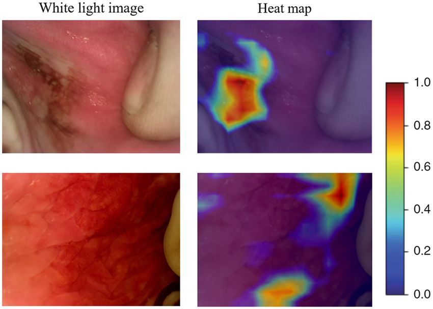
Song et al.: Mobile-based oral cancer classification for point-of-care screening

1 Introduction
Oral cancer is a highly prevalent cancer; 377,713 new cases and 177,757 deaths are reported each
year worldwide.1 India alone accounts for more than 33% (135,929) of new cases and 20%
(75,290) of deaths, according to Globocan 2020.1 The second most common cancer in India
is oral cancer,2 and its pervasiveness is increasing rapidly. From 2018 to 2020, the number of
new cases increased by 12% (from 119,992 to 135,929) according to comparisons from
Globocan 2018 and Globocan 2020.1,3 According to the World Health Organization, the esti-
mated number of new cases and deaths in Asia will increase to 374,000 and 208,000 by 2040.4
It is a growing problem and a leading cause of mortality in specific regions such as India.5,6
Detecting oral cancer at an early stage is believed to be the most effective way of (1) reducing
the individual burden, (2) decreasing morbidity and mortality, and (3) improving quality of life.7
Most high-risk individuals live in resource-limited settings that lack trained specialists and health
services.8,9 Conventional oral examination and biopsy, the clinical and gold standard for oral
lesion detection, are not ideal as screening tools in these regions.10 Therefore, the chances
of survival are reduced due to delayed diagnosis.11
    Convolutional neural networks (CNNs)12 are a powerful tool to solve image classification
problems13 and have been widely used for many medical image analysis tasks with great
performance.14,15 Deep CNNs show potential for automated classification of different types
of cancer lesions, such as skin cancer,16 cervical cancer,17,18 and oral cancer.19,20 A smartphone-
based, point-of-care platform outfitted with automated classification powered by deep learning
techniques can extend the reach of trained specialists outside of the clinic to provide low-cost
universal access to vital diagnostic care. Deep learning models suffer from being large, com-
plicated, slow, and typically need high-performance computing to run. Mobile platforms are
small and inexpensive; however, they do not have high-performance computing capabilities.
One common solution is to host an AI cloud server, upload the image to the cloud server for
processing, and then download the results.21 For this approach, internet connection is the bottle-
neck. It is not easy to get internet access everywhere, especially for resource-limited settings,
much less at high-speed upload and download data rates. Rural India only has 18% mobile
internet penetration, according to a report by the Internet and Mobile Association of India.22
    This paper describes the development of an automated, dual-modality, oral potentially malig-
nant lesion (OPML) and malignant lesion image classification system using the deep learning
model MobileNet.23 The platform can run quickly with high accuracy in resource-constrained
settings where computation power and internet access is limited. The deep learning-based clas-
sification and image preprocessing user interface can be accessed by clinicians through an
easy-to-use Android smartphone application for real-time classification results of patients in
resource-limited settings, regardless of internet connection. The network is trained to identify
clinically suspicious lesions based on a gold standard of clinical impression. Experimental
results with the developed application confirm the effectiveness and efficiency of the automated,
dual-modality, oral potentially, and malignant image classification system approach on mobile
devices for identifying clinically suspicious lesions.

2 Methods
Figure 1 shows our smartphone-based, dual-modal oral cancer screening platform that runs our
deep learning-based approach.24,25 The platform consists of a commercial Moto G5 Android
smartphone, an intraoral imaging probe, an light-emitting diode (LED) driver, a rechargeable
lithium battery, and mobile application. The system utilizes four 405-nm Luxeon UV U1 LEDs
(Lumileds, Amsterdam, Netherlands) to enable intraoral autofluorescence imaging (AFI) and four
4000-K Luxeon Z ES LEDs for intraoral white-light imaging (WLI). The illumination LEDs
are driven by a switching boost voltage regulator (LT1815, Linear Technology, Milpitas, CA).
The custom Android application provides the user interface, controls the phone and probe, cap-
tures dual-modal intraoral images, and implements our oral image classification approach.
    For visual oral cancer diagnosis, AFI can help detect some OPMLs and malignant lesions not
visible by WLI and vice versa.26–28 Previous studies conclude that better accuracy could be
achieved by combining AFI and WLI.29 Dual-modal images using AFI and WLI have proven

Journal of Biomedical Optics                    065003-2                           June 2021    •   Vol. 26(6)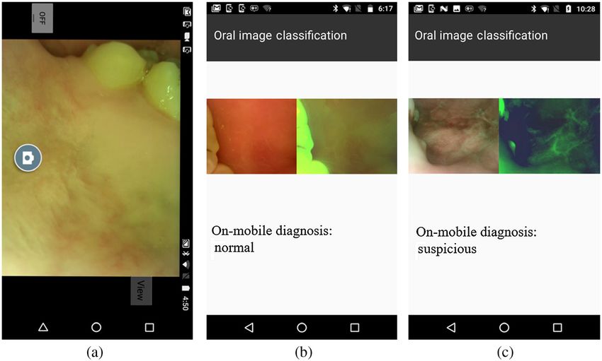
Song et al.: Mobile-based oral cancer classification for point-of-care screening

Fig. 1 The (a) back and (b) front side: the dual-modal oral cancer screening system consists of a
Moto G5 smartphone, a phone case with battery and electronics, an intraoral probe, an OTG cable
connection from the phone case to the smartphone, and a cable connection from the intraoral
probe to the phone case. The head of the intraoral probe has white-light LEDs, violet LEDs, and
a camera module. Narrowband filters and a long-pass filter are placed in front of the violet LEDs
and camera to enable intraoral autofluorescence imaging. The polarizing filter has been put in front
of the white-light LEDs and camera to suppress glare from the saliva on mucosa surface. A custom
Android application controls the system, captures dual-modal images, and implements the
MobileNet based on-device dual-modal classification system in an easy-to-use format. (c) The
three example image pairs of the WLI and AFI are shown in the figure labeled “normal,” “potentially
malignant,” and “malignant” by oral specialists. The image pair labeled normal would be catego-
rized as negative by the gold standard (normal/benign). The image pairs labeled potentially malig-
nant and malignant would both be categorized as positive by the gold standard [suspicious
(OPMD/malignant)]. The algorithm is trained to classify normal/benign versus suspicious.

to improve accuracy in oral cancer image classification, as shown in our previous study.24 Dual-
modality images fuse information from AFI and WLI to feed the CNN. For AFI, a red channel
to green channel ratio map is created because it is an important feature correlated with OPMLs
(oral premalignant lesions) and malignant lesions. For WLI, the blue channel is excluded
because the long-pass filter blocks light beneath the 470-nm wavelength. The normalized ratio
map from AFI and two remaining channels from WLI were combined to feed the MobileNet
neural network.
    The workflow of the proposed mobile-based oral cancer classification system is shown in
Fig. 2. The autofluorescence and WLI pairs are captured on-site and analyzed directly on the
smartphone device if the internet connection is poor, such as in a resource-limited setting. The
data will then be uploaded to a cloud server when internet connection is available. The deep
learning model deployed on the cloud server will also classify the data. Next, remote specialists
can log into the platform and provide diagnosis via a web browser.
    MobileNet operates by converting the standard convolutional layers to a more efficient for-
mat, depth-wise separable convolution.30 Depth-wise separable convolution is small, has low
latency, and has low power consumption, characteristics that meet the needs of real-time,
high-accuracy analysis for on-device embedded applications. The depth-wise separable convo-
lution layer is made of a depth-wise convolution and a pointwise convolution. The depth-
wise convolution layer filters each of the input channels, and the pointwise convolution layer

Journal of Biomedical Optics                     065003-3                           June 2021    •   Vol. 26(6)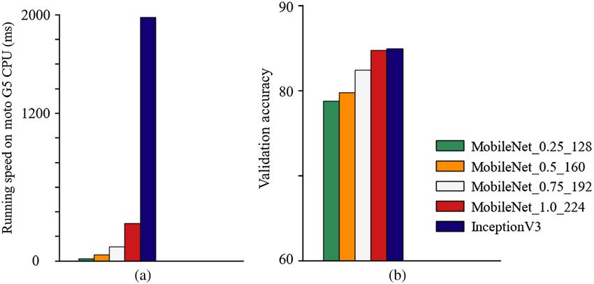
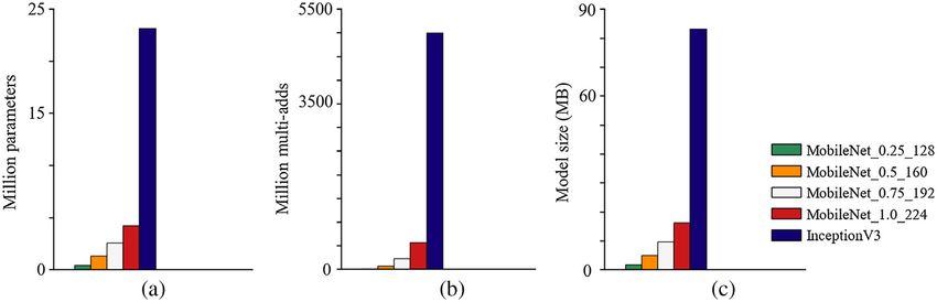
Song et al.: Mobile-based oral cancer classification for point-of-care screening

Fig. 2 Workflow of our mobile-based oral cancer screening and classification system. The images
captured from smartphone could be classified directly on the device in resource-limited setting
with poor internet connection.

combines the results through the depth-wise convolution layer. The computational cost and
model size are drastically reduced because of the conversion. The computational cost of standard
convolution is Df × Df × M × N × Dk × Dk , where Df is the spatial width and height of the
input feature map, M is the number of input channels, Dk is the spatial dimension of the kernel,
and N is the number of output channels. However, the computational cost of the depth-wise
separable convolution is Df × Df × M × Dk × Dk þ Df × Df × M × N. By converting standard
convolution to the depthwise separable convolution, we get a reduction in the computation of
ðDf × Df × M × Dk × Dk þ Df × Df × M × NÞ ¼ 1∕N þ 1∕D2k . Since the aim of lightweight
efficient neural networks is to achieve the highest accuracy with the fastest speed, which is a
contradiction in nature, MobileNet introduces two hyperparameters to trade-off between speed
and accuracy. First, the width multiplier reduces the number of input/output channels of each
layer. Second, the resolution multiplier reduces the resolution of every layer input. The
InceptionV3 network was used for comparison. InceptionV3, based on inception deep convolu-
tional architecture with additional factorization ideas, has been widely used for many applica-
tions. Figure 3 shows the comparison of operations, parameters, and model sizes of MobileNet
with InceptionV3. The operations were quantified using Multi-Adds, which represents the num-
ber of multiply-add operations of a deep learning model, smaller Multi-Adds means the model

Fig. 3 Comparison of (a) parameters, (b) operations (Multi-Adds), and (c) model sizes of
MobileNet (different hyperparameters settings) with InceptionV3.

Journal of Biomedical Optics                    065003-4                           June 2021    •   Vol. 26(6)
Song et al.: Mobile-based oral cancer classification for point-of-care screening

needs fewer operations and leads to lower computational costs. MobileNet with depth-wise sepa-
rable convolutions tremendously reduces the computational cost, number of parameters, and
model size with only small accuracy compromises.
    The performance of the proposed MobileNet-based dual-modality approach for classification
of normal/benign versus suspicious lesions based on a gold standard of clinical impression was
validated using the image dataset collected in India with the dual-modality mobile oral screening
devices. The dataset was captured among 5025 patients attending the outpatient clinics of the
Department of Oral Medicine and Radiology at KLE Society Institute of Dental Sciences, Head
and Neck Oncology Department of Mazumdar Shaw Medical Center (MSMC), and Christian
Institute of Health Sciences and Research (CIHSR), India. Institutional ethical committee appro-
val was obtained from all participating hospitals and written informed consent was collected
from all subjects enrolled. Oral oncology specialists from MSMC, KLE, and CIHSR labeled
all the dual-modality image pairs and separated them into two categories: (A) “normal” which
contains normal and benign mucosal lesion images and (B) “suspicious” which contains OPML
and malignant lesion images. In a previous study, we showed that oral oncology specialists’
interpretation of classifying normal/benign versus OPML/malignant has high accuracy with
biopsy-confirmed cases.31
    We collected a total of 7970 pair of intraoral images from 5025 patients. Undiagnosable and
low image quality images such as “unclear” or “not well focused” were excluded from the data-
set. After exclusion, only 6211 image pairs remained. We used 5329 image pairs for training and
validation. Through random assignment, 75% of the image pairs were allocated to training and
25% of the image pairs were allocated to validation. For the standalone test, we used 882 image
pairs that were stratified at the patient level. Tables 1 and 2 show the lesion type distribution
of the training/validation dataset and the standalone test dataset. Table 3 shows the lesion site
distribution of the standalone test dataset.
    We trained the network with the focal loss and the RMSProp optimization algorithm imple-
mented on Tensorflow. The batch size for each iteration was 32, the learning rate was 0.0001, and
the epoch number was 300. We used a desktop computer with an Nvidia 1080Ti GPU to train the
network and achieved 85% accuracy (85% sensitivity and 84% specificity) on the validation
dataset. If the likelihood score (generated by the model’s softmax layer) of a case belonging
to suspicious class was >0.5, the result would be predicted to be suspicious. Then we converted
the TensorFlow model into TensorFlow Lite format using TFLite Converter.32 The TensorFlow

                   Table 1 The lesion type distribution of the training/validation
                   dataset.

                   Lesion types                                 Number of image pairs

                   Normal                                                3211

                   Benign                                                 740

                   OPML                                                  1346

                   Malignancy                                             32

                   Table 2 The lesion type distribution of the standalone test
                   dataset.

                   Lesion types                                 Number of image pairs

                   Normal                                                 393

                   Benign                                                 69

                   OPML                                                   397

                   Malignancy                                             23

Journal of Biomedical Optics                     065003-5                           June 2021    •   Vol. 26(6)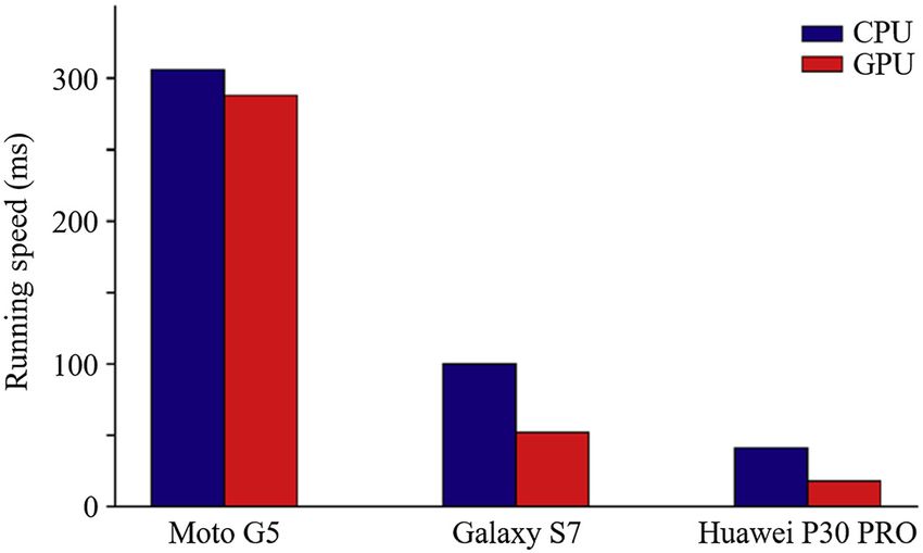
Song et al.: Mobile-based oral cancer classification for point-of-care screening
Table 3 The lesion site distribution of the standalone test
dataset.
Lesion sites Number of image pairs
Cheek mucosa 604
Lower vestibule 42
Tongue 46
Lip 130
Floor of mouth 3
Palate 10
RMT 29
Other 18
Lite format is designed to execute models efficiently on devices, which reduces the file size and
introduces optimizations that do not affect accuracy.
We have developed an Android smartphone application that implements the MobileNet-
based dual-modality OPML and malignancy image classification approach in an easy-to-use
format. The application controls the intraoral probe’s camera and LEDs to capture the dual-
modality image pair. After the dual-modality oral image pair is obtained, the application can
be used to analyze the image pair by our proposed deep learning-based classification method.
The Android application uses the TensorFlow Lite Library. The image preprocessing is also
implemented on the mobile application, which uses the OpenCV Android library.
We have tested the running speed on three different smartphone CPU and GPU platforms of
our proposed method: (1) the Moto G5 smartphone with an Octa-core 1.4 GHz CPU and Adreno
505 GPU, (2) the Samsung Galaxy S7 smartphone with Octa-core (4 × 2.3 GHz Mongoose
and 4 × 1.6 GHz Cortex-A53) CPU and Mali-T880 MP12 GPU, and (3) the Huawei P30 pro
smartphone with Octa-core (2 × 2.6 GHz Cortex-A76 and 2 × 1.92 GHz Cortex-A76 and
4 × 1.8 GHz Cortex-A55) CPU and Mali-G76 MP10 GPU.
3 Results
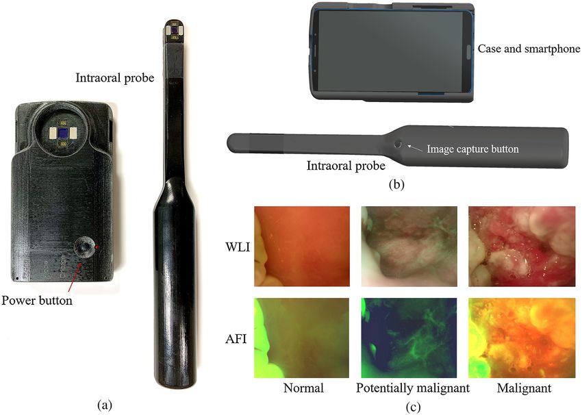
The finalized lite format MobileNet model is ∼16.3 MB and ideal for smartphone platform oper-
ation. Figure 4 shows the screenshots of our customized Android application. It takes ∼300 ms
to process one image pair with the Moto G5 Android smartphone.
We have also implemented our proposed dual-modality method with the InceptionV3
network and converted the trained model to the Tensorflow Lite format to run directly on our
smartphone platform. The validation accuracy is 85% (85% sensitivity and 85% specificity).
The InceptionV3 performance is almost the same as the MobileNet-based approach. However,
the model file size is ∼5 times larger (83.3 MB) and 5 times slower running on the Moto G5
smartphone (1981 ms). This experiment shows the MobileNet-based approach can deliver state-
of-the-art performance with smaller file size and faster processing speed.
In our experiment, we compared the effect of different optimization algorithms for the dual-
modality MobileNet network. To efficiently and effectively train the model to produce accurate
results, the optimization strategies and algorithms were used to calculate appropriate and opti-
mum values of the internal parameters of the model. The choice of optimization algorithms for
a model plays a significant role in the training process. There are mainly two categories of opti-
mization algorithms: constant learning rate algorithms such as stochastic gradient descent and
adaptive learning algorithms such as Adam, Adagrad, and RMSprop. To fairly compare the per-
formances of different optimization algorithms, we kept the learning rate, loss function, and all
other settings unchanged. Table 4 shows the RMSProp algorithm having the best performance
for this application and the Adadelta optimization algorithm having the longest training time.
Journal of Biomedical Optics 065003-6 June 2021 • Vol. 26(6)Song et al.: Mobile-based oral cancer classification for point-of-care screening
Fig. 4 Screenshots of the custom Android application: (a) capture images use our custom appli-
cation and device; (b), (c) efficient mobile deep learning result.
Table 4 Comparison of validation accuracy and training
time by using different optimization algorithms.
Optimization Training Performance
algorithm speed (s) (validation accuracy) (%)
Adadelta 3771 76.60
Adagrad 3855 81.41
Adam 1867 83.32
GradientDescent 1853 82.63
RMSProp 2057 84.72
In our experiment, different width multiplier settings of 1, 0.75, 0.5, and 0.25, as well as
resolution multiplier settings of 224, 192, 160, and 128 were explored to compare the accuracy,
size, and on-device processing time of the model. Figure 5 shows the model with a width multi-
plier of 1.0 and a resolution multiplier of 224 as having the best accuracy but largest file size and
the slowest on-device processing time as compared to other settings (but still smaller and faster
than Inception V3). The model with a width multiplier of 0.25 and a resolution multiplier of 128
has the smallest file size and shortest on-device process time but has the worst accuracy per-
formance. Introduction of the hyperparameters that show accuracy trade-offs demonstrate that
the speed and file size of the proposed method could be further improved to run faster on more
restricted and cost-effective mobile platforms.
The comparison of running speeds on three different smartphone CPU and GPU platforms of
our proposed method is shown in Fig. 6. The results indicate that the MobileNet-based method
could run on different smartphone platforms (different CPUs, different GPUs, and different man-
ufacturers). The runtime is shorter on a more powerful CPU and GPU. However, good perfor-
mance can still be achieved on a cost-effective smartphone. The results further indicate the
running speed is faster on a mobile GPU instead of a mobile CPU.
For investigating the interpretability of the deep learning model, we used gradient-weighted
class activation mapping to visualize the network by highlighting the important regions for clas-
sification. This helps to visually validate where our network is looking and verify that it looks at
the correct patterns in the image and activates around those patterns. It does not represent pixel-
wise probability. Examples of two suspicious images are shown in Fig. 7 where the highlighted
Journal of Biomedical Optics 065003-7 June 2021 • Vol. 26(6)Song et al.: Mobile-based oral cancer classification for point-of-care screening Fig. 5 Comparison of running speed on Moto G5 CPU and validation accuracy on our dual-modal oral cancer dataset. Fig. 6 Comparison of running speed (ms per frame) of MobileNet with 1.0 width multiplier and 224 resolution multiplier on different smartphone CPU and GPU platforms. Fig. 7 Visualization of two suspicious examples: each example shows the white-light image and corresponding heat map. The red region represents where the neural network is most concentrated. Journal of Biomedical Optics 065003-8 June 2021 • Vol. 26(6)
Song et al.: Mobile-based oral cancer classification for point-of-care screening

features in red show where the deep learning model is influenced most. We found that the lesion
areas were activated for predicting the result. This demonstrates an encouraging direction for
future investigation.
    To further challenge and validate the proposed approach, we have tested the proposed meth-
ods on the standalone test dataset. Our proposed method achieved 81% accuracy, 79% sensi-
tivity, and 82% specificity on the standalone dataset.

4 Conclusion
For oral cancer screening in resource-limited settings, the ideal solution needs to have (1) quick
response time, (2) capability to work under restricted conditions such as lack of good medical
facilities and a bad internet connection, and (3) high overall performance. Our proposed
MobileNet-based dual-modality approach can run quickly with high accuracy on a resource-
constrained mobile platform using limited computational power and space. This approach can
enable the delivery of deep learning-based oral cancer screening technology into the hands of
low-income patients. Our MobileNet-based approach is much more efficient than the conven-
tional deep CNN model. The computational costs and memory requirements are much lower.
Our experiment demonstrates the high classification accuracy, fast computing speed, and robust-
ness of the method, which can run on different mobile platforms.
    In conclusion, an end-to-end dual-modality deep CNN approach is proposed for automated
oral dysplasia and malignancy image classification. This approach classifies dual-modality oral
dysplasia and malignancy images fast on mobile platforms. In our preliminary experiments, the
approach was able to show accurate results running on different mobile platforms very close
to real time. Our method paves the way for oral cancer screening in resource-limited settings.
We hope that its power can be further demonstrated and developed for future studies by deliv-
ering it to clinicians for use on patients in real time where trained specialists, health facilities, and
internet access are limited.

Disclosures
There are no conflicts of interest to declare.

Acknowledgments
This work was supported by the National Institute of Biomedical Imaging and Bioengineering
(NIBIB) (No. UH2EB022623) and the National Cancer Institute (NCI) (No. UH3CA239682) of
the National Institutes of Health (NIH).

References
 1. H. Sung et al., “Global cancer statistics 2020: GLOBOCAN estimates of incidence and
    mortality worldwide for 36 cancers in 185 countries,” CA: Cancer J. Clin. 71, 209–249
    (2021).
 2. International Agency for Research on Cancer of World Health Organization, “Cancer
    today – population fact sheets – India,” http://gco.iarc.fr/today/data/factsheets/populations/
    356-india-fact-sheets.pdf (2021).
 3. F. Bray et al., “Global cancer statistics 2018: GLOBOCAN estimates of incidence and mor-
    tality worldwide for 36 cancers in 185 countries,” CA: Cancer J. Clin. 68, 394–424 (2018).
 4. International Agency for Research on Cancer of World Health Organization, “Cancer
    Tomorrow – The future of cancer burden,” https://gco.iarc.fr/tomorrow/home (2021).
 5. B. Gupta and N. W. Johnson, “Oral cancer: Indian pandemic,” Br. Dent. J. 222, 497–497
    (2017).
 6. A. Varshitha, “Prevalence of oral cancer in India,” J. Pharmaceut. Sci. Res. 7, 845 (2015).
 7. T. Baykul et al., “Early diagnosis of oral cancer,” J. Int. Med. Res. 38, 737–749 (2010).

Journal of Biomedical Optics                     065003-9                           June 2021    •   Vol. 26(6)
Song et al.: Mobile-based oral cancer classification for point-of-care screening
8. P. E. Petersen, “Oral cancer prevention and control—the approach of the World Health
Organization,” Oral Oncol. 45, 454–460 (2009).
9. N. W. Johnson et al., “Global oral health inequalities in incidence and outcomes for oral
cancer: causes and solutions,” Adv. Dent. Res. 23, 237–246 (2011).
10. K. D. Shield et al., “The global incidence of lip, oral cavity, and pharyngeal cancers by
subsite in 2012,” CA: Cancer J. Clin. 67, 51–64 (2017).
11. K. R. Coelho, “Challenges of the oral cancer burden in India,” J. Cancer Epidemiol. 2012,
701932 (2012).
12. Y. LeCun, Y. Bengio, and G. Hinton, “Deep learning,” Nature 521, 436–444 (2015).
13. A. Krizhevsky, I. Sutskever, and G. E. Hinton, “ImageNet classification with deep convolu-
tional neural networks,” Commun. ACM 60, 84–90 (2017).
14. G. Hinton, “Deep learning—a technology with the potential to transform health care,”
JAMA 320, 1101–1102 (2018).
15. R. Miotto et al., “Deep learning for healthcare: review, opportunities and challenges,” Brief.
Bioinf. 19, 1236–1246 (2018).
16. A. Esteva et al., “Dermatologist-level classification of skin cancer with deep neural net-
works,” Nature 542, 115–118 (2017).
17. M. Wu et al., “Automatic classification of cervical cancer from cytological images by using
convolutional neural network,” Biosci. Rep. 38, BSR20181769 (2018).
18. L. Hu et al., “An observational study of deep learning and automated evaluation of cervical
images for cancer screening,” J. Natl. Cancer Inst. 111, 923–932 (2019).
19. H. Martin et al., “Deep convolutional neural networks for classifying head and neck cancer
using hyperspectral imaging,” J. Biomed. Opt. 22, 060503 (2017).
20. M. Aubreville et al., “Automatic classification of cancerous tissue in laserendomicroscopy
images of the oral cavity using deep learning,” Sci. Rep. 7, 11979 (2017).
21. R. D. Uthoff et al., “Point-of-care, smartphone-based, dual-modality, dual-view, oral cancer
screening device with neural network classification for low-resource communities,” PLoS
One 13, e0207493 (2018).
22. Internet and Mobile Association of India, “Mobile Internet Report,” https://cms.iamai.in/
Content/ResearchPapers/2b08cce4-e571-4cfe-9f8b-86435a12ed17.pdf (2017).
23. A. G. Howard et al., “Mobilenets: efficient convolutional neural networks for mobile vision
applications,” https://arxiv.org/abs/1704.04861 (2017).
24. B. Song et al., “Automatic classification of dual-modalilty, smartphone-based oral dysplasia
and malignancy images using deep learning,” Biomed. Opt. Express 9, 5318–5329 (2018).
25. R. Uthoff et al., “Small form factor, flexible, dual-modality handheld probe for smartphone-
based, point-of-care oral and oropharyngeal cancer screening,” J. Biomed. Opt. 24, 106003
(2019).
26. I. Pavlova et al., “Understanding the biological basis of autofluorescence imaging for oral
cancer detection: high-resolution fluorescence microscopy in viable tissue,” Clin. Cancer
Res. 14, 2396 (2008).
27. R. Mehrotra et al., “A cross-sectional study evaluating chemiluminescence and autofluor-
escence in the detection of clinically innocuous precancerous and cancerous oral lesions,”
J. Am. Dent. Assoc. 141, 151–156 (2010).
28. M. Rana et al., “Clinical evaluation of an autofluorescence diagnostic device for oral cancer
detection: a prospective randomized diagnostic study,” Eur. J. Cancer Prevent. 21, 460–466
(2012).
29. C. S. Farah et al., “Efficacy of tissue autofluorescence imaging (velscope) in the visuali-
zation of oral mucosal lesions,” Head Neck 34, 856–862 (2012).
30. L. Sifre and S. Mallat, “Rigid-motion scattering for texture classification,” https://arxiv.org/
abs/1403.1687 (2014).
31. B. Praveen et al., “Role of community health worker in a mobile health program for early
detection of oral cancer,” Indian J. Cancer 56, 107–113 (2019).
32. Google TensorFlow, “TensorFlow Lite converter,” https://www.tensorflow.org/lite/convert
(2021).
Biographies of the authors are not available.
Journal of Biomedical Optics 065003-10 June 2021 • Vol. 26(6)You can also read