MicroRNA 615 5p regulates the proliferation and apoptosis of breast cancer cells by targeting HSF1
←
→
Page content transcription
If your browser does not render page correctly, please read the page content below
EXPERIMENTAL AND THERAPEUTIC MEDICINE 21: 192, 2021

                  MicroRNA‑615‑5p regulates the proliferation and
                  apoptosis of breast cancer cells by targeting HSF1
                                               KAISHENG LIU and RONG MA

           Department of Breast Surgery, Qilu Hospital of Shandong University, Jinan, Shandong 250012, P.R. China

                                       Received July 25, 2018; Accepted January 31, 2019

                                                  DOI: 10.3892/etm.2021.9624

Abstract. Breast cancer, which commonly occurs in the epithe‑      of Bax were significantly increased, whereas those of Bcl‑2,
lium of the mammary gland, is a malignant tumor. MicroRNAs         cyclin D1 and PCNA were decreased in the cells transfected
are involved in various cancer‑associated processes, and           with microRNA‑615‑5p mimics compared with the control
microRNA‑615‑5p has been identified to be decreased in the         and NC cells (P
2 LIU and MA: miR-615-5p REGULATES CELL PROLIFERATION AND APOPTOSIS IN BREAST CANCER
Materials and methods i) NC group (microRNA‑615‑5p mimics NC); ii) mimics
group (transfected with microRNA‑615‑5p mimics); and
Specimens. A total of 40 pairs of breast cancer tissues and iii) mimics + HSF1 group (transfected with microRNA‑615‑5p
normal adjacent breast tissues were obtained from Qilu Hospital mimics and pcDNA3.1‑HSF1). Briefly, cells were seeded onto
of Shandong University (Jinan, China) between June 2015 six‑well plates at a density of 1x10⁵ cells/well, and co‑trans‑
and January 2016. The patients comprised 40 females, whose fected with 50 nmol/l NC or microRNA‑615‑5p mimics with or
mean age was 47.2±7.3 years. All patients were pathologically without 2 µg/ml pcDNA3.1‑HSF1 using Lipofectamine® 2000
diagnosed with breast cancer (Table 1) and had not received (Invitrogen; Thermo Fisher Scientific, Inc.), according to the
chemotherapy or radiotherapy prior to the present study. manufacturer's protocol.
Written consent was obtained from each patient. The present
study was approved by the Ethics Committee of Qilu Hospital Analysis of cell proliferation. Following 24 h of transfection,
of Shandong University. the cells were washed with PBS, re‑seeded onto a 96‑well
plate at a density of 1x10 4 cells/well and cultured in fresh
Cell culture. The MCF‑7 human breast cancer cell line (Type RPMI‑1640 medium for 12, 24 or 48 h. Subsequently, a Cell
Culture Collection of the Chinese Academy of Sciences, Counting kit‑8 (CCK‑8) cell proliferation assay was conducted
Shanghai, China) was cultured in RPMI‑1640 (Gibco; Thermo using the CCK‑8 kit (Dojindo Molecular Technologies Inc.,
Fisher Scientific, Inc., Waltham, MA, USA) supplemented Kumamoto, Japan). The absorbance of each well was measured
with 10% fetal bovine serum (Invitrogen; Thermo Fisher at 450 nm using a microplate reader.
Scientific, Inc.), and 1% penicillin and streptomycin. The cells
were cultured at 37˚C in a 5% CO2 humidified incubator. Analysis of cell apoptosis. At 48 h post‑transfection, the MCF‑7
cells were washed with PBS. An Annexin V‑fluorescein
Transfection efficiency of microRNA‑615‑5p. The MCF‑7 isothiocyanate (FITC) apoptosis kit (BD Biosciences, San
cells were divided into three groups: i) Control group Jose, CA, USA) was used to quantify apoptotic MCF‑7 breast
(untreated); ii) negative control (NC) group (transfected with cancer cells by flow cytometry (FACSCalibur, BD Biosciences)
microRNA‑615‑5p mimics NC); and iii) mimics group (trans‑ using Cell Quest Pro software (version 6.0; BD Biosciences),
fected with microRNA‑615‑5p mimics). The MCF‑7 cells were according to the manufacturer's protocol.
seeded onto six‑well plates at a density of 1x10⁵ cells/well,
and 50 nmol/l oligonucleotide (microRNA‑615‑5p mimics RNA extraction and reverse transcription‑quantitative PCR
or NC) were subsequently transfected into cells using (RT‑qPCR). Total RNA was extracted from patient tissue
Lipofectamine® 2000 (Invitrogen; Thermo Fisher Scientific, samples and MCF‑7 cells using a microRNAeasy kit (Invitrogen;
Inc.), according to the manufacturer's protocol. The Thermo Fisher Scientific, Inc.), according to the manufacturer's
microRNA‑615‑5p mimics and NC oligonucleotides were protocol. To quantify the microRNA‑615‑5p and HSF1 mRNA
purchased from Shanghai GenePharma Co., Ltd. (Shanghai, expression in the 40 paired tumor and adjacent tissue samples
China). The sequences of the miR‑615‑5p mimics were as and MCF‑7 cells at 48 h post‑transfection. In total, 2 µl RNA
follows: 5'‑GGGG GUCCCCGGUGCUCGGAUC‑3' (sense) was isolated from the adjacent and tumor samples and reverse
and 5'‑UCCGAGCACCGGG GACCCCCUU‑3' (anti‑sense). transcribed into cDNA using the TaqMan MicroRNA RT kit
The sequences of the NC were as follows: 5'‑UUCUCCGAA (Applied Biosystems; Thermo Fisher Scientific, Inc.), according
CGUGUCACGU TT‑3' (sense) and 5'‑ACGUGACACGUU to the manufacturer's protocol. qPCR step was subsequently
CGGAGAATT‑3' (anti‑sense). performed using TaqMan Universal Master mix (Thermo Fisher
Scientific, Inc.). In total, 2 µl cDNA, 10 µl TaqMan Universal
Transfection efficiency of HSF1. The MCF‑7 cells were divided Master mix, 1 µl primers and nuclease‑free H2O. The primer
into three groups: i) Control group (untreated); ii) pcDNA3.1 sequences used were as follows: microRNA‑615‑5p forward,
group (transfected with pcDNA3.1); and iii) pcDNA3.1‑HSF1 5'‑GCCAGCCACCAAGAAGC‑3' and reverse, 5'‑GCTCCC
group (transfected with pcDNA3.1‑HSF1). The cells were GCTGTTTACTCTG‑3'; U6 forward, 5'‑GCTTCGGCAGCA
seeded onto six‑well plates at a density of 2x10⁴ cells/well, and CATATACTAA AAT‑3' and reverse, 5'‑CGCT TCACGA AT
2 µg/ml pcDNA3.1 (Thermo Fisher Scientific, Inc., Waltham, TTGCGTGTCAT‑3'; HSF1 forward, 5'‑GCCT TCCTGACC
MA, USA) or pcDNA3.1‑HSF1 was subsequently transfected AAGCTGT‑3' and reverse, 5'‑GTCGAACACGTGGAAGCT
into the cells using Lipofectamine® 2000 (Invitrogen; Thermo GT‑3'; Bcl‑2 forward, 5'‑ACAACATCGCCCTGTG GATGA
Fisher Scientific, Inc.) according to the manufacturer's protocol. C‑3' and reverse, 5'‑ATAG CTGATTCGACGT TTTGCC‑3';
cyclin D1 forward, 5'‑CCTGTCCTACTACCGCCTCA‑3' and
Vector construction and co‑transfection assay. The reverse, 5'‑TCCTCCTCTTCCTCCTCCTC‑3'; PCNA forward,
HSF1‑expression vector was constructed by inserting HSF1 5'‑CTCCAACTTCTGG GCTCAAG‑3' and reverse, 5'‑GTA
into a pcDNA3.1 vector. In brief, HSF1 cDNA was ampli‑ AACG GACTGCTGGAGGA‑3'; Bax forward, 5'‑GGAATT
fied by polymerase chain reaction (PCR) from the cDNA of CTGACGG CA ACTTCA ACTG GG‑3' and reverse, 5'‑GGA
MCF‑10A cells (Bena Culture Collection, Beijing, China). ATTCTTCCAGATG GTGAGCGAG G‑3'; GAPDH forward,
The HSF1 cDNA was then inserted into pcDNA3.1 (Thermo 5'‑GAAG GTGAAG GTCGGAGTC‑3' and reverse 5'‑GAA
Fisher Scientific, Inc.) to construct a pcDNA3.1‑HSF1 expres‑ GATG GTGATG GGATTTC‑3'. The thermocycling condi‑
sion vector. To evaluate whether or not HSF1 overexpression tions used were as follows: Initial denaturation at 95˚C for
attenuates the microRNA‑615‑5p‑induced suppression of 10 min; 40 cycles of 95˚C for 10 sec and 62˚C for 15 sec. U6
HSF1, MCF‑7 cells were divided into three different groups: small nuclear RNA and GAPDH were used as the endogenousEXPERIMENTAL AND THERAPEUTIC MEDICINE 21: 192, 2021 3
Table Ⅰ. Association between miR‑615‑5p expression status and clinicopathological features of patients with breast cancer.
Expression level
‑‑‑‑‑‑‑‑‑‑‑‑‑‑‑‑‑‑‑‑‑‑‑‑‑‑‑‑‑‑‑‑‑‑‑‑‑‑‑‑‑‑‑‑‑‑‑‑‑‑‑‑‑‑‑‑‑‑‑‑‑‑‑‑‑‑‑‑‑‑‑‑‑‑‑‑‑‑‑‑‑‑‑‑‑‑‑‑‑‑‑‑‑‑‑‑‑‑‑‑‑‑‑‑‑‑‑‑‑‑‑‑‑‑
Clinicopathological feature Cases High miR‑615‑5p (n=10) Low miR‑615‑5p (n=30) P‑value
Age 0.271
≤60 years 22 4 18
>60 years 18 6 12
Tumor size 0.000
≥2 cm 29 3 26
>2 cm 11 7 4
Tumor location 0.361
Left 21 4 17
Right 19 6 13
ER status 0.714
Negative 22 6 16
Positive 18 4 14
TNM stage 0.126
I/II 31 6 25
III/IV 9 4 5
Lymph node metastasis 0.002
Negative 7 5 2
Positive 33 5 28
ER, estrogen receptor; TNM, tumor, node and metastasis.
controls. The relative expression level of microRNA‑615‑5p was Abcam Chemiluminescent Horseradish Peroxidase Substrate
normalized to U6, while the expression levels of HSF1, B‑cell (Abcam) was added to the membrane surface. The signals were
lymphoma 2 (Bcl‑2), cyclin D1, proliferating cell nuclear antigen captured and the intensity of the bands was quantified. ImageJ
(PCNA) and Bcl‑2‑associated X protein (Bax) were normalized software (version 1.49; National Institutes of Health, Bethesda,
to GAPDH using the 2‑ΔΔCq method (10). MD, USA) was used to determine the protein expression levels
of HSF1, Bcl‑2, PCNA, cyclin D1 and Bax relative to those of
Western blot analysis. Total protein was extracted from different GAPDH. GAPDH served as the internal control.
groups of treated MCF‑7 cells 48 h post‑transfection using M‑PER
protein extraction reagent (Pierce; Thermo Fisher Scientific, Inc.) Dual luciferase reporter assay. TargetScan bioinformatics
supplemented with a protease mimics cocktail (Thermo Fisher analysis (www.targetscan.org) was used to identify HSF1 as
Scientific, Inc.). Total protein was quantified using the Bradford a potential target of miR‑615‑5p (11). The 293 cells (Type
method and 10 µg protein/lane was separated via SDS‑PAGE Culture Collection of the Chinese Academy of Sciences) were
on a 10% gel (Thermo Fisher Scientific, Inc.), according to the seeded onto a six‑well plate at a density of 1x10⁵ cells/well
manufacturer's protocol. The separated proteins were transferred and transfected with the wild‑type (WT) HSF1 3'untranslated
onto polyvinylidene fluoride membranes (Shanghai Ofluorine region (UTR; WT HSF1‑3'UTR) or mutant HSF1 3'UTR
Chemical Technology Co., Ltd., Shanghai, China) and blocked in (MUT HSF1 3'UTR) in combination with either NC or
5% nonfat milk for 2 h at 37˚C. The membranes were incubated microRNA‑615‑5p mimics using Lipofectamine® RNAi Max
overnight at 4˚C with the following primary antibodies: Mouse (Thermo Fisher Scientific, Inc.). Transfected cells were subse‑
anti‑HSF1 (1:1,000; cat. no. ab201978; Abcam, Cambridge, quently incubated at 37˚C for 48 h and the luciferase activity
UK), mouse anti‑Bcl‑2 (1:1,000; cat. no. ab692; Abcam), was examined using a Dual‑Luciferase Reporter kit (Beyotime
rabbit anti‑cyclin D1 (1:100; cat. no. ab16663; Abcam), mouse Institute of Biotechnology, Haimen, China). Firefly luciferase
anti‑PCNA (1:1,000; cat. no. ab29; Abcam), rabbit anti‑Bax activity was normalized to Renilla luciferase activity.
(1:1,000; cat. no. ab32503; Abcam) and mouse anti‑GAPDH
(1:1,000; cat. no. ab8245; Abcam). Subsequent to washing, the Statistical analysis. SPSS version 22.0 (IBM Corp., Armonk,
membranes were incubated for 1 h at 37˚C with horseradish NY, USA) was used to analyze the results. Data are presented
peroxidase (HRP)‑labeled goat anti‑rabbit IgG (1:1,000; as the mean ± standard deviation. Statistical comparisons
cat. no. A0208; Beyotime, Shanghai, China) and HRP‑labeled between two groups were conducted using Student's t‑test, while
goat anti‑mouse secondary antibody (1:1,000; cat. no. A0216; one‑way analysis of variance followed by Newman‑Keuls tests
Beyotime, Shanghai, China). Subsequent to washing, 200 µl was used to analyze differences among three or more groups.4 LIU and MA: miR-615-5p REGULATES CELL PROLIFERATION AND APOPTOSIS IN BREAST CANCER Figure 1. Comparison of the relative expression of miR‑615‑5p and HSF1 in breast cancer tissues and adjacent tissues. Relative expression of (A) miR‑615‑5p and (B) HSF1 mRNA in breast cancer tissues and adjacent tissues. (C) Results of Pearson's correlation analysis between the expression of miR‑615‑5p and the expression of HSF1 in patients with breast cancer. **P
EXPERIMENTAL AND THERAPEUTIC MEDICINE 21: 192, 2021 5
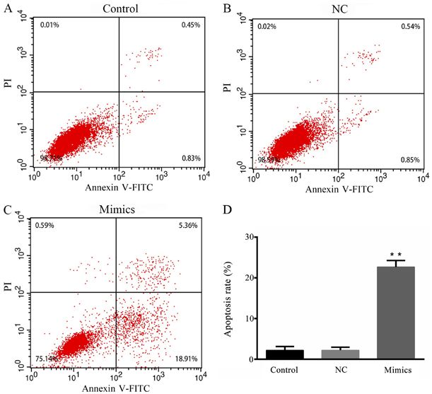
Figure 3. Effect of microRNA‑615‑5p on the proliferation of MCF‑7 cells.
Proliferation rate of MCF‑7 cells in different groups. **P6 LIU and MA: miR-615-5p REGULATES CELL PROLIFERATION AND APOPTOSIS IN BREAST CANCER Figure 4. Effect of microRNA‑615‑5p on the apoptosis of MCF‑7 cells. Apoptosis of the (A) control group, (B) NC and (C) microRNA‑615‑5p mimics group. (D) Quantified results of the flow cytometric analysis. **P
EXPERIMENTAL AND THERAPEUTIC MEDICINE 21: 192, 2021                                                                7

Figure 7. Effect of microRNA‑615‑5p on the mRNA expression levels of proliferation‑ and apoptosis‑associated factors. The mRNA expression levels
of (A) Bcl‑2, (B) cyclin D1, (C) PCNA and (D) Bax. **P
8                   LIU and MA: miR-615-5p REGULATES CELL PROLIFERATION AND APOPTOSIS IN BREAST CANCER

breast cancer cell proliferation and apoptosis requires further              3. Farazi TA, Hoell JI, Morozov P and Tuschl T: MicroRNAs in
                                                                                human cancer. Adv Exp Med Biol 774: 1‑20, 2013.
investigation. Other studies have indicated that HSF1 is down‑               4. Dalmay T: Mechanism of miRNA‑mediated repression of mRNA
stream of human epidermal growth factor receptor 2 (HER2)                       translation. Essays Biochem 54: 29‑38, 2013.
and could be activated by HER2 (48,49). Therefore, the inter‑                5. Ji CZ, Han SH and Xing YF: Overexpression of miR‑3196
                                                                                suppresses cell proliferation and induces cell apoptosis through
action of microRNA‑615‑5p and HER2 in the regulation of                         targeting ERBB3 in breast cancer. Eur Rev Med Pharmacol
breast cancer cell proliferation and apoptosis remains unclear                  Sci 22: 8383‑8390, 2018.
and also requires further investigation.                                     6. Lu K, Wang J, Song Y, Zhao S, Liu H, Tang D, Pan B, Zhao H and
                                                                                Zhang Q: miRNA‑24‑3p promotes cell proliferation and inhibits
    In conclusion, the present results demonstrated that                        apoptosis in human breast cancer by targeting p27Kip1. Oncol
microRNA‑615‑5p promoted the apoptosis of breast cancer                         Rep 34: 995‑1002, 2015.
cells and inhibited their growth by downregulating the expres‑               7. Bai Y, Li JY, Li J, Liu YH and Zhang B: miR‑615 inhibited
                                                                                cell proliferation and cell cycle of human breast cancer cells
sion levels of HSF1, Bcl‑2, PCNA and cyclin D1, and increasing                  by suppressing of AKT2 expression. Int J Clin Exp Med 8:
the expression of Bax. Therefore, targeting microRNA‑615‑5p                     3801‑3808, 2015.
is potentially a promising method for the treatment of breast                8. Dong Y, Huo X, Sun R, Liu Z, Huang M and Yang S: lncRNA
                                                                                Gm15290 promotes cell proliferation and invasion in lung cancer
cancer. However, two limitations remain: i) The underlying                      through directly interacting with and suppressing the tumor
targeted relationship between microRNA‑615‑5p and HER2                          suppressor miR‑615‑5p. Biosci Rep 38: pii: BSR20181150, 2018.
requires further elucidation, and ii) further investigation of               9. Sun Y, Zhang T, Wang C, Jin X, Jia C, Yu S and Chen J:
                                                                                miRNA‑615‑5p functions as a tumor suppressor in pancreatic
HER‑positive breast cancer cell lines is necessary.                             ductal adenocarcinoma by targeting AKT2. PLoS One 10:
                                                                                e0119783, 2015.
Acknowledgements                                                            10. Livak KJ and Schmittgen TD: Analysis of relative gene expres‑
                                                                                sion data using real‑time quantitative PCR and the 2(‑Delta Delta
                                                                                C(T)) method. Methods 25: 402‑408, 2001.
Not applicable.                                                             11. Agarwal V, Bell GW, Nam JW and Bartel DP: Predicting effec‑
                                                                                tive microRNA target sites in mammalian mRNAs. Elife 4, 2015.
                                                                            12. Pu HY, Xu R, Zhang MY, Yuan LJ, Hu JY, Huang GL and
Funding                                                                         Wang HY: Identification of microRNA‑615‑3p as a novel
                                                                                tumor suppressor in non‑small cell lung cancer. Oncol Lett 13:
No funding was received.                                                        2403‑2410, 2017.
                                                                            13. Asano Y, Kawase T, Okabe A, Tsutsumi S, Ichikawa H, Tatebe S,
                                                                                Kitabayashi I, Tashiro F, Namiki H, Kondo T, et al: IER5 gener‑
Availability of data and materials                                              ates a novel hypo‑phosphorylated active form of HSF1 and
                                                                                contributes to tumorigenesis. Sci Rep 6: 19174, 2016.
                                                                            14. Gao W, Gu Y, Li Z, Cai H, Peng Q, Tu M, Kondo Y, Shinjo K,
The datasets used and/or analyzed during the current study                      Zhu Y, Zhang J, et al: miR‑615‑5p is epigenetically inactivated
are available from the corresponding author on reasonable                       and functions as a tumor suppressor in pancreatic ductal adeno‑
request.                                                                        carcinoma. Oncogene 34: 1629‑1640, 2015.
                                                                            15. Huang FY, Zhao HJ, Du ZJ and Jiang H: miR‑615 inhibits pros‑
                                                                                tate cancer cell proliferation and invasion by directly targeting
Authors' contributions                                                          Cyclin D2. Oncol Res 27: 293‑299, 2019.
                                                                            16. Naresh DJ and Nadav B: Reconstruction of temporal activity of
                                                                                microRNAs from gene expression data in breast cancer cell line.
KL designed the experiments, analyzed the patient data and                      BMC Genomics 16: 1077, 2015.
purchased the reagents. RM performed the examination,                       17. Fang F, Chang R and Yang L: Heat shock factor 1 promotes
and was a major contributor in writing the manuscript. Both                     invasion and metastasis of hepatocellular carcinoma in vitro and
                                                                                in vivo. Cancer 118: 1782‑1794, 2012.
authors read and approved the final manuscript.                             18. Cen H, Zheng S, Fang YM, Tang XP and Dong Q: Induction of
                                                                                HSF1 expression is associated with sporadic colorectal cancer.
Ethics approval and consent to participate                                      World J Gastroenterol 10: 3122‑3126, 2004.
                                                                            19. Santagata S, Hu R, Lin NU, Mendillo ML, Collins LC,
                                                                                Hankinson SE, Schnitt SJ, Whitesell L, Tamimi RM, Lindquist S
The present study was approved by the Ethics Committee of                       and Ince TA: High levels of nuclear heat‑shock factor 1 (HSF1)
Qilu Hospital of Shandong University (Jinan, China). Written                    are associated with poor prognosis in breast cancer. Proc Natl
                                                                                Acad Sci USA 108: 18378‑18383, 2011.
consent was obtained from each patient.                                     20. Chen FY, Dong Z, Xia Y, Tang J, Peng L, Wang S and Lai D:
                                                                                Nucleoside analog inhibits microRNA‑214 through targeting
Patient consent for publication                                                 heat‑shock factor 1 in human epithelial ovarian cancer. Cancer
                                                                                Sci 104: 1683‑1689, 2013.
                                                                            21. Dai C, Santagata S, Tang Z, Shi J, Cao J, Kwon H, Bronson RT,
Not applicable.                                                                 Whitesell L and Lindquist S: Loss of tumor suppressor NF1
                                                                                activates HSF1 to promote carcinogenesis. J Clin Invest 122:
                                                                                3742‑3745, 2012.
Competing interests                                                         22. Heimberger T, Andrulis M, Riedel S, Stühmer T, Schraud H,
                                                                                Beilhack A, Bumm T, Bogen B, Einsele H, Bargou RC and
The authors declare that they have no competing interests.                      Chatterjee M: The heat shock transcription factor 1 as a potential
                                                                                new therapeutic target in multiple myeloma. Br J Haematol 160:
                                                                                465‑476, 2013.
References                                                                  23. Ishiwata J, Kasamatsu A, Sakuma K, Iyoda M, Yamatoji M,
                                                                                Usukura K, Ishige S, Shimizu T, Yamano Y, Ogawara K, et al:
                                                                                State of heat shock factor 1 expression as a putative diagnostic
 1. Torre LA, Bray F, Siegel RL, Ferlay J, Lortet‑Tieulent J and Jemal A:       marker for oral squamous cell carcinoma. Int J Oncol 40: 47‑52,
    Global cancer statistics, 2012. CA Cancer J Clin 65: 87‑108, 2015.          2012.
 2. Hesari A, Azizian M, Darabi H, Nesaei A, Hosseini SA, Salarinia R,      24. Hoang AT, Huang J, Rudra‑Ganguly N, Zheng J, Powell WC,
    Motaghi AA and Ghasemi F: Expression of circulating miR‑17,                 Rabindran SK, Wu C and Roy P: A novel association between
    miR‑25, and miR‑133 in breast cancer patients. J Cell Biochem,              the human heat shock transcription factor 1 (HSF1) and prostate
    Nov 28, 2018 (Epub ahead of print). doi: 10.1002/jcb.27984.                 adenocarcinoma. Am J Pathol 156: 857‑864, 2000.
EXPERIMENTAL AND THERAPEUTIC MEDICINE 21: 192, 2021 9
25. Miyachi K, Fritzler MJ and Tan EM: Autoantibody to a nuclear 38. de Ruijter TC, Veeck J, de Hoon JP, van Engeland M and
antigen in proliferation cells. J Immunol 121: 2228‑2234, 1978. Tjan‑Heijnen VC: Characteristics of triple‑negative breast cancer.
26. Liao XH, Lu DL, Wang N, Liu LY, Wang Y, Li YQ, Yan TB, Sun XG, J Cancer Res Clin Oncol 137: 183‑192, 2011.
Hu P and Zhang TC: Estrogen receptor α mediates proliferation 39. Wang X, Zhang D, Cao M, Ba J, Wu B, Liu T and Nie C: A study
of breast cancer MCF‑7 cells via a p21/PCNA/E2F1‑dependent on the biological function of heat shock factor 1 proteins in breast
pathway. FEBS J 281: 927‑942, 2014. cancer. Oncol Lett 16: 3821‑3825, 2018.
27. Li T, Zhang C, Ding Y, Zhai W, Liu K, Bu F, Tu T, Sun L, 40. Du L, Fei Z, Song S and Wei N: Antitumor activity of
Zhu W, Zhou F, et al: Umbilical cord‑derived mesenchymal Lobaplatin against esophageal squamous cell carcinoma through
stem cells promote proliferation and migration in MCF‑7 and caspase‑dependent apoptosis and increasing the Bax/Bcl‑2 ratio.
MDA‑MB‑231 breast cancer cells through activation of the ERK Biomed Pharmacother 95: 447‑452, 2017.
pathway. Oncol Rep 34: 1469‑1477, 2015. 41. Yang F, Yu Y, Lei Q, Zeng A, Li Y, Xie Y, Ye T and Wei Y:
28. Wu J, Li H, Wang X, Zhang X, Liu W, Wang Y, Zhang Y, Pan H, Lobaplatin arrests cell cycle progression, induces apoptosis and
Wang Q and Han Y: Effect of polysaccharide from Undaria impairs migration and invasion in B16‑F10 melanoma cell line
pinnatifida on proliferation, migration and apoptosis of breast in vitro. Biomed Pharmacother 69: 402‑408, 2015.
cancer cell MCF7. Int J Biol Macromol 121: 734‑742, 2019. 42. Lapierre M, Linares A, Dalvai M, Duraffourd C, Bonnet S,
29. Min JN, Huang L, Zimonjic DB, Moskophidis D and Mivechi NF: Boulahtouf A, Rodriguez C, Jalaquier S, Assou S, Orsetti B, et al:
Selective suppression of lymphomas by functional loss of Histone deacetylase 9 regulates breast cancer cell proliferation
Hsf1 in a p53‑deficient mouse model for spontaneous tumors. and the response to histone deacetylase inhibitors. Oncotarget 7:
Oncogene 26: 5086‑5097, 2007. 19693‑19708, 2016.
30. Diehl JA: Cycling to cancer with cyclin D1. Cancer Biol Ther 1: 43. Quisbert‑Valenzuela EO and Calaf GM: Apoptotic effect of
226‑231, 2002. noscapine in breast cancer cell lines. Int J Oncol 48: 2666‑2674,
31. He Y, Liu Z, Qiao C, Xu M, Yu J and Li G: Expression and 2016.
significance of Wnt signaling components and their target genes 44. Lou Q, Hu Y, Ma Y and Dong Z: Heat shock factor 1 induces
in breast carcinoma. Mol Med Rep 9: 137‑143, 2014. crystallin‑αB to protect against cisplatin nephrotoxicity. Am J
32. Qin H and Liu W: MicroRNA‑99a‑5p suppresses breast cancer Physiol Renal Physiol 311: F94‑F102, 2016.
progression and cell‑cycle pathway through downregulating 45. Jiang Y, Zhang Y, Li F, Du X and Zhang J: CDX2 inhibits
CDC25A. J Cell Physiol 234: 3526‑3537, 2019. pancreatic adenocarcinoma cell proliferation via promoting
33. Song X, Wu JQ, Yu XF, Yang XS and Yang Y: Trichostatin A tumor suppressor miR‑615‑5p. Tumor Biol 37: 1041‑1049, 2016.
inhibits proliferation of triple negative breast cancer cells by 46. Song LJ, Zhang WJ, Chang ZW, Pan YF, Zong H, Fan QX and
inducing cell cycle arrest and apoptosis. Neoplasma 65: 898‑906, Wang LX: PU. 1 is identified as a novel metastasis suppressor in
2018. hepatocellular carcinoma regulating the miR‑615‑5p/IGF2 axis.
34. Chi Y, Xu H, Wang F, Chen X, Shan Z, Sun Y and Fan Q: Asian Pac J Cancer Prev 16: 3667‑3671, 2015.
ZKSCAN3 promotes breast cancer cell proliferation, migration 47. Shetty PJ, Movva S, Pasupuleti N, Vedicherlla B, Vattam KK,
and invasion. Biochem Biophys Res Commun 503: 2583‑2589, Venkatasubramaniam S, Ahuja YR and Hasan Q: Regulation of
2018. IGF2 transcript and protein expression by altered methylation in
35. Sawai M, Ishikawa Y, Ota A and Sakurai H: The proto‑oncogene breast cancer. J Cancer Res Clin Oncol 137: 339‑345, 2011.
JUN is a target of the heat shock transcription factor HSF1. 48. Schulz R, Streller F, Scheel AH, Rüschoff J, Reinert MC,
FEBS J 280: 6672‑6680, 2013. Dobbelstein M, Marchenko ND and Moll UM: HER2/ErbB2
36. Escórcio‑Dourado CS, Martins LM, Simplício‑Revoredo CM, activates HSF1 and thereby controls HSP90 clients including
Sampaio FA, Tavares CB, da Silva‑Sampaio JP, Borges US, MIF in HER2‑overexpressing breast cancer. Cell Death Dis 5:
Alves‑Ribeiro FA, Lopes‑Costa PV, Lima‑Dourado JC and da e980, 2014.
Silva BB: Bcl‑2 antigen expression in luminal A and triple‑nega‑ 49. Guettouche T, Boellmann F, Lane WS and Voellmy R: Analysis
tive breast cancer. Med Oncol 34: 161, 2017. of phosphorylation of human heat shock factor 1 in cells experi‑
37. Kallel‑Bayoundh I, Hassen HB, Khabir A, Boujelbene N, encing a stress. BMC Biochem 6: 4, 2005.
Daoud J, Frikha M, Sallemi‑Boundawara T, Aifa S and Rebai A:
Bcl‑2 expression and triple negative profile in breast carcinoma.
Med Oncol 28 (Suppl 1): S55‑S61, 2001.You can also read