NS5A-ISGylation via Lysine 26 Has a Critical Role for Efficient Propagation of Hepatitis C Virus Genotype 2a
←
→
Page content transcription
If your browser does not render page correctly, please read the page content below
Kobe J. Med. Sci.,   Vol. 67, No. 2, pp. E38-E47,    2021

      NS5A-ISGylation via Lysine 26 Has a Critical Role for Efficient
             Propagation of Hepatitis C Virus Genotype 2a
            RHEZA GANDI BAWONO, TAKAYUKI ABE, YASUAKI SHIBATA,
                   CHIEKO MATSUI, LIN DENG, and IKUO SHOJI*
  Division of Infectious Disease Control, Center for Infectious Diseases, Kobe University Graduate School of
                                Medicine, Kobe, Japan *Corresponding author

                                 Received 29 June 2021/ Accepted 15 July 2021

Keywords: hepatitis C virus, Lys residue, viral propagation

    We previously reported that hepatitis C virus (HCV) NS5A (1b, Con1) protein accepts covalent ISG15
conjugation at specific lysine (Lys) residues (K44, K68, K166, K215 and K308), exhibiting proviral effects
on HCV RNA replication. Here we investigated a role of NS5A-ISGylation via Lys residues in HCV
propagation using HCV infectious clone. The alignment of amino acid sequences revealed that 5 Lys
residues (K20, K26, K44, K139, and K166) of the 13 Lys residues within NS5A (genotype 2a, JFH1 strain)
were conserved compared to those of HCV (genotype 1b, Con1 strain). The cell-based ISGylation assay
revealed that the K26 residue in the amphipathic helix (AH) domain and the K139 residue in domain I of
NS5A (2a, JFH1) had the potential to accept ISGylation. Use of the HCV replicon carrying luciferase gene
revealed that the K26 residue but not K139 residue of NS5A (2a, JFH1) was important for HCV RNA
replication. Furthermore, cell culture HCV revealed that the mutation with the K26 residue in
combination with K139 or K166 on NS5A (2a, JFH1) resulted in complete abolishment of viral
propagation, suggesting that the K26 residue collaborates with either the K139 residue or K166 residue
for efficient HCV propagation. Taken together, these results suggest that HCV NS5A protein has the
potential to accept ISGylation via specific Lys residues, involving efficient viral propagation in a
genotype-specific manner.

                                               INTRODUCTION
    Hepatitis C virus (HCV) is a major causative agent causing chronic hepatitis, liver cirrhosis, and
hepatocellular carcinoma and still remains a public health burden worldwide. For a long term, interferon
(IFN)-based therapy has been used as a standard therapy for patients with chronic HCV infection. However, the
IFN-based therapy is restrictive and can cure only approximately 50% of the patients, especially for the patients
infected with genotype 1 strains (29, 33). Recent basic and clinical advances in HCV research have developed
novel anti-HCV therapeutics, direct-acting antivirals (DAAs) therapy, and achieved sustained virological
response rates over 95 % in patients with several HCV genotypes (7). Several approved DAAs targeted to
nonstructural proteins, including NS3/4A protease, NS5A, and NS5B polymerase, are currently available for the
treatment of chronic hepatitis C.

    HCV is an enveloped, single-stranded positive-sense RNA virus classified into the Hepacivirus genus of the
Flaviviridae family. HCV is divided into seven major genotypes (genotype 1-7) and dozens of subtypes. Among
the HCV genotypes, genotypes 1 and 3 are the most prevalent globally, while the other genotypes are typically
geographically restricted. HCV genotypes 1 and 2 are widely spread in Asia, with the subtypes 1b and 2a
common in Japan (18). The HCV genome consists of 9.6-kb RNA encoding a single polyprotein which is
processed by viral proteases and cellular signalases to produce three structural proteins (Core, E1, and E2) and
seven nonstructural proteins (p7, NS2, NS3, NS4A, NS4B, NS5A, and NS5B) (21). HCV replication occurs on
the endoplasmic reticulum (ER)-derived double membrane vesicles associated with lipid droplet (LD) and
requires the involvement of NS5A protein through the recruitment of several cellular host factors in the viral
replication complexes (RCs) (15, 20, 21).

     NS5A protein is a membrane-associated RNA-binding protein that is involved in HCV replication and virus
assembly. Although NS5A protein does not possess any enzymatic motifs like NS3 and NS5B protein, NS5A
protein plays an essential role in viral replication. NS5A is divided into three distinct domains (Domain I, II, and
III) in addition to the N-terminal membrane anchor domain forming an amphipathic alpha-helix (AH). Domain I
(DI) contains the binding site for daclatasvir, which is an NS5A inhibitor, and various host factors required for

Phone: +81-78-382-5500        Fax: +81-78-382-5519       E-mail: ishoji@med.kobe-u.ac.jp
E38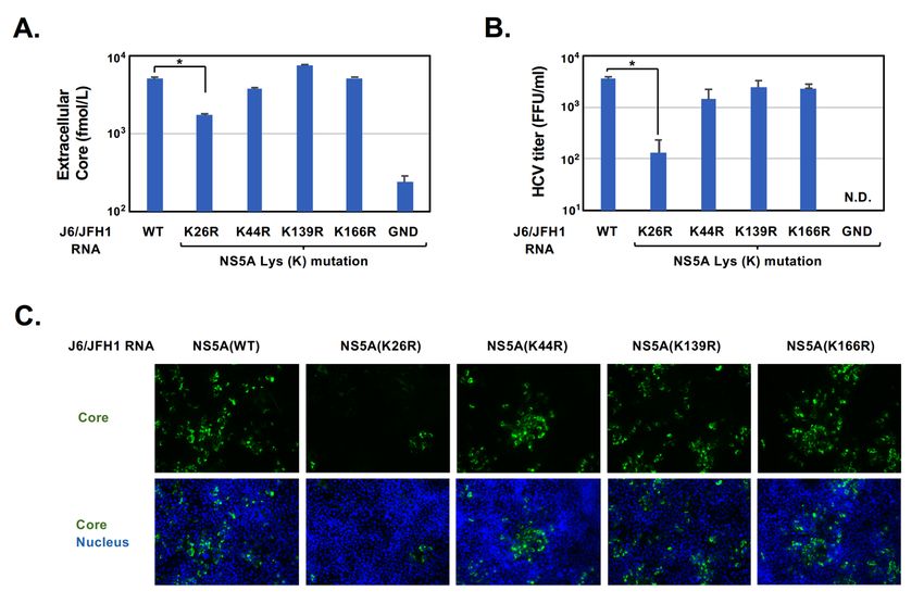
NS5A-ISGYLATION VIA LYSINE 26 IN HCV GENOTYPE 2A

HCV RNA replication (9, 11). Domain II (DII) is involved in the viral replication (8, 23), while Domain III
(DIII) is required for the process of viral release (2, 10, 12).

    IFN-stimulated gene 15 (ISG15) is a ubiquitin-like protein that is induced by IFN production upon
stimulation with viral and bacterial infection (28). ISG15 is covalently conjugated to the target substrate proteins
via specific lysine (Lys) residues by three enzymes: E1 (UBE1L), E2 (UbcH8), and E3 ligase (HERC5). This
process is called 'ISGylation' and is a post-translational protein modification that is similar to ubiquitylation. The
function of ISG15 has been suggested to be antiviral function to several viruses (31). However, there were
conflicting observations, suggesting that ISG15 has proviral function in HCV infection (1, 3, 4, 5, 22). We
previously reported that HCV NS5A (1b, Con1) protein accepts ISGylation to promote HCV RNA replication
through the recruitment of cyclophilin A (CypA), which is the critical host factor for HCV RNA replication (1).

    In the present study, we sought to elucidate the role of NS5A-ISGylation in HCV genotype 2a replication.
We demonstrate that the K26 residue in the AH domain and the K139 residue in DI of NS5A (2a, JFH1) have
the potential to accept ISGylation. Furthermore, we demonstrate that the K26 residue in combination with the
K139 residue or K166 residue is required for efficient HCV propagation.

                                        MATERIALS AND METHODS
Cell culture and transfection
   Huh-7.5 cells (kindly provided by Dr. C.M. Rice, The Rockefeller University, NY), which are highly
permissive for the culture-adapted HCV of genotype 2a JFH1 strain propagation (25, 26). Huh-7.5 cells were
maintained in Dulbecco's modified Eagle's medium (DMEM) (High Glucose) with L-glutamine (Wako, Osaka,
Japan) supplemented with 50 IU/ml penicillin, 50 µg/ml streptomycin (Gibco, Grand Island, NY), and 10%
heat-inactivated fetal bovine serum (Biowest, Nuaillé, France) at 37°C in a 5% CO2 incubator.

   Cells were transfected with plasmid DNA using FuGene 6 transfection reagents (Promega, Madison, WI).
The pFL-J6/JFH1 plasmid that encodes the entire viral genome of a chimeric strain of HCV-2a, JFH1 (14), was
kindly provided by Dr. C.M. Rice. The HCV genome RNA was synthesized in vitro using pFL-J6/JFH1 as a
template and was transfected into Huh-7.5 cells by electroporation. The virus produced in the culture supernatant
was used for the titration of virus infectivity.

Plasmids
    The cDNA fragment of NS5A (2a, JFH1) was inserted into the NotI site of pCAG-HA using the In-Fusion
HD-Cloning kit (Clontech, Mountain View, CA). The HCV (2a, JFH1) reporter subgenomic replicon (SGR)
plasmid encoding the firefly luciferase gene (pSGR-luc) was kindly provided by Dr. T. Wakita (NIID, Japan).
The cDNA fragments of NS5A (2a, JFH1) with the Lys residue mutated to Arg residue were generated by the
mutagenesis PCR method using pCAG-HA-NS5A, pSGR-luc, or pFL-J6/JFH1 as a template. The specific
primers used for the PCR were as follows: sense primer (K26R), 5'-TGGCTGACCTCTAGATTGTTCCCC-3';
antisense primer (K26R), 5'-GGGGAACAATCTAGAGGTCAGCCA-3', sense primer (K44R),
5'-CAAAAGGGGTACAGGGGTGTGTGG-3';                              antisense              primer         (K44R),
5'-CCACACACCCCTGTACCCCTTTTG-3',                             sense                primer           (K139R),
5'-ACTGACAATCTGAGAATTCCTTGC-3';                            antisense              primer          (K139R),
5'-GCAAGGAATTCTCAGATTGTCAGT-3',                              sense               primer           (K166R),
5'-GCACCCACACCAAGGCCGTTTTTC-3';                            antisense              primer          (K166R),
5'-GAAAAACGGCCTTGGTGTGGGTGC-3'. The expression plasmid for pCAG-FLAG-ISG15 was previously
described (19). The cDNA fragments encoding UBE1L and UbcH8, or HERC5 were cloned into the NotI/BglII
or SmaI/KpnI site of pCAG-MCS2, respectively using the In-Fusion HD-Cloning kit.
    The various genotypes of HCV subgenomic replicon plasmids (SG-Feo) possessing a chimeric gene
encoding the firefly luciferase and neomycin resistance gene, including H77c (L+8), S52 (AII), ED43 (VYG),
and SA1 (SKIP), were kindly provided by Dr. C.M. Rice (24, 34). The insertions of NS5A with the Lys26
residue mutation (K26R) from each of HCV genotypes were generated by the mutagenesis PCR method using
SG-Feo as a template. The specific primers used for the PCR were as follows: sense primer (H77c, K26R),
5'-TGGCTGAAAGCCAGGCTCATGCCA-3';                       antisense          primer          (H77c,     K26R),
5'-TGGCATGAGCCTGGCTTTCAGCCA-3',                        sense            primer           (S52,      K26R),
5'-CTCTCTGCTAGGATTATGCCAGCA-3';                       antisense           primer          (S52,     K26R),
5'-TGCTGGCATAATCCTAGCAGAGAG-3',                        sense           primer           (ED43,      K26R),
5'-ACGTGGCTAAAAGCCAGGTTGCTG-3';                       antisense          primer          (ED43,     K26R),

                                                                                                                 E39
R. G. BAWONO et al.

5'-CAGCAACCTGGCTTTTAGCCACGT-3',                       sense         primer           (SA1,          K26R),
5'-TGGTTGCAGGCAAGACTCCTCCCG-3';                      antisense         primer         (SA1,         K26R),
5'-CGGGAGGAGTCTTGCCTGCAACCA-3'.                 The sequences of the inserts were extensively confirmed by
sequencing (Eurofins Genomics).

Antibodies and reagents
    The mouse monoclonal antibodies (mAbs) used in this study were anti-FLAG (M2) mAb (F-3165,
Sigma-Aldrich, St. Louis, MO), anti-NS3 (MAB8691, Millipore), anti-glyceraldehyde-3-phosphate
dehydrogenase (GAPDH) mAb (MAB374, Millipore), and anti-Core mAb (2H9 clone, kindly provided by Dr. T.
Wakita, NIID, Japan). The rabbit polyclonal antibodies (pAbs) used in this study were anti-HA pAb (H-6908,
Sigma-Aldrich) and anti-NS5A pAb (2914-1 clone; kindly provided by Dr. Wakita). Horseradish peroxidase
(HRP)-conjugated goat anti-mouse IgG and goat anti-rabbit IgG antibody (Cell Signaling Technology, Beverly,
MAR), and HRP-conjugated donkey anti-goat IgG (Santa Cruz Biotechnology) were used as secondary
antibodies. CsA was purchased from Sigma-Aldrich.

Immunoprecipitation and immunoblot analysis
   Cells were transfected with the plasmids using FuGene 6 (Promega), harvested at 48 h post-transfection, and
suspended in 0.5 ml of RIPA buffer containing 50 mM Tris-HCl (pH 8.0), 150 mM NaCl and 0.1% SDS, 1%
NP-40, 0.5% deoxycholate (DOC), and protease inhibitor cocktail tablets (Roche Molecular Biochemicals,
Mannheim, Germany). Cell lysates were incubated for 2 h at 4°C and centrifuged at 20,400 × g for 30 min at 4°C
(TOMY centrifuge MX-307, Rotor Rack AR015-SC24, TOMY, Tokyo). The supernatant was
immunoprecipitated with protein G Sepharose 4 fast flow (GE Healthcare, Buckinghamshire, UK) and incubated
with appropriate antibodies at 4°C overnight.

    After being washed with the RIPA buffer five times, the samples were boiled in 15 µl of sodium dodecyl
sulfate (SDS) sample buffer and then subjected to SDS-10% polyacrylamide gel electrophoresis (SDS-PAGE)
and transferred to polyvinylidene difluoride membranes (PVDF) (Millipore). The membranes were blocked with
Tris-buffered saline containing 20 mM Tris-HCl (pH 7.6), 135 mM NaCl, and 0.05% Tween 20 (TBST)
containing 5% skim milk at room temperature for 2 h and incubated with corresponding antibodies. The
membranes were then incubated with HRP-conjugated secondary antibody at room temperature for 2 h. The
immune complexes and cell lysates were visualized with ECL Western blotting detection reagents (GE
Healthcare) and detected by the LAS-4000 image analyzer system (GE Healthcare). The band intensities were
quantified using ImageQuant TL software (ver.7.0).

Quantification of extracellular core protein
    The HCV core protein in the culture supernatants was quantified by highly sensitive enzyme immunoassay
using the Ortho HCV core antigen ELISA test (Ortho-Clinical Diagnostics, Raritan, NJ).

HCV infectivity assay
    Briefly, culture supernatants were serially diluted 10-fold and used to infect duplicate 24-well cultures of
Huh-7.5 cells. At 24 h post-inoculation, the cultures were overlaid with complete DMEM containing a final
concentration of 0.25% methylcellulose (Sigma-Aldrich). At 72 h of incubation, the cells were fixed in 4%
paraformaldehyde (PFA) and immunohistochemically stained using anti-Core mAb (2H9 clone). The infectious
HCV titers were determined based on the focus-forming units (FFU)/ml (32).

In vitro transcription of HCV RNA, electroporation into the cells, and reporter analysis
    The HCV replicon plasmid pSGR (2a, JFH1)-Luc, and pFL-J6/JFH1 plasmids were digested with XbaI,
respectively, and transcribed in vitro using a MEGAscript T7 kit (Ambion, Austin, TX). Then, 10 µg of in
vitro-transcribed HCV RNA was electroporated at 270 V and 960 µF by a GenePulser Xcells™ (Bio-Rad,
Hercules, CA) into 4×10⁶ Huh-7.5 cells treated with BTXpress buffer (BTX, Holliston, MA). The electroporated
cells were seeded into 24-well plates and harvested at the indicated time points, and luciferase activity was
determined in triplicate using a GloMax™ 96 microplate luminometer (Promega). The luciferase activity at 4 h
after electroporation was used for normalization to account for the varying transduction efficiency of HCV RNA.

Statistics
    Results are expressed as the mean ± standard error. The statistical significance was determined by Dunnett’s
test for the results shown in Fig. 3A, 3B, 4A, and 4B. P-values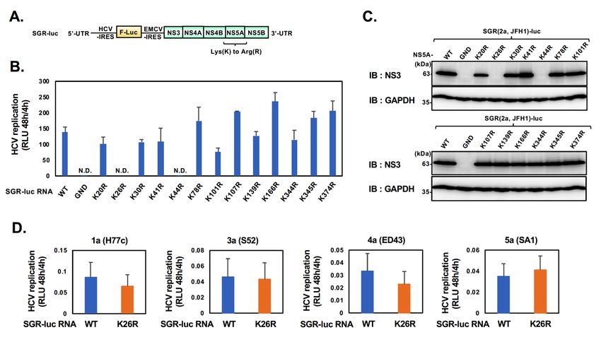
NS5A-ISGYLATION VIA LYSINE 26 IN HCV GENOTYPE 2A

                                                     RESULTS
The Lys residues K26 and K139 on NS5A from genotype 2a are acceptor sites for ISGylation
    The alignment of amino acid sequences revealed that 5 Lys residues (K20, K26, K44, K139, and K166) of
the 13 Lys residues within NS5A (2a, JFH1) are conserved compared to those of genotype 1b (Con1) (Fig. 1A).
In contrast, the K308 residue within domain II (DII) of NS5A (1b, Con1), which is important for both
NS5A-ISGylation and HCV RNA replication (1), is not conserved in genotype 2a due to lack of Lys residues in
DII of NS5A (2a, JFH1) (Fig. 1A). On the other hand, three Lys residues, including K20 and K26 in the
amphipathic helix (AH) domain and K139 in domain I (DI), are well conserved among the HCV genotypes
examined in this study (Fig. 1A). These results imply the possibility that there are distinct roles of Lys residues
in NS5A-ISGylation to regulate HCV RNA replication between genotype 1b and 2a.

    Next, to identify the acceptor sites for ISGylation on NS5A (2a, JFH1) protein, we constructed a series of
NS5A mutants, containing a point mutation of Arginine (R) at a corresponding Lys (K) (K to R mutant series).
We then co-transfected HEK293T cells with pCAG-HA-NS5A (2a) or its Lys (K) mutants and
pCAG-FLAG-ISG15 together with E1 (UBE1L), E2 (UbcH8), and E3 (HERC5), followed by
immunoprecipitation with anti-HA antibody and detection with anti-NS5A (2a, JFH1) specific rabbit pAb. The
immunoprecipitation analysis coupled with immunoblotting revealed that the slowly migrating forms of
HA-NS5A (indicated as NS5A-ISGylation) were clearly detected in the co-transfected cells compared to the
cells expressed with HA-NS5A alone (Fig. 1B, upper panel, lanes 1 and 2). On the other hand, ISGylation of
NS5A (2a) protein was markedly reduced by the mutations of K26R and K139R but not K44R, K166R
compared to HA-NS5A (2a) (WT) (Fig. 1B, upper panel, lanes 2 to 6). These results suggest that K26 and K139
residues are acceptor sites for ISGylation on NS5A (2a) protein.

Fig. 1. Schematic diagrams of the distribution of Lys residues on NS5A protein from HCV genotypes and the
         cell-based ISGylation assay for HA-NS5A (2a) WT and mutants.
      A: Schematic diagrams of the distribution of Lys (K) residues on NS5A protein from HCV genotypes (1b: Con1, 1a:
      H77c, 2a: JFH1, 3a: S52, 4a: ED43, 5a: SA1). AH: amphipathic helix. B: The expression vector encoding HA-NS5A
      (2a, JFH1) or HA-NS5A mutants, in which the K26, K44, K139, and K166 residues were mutated to Arg (R)
      (indicated as K26R, K44R, K139R, and K166), was co-transfected with FLAG-ISG15 together with E1 (UBE1L), E2
      (UbcH8), and E3 ligase (HERC5) in HEK293T cells, followed by immunoprecipitation with anti-HA rabbit pAb and
      detection with anti-NS5A specific rabbit pAb. Input samples (indicate as Lysate) were detected with anti-HA rabbit
      pAb and anti-FLAG mouse mAb.

The K26 residue on NS5A from genotype 2a is involved in the positive regulation of HCV replication
    To determine the role of the K26 and K139 residues on NS5A (2a, JFH1) in HCV RNA replication, we
constructed plasmids expressing firefly luciferase (F-luc) reporter subgenomic RNA replicons (indicated as
SGR-luc RNA), with a point mutation of NS5A that replaces one of the 13 Lys residues with Arg (Fig. 2A). We
then electroporated in vitro-transcribed RNAs into Huh-7.5 cells to evaluate the viral replication by measuring
the luciferase activity. As shown in Fig. 2B, the electroporation of SGR-Luc RNA with the mutation of K26R

                                                                                                                   E41
R. G. BAWONO et al.
but not K139R exhibited undetectable luciferase activities at 48 h compared to that of WT. The luciferase
activities of the other mutants, except for the K44R, were comparable to that of the WT (Fig. 2B). Immunoblot
analysis revealed undetectable NS3 protein levels in the cells electroporated with SGR-Luc RNA with the
mutations of K26R and K44R on NS5A protein and the polymerase-inactive mutant (GND) on NS5B protein
(Fig. 2C) (16). These results suggest that the K26 residue is involved in the positive regulation of HCV
replication via the NS5A-ISGylation, while the K44 residue may contribute to HCV RNA replication in the
NS5A-ISGylation-independent manner.
The K26 residue on NS5A is well conserved among HCV genotypes: 1a (H77c), 1b (Con1), 2a (JFH1), 3a
(S52), 4a (ED43), and 5a (SA1) (Fig. 1A). The K26 residue on NS5A (1b, Con1) has no impact on HCV RNA
replication (1). To test whether the K26 residue on NS5A protein is involved in HCV RNA replication in other
HCV genotypes, we electroporated HCV-SGR-Luc RNA with the mutation of K26R from other HCV genotypes
into Huh-7.5 cells to evaluate viral replication. Interestingly, the levels of luciferase activity in the cells
electroporated with SGR-Luc RNA (K26R) from other HCV genotypes were comparable to those of WT (Fig.
2D). These results suggest that the K26 on NS5A (2a, JFH1) plays an important role in HCV RNA replication in
genotype 2a-specific manner.
Fig. 2. HCV replication assays using subgenomic replicon RNA possessing the firefly luciferase gene.
A: Schematic diagram of HCV reporter subgenomic replicon (SGR-luc) RNA comprising NS3 to NS5B of the
genotype 2a (JFH1). HCV reporter replicon RNA possesses the firefly luciferase (F-Luc) gene. IRES: internal
ribosomal entry site, UTR: untranslated region, EMCV: encephalomyocarditis virus. B: Huh-7.5 cells were
electroporated with HCV-SGR-luc RNA or the replicon possessing NS5A Lys mutants in which a Lys (K) is replaced
with Arg (R) (indicated as K to R mutant series). The luciferase activity was measured at 48 h after electroporation.
The luciferase activity measured at 4 h after electroporation was used to normalize for the input RNA. C: The cell
lysates from luciferase assay were subjected to immunoblotting analysis with anti-NS3 mouse mAb or anti-GAPDH
mouse mAb. D: Huh-7.5 cells were electroporated with either HCV SGR-luc from HCV genotypes or their mutant
replicon possessing NS5A K26 mutant, in which a Lys (K) is replaced with Arg (R) (indicated as K26R). The
luciferase activity was measured at 48 h after electroporation. The luciferase activity measured at 4 h after
electroporation was used to normalize for the input RNA.
The K26 residue on NS5A from genotype 2a is involved in the positive regulation of HCV RNA replication
To further examine the role of the K26 residue on NS5A (2a, JFH1) in HCV propagation, we constructed an
infectious HCV RNA clone (indicated as J6/JFH1 RNA) carrying the K26R mutation on NS5A protein. We
electroporated in vitro-transcribed infectious HCV RNA into Huh-7.5 cells to evaluate viral propagation by
measuring several virological parameters. The extracellular core protein levels were significantly decreased in
the cells electroporated with J6/JFH1 RNA carrying K26R mutation compared to those in the cells
E42NS5A-ISGYLATION VIA LYSINE 26 IN HCV GENOTYPE 2A
electroporated with WT and other Lys mutations, including K44R, K139R, and K166R at 5 days
post-electroporation (Fig. 3A). Consistent with this result, in the cells electroporated with J6/JFH1 RNA with
K26R mutation, we found marked reduction in the extracellular virus titers (Fig. 3B) and the intracellular core
protein levels (Fig. 3C). Taken together, these results suggest that the K26 residue on NS5A (2a, JFH1) plays an
important role in HCV propagation.
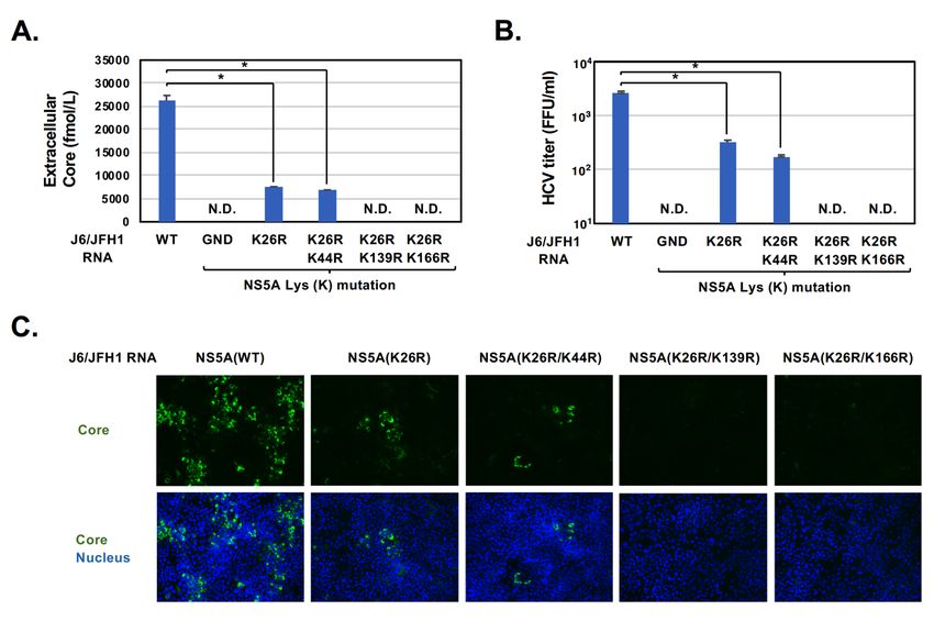
Fig. 3. The amounts of extracellular core protein, the infectious HCV titers, and intracellular core protein levels in
Huh-7.5 cells electroporated with HCV J6/JFH1 RNA wild-type and NS5A Lys mutants.
HCV J6/JFH1 RNAs derived from WT, a replication-defective mutant (GND), or a series of NS5A Lys (K) mutants
(indicated as K26R, K44R, K139R, and K166R) were electroporated into Huh-7.5 cells. At 5 days post-electroporation,
supernatants were collected and subjected to measure the various virological parameters. The supernatants were
inoculated to naïve Huh7.5 cells, and the amounts of extracellular core protein (A), the infectious HCV titer (B), and
intracellular core protein levels (C) were determined at 3 days post-inoculation using core ELISA, a focus-forming
assay (FFA) and an indirect immunofluorescent analysis, respectively. Results are the mean values of triplicates ± S.E.
*pR. G. BAWONO et al.
(Fig. 4B) and intracellular core protein levels (Fig. 4C). Taken together, these results suggest that the K26
residue may collaborate with the K139 or K166 residues to facilitate efficient HCV propagation.
Collectively, these results suggest that HCV NS5A (2a, JFH1) protein has the potential to accept
ISG15-conjugation via the specific Lys residues, leading to the facilitation of HCV propagation.
Fig. 4. The amounts of extracellular core protein, the infectious HCV titers, and intracellular core protein levels in
Huh-7.5 cells electroporated with HCV J6/JFH1 RNA wild-type and NS5A Lys double mutants.
HCV J6/JFH1 RNAs derived from WT, a replication-defective mutant (GND), or a series of NS5A Lys (K) mutants
(indicated as K26R, K26R/K44R, K26R/K139R, and K26R/K166R) were electroporated into Huh-7.5 cells. At 5 days
post-electroporation, supernatants were collected and subjected to measure the various virological parameters. The
supernatants were inoculated to naïve Huh7.5 cells, and the amounts of extracellular core protein (A), the infectious
HCV titer (B), and intracellular core protein levels (C) were determined at 3 days post-inoculation by core ELISA, a
focus-forming assay (FFA) and an indirect immunofluorescent analysis, respectively. Results are the mean values of
triplicates ± S.E. *pNS5A-ISGYLATION VIA LYSINE 26 IN HCV GENOTYPE 2A
compensate for the role of the K26 residue in genotype 2a of HCV. Further studies are needed to clarify the
detailed contribution of the K20 residue on NS5A from other HCV genotypes.
Crystal structure of NS5A DI was analyzed (13, 17, 27), while other structures, including DII and DIII,
remain to be analyzed. The N-terminal AH domain of NS5A protein are believed to mediate its membrane
anchoring (6). The recent report by Zhang and colleagues suggests that the AH domain of NS5A protein may be
involved in the facilitation of both the viral polyprotein cleavage and the viral replicase assembly formation (30).
These results suggest that the AH domain of NS5A has potential as a functional regulatory domain for efficient
HCV propagation. Our results indicate that the mutation of the K26 residue in the AH-domain results in
reduction of HCV propagation (Fig. 2B, Fig. 3 and Fig. 4). We thus speculate that the ISGylated NS5A within
the AH domain may facilitate both viral polyprotein cleavage and viral replicase assembly formation.
Additionally, we found that the K26 residue may collaborate with the K139 residue or K166 residue for
efficient HCV propagation (Fig. 4). The Lysine residue K139 but not K166 exhibited the involvement of
NS5A-ISGylation, which is similar to the K26 residue (Fig. 1B). These results may imply the role of
ISGylation-dependent and -independent manners in the collaboration of their Lys residues on HCV propagation.
Further studies are needed to clarify the intermolecular interactions of the K26 residue together with K139 and
K166 residues for efficient HCV propagation.
There was discrepancy in the role of the K44 residue between HCV subgenomic replicon and cell culture
HCV (Fig. 2B, Fig. 3, and Fig. 4). We speculate that the role of the K44 residue in the cell culture HCV might be
compensated by another lysine residue, as we discussed the role of the K26 residue in HCV genotypes.
In summary, using an HCV infectious clone, we demonstrated that NS5A (2a, JFH1) protein accepts
ISGylation via the K26 residue in AH domain and participates in the positive regulation of HCV propagation in
genotype 2a-specific manner. The targeting of the ISGylation machinery may lead to development of novel
therapeutics for HCV infection.
ACKNOWLEDGEMENTS
We are grateful to Dr. C.M. Rice (The Rockefeller University, New York, NY) for providing the Huh-7.5
cells, pFL-J6/JFH1, and the series of SG-Feo constructions. We thank Y. Kozaki for the secretarial work. This
work was supported by the Program for Basic and Clinical Research on Hepatitis from the Japan Agency for
Medical Research and Development (AMED) under Grant no. JP18fk0210006, JP20fk0210040, and
JP20fk0210053. This work was also supported by the Ministry of Education, Culture, Sports, Science, and
Technology (MEXT) of Japan, the Japan Society for the Promotion of Science (KAKENHI), Grant no.
JP17K08857, and the Takeda Science Foundation.
AUTHOR CONTRIBUTIONS
R.G.B., T.A., and I.S. conceived and designed the experiments. R.G.B., Y.S. and T.A. carried out most of the
experiments. C.M., and L.D. assisted the constructions and the data analysis. T.A. and I.S. wrote the manuscript.
CONFLICTS OF INTEREST
The authors have no conflicts of interest to declare.
REFERENCES
1. Abe, T., Minami, N., Rheza Gandi Bawono., Matsui, C., Deng, L., Fukuhara, T., Matsuura, Y., and
Shoji, I. 2020. ISGylation of hepatitis C virus NS5A protein promotes viral RNA replication via
recruitment of cyclophilin A. J Virol 94:e00532-20.
2. Appel, N., Zayas, M., Miller, S., Krijnse-Locker, J., Schaller, T., Friebe, P., Kallis, S., Engel, U., and
Bartenschlager, R. 2008. Essential role of domain III of nonstructural protein 5A for hepatitis C virus
infectious particle assembly. PLoS Pathog 4:e1000035.
3. Broering, R., Zhang, X., Kottilil, S., Trippler, M., Jiang, M., Lu, M., Gerken, G., and Schlaak, J.F.
2010. The interferon stimulated gene 15 functions as a proviral factor for the hepatitis C virus and as a
regulator of the IFN response. Gut 59:1111-1119.
4. Chen, L., Sun, J., Meng, L., Heathcote, J., Edwards, A.M., and McGilvray, I.D. 2010. ISG15, a
ubiquitin-like interferon-stimulated gene, promotes hepatitis C virus production in vitro: implications for
E45R. G. BAWONO et al.
chronic infection and response to treatment. J Gen Virol 91:382-388.
5. Chua, P.K., McCown, M.F., Rajyaguru, S., Kular, S., Varma, R., Symons, J., Chiu, S.S., Cammack,
N., and Najera, I. 2009. Modulation of alpha interferon anti-hepatitis C virus activity by ISG15. J Gen
Virol 90:2929-2939.
6. Elazar, M., Cheong, K.H., Liu, P., Greenberg, H.B., Rice, C.M., and Glenn, J.S. 2003. Amphipathic
helix-dependent localization of NS5A mediates hepatitis C virus RNA replication. J Virol 77:6055-6061.
7. Falade-Nwulia, O., Suarez-Cuervo, C., Nelson, D.R., Fried, M.W., Segal, J.B., and Sulkowski, M.S.
2017. Oral direct-acting agent therapy for hepatitis C virus infection: a systematic review. Ann Intern Med
166:637-648.
8. Foster, T.L., Gallay, P., Stonehouse, N.J., and Harris, M. 2011. Cyclophilin A interacts with domain II
of hepatitis C virus NS5A and stimulates RNA binding in an isomerase-dependent manner. J Virol
85:7460-7464.
9. Gao, M., Nettles, R.E., Belema, M., Snyder, L.B., Nguyen, V.N., Fridell, R.A., Serrano-Wu, M.H.,
Langley, D.R., Sun, J.H., O'Boyle, D.R. 2nd., Lemm, J.A., Wang, C., Knipe, J.O., Chien, C., Colonno,
R.J., Grasela, D.M., Meanwell, N.A., and Hamann, L.G. 2010. Chemical genetics strategy identifies an
HCV NS5A inhibitor with a potent clinical effect. Nature 465:96-100.
10. Hanoulle, X., Verdegem, D., Badillo, A., Wieruszeski, J.M., Penin, F., and Lippens, G. 2009. Domain
3 of non-structural protein 5A from hepatitis C virus is natively unfolded. Biochem Biophys Res Commun
381:634-638.
11. Harak, C., and Lohmann, V. 2015. Ultrastructure of the replication sites of positive-strand RNA viruses.
Virology 479-480:418-433.
12. Hughes, M., Griffin, S., and Harris, M. 2009. Domain III of NS5A contributes to both RNA replication
and assembly of hepatitis C virus particles. J Gen Virol 90:1329-1334.
13. Lambert, S.M., Langley, D.R., Garnett, J.A., Angell, R., Hedgethorne, K., Meanwell, N.A., and
Matthews, S.J. 2014. The crystal structure of NS5A domain 1 from genotype 1a reveals new clues to the
mechanism of action for dimeric HCV inhibitors. Protein Sci 23:723-734.
14. indenbach, B.D., Evans, M.J., Syder, A.J., Wolk, B., Tellinghuisen, T.L., Liu, C.C., Maruyama, T.,
Hynes, R.O., Burton, D.R., Mckeating, J.A., and Rice, C.M. 2005. Complete replication of hepatitis C
virus in cell culture. Science 309:623-626.
15. Lohmann, V. 2013. Hepatitis C virus RNA replication. Curr Top Microbiol Immunol 369:167-198.
16. Lohmann, V., Korner, F., Herian, U., and Bartenschlager, R. 1997. Biochemical properties of hepatitis
C virus NS5B RNA-dependent RNA polymerase and identification of amino acid sequence motifs essential
for enzymatic activity. J Virol 71:8416-8428.
17. Love, R.A., Brodsky, O., Hickey, M.J., Wells, P.A., and Cronin, C.N. 2009. Crystal structure of a novel
dimeric form of NS5A domain I protein from hepatitis C virus. J Virol 83:4395-4403.
18. Messina, J.P., Humphreys, I., Flaxman, A., Brown, A., Cooke, G.S., Pybus, O.G., and Barnes, E. 2015.
Global distribution and prevalence of hepatitis C virus genotypes. Hepatology 61:77-87.
19. Minami, N., Abe, T., Deng, L., Matsui, C., Fukuhara, T., Matsuura, Y., and Shoji, I. 2017.
Unconjugated interferon-stimulated gene 15 specifically interacts with the hepatitis C virus NS5A protein
via domain I. Microbiol Immunol 61:287-292.
20. Moradpour, D., Penin, F., and Rice, C.M. 2007. Replication of hepatitis C virus. Nat Rev Microbiol
5:453-463.
21. Neufeldt, C.J., Cortese, M., Acosta, E.G., and Bartenschlager, R. 2018. Rewiring cellular networks by
members of the Flaviviridae family. Nat Rev Microbiol 16:125-142.
22. Real, C.I., Megger, D.A., Sitek, B., Jahn-Hofmann, K., Ickenstein, L.M., John, M.J., Walker, A.,
Timm, J., Kuhlmann, K., Eisenacher, M., Meyer, H.E., Gerken, G., Broering, R., and Schlaak, J.F.
2013. Identification of proteins that mediate the pro-viral functions of the interferon stimulated gene 15 in
hepatitis C virus replication. Antiviral Res 100:654-661.
23. Ross-Thriepland, D., Amako, Y., and Harris, M. 2013. The C terminus of NS5A domain II is a key
determinant of hepatitis C virus genome replication, but is not required for virion assembly and release. J
Gen Virol 94:1009-1018.
24. Saeed, M., Scheel, T.K., Gottwein, J.M., Marukian, S., Dustin, L.B., Bukh, J., and Rice, C.M. 2012.
Efficient replication of genotype 3a and 4a hepatitis C virus replicons in human hepatoma cells. Antimicrob
Agents Chemother 56:5365-5373.
25. Sainz, B. Jr., and Chisari, F.V. 2006. Production of infectious hepatitis C virus by well-differentiated,
growth-arrested human hepatoma-derived cells. J Virol 80:10253-10257.
26. Sainz, B. Jr., Barretto, N., and Uprichard, S.L. 2009. Hepatitis C virus infection in phenotypically
distinct Huh7 cell lines. PLoS One 4:e6561.
E46NS5A-ISGYLATION VIA LYSINE 26 IN HCV GENOTYPE 2A
27. Tellinghuisen, T.L., Marcotrigiano, J., and Rice, C.M. 2005. Structure of the zinc-binding domain of an
essential component of the hepatitis C virus replicase. Nature 435:374-379.
28. Zhang, D., and Zhang, D.E. 2011. Interferon-stimulated gene 15 and the protein ISGylation system. J
Interferon Cytokine Res 31:119-130.
29. Zoulim, F., Liang, T.J., Gerbes, A.L., Aghemo, A., Deuffic-Burban, S., Dusheiko, G., Fried, M.W.,
Pol, S., Rockstroh, J.K., Terrault, N.A., and Wiktor, S. 2015. Hepatitis C virus treatment in the real
world: optimising treatment and access to therapies. Gut 64:1824-1833.
30. Zhang, Y., Zhao, X., Zou, J., Yuan, Z., and Yi, Z. 2019. Dual role of the amphipathic helix of hepatitis C
virus NS5A in the viral polyprotein cleavage and replicase assembly. Virology 535:283-296.
31. Perng, Y.C., and Lenschow, D.J. 2018. ISG15 in antiviral immunity and beyond. Nat Rev Microbiol
16:423-439.
32. Wakita, T., Pietschmann, T., Kato, T., Date, T., Miyamoto, M., Zhao, Z., Murthy, K., Habermann, A.,
Krausslich, H.G., Mizokami, M., Bartenschlager, R., and Liang, T.J. 2005. Production of infectious
hepatitis C virus in tissue culture from a cloned viral genome. Nat Med 11:791-796.
33. Webster, D.P., Klenerman, P., and Dusheiko, G.M. 2015. Hepatitis C. The Lancet 385:1124-1135.
34. Wose Kinge, C.N., Espiritu, C., Prabdial-Sing, N., Sithebe, N.P., Saeed, M., and Rice, C.M. 2014.
Hepatitis C virus genotype 5a subgenomic replicons for evaluation of direct-acting antiviral agents.
Antimicrob Agents Chemother 58:5386-5394.
E47You can also read