PEP-1-GLRX1 protein exhibits anti-inflammatory effects by inhibiting the activation of MAPK and NF- B pathways in Raw 264.7 cells
←
→
Page content transcription
If your browser does not render page correctly, please read the page content below
BMB Rep. 2020; 53(2): 106-111
BMB www.bmbreports.org
Reports
PEP-1-GLRX1 protein exhibits anti-inflammatory effects by
inhibiting the activation of MAPK and NF-B pathways in Raw
264.7 cells
1,# 2,# 1 1 1,3 4 1
Min Jea Shin , Dae Won Kim , Yeon Joo Choi , Hyun Ju Cha , Sung Ho Lee , Sunghou Lee , Jinseu Park ,
Kyu Hyung Han , Won Sik Eum * & Soo Young Choi *
1 1, 1,
1
Department of Biomedical Science and Research Institute of Bioscience and Biotechnology, Hallym University, Chuncheon 24252,
2
Department of Biochemistry and Molecular Biology, Research Institute of Oral Sciences, College of Dentistry, Gangneung-Wonju National
University, Gangneung 25457, 3Genesen Inc., Seoul 06181, 4Department of Green Chemical Engineering, Sangmyung University, Cheonan
31066, Korea
Glutaredoxin 1 (GLRX1) has been recognized as an important contributes to various disorders, including cardiovascular
regulator of redox signaling. Although GLRX1 plays an diseases, cancer, arthritis, and neuronal diseases (1, 2). It is
essential role in cell survival as an antioxidant protein, the well known that macrophages, which are important immune
function of GLRX1 protein in inflammatory response is still cells, regulate the inflammatory response and produce
under investigation. Therefore, we wanted to know whether pro-inflammatory mediators and cytokines. These mediators
transduced PEP-1-GLRX1 protein inhibits lipopolysaccharide and cytokines contribute to the pathogenesis of inflammation
(LPS)- and 12-O-tetradecanoyl phorbol-13-acetate (TPA)-induced (3-6). Therefore, Gue et al. (2016) suggest that inhibition of
inflammation. In LPS-exposed Raw 264.7 cells, PEP-1-GLRX1 pro-inflammatory mediators and cytokines is important for
inhibited cyclooxygenase-2 (COX-2), inducible nitric oxide preventing the progression of inflammatory diseases (7).
synthase (iNOS), activation of mitogen activated protein kinases Nuclear factor-kappaB (NF-B) is well known to modulate
(MAPKs) and nuclear factor-kappaB (NF-B) expression levels. the inflammatory response. NF-B is located in the cytoplasm
In a TPA-induced mouse-ear edema model, topically applied as a complex with IB under normal conditions. However,
PEP-1-GLRX1 transduced into ear tissues and significantly NF-B is translocated to the nucleus under IB degradation
ameliorated ear edema. Our data reveal that PEP-1-GLRX1 when exposed to inflammatory stimuli, such as LPS (8). Also,
attenuates inflammation in vitro and in vivo, suggesting that other studies have shown that NF-B is an essential pathway
PEP-1-GLRX1 may be a potential therapeutic protein for associated with pro-inflammatory mediator production in the
inflammatory diseases. [BMB Reports 2020; 53(2): 106-111] inflammatory response (9, 10). In addition, mitogen-activated
protein kinase (MAPK) has been considered to be a typical
molecular target for the development of anti-inflammatory
INTRODUCTION agents. Several studies have reported that the activation of
MAPKs leads to increased production of the pro-inflammatory
Inflammatory response is known to be a defense against mediators. Therefore, NF-B and MAPKs signaling pathways
external harmful factors, such as microbial pathogens and have been considered to be potential targets for anti-
chemicals. However, an excessive inflammatory response inflammatory drugs (9, 11-13).
Human glutaredoxin (GLRX) is a small molecular-weight
protein and a member of the thioredoxin family. In humans,
*Corresponding authors. Soo Young Choi, Tel: +82-33-248-2112; GLRX1 is located in the cytosol, whereas GLRX2 is located in
Fax: +82-33-248-3202; E-mail: sychoi@hallym.ac.kr; Won Sik Eum, the mitochondria (14). Several studies have described GLRX1
Tel: +82-33-248-3221; Fax: +82-33-248-3202; E-mail: wseum@ as being distributed in various tissues and as regulating
hallym.ac.kr
# redox-dependent signaling pathways. GLRX1 has a protective
These authors contributed equally to this work.
role against oxidative stress (15-17). Also, Cater et al. (2014)
https://doi.org/10.5483/BMBRep.2020.53.2.180 have shown that GLRX1 plays an important role as an
antioxidant protein and protects against copper-induced
Received 15 July 2019, Revised 26 July 2019, toxicity and neuronal cell death (18). We also have reported
Accepted 28 August 2019 that PEP-1-GLRX1 markedly protects against hippocampal
neuronal cell damage by inhibiting oxidative stress (19). In this
Keywords: Inflammation, MAPK, NF-B, PEP-1-GLRX1, Protein
study, we investigated the function of PEP-1-GLRX1 against
therapy
ISSN: 1976-670X (electronic edition)
Copyright ⓒ 2020 by the The Korean Society for Biochemistry and Molecular Biology
This is an open-access article distributed under the terms of the Creative Commons Attribution Non-Commercial License (http://creativecommons.org/li-
censes/by-nc/4.0) which permits unrestricted non-commercial use, distribution, and reproduction in any medium, provided the original work is properly cited.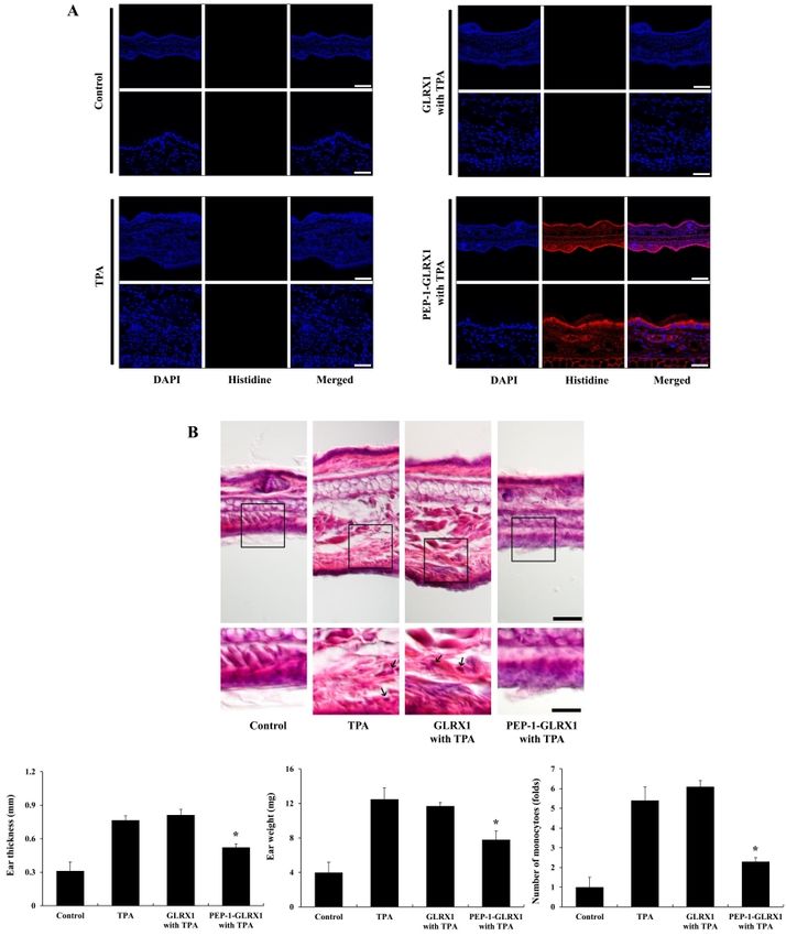
Transduced PEP-1-GLRX1 inhibits inflammation
Min Jea Shin, et al.
inflammatory responses in vitro and in vivo. (Fig. 2A). Next, to identify the location of transduced PEP-1-
GLRX1, we prepared nuclear and cytosolic fractions from the cells
RESULTS AND DISCUSSION and did Western blotting using subcellular specific marker
antibodies. Fig. 2B shows that transduced PEP-1-GLRX1 was
Transduction of PEP-1-GLRX1 into Raw 264.7 cells distributed into the cytosol and nuclei of the cells.
PTDs including PEP-1 peptide are known to be small peptides that To find out whether PEP-1-GLRX1 has anti-inflammatory roles
can transduce the plasma membrane either alone or combined against LPS-exposed Raw 264.7 cells, we examined the expression
with various macromolecules, such as proteins (20). PEP-1-GLRX1 of COX-2 and iNOS levels. Treatment with LPS markedly increased
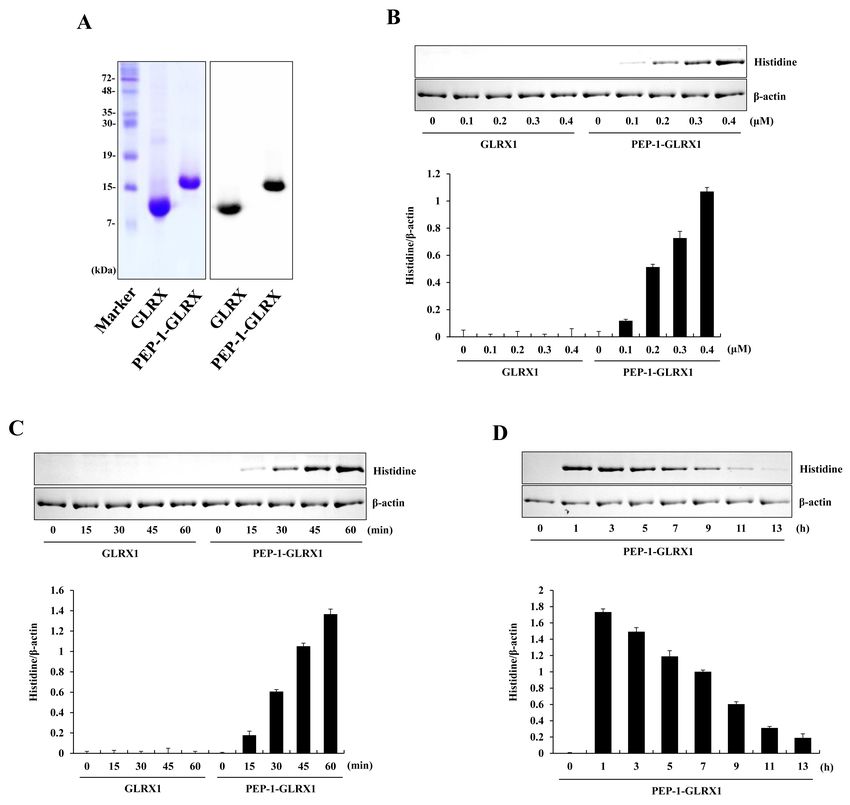
protein was produced as described previously (19). Purified COX-2 and iNOS expressions levels more than did those in the
PEP-1-GLRX1 was confirmed through SDS-PAGE and Western control. PEP-1-GLRX1 treatment significantly reduced the COX-2
blotting with a histidine antibody (Fig. 1A). Next, we measured the and iNOS expression levels in the LPS-treated cells (Fig. 2C). These
transduction efficiency of purified protein into Raw 264.7 cells. results suggest that transduced PEP-1-GLRX1 has anti-inflam-
Figs. 1B-1D show that PEP-1-GLRX1 concentration- and time- matory effects by inhibiting pro-inflammatory mediators. Chung
dependently transduced into the cells. Transduced PEP-1-GLRX1 et al. (2010) showed that pro-inflammatory mediators increased
levels remained for up to a maximum of 9 h. We have also reported in GLRX1 knockout mice more than in wild-type mice in a cigarette
that this protein was transduced into HT-22 cells (19). However, smoke-induced lung inflammation model, suggesting that the
the transduced levels of this protein into HT-22 cells remained GLRX1 plays an important role in lung inflammation (31). In
longer than in Raw 264.7 cells. addition, other studies have shown that a pro-inflammatory
response was increased in the lens and heart of GLRX1-deficient
Effect of PEP-1-GLRX1 on expression of COX-2 and iNOS mice (32, 33). On the other hand, Aesif et al. (2011) reported that
Since protein transduction is important for the development of ablation of the GLRX1 protein attenuates LPS-induced pro-
therapeutic proteins, PTD-fused protein transduction can be used inflammatory responses by controlling S-glutathionylation-
for the intracellular application of therapeutic proteins (20-24). In sensitive signaling pathways, suggesting that GLRX1 expression
previous studies, we have demonstrated that PTD-fusion proteins is critical for activating alveolar macrophages in an LPS-induced
were transduced into various cells (25-30). In this study, we first lung inflammation mice model (34).
identified the distribution of transduced PEP-1-GLRX1 using
fluorescence analysis. Green fluorescence signals were strongly
observed in the cells treated with PEP-1-GLRX1, whereas cell
treated with control GLRX1 did not show the fluorescence signals
Fig. 2. Effects of transduced PEP-1-GLRX1 protein on LPS-induced
Fig. 1. Purification and transduction of PEP-1-GLRX1 protein into Raw inflammatory response in Raw 264.7 cells. The cells were treated with
264.7 cells. Purified PEP-1-GLRX1 and control GLRX1 proteins were PEP-1-GLRX1 protein (0.4 M) for 1 h, and the cellular distribution
confirmed by 15% SDS-PAGE and Western blot analysis (A). Trans- of transduced PEP-1-GLRX1 protein was confirmed by fluorescence
duction of PEP-1-GLRX1 proteins into Raw 264.7 cells. PEP-1-GLRX1 microscopy (A). Scale bar = 50 m. Subcellular localization of PEP-
protein (0.1-0.4 M) was added to the culture media for 1 h (B), 1-GLRX1 (B). The nuclear and cytosolic extracts were prepared from
PEP-1-GLRX1 protein (0.4 M) was added to the culture media for transduced and normal Raw 264.7 cells and analyzed by Western
15-60 min (C). The stability of transduced PEP-1-GLRX1 protein was blotting. The cells were pretreated with PEP-1-GLRX1 protein (0.4 M)
assessed after various time periods. The cells were treated with for 1 h and then treated with LPS (1 g/ml). Expression levels of COX-2
PEP-1-GLRX1 protein (0.4 M), incubated with 1-13 h, and analyzed and iNOS protein were measured by Western blot analysis (C). The
by Western blot analysis (D). The band intensities were measured by band intensity was measured by densitometer. The data are presented as
densitometer. The data are presented as mean values ± SD (n = 3). mean values ± SD (n = 3). *P < 0.01 compared with LPS-treated cells.
http://bmbreports.org BMB Reports 107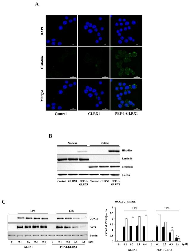
Transduced PEP-1-GLRX1 inhibits inflammation
Min Jea Shin, et al.
Effect of PEP-1-GLRX1 on the NF-B and MAPK signaling cells (43-45). Therefore, these findings suggest that NF-B and
pathways GLRX1 may be regulated in a coordinated fashion. However, the
NF-B and MAPK signaling pathway promote the production of role of GLRX1 in regulation of NF-B signaling is not completely
pro-inflammatory mediators. NF-B is one of the key transcription understood yet. Further studies are needed.
factors that control gene expression of pro-inflammatory
mediators and cytokines (35, 36). Also, MAPK signaling pathways Effect of PEP-1-GLRX1 on mouse-ear edema
(ERK, p38, JNK) are crucial for NF-B activation and regulate the PTDs can effectively transduce exogenous macromolecules into
inflammatory response (37, 38). Therefore, we measured the cells and tissues (46). TPA is known as an inducer of skin
effects of PEP-1-GLRX1 against LPS-induced phosphorylation of inflammation, and several studies have used it to find out the effects
NF-B and MAPKs in Raw 264.7 cells. In the LPS-treated cells, of different treatments against skin inflammation (47-50).
phosphorylated NF-B and MAPKs expression levels were Therefore, we wanted to find out whether PEP-1-GLRX1 was
increased more than in the control. In contrast, PEP-1-GLRX1 transduced into mice ear tissue and whether it demonstrated
significantly reduced phosphorylated NF-B and MAPKs protective effects against TPA-induced edema. After mice ears
expression levels more than in the LPS-treated cells. Control were treated with PEP-1-GLRX and TPA as described in Methods,
GLRX1 did not alter phosphorylated NF-B and MAPKs expression we confirmed PEP-1-GLRX1 levels using immunohistochemical
levels (Fig. 3). These results suggest that transduced PEP-1-GLRX1 analysis. As shown in Fig. 4A, there were no changes in the His
inhibits inflammatory responses by regulating NF-B and MAPKs antibody staining in the control-, TPA-, and TPA + control
signaling pathways. In agreement with our results, Ryu et al. (2018) GLRX1-treated groups. However, fluorescent signals were
have reported that transduced PEP-1-GLRX1 inhibited markedly increased in the PEP-1-GLRX1-treated groups compared
phosphorylation of MAPK signaling in H2O2-exposed HT-22 cells to that in the control and other groups.
(19). Other studies have reported that phosphorylated IB and Further, we measured the effects of PEP-1-GLRX1 against
p-65 protein expression levels were markedly increased in TPA-induced mouse-ear edema. As shown in Fig. 4B, TPA-treated
cigarette-smoke-induced lung inflammation GLRX1 knockout mice showed markedly increased ear thickness and weights
mice (31). In addition, several studies have reported that the compared to those in the control mice. PEP-1-GLRX1 drastically
phosphorylated NF-B and MAPKs expression levels were reduced ear thickness and weights more than in TPA-treated mice.
increased in LPS-treated macrophages (39-41). In contrast, However, there were no significant changes in the control
overexpression of GLRX1 decreased S-glutathionylation IKK GLRX1-treated mice compared to the TPA-treated mice. In
Cys179 and increased NF-B activation after oxidation induced addition, PEP-1-GLRX1 drastically reduced infiltration of
by oxidative stress, suggesting that cellular content of GLRX1 monocytes, which is an early event in skin inflammation. Control
regulates the activation of NF-B under oxidative stress (42). Also,
other studies have demonstrated that the cellular content of GLRX1
is regulated by the proinflammatory stimuli. GLRX1 level was
increased in bronchial epithelial cells of in mice with allergic
airway inflammation, activation of NF-B and GLRX1 is also
increased concomitant with activation of NF-B in the retinal glial
Fig. 4. Effects of PEP-1-GLRX1 protein on ear edema in a TPA-induced
mice model of inflammation. Ears of mice were exposed to TPA (1
g/ear), and PEP-1-GLRX1 protein (10 g) was topically applied to mice
ears 1 h after TPA treatment for 3 days. Transduced PEP-1-GLRX1 protein
Fig. 3. Effects of transduced PEP-1-GLRX1 protein on LPS-induced was confirmed by Histidine antibody (A). Scale bar = 50 m (top
NF-B and MAPK phosphorylation in Raw 264.7 cells. The cells were panel) and 25 m (bottom panel). The effects of PEP-1-GLRX1 protein
pretreated with PEP-1-GLRX1 protein (0.4 M) for 1 h and then treated against TPA-induced ear edema was analyzed by hematoxylin and eosin
with LPS (1 g/ml). Phosphorylation of NF-B (A) and MAPK (B) levels immunostaining, changes of ear thickness and ear weights, and monocyte
was measured by Western blot analysis. The band intensity was infiltration folds (B). Scale bar = 50 m (top panel) and 25 m (bottom
measured by densitometer. The data are presented as mean values panel). *P < 0.01 compared with TPA-treated mice. The data are
± SD (n = 3). *P < 0.01 compared with LPS-treated cells. presented as mean values ± SD (n = 5).
108 BMB Reports http://bmbreports.orgTransduced PEP-1-GLRX1 inhibits inflammation
Min Jea Shin, et al.
GLRX had a minimal effect. These results indicate that membranes were immunoblotted with the indicated primary and
PEP-1-GLRX1 plays an anti-inflammatory role in TPA-induced skin HRP-conjugated secondary antibodies as recommended by the
inflammation. We have demonstrated that cell-permeable manufacturer. The protein bands were detected using enhanced
antioxidant PTD-fusion protein markedly inhibits TPA-induced chemiluminescent reagents (Amersham, Franklin Lakes, NJ, USA).
inflammation in a mice model (27, 48). In addition, other studies
have reported that antioxidant proteins play protective roles in Fluorescence microscopy analysis
inflammation (51, 52). However, the exact function of GLRX1 in Fluorescence microscopy analysis was done as described
inflammation requires further study. previously (27, 48). Raw 264.7 cells were grown on coverslips and
In summary, we have demonstrated that transduced PEP-1- treated with 0.4 M of PEP-1-GLRX1 for 1 h at 37oC. Then, the cells
GLRX1 markedly inhibits inflammation in vitro and in vivo by the were washed twice with PBS and fixed with 4% paraformaldehyde
reduction of inflammatory responses. Therefore, we suggest that for 5 min at room temperature. Then the cells were permeabilized
PEP-1-GLRX1 may have applications in inflammatory disorders. and blocked for 40 min with 3% bovine serum albumin, 0.1%
Triton X-100 in PBS (PBS-BT) and washed with PBS-BT. The
MATERIALS AND METHODS primary antibody (Histidine) was diluted 1:2000 and incubated
for 1 h at room temperature. The secondary antibody (Alexa Fluor
Materials and cell culture 488, Invitrogen) was diluted 1:15000 and incubated for 1 h at room
We obtained PEP-1-GLRX1 and control GLRX1 protein were temperature in the dark. Nuclei were stained for 2 min with 1 g/ml
prepared as described previously (19). Histidine, COX-2, iNOS, DAPI (Roche, Mannheim, Germany). We analyzed the
p-p65, p65, p-IB, IB, p-p38, p38, p-JNK, JNK, p-ERK, ERK, distributions of fluorescence using a fluorescence microscope
and -actin antibodies from Santa Cruz Biotechnology (Santa Cruz, (Nikon Eclipse 80i; Nikon, Tokyo, Japan).
CA, USA) and Cell Signaling Technology (Beverly, MA, USA). LPS
and TPA was purchased from Sigma-Aldrich (St. Louis, MO, USA). Subcellular fractionation of the transduced cells
We obtained male ICR mice (4-6 weeks old) from the Experimental The nuclear and cytosolic fractions were prepared as previously
Animal Center at Hallym University. All other agents were of the described (54). Transduced Raw 264.7 cells were washed with
highest grade available unless otherwise stated. PBS, acid-washed with 0.2 M glycine-HCl, pH 2.2, and trypsinized
o
Raw 264.7 murine macrophage cells were cultured in Dulbecco’s for 10 min at 37 C. The cells were harvested after washing with
modified Eagle’s medium (DMEM) containing 20 mM HEPES/ cold PBS and pelleted. The cells were then resuspended in 1 ml
NaOH (pH 7.4), 5 mM NaHCO3, 10% fetal bovine serum (FBS) of NP-40 buffer by gentle pipetting and incubated on ice for 10
and antibiotics (100 g/ml streptomycin, 100 U/ml penicillin) at min. Cells were spun through a sucrose cushion at 1000 g for 10
37oC under humidified conditions of 95% air and 5% CO2. min, and the cytosolic fractions were collected from the
supernatants. Pellets were washed with 1 ml of NP-40 buffer to
Purification and transduction of PEP-1-GLRX1 protein completely remove cytosolic fractions. The nuclei were lysed in
PEP-1-GLRX1 and control GLRX1 proteins were purified as a lysis buffer (50 mM Tris-HCl, pH 8.0, 150 mM NaCl, 0.02%
described previously (19). To remove endotoxins from purified sodium azide, 100 g/ml PMSF, 1% Triton X-100). The resulting
TM
proteins, we treated the proteins with Detoxi-Gel (Pierce, nuclear and cytosolic lysates were analyzed by Western blotting.
Rockford, IL, USA) according to the manufacturer’s instruction and
confirmed the proteins (< 0.03 EU/ml) using a Limulus TPA-induced mouse ear edema model
amoebocyte lysate assay (BioWhitaker, Walkersville, MD, USA) Male ICR mice were housed at a constant temperature (23ºC) and
(27, 48). Then, the protein concentration was measured using the relative humidity (60%) with a fixed 12 h light:12 h dark cycle and
Bradford assay (53). had free access to food and water. All experimental procedures
To detect the transduction of PEP-1-GLRX1, we treated Raw 26.7 involving animals and their care conformed to the Guide for the
cells with various concentrations of PEP-1-GLRX1 and control Care and Use of Laboratory Animals of the National Veterinary
GLRX1 (0.1-0.4 M) for 1 h. Also, the cells were treated with Research and Quarantine Service of Korea and were approved by
PEP-1-GLRX1 and control GLRX1 (0.4 M) for various times (15-60 the Hallym Medical Center Institutional Animal Care and Use
min). Then the cells were treated with trypsin-EDTA, washed with Committee.
phosphate-buffered saline (PBS), and harvested for Western blot TPA-induced mouse-ear edema models were prepared as
analysis. described previously (27, 48). To examine the effects of
PEP-1-GLRX1 against TPA-induced ear edema, we divided the
Western blot analysis mice into four groups (n = 5 per group). The experimental groups
We did Western blot analysis as described previously (27, 48). were as follows: (1) normal control mice; (2) TPA-induced
Equal amounts of sample proteins were separated with 15% ear-edema mice; (3) TPA + control GLRX1-treated mice; and (4)
SDS-PAGE and transferred to a nitrocellulose membrane, which TPA + PEP-1-GLRX1-treated mice. TPA (1.0 g) dissolved in 20
was blocked with 5% nonfat dry milk in TBST buffer (25 mM l of acetone was applied to the inner and outer surfaces of the
Tris-HCl, 140 mM NaCl, 0.1% Tween 20, pH 7.5) for 1 h. The ears of the mice every day for 3 days. PEP-1-GLRX1 (10 g) protein
http://bmbreports.org BMB Reports 109Transduced PEP-1-GLRX1 inhibits inflammation
Min Jea Shin, et al.
was topically applied to the ears of mice every day 1 h after TPA factor NF-kappaB requires ELKS, an IkappaB kinase regulatory
treatment. After the final treatment with TPA and PEP-1-GLRX1, subunit. Science 304, 1963-1967
we sacrificed the mice to obtain ear biopsies . Ear thicknesses were 9. Wang Y, Cui Y, Cao F, Qin Y, Li W and Zhang J (2015)
measured using a digital thickness gauge (Mitutoyo Corporation, Gangliosdie GD 1a suppresses LPS-induced pro-inflammatory
Toyko, Japan). Ear weights were measured after 5-mm diameter cytokines in RAW 264.7 macrophages by reducing MAPKs
and NF-kappaB signaling pathways through TLR4. Int
ear biopsies were obtained from each group using a punch (Kai
Immunopharmacol 28, 136-145
Industries, Gifu, Japan). For histological analysis, ear biopsies were 10. Li MY, Sun L, Niu XT et al (2018) Astaxanthin protects
fixed in 4% paraformaldehyde, embedded in paraffin, sectioned lipopolysaccharide-induced inflammatory response in
at a thickness of 5 m, and then stained with Histidine and Channa argus through inhibiting NF-B and MAPKs signaling
hematoxylin and eosin. pathways. Fish Shellfish Immunol 86, 280-286
11. Haque MA, Jantan I and Harikrishnan H (2018) Zerumbone
Statistical analysis suppresses the activation of inflammatory mediators in
Data represent the mean of three experiments ± SD. Differences LPS-stimulated U937 macrophages through MyD88-dependent
between groups were analyzed by one-way analysis of variance NF-B/MAPK/PI3K-Akt signaling pathways. Int Immuno-
pharmacol 55, 312-322
followed by a Bonferroni’s post-hoc test using GraphPad Prism
12. Arthur JS and Ley SC (2013) Mitogen-activated protein kinases
software (version 5.01; GraphPad Software Inc., San Diego, CA, in innate immunity. Nat Rev Immunol 13, 679-692
USA); P < 0.05 was considered to indicate a statistically significant 13. Hwang PA, Chien SY, Chan YL et al (2011) Inhibition of
difference. lipopolysaccharide (LPS)-induced inflammatory responses
by Sargassum hemiphyllum sulfated polysaccharide extract
ACKNOWLEDGEMENTS in RAW 264.7 macrophage cells. J Agric Food Chem 59,
2062-2068
This research was supported by the Bio & Medical Technology 14. Lillig CH, Berndt C and Holmgren A (2008) Glutaredoxin
Development Program of the National Research Foundation systems. Biochim Biophys Acta 1780, 1304-1317
15. Okuda M, Inoue N, Azumi H et al (2001) Expression of
(NRF) funded by the Korean government (MSIT) (2018M3A
glutaredoxin in human coronary arteries: its potential role in
9C8023568). antioxidant protection against atherosclerosis. Arterioscler
Thromb Vasc Biol 21, 1483-1487
CONFLICTS OF INTEREST 16. Pai HV, Starke DW, Lesnefsky EJ, Hoppel CL and Mieyal JJ
(2007) What is the functional significance of the unique
The authors have no conflicting interests. location of glutaredoxin 1 (GRx1) in the intermembrane space
of mitochondria? Antioxid Redox Signal 9, 2027-2033
17. Peltoniemi M, Kaarteenaho-Wiik R, Saily M et al (2004)
REFERENCES Expression of glutaredoxin is highly cell specific in human
lung and is decreased by transforming growth factor-beta in
1. Medzhitov R (2008) Origin and physiological roles of vitro and in interstitial lung diseases in vivo. Hum Pathol 35,
inflammation. Nature 454, 428-435 1000-1007
2. Tsaryk R, Peters K, Barth S, Unger RE, Schamweber D and 18. Cater MA, Materia S, Xiao Z et al (2014) Glutaredoxin1
Kirkpatrick CJ (2013) The role of oxidative stress in protects neuronal cells from copper-induced toxicity.
pro-inflammatory activation of human endothelial cells on Biometals 27, 661-672
Ti6A14V alloy. Biomaterials 34, 8075-8085 19. Ryu EJ, Kim DW, Shin MJ et al (2018) PEP-1-glutaredoxin 1
3. Fujiwara N and Kobayashi K (2005) Macrophages in protects against hippocampal neuronal cell damage from
inflammation. Curr Drug Targets Inflamm Allergy 4, 281-286 oxidative stress via regulation of MAPK and apoptotic
4. Hiraiwa K and van Eeden SF (2013) Contribution of lung signaling pathways. Mol Med Rep 18, 2216-2228
macrophages to the inflammatory responses induced by 20. Joliot A and Prochiantz A (2004) Transduction peptides: from
exposure to air pollutants. Mediators Inflamm 2013, 619523 technology to physiology. Nat Cell Biol 6, 189-196
5. Jin SE, Kim OS, Yoo SR et al (2016) Anti-inflammatory effect 21. Dolgova NV, Nokhrin S, Yu CH, George GN and Dmitriev
and action mechanisms of traditional herbal formula OY (2013) Copper chaperone Atox1 interacts with the
Gamisoyo-san in RAW 264.7 macrophages. BMC metal-binding domain of Wilson's disease protein in cisplatin
Complement Altern Med 16, 219 detoxification. Biochem J 454, 147-156
6. Shen YZ, Sun Z and Guo X (2015) Citral inhibits 22. Wadia JS and Dowdy SF (2002) Protein transduction
lipopolysaccharide-induced acute lung injury by activating technology. Curr Opin Biotechnol 13, 52-56
PPAR-gamma. Eur J Pharmacol 747, 45-51 23. van den Berg A and Dowdy SF (2011) Protein transduction
7. Guo C, Yang L, Luo J et al (2016) Sophoraflavanone G from domain delivery of therapeutic macromolecules. Curr Opin
Sophora alopecuroides inhibits lipopolysaccharide-induced Biotechnol 22, 888-893
RAW 264.7 cells by targeting PI3K/Akt, JAK/STAT and 24. Morris MC, Depollier J, Mery J, Heitz F and Divita G (2001)
Nrf2/HO-1 pathways. Int Immunopharmacol 38, 349-356 A peptide carrier for the delivery of biologically active
8. Ducut Sigala JL, Bottero V, Young DB, Shevchenko A, proteins into mammalian cells. Nat Biotechonol 19,
Mercurio M and Verma IM (2004) Activation of transcription 1173-1176
110 BMB Reports http://bmbreports.orgTransduced PEP-1-GLRX1 inhibits inflammation
Min Jea Shin, et al.
25. Yeo HJ, Yeo EJ, Shin MJ et al (2018) Protective effects of in macrophages via suppression of MAPK pathways. Chin J
Tat-DJ-1 protein against streptozotocin-induced diabetes in Nat Med 16, 481-489
a mice model. BMB Rep 51, 362-367 40. Harikrishnan H, Jantan I, Haque MA and Kumolosasi E (2018)
26. Yeo HJ, Shin MJ, Yeo EJ et al (2019) Tat-CIAPIN1 inhibits Anti-inflammatory effect of phyllanthus amarus Schum. &
hippocampal neuronal cell damage through the MAPK and Thonn. Through inhibition of NF-B, MAPK, and PI3K-Akt
apoptotic signaling pathways. Free Radic Biol Med 135, signaling pathways in LPS-induced human macrophages.
68-78 BMC Complement Altern Med 18, 224
27. Kim DW, Shin MJ, Choi YJ et al (2018) Tat-ATOX1 inhibits 41. Islam SU, Lee JH, Shehzad A, Ahn EM, Lee YM and Lee YS
inflammatory responses via regulation of MAPK and NF-B (2018) Decursinol angelate inhibits LPS-induced macrophages
pathways. BMB Rep 51, 654-659 polarization through modulation of the NF-B and MAPK
28. Kim MJ, Park M, Kim DW et al (2015) Transduced signaling pathways. Molecules 23, 1880
PEP-1-PON1 proteins regulate microglial activation and 42. Reynaert NL, van der Vliet A, Gulal AS et al (2006) Dynamic
dopaminergic neuronal death in a Parkinson's disease model. redox control of NF-kappaB through glutaredoxin-regulated
Biomaterials 64, 45-56 S-glutathionylation of inhibitory kappaB kinase beta. Proc
29. Kim YN, Jung HY, Eum WS et al (2014) Neuroprotective Natl Acad Sci U S A 103, 13086-13091
effects of PEP-1-carbonyl reductase 1 against oxidative- 43. Reynaert NL, Wouters EF and Janssen-Heininger YM (2007)
stress-induced ischemic neuronal cell damage. Free Radic Modulation of glutaredoxin-1 expression in a mouse model
Biol Med 69, 181-196 of allergic airway disease. Am J Respir Cell Mol Biol 36,
30. Lee SJ, Kang HK, Choi YJ et al (2018) PEP-1-paraoxonase 1 147-151
fusion protein prevents cytokine-induced cell destruction and 44. Poynter ME, Irvin CG and Janssen-Heininger YM (2002) Rapid
impared insulin secretion in rat insulinoma cells. BMB Rep activation of nuclear factor-kappaB in airway epithelium in
51, 538-543 a murine model of allergic airway inflammation. Am J Pathol
31. Chung S, Sunder IK, Yao H, Ho YS and Rahman I (2010) 160, 1325-1334
Glutaredoxin 1 regulates cigarette smoke-mediated lung 45. Shelton MD, Distler AM, Kern TS and Mieyal JJ (2009)
inflammation through differential modulation of IB kinases Glutaredoxin regulates autocrine and paracrine proinflam-
in mice: impact on histone acetylation. Am J Physiol Lung Cell matory responses in retinal glial (Muller) cells. J Biol Chem
Mol Physiiol 299, L192-L203 284, 4760-4766
32. Malik G, Nagy N, Ho YS, Maulik N and Das DK (2008) Role 46. El-Andaloussi S, Holm T and Langel U (2005) Cell-penetrating
of glutaredoxin-1 in cardioprotection: an insight with Glrx1 peptides: mechanisms and applications. Curr Pharm Des 11,
transgenic and knockout animals. J Mol Cell Cardiol 44, 3597-3611
261-269 47. Stanley PL, Steiner S, Havens M and Tramposch KM (1991)
33. Meyer LM, Lofgren S, Ho YS et al (2009) Absence of Mouse skin inflammation induced by multiple topical
glutaredoxn1 increase lens susceptibility to oxidative stress applications of 12-O-tetradecanoyl phorbol-13-acetate. Skin
induced by UVR-B. Exp Eye Res 89, 833-839 Pharmacol 4, 262-271
34. Aesif SW, Anathy V, Kuipers I et al (2011) Ablation of 48. Kim MJ, Jeong HJ, Kim DW et al (2014) PEP-1-PON1 protein
glutaredoxin-1 attenuates lipopolysaccharide-induced lung regulates inflammatory response in raw 264.7 macrophages
inflammation and alveolar macrophage activation. Am J and ameliorates inflammation in a TPA-induced animal
Respir Cell Mol Biol 44, 491-499 model. PLoS One 9, e86034
35. Shi Q, Cao J, Fang L et al (2014) Geniposde suppresses 49. Park MK, Cho SA, Lee HJ et al (2012) Suppression of
LPS-induced nitric oxide, PGE2 and inflammatory cytokine transglutaminase-2 involved in anti-inflammatory actions of
by downregulating NF-kappaB, MAPK and AP-1 signaling glutasamine in 12-O-tetradecanoyl phorbol-13-acetate-
pathways in macrophages. Int Immunopharmacol 20, induced skin inflammation. Biomol Ther 20, 380-385
298-306 50. Kulkarni NM, Muley MM, Jaji MS et al (2015) Topical
36. Chen H, Sohn J, Zhang L, Tian J, Chen S and Bjeldanes LF atorvastatin ameliorates 12-O-tetradecanoylphorbol-13-
(2014) Anti-inflammatory effects of chicanine on murine acetate induced skin inflammation by reducing cutaneous
macrophage by down-regulating LPS-induced inflammatory cytokine levels and NF-B activation. Arch Pharm Res 28,
cytokines in IkappaBalpha/MAPK/ERK signaling pathways. 1238-1247
Eur J Pharmacol 724, 168-174 51. Kamiya T, Takeuchi K, Fukudome S, Hara H and Adachi T
37. Yodkeeree S, Ooppachai C, Pompimon W and Limtrakul P (2018) Copper chaperone antixodiant-1, Atox-1, is involved
(2018) O-methybulbocapnine and dicentrine suppress in the induction of SOD3 in THP-1 cells. Biometals 31, 61-68
LPS-induced inflammatory responses by blocking NF-B and 52. Kim SH, Kim MO, Gao P et al (2005) Overexpression of
AP-1 inactivation through inhibition MAPKs and Akt extracellular superoxide dismutase (EC-SOD) in mouse skin
signaling in RAW264.7 macrophages. Biol Pharm Bull 41, plays a protective role in DMBA/TPA-induced tumor
1219-1227 formation. Oncol Res 15, 333-341
38. Plotnikov A, Zehorai E, Procaccia S and Seger R (2011) The 53. Bradford M (1976) A rapid and sensitive method for the
MAPK cascades: signaling components, nuclear roles and quantitation of microgram quantities utilizing the principle
mechanisms of nuclear translocation. Biochim Biophys Acta of protein-dye binding. Anal Biochem 72, 248-254
1813, 1619-1633 54. Ahn EH, Kim DW, Shin MJ et al (2014) PEP-1-PEA-15 protects
39. Qiang Z, Ko CH, Siu WS et al (2018) Inhibitory effect of against toxin-induced neuronal damage in a mouse model of
different Dendrobium species on LPS-induced inflammation Parkinson’s disease. Biochim Biophys Acta 1840, 1686-1700
http://bmbreports.org BMB Reports 111You can also read