Spontaneous and Age-Related Testicular Findings in Beagle Dogs
←
→
Page content transcription
If your browser does not render page correctly, please read the page content below
Toxicologic Pathology, 36: 465-471, 2008
Copyright © 2008 by Society of Toxicologic Pathology
ISSN: 0192-6233 print / 1533-1601 online
DOI: 10.1177/0192623308315670

                                   Spontaneous and Age-Related Testicular
                                          Findings in Beagle Dogs
                                                MICHAEL J. GOEDKEN, ROY L. KERLIN, AND DANIEL MORTON
                                                          Pfizer, Inc., Groton, Connecticut, USA

                                                                                  ABSTRACT

            This study was conducted to characterize spontaneous testicular and epididymal microscopic findings in eighty control beagle dogs from toxicity
        studies. Hypospermatogenesis, characterized by randomly scattered missing spermatids and/or spermatocytes within seminiferous tubules, was observed
        in 75% of dogs six to seven months of age and declined to fewer than 10% in dogs over eleven months of age. Atrophy/hypoplasia of seminiferous
        tubules, characterized by subcapsular triangular clusters of tubules containing no germ cells, was observed in 25 to 40% of dogs under twelve months
        old, decreasing with age to 14 to 17% in dogs twelve to thirty-six months old. Retained spermatids, multinucleate giant cells, intracytoplasmic vac-
        uoles (presumably in Sertoli cells), and swollen spermatocytes were common findings of minimal severity. Six- and seven-month-old dogs had lower
        testicular weights, less filling of the epididymal tails with sperm, and a two-fold higher incidence of abnormal epididymal content compared to dogs
        more than eight months of age. Most male beagles were histologically sexually mature by eight to nine months of age. This study confirms published
        reports that dogs at least ten months of age at necropsy usually are adequate for routine microscopic evaluation of the testes. If evaluation of
        spermatogenesis is critical, the incidental findings can be minimized by using males over twelve months of age.

            Keywords: testis; epididymis; background findings; spontaneous; beagle dogs; hypospermatogenesis.

                                INTRODUCTION                                                                             MATERIALS AND METHODS
   Histopathology is a sensitive method for evaluating adverse                              Animals
effects of chemicals on spermatogenesis (Rehm 2000;
                                                                                               Testes from eighty control beagle dogs (aged six to thirty-
Takayama et al. 1995; Ulbrich and Palmer 1995). Knowledge
                                                                                            six months) were obtained from toxicology studies conducted
of spontaneous microscopic findings in normal dog testes is
                                                                                            between 2000 and 2005 at Pfizer Global Research and
required before drug-related lesions can be distinguished from
                                                                                            Development, Groton, Connecticut. All dogs were obtained
spontaneous or age-related testicular findings. Although beagle
                                                                                            from Marshall Farms (North Rose, NY). Animals were housed
dogs are commonly used as a nonrodent model for evaluating
                                                                                            singly in stainless steel cages in environmentally stable rooms
chemical toxicity, there are limited data on spontaneous testic-
                                                                                            (18°C –22°C, 40 to 60% humidity, and twelve-hour light cycle).
ular findings in these animals (Hottendorf and Hirth 1974;
                                                                                            They were fed Advanced Protocol High Density Diet 5L66
James and Heywood 1979; Rehm 2000). Rehm described the
                                                                                            (PMI Nutrition International) once daily and had ad libitum
features and incidences of common spontaneous findings in the
                                                                                            tap water. Procedures were performed in a facility accredited
testes of fifty beagle dogs from eight to twenty months of age
                                                                                            by the Association for Assessment and Accreditation of
with a relatively small sample of dogs in each age group,
                                                                                            Laboratory Animal Care, International, and were approved by
specifically excluding dogs considered immature from her
                                                                                            the Institutional Animal Care and Use Committee.
report (Rehm 2000). Our report extends Rehm’s observations
by describing microscopic findings in testes from eighty addi-
tional control beagles ranging from six months to three years                               Histologic Evaluation
of age. Some of the beagles in our report were younger and
older than those described by Rehm, and we did not selectively                                  Testes were dissected free of epididymides and adjacent
exclude any animals based on age. This manuscript demon-                                    tissue at necropsy, weighed, and fixed by immersion in either
strates novel relationships regarding testicular weight and sper-                           10% neutral buffered formalin (eight of eighty total dogs) or
matogenic changes in control beagle dogs six to seven months                                modified Davidson’s fluid (seventy-two of eighty total dogs).
of age. This information will assist toxicologic pathologists in                            Following fixation, cross sections through the middle of the
distinguishing spontaneous and treatment-related findings of                                testes and longitudinal sections of the epididymides were
the testes and epididymides of laboratory beagles.                                          embedded in paraffin, sectioned at 5 µm, stained with hema-
                                                                                            toxylin and eosin, and evaluated by a single pathologist using
                                                                                            light microscopy. In almost all cases, the full testicular cross
   Address correspondence to: Daniel Morton, DVM, PhD, Pfizer, Inc.,                        section was available for examination. In a few cases with very
Eastern Point Rd., Groton, CT 06340; e-mail: dan.g.morton@pfizer.com.                       large testes, one edge of the testis was trimmed away so that

                                                                                     465

                                                          Downloaded from tpx.sagepub.com by guest on October 24, 2015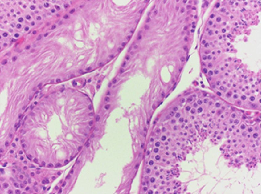
466                                                              GOEDKEN ET AL.                                                 TOXICOLOGIC PATHOLOGY

the specimen would fit into a standard cassette. Most epididy-                    Testicular Microscopic Findings
mal sections contained head, body, and tail. Histologic exami-
                                                                                      Hypospermatogenesis (absence of some or all of the germ
nation was conducted using nomenclature and semiquantitative
                                                                                  cells within individual tubules) was a common finding, occur-
severity grades as previously published (Rehm 2000; Lanning
                                                                                  ring in 20% of all dogs (sixteen of eighty dogs). Of these six-
et al. 2002). This semiquantitative scoring system catego-
                                                                                  teen animals, five dogs (31.3%) had bilateral changes (Table 1).
rized severity as minimal (score 1) for fewer than 5% tubules
                                                                                  Testicular and epididymal changes in control dogs are shown in
affected, slight (score 2) for 5%–25%, moderate (score 3) for
                                                                                  Figures 2–9. Testes with hypospermatogenesis contained ran-
25%–50%, marked (score 4) for 50%–75%, and severe (score
                                                                                  domly scattered groups of one to fifteen tubules with partial to
5) for more than 75% tubules affected. An additional category
                                                                                  complete absence of germ cells. Multifocal absence of sperma-
(score 0) representing absence of the finding was included.
                                                                                  tocytes was observed in six dogs, whereas lower numbers of
The spermatogenic stages of tubules containing findings were
                                                                                  round spermatids were seen in twelve dogs. All sixteen dogs
qualitatively assessed when possible (Creasy 1997; Russell
                                                                                  demonstrated multifocal absence of elongate spermatids
et al. 1990).
                                                                                  (Figures 2 and 3). Hypospermatogenic tubules often contained
    Each testis was evaluated independently, and findings were
                                                                                  small amounts of intraluminal debris, giant cells, and/or swollen
categorized as either bilateral or unilateral. Hypospermatogenesis
                                                                                  spermatocytes. The severity of hypospermatogenesis was mini-
was further characterized according to the germ cell layer (sper-
                                                                                  mal (score 1) in 5% (four of eighty dogs), slight (score 2) in
matocyte, round spermatid, and/or elongate spermatid) that
                                                                                  10% (eight of eighty dogs), moderate (score 3) in 13.8% (eleven
exhibited the greatest cell loss. Assessment of epididymides
                                                                                  of eighty dogs), and marked (score 4) in 3.8% (three of eighty
included a semiquantitative estimate of the percentage of all
                                                                                  dogs) (Table 2). In addition, there was an age distribution to the
ductular lumens filled with sperm and qualitative assessment of
                                                                                  incidence of hypospermatogenesis, which was observed in 75%
abnormal intraluminal cell types, epithelial changes, and inter-
                                                                                  of dogs six to seven months of age, declining in incidence with
stitial changes.
                                                                                  age to less than 10% in dogs over eleven months of age.
                                                                                      Tubules with atrophy/hypoplasia were typically arranged in
Imaging                                                                           well-demarcated, subcapsular, triangular clusters of three to
                                                                                  fifty seminiferous tubules lacking all germ cells, as described
    Slides were examined with an Olympus BX50 light micro-                        by Rehm (2000) (Figures 4 and 5). These clusters were readily
scope, and selected images were captured using a high-resolu-                     observed at low magnification and immediately juxtaposed
tion camera (Qimaging 5.3RTV). Images were captured using                         with normal tubules at the borders of the clusters. The tubules
QCapture Pro 5.0.1.25 software (QImaging, Surrey, BC, Canada)                     in these clusters were lined only by elongate Sertoli cells with
at 2048 x 1536 dpi resolution. Grayscale conversion, brightness/                  round nuclei, were smaller in diameter with smaller tubular
contrast adjustments, and plate production were performed in                      lumina than normal tubules, contained no luminal content, and
Adobe Photoshop 6.0 for Windows (Adobe Systems, Inc., San                         lacked evidence of interstitial inflammation or basement mem-
Jose, CA, 2007).                                                                  brane thickening. Atrophic/hypoplastic tubules were seen in
                                                                                  26.3% of all dogs (twenty-one of eighty dogs); four of these
Statistical Analysis                                                              (19%) exhibited bilateral lesions. The incidence was clearly
                                                                                  affected by age, with 25%–40% of dogs under twelve months
   Combined testes weights and incidences of hypospermato-                        old having this finding, decreasing with age to 14%–17% in
genesis and atrophy/hypoplasia were analyzed independently                        dogs twelve to thirty-six months old. Eleven dogs exhibited
by age group. Data were analyzed using one-way analysis of                        both atrophy/hypoplasia and hypospermatogenesis.
variance followed by Newman-Keul’s multiple range post hoc                            All other histopathological changes were of minimal sever-
test (p < .05). Histopathologic grades were rank-ordered prior                    ity. These changes included retained spermatids, multinucleate
to statistical analysis. Quantitative results were expressed as                   giant cells, swollen spermatocytes, apoptotic cells, intracyto-
means ± standard deviations.                                                      plasmic vacuoles within tubules (presumably within Sertoli
                                                                                  cell), and dilated tubules.
                             RESULTS                                                  Retained spermatids had elongated hyperchromatic nuclei
                                                                                  and were located either basally (in or between Sertoli cells)
Testicular Weight
                                                                                  or apically (adhered to the surface). These retained cells had
   Significantly lower paired testicular weights were seen in                     condensed cytoplasm and were found in later stages of sper-
six- and seven-month-old dogs (8.1 ± 1.2 g) compared to testic-                   matogenesis (six and seven, rarely eight). Spermatid retention
ular weights from dogs aged eight to thirty-six months (range:                    consisting of two to seven spermatids in an average of six tubules
11.0 ± 3.9 g to 14.8 ± 2.3 g) (Figure 1). Testicular weights                      per testis was seen in 47.5% of dogs (thirty-eight of eighty). Of
appeared to plateau at eight months of age, with ranges as follows:               the thirty-eight dogs with retained spermatids, 79% (thirty of
six and seven months (6.5–10.4 g), eight months (10.6–18.3 g),                    eighty) had bilateral findings.
nine months (5.5–16.2 g), ten months (9.3–12.7 g), eleven                             Multinucleated giant cells were seen in 73.8% of dogs
months (9.3–17 g), twelve to twenty-four months (8.3–19.1 g),                     (fifty-nine of eighty) (Figure 6), and 85% of these dogs (fifty
and twenty-four to thirty-six months (11.5–17.7 g).                               of fifty-nine) had a bilateral distribution. In each case, there

                                                 Downloaded from tpx.sagepub.com by guest on October 24, 2015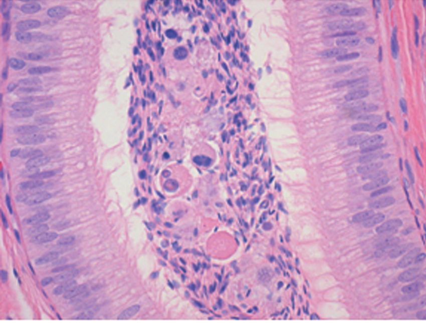
Vol. 36, No. 3, 2008                                       CONTROL BEAGLE TESTICULAR FINDINGS                                                                467

FIGURE 1.—Relationship of control beagle dog age and testes weight.                            FIGURE 3.—Lower numbers of spermatocytes and round spermatids in
Testicular weight was measured in control beagle dogs of varying                               tubule with hypospermatogenesis. H&E, x400.
age (n = 5–23 dogs). Asterisks (*) represent a statistical difference (p
< .05) compared to six- and seven-month-old dogs.

      TABLE 1.—Incidence of spontaneous spermatogenic changes.a                                cells per tubule, and there were typically fewer than ten affected
                                                                                               tubules per testis (Figure 7). Swollen cells had abundant pale
Pattern                      No. affected dogs      Total no. dogs       Percentage
                                                                                               cytoplasm and marginated nuclei. Some swollen spermatocytes
Atrophy / hypoplasia                 21                  80                   26.3             had condensed chromatin (pyknosis).
Unilateral                           17                  21                                       Apoptotic germ cells within the seminiferous epithelium
Bilateral                             4                  21                                    were found in 10% of dogs (eight of eighty), with no apparent
Hypospermatogenesis                  16                  80                   20.0
                                                                                               age correlation. Of the eight dogs with apoptotic cells, only
Unilateral                           11                  16
Bilateral                             5                  16                                    two animals had this lesion bilaterally.
                                                                                                  Rare intracytoplasmic vacuoles, presumed to be in Sertoli
  a
   Eleven dogs exhibited both patterns of change.                                              cells based on cellular morphology, were seen in 55% of dogs
                                                                                               (forty-four of eighty).
                                                                                                  Five percent (four of eighty) of dogs had dilated seminifer-
                                                                                               ous tubules. Some dilated tubules in three of the four affected
                                                                                               dogs contained aggregates of desquamated immature intralu-
                                                                                               minal germinal cells.
                                                                                                  One dog had unilateral subacute coagulative necrosis of one
                                                                                               fourth of the parenchyma, suggesting a spontaneous infarct or
                                                                                               focal trauma, although the inciting cause was not determined.

                                                                                               Epididymal Changes
                                                                                                   Epididymal ducts from most dogs contained sperm admixed
                                                                                               with rare multinucleate giant cells and a few immature sper-
                                                                                               matids. Older dogs (eight to thirty months) had between 40 and
                                                                                               90% of all epididymal duct profiles filled with sperm with con-
                                                                                               sistent filling of the tails. Dogs from six to seven months of age
FIGURE 2.—Hypospermatogenesis with decreased numbers of sperma-                                had 0%–10% of epididymal duct profiles filled with a mixture of
tocytes and spermatids. H&E, x100.                                                             sperm, round spermatids, spermatocytes, multinucleated giant
                                                                                               cells, and cell debris (Figure 8). Most dogs six to seven months
were from one to six multinucleate cells per affected tubule with                              of age had little or no sperm in their epididymal tails, and ducts
fewer than ten affected tubules per testis and no predilection for                             of the epididymal head and body were incompletely expanded
specific stages.                                                                               with sperm. Interestingly, incomplete filling of tubules in the
   Tubules with swollen spermatocytes were observed in seventy-                                head and body also was seen in three of the nine-month-old dogs.
four of eighty dogs (93%), and in most cases were observed                                     Filling of the tail of the epididymis with normal sperm was deter-
bilaterally (fifty-two of seventy-four). These swollen spermato-                               mined to be the most appropriate epididymal finding for evalu-
cytes were found singly or in loose aggregates of two to eight                                 ating maturity. One to four small (less than one tubule diameter),

                                                              Downloaded from tpx.sagepub.com by guest on October 24, 2015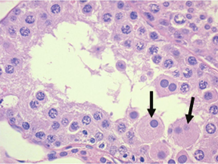
468 GOEDKEN ET AL. TOXICOLOGIC PATHOLOGY
TABLE 2.—Incidence of hypospermatogenesis and atrophy/hypoplasia by age group.
No. with Total Percentage
Age (mo.) n Finding Grade 0 Grade 1 Grade 2 Grade 3 Grade 4 both incidence incidence
6–7 8 Hypospermatogenesis 2 0 2 1 3 2 6/8 75
Atrophy/hypoplasia 5 0 1 1 1 3/8 37.5
8 5 Hypospermatogenesis 3 0 1 1 0 2 2/5 40
Atrophy/hypoplasia 3 0 1 1 0 2/5 40
9 15 Hypospermatogenesis 12 0 2 1 0 3 3/15 20a
Atrophy/hypoplasia 10 1 2 2 0 5/15 33.3
10 8 Hypospermatogenesis 5 1 1 1 0 2 3/8 37.5
Atrophy/hypoplasia 6 0 1 1 0 2/8 25
11 14 Hypospermatogenesis 14 0 0 0 0 0 0/14 0a
Atrophy/hypoplasia 10 1 1 2 0 4/14 29
12–23 23 Hypospermatogenesis 21 0 0 2 0 2 2/23 8.7a
Atrophy/hypoplasia 19 0 1 3 0 4/23 17.3
24–36 7 Hypospermatogenesis 7 0 0 0 0 0 0/7 0a
Atrophy/hypoplasia 6 1 0 0 0 1/7 14.3
Note: There were no statistical differences among dogs with atrophy/hypoplasia. There were no statistical differences comparing dogs with atrophy/hypoplasia and hypospermatogenesis
at any age.
a
Statistically different from six- to seven-month-old dogs with hypospermatogenesis.
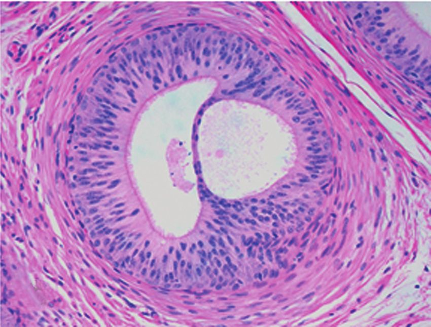
FIGURE 4.—Atrophy/hypoplasia demonstrated smaller, well-demarcated FIGURE 6.—Multinucleated giant cell composed of round spermatids.
tubules immediately adjacent to normal tubules. H&E, x100. H&E, x200.
FIGURE 5.—Atrophy/hypoplasia. Tubules contain only columnar Sertoli FIGURE 7.—Swollen spermatocytes (arrow). H&E, x400.
cells. H&E, x200.
Downloaded from tpx.sagepub.com by guest on October 24, 2015Vol. 36, No. 3, 2008 CONTROL BEAGLE TESTICULAR FINDINGS 469
unilateral, interstitial lymphoid aggregates were present in four
dogs. In four other dogs, the epididymal epithelium bilaterally
contained rare small intracytoplasmic vacuoles. Lastly, two dogs
had bilateral cribriform change characterized by 50–200 μm
intraepithelial spaces rimmed by compressed columnar cells,
primarily in the tail of the epididymis (Figure 9).
DISCUSSION
Focal hypospermatogenesis and/or tubular atrophy/hypoplasia
occurred in all age groups, but they were more common in
younger animals. Eighty-seven percent of dogs six to seven
months old and 26.4% of dogs eight months through three years
old had either hypospermatogenesis and/or atrophy/hypoplasia.
Although the interactions of Sertoli cells and germ cells in
normal and abnormal spermatogenesis are not completely FIGURE 8.—Cell debris, spermatocytes, and mature sperm in epididymal
characterized, recent information from rodents and nonhuman lumen. H&E, x200.
primates suggests that interaction between Sertoli cells and
germinal cells is bidirectional. Immature Sertoli cells are iden-
tified by positive staining for anti-Müllerian hormone, neural
cell adhesion molecule, and cytokeratin 18, whereas mature
Sertoli cells are labeled with inhibin B (Rajpert-De Meyts et al.
1999; Rey et al. 1994; Rey et al. 1999; Stosiek et al. 1990).
Positive staining for immature Sertoli cell markers is found in
tubules with hypospermatogenesis, suggesting that incomplete
maturation of Sertoli cells is associated with partial or complete
absence of germ cells (Cortes et al. 1987; Mann et al. 1997;
Marshall and Plant 1996; Sharpe et al. 2003; Sharpe et al. 2000;
Wang et al. 1989). On the other hand, it is known that germ cell
loss triggers mature Sertoli cells to develop characteristics of
immature cells (Foley 2001; Sharpe et al. 2003; Sharpe et al.
1993; Steger et al. 1999). If future studies in beagle dogs are
consistent with information from rodents and primates, the
terms atrophy/hypoplasia and hypospermatogenesis may not
adequately reflect the mechanisms of action responsible for FIGURE 9.—Epididymal cribriform change. Intraepithelial lumens often
are partially lined by ciliated cells and may have eosinophilic content.
these findings. Abnormal tubules with similar appearances could
H&E, x400.
develop through multiple mechanisms, and a single mechanism
could produce morphologic findings compatible with both
hypospermatogenesis and atrophy/hypoplasia. More descrip- result from prior testicular toxicity of two to fourteen days in
tive terminology, such as “Sertoli-cell-only tubules” for atrophy/ duration (Abbott 1993; Foley 2001). In our study, six- to seven-
hypoplasia and “missing germ cells” for hypospermatogenesis, month-old dogs consistently had a much lower percentage of the
may be more appropriate. overall epididymal lumen filled with spermatozoa (0%–10%)
Multinucleate giant cells, swollen germ cells, vacuolation compared with animals at least eight months of age (40%–90%),
of Sertoli cells, and retained spermatids of mild severity were and the epididymal tails of most six- to seven-month-old dogs
common findings in the testes of normal beagles. Apoptosis and contained little or no sperm compared to older dogs. Filling of
intraluminal cell debris were relatively uncommon, and when the tail of the epididymides with normal sperm was one indica-
present they were quite mild in severity. Multinucleate giant tor of sexual maturity. Dogs six to seven months of age had
cells are a common finding in clinically normal male dogs, rats, proportionately more epididymal intraluminal eosinophilic debris,
rabbits, and humans, but they also may be linked to starvation, intraluminal spermatocytes, multinucleate giant cells, and degen-
thermal stress, surgical procedures, local injury, and toxicity erate immature spermatids than observed in the older dogs.
(Russell et al. 1990). Swollen and degenerate spermatocytes These findings correlated well with lower testicular weights and
near the basal cell layer of seminiferous tubules were common the increased incidence of hypospermatogenesis in the younger
spontaneous findings that may be confused with toxicity. group. Although there was at least four-fold greater epididymal
The major epididymal change was high variability in filling filling in older dogs (eight to thirty-six months), the high vari-
of the epididymal tail with sperm. Reduced luminal filling with ability (40%–90%) may have been partly a result of artifactual
increased numbers of immature germ cells and cell debris may loss of sperm during processing. Epithelial cell cytoplasmic vac-
be seen in healthy, peripubertal dogs, but such findings may also uoles, cribriform change, and interstitial mononuclear infiltrates
Downloaded from tpx.sagepub.com by guest on October 24, 2015470 GOEDKEN ET AL. TOXICOLOGIC PATHOLOGY
were considered spontaneous findings. The cause of spontaneous months of age will reduce the incidence of spontaneous back-
interstitial lymphocytic aggregates is unknown and may be diffi- ground findings that may be confused with toxicity.
cult to distinguish from a drug effect if there was increased inci- Every study must be evaluated on a case-by-case basis to dis-
dence or severity in treated animals compared with controls. tinguish primary treatment-related effects, effects secondary to
However, small testicular or epididymal lymphocytic aggregates nonspecific stress or morbidity, and spontaneous findings. In
in the absence of other epididymal findings likely would be of some cases, a finding cannot be easily classified into a specific
negligible toxicological significance. category. Testicular and/or epididymal findings in one or more
Testicular histology and testicular weights are routinely treated dogs are more likely to be spontaneous and incidental if
used to detect drug toxicity in male reproductive organs the findings: are known to occur as spontaneous findings; are
(Ulbrich and Palmer 1995). Knowledge of the timing of sexual unilateral rather than bilateral; occur in tubules intermixed with
maturation is important to the design of preclinical toxicity many normal tubules of the same stage; occur in the absence of
studies, since sexually immature beagle dogs exhibit histologic other clearly treatment-related findings in the reproductive tract;
findings similar to drug toxicity end points such as low testic- are found in control animals as well as treated animals; demon-
ular weight, hypospermatogenesis, multinucleate giant cells, strate no dose response in incidence or severity; and/or are found
reduced epididymal filling, intraluminal cell debris, and inconsistently in a minority of the treated animals. When a dose
swollen, retained, and apoptotic germ cells (James and response exists in incidence and/or severity of a finding that has
Heywood 1979). Sexual maturity in male beagle dogs has been been reported to occur spontaneously, the pathologist must weigh
estimated to occur between thirty-five and forty-one weeks of knowledge of spontaneous findings, mechanism(s) of intended
age based on hormone levels, sperm analysis, histology, and and secondary actions of the test article, and all other study data
testes weight (James et al. 1979; James and Heywood 1979; (gross and microscopic findings, testicular and body weights,
Kawakami et al. 1991). food consumption, etc.) with a thorough examination of treated
Our data indicated that 75% of six- and seven-month-old and control animal tissues to determine if a treatment-related
dogs had microscopically evident hypospermatogenesis which effect exists and is toxicologically meaningful.
sharply declined in incidence after eleven months of age. In summary, hypospermatogenesis and atrophy/hypoplasia
Similarly, 40% of six- to seven-month-old dogs had focal in seminiferous tubules are more common in young dogs than
atrophy/hypoplasia, declining to about half this level in dogs in older dogs. Dogs less than eight months of age have high
older than twelve months of age. Interestingly, the testes weight incidences of hypospermatogenesis, lower testicular weights,
range for nine-month-old dogs was quite variable, ranging from and incomplete filling of epididymal tails with sperm, all com-
5.5 to 16.2 g (Table 1). The three dogs (20% of nine-month-old patible with immaturity. If evaluation of spermatogenesis is a
dogs) that had microscopic findings of both hypospermatogen- critical end point in a toxicity study, males should be necrop-
esis and atrophy/hypoplasia accounted for the lowest testes sied at ages no younger than ten months so that low testicular
weights (5.5, 6.6, and 7.6 g) in this age group. The remaining weights and microscopic findings found commonly in immature
dogs that did not exhibit both lesions had a testes weight range dogs do not complicate study interpretation.
of 8 to 16.2 g (mean 12.1 ± 0.6 g), which is similar to the
remaining age groups. These findings, along with evidence of
REFERENCES
reduced testis weights and microscopically reduced epididymal
sperm content in the six- to seven-month age group compared Abbott, D. P. (1993). Cribriform intra-tubular epididymal change and testicular
with older dogs, are in line with data reporting that the greatest atrophy. Histopathology 23, 293.
Cortes, D., Muller, J., and Skakkebaek, N. E. (1987). Proliferation of Sertoli
testicular development and maturation in dogs occurs between cells during development of the human testis assessed by stereological
five and eight months of age (James and Heywood 1979; methods. Int J Androl 10, 589–96.
Kawakami et al. 1991). Creasy, D. M. (1997). Evaluation of testicular toxicity in safety evaluation
The age-related differences in the incidences of atrophy/ studies: the appropriate use of spermatogenic staging. Toxicol Pathol
hypoplasia vary from a previous report, perhaps because our 25, 119–31.
Foley, G. L. (2001). Overview of male reproductive pathology. Toxicol Pathol
study included younger and older dogs than the previous study 29, 49–63.
(Rehm 2000). Taken together, more frequent hypospermatoge- Hottendorf, G. H., and Hirth, R. S. (1974). Lesions of spontaneous subclinical
nesis, reduced testicular weights, and reduced epididymal filling disease in Beagle dogs. Vet Pathol 11, 240–58.
with sperm in six- and seven-month-old dogs suggested that James, R. W., Crook, D., and Heywood, R. (1979). Canine pituitary-testicular
histologic sexual maturity occurred in most dogs by eight function in relation to toxicity testing. Toxicology 13, 237–47.
James, R. W., and Heywood, R. (1979). Age-related variations in the testes and
months of age. Occasional dogs nine months of age may still prostate of beagle dogs. Toxicology 12, 273–79.
have features of immaturity, including decreased testes weights Kawakami, E., Tsutsui, T., and Ogasa, A. (1991). Histological observations of the
and decreased filling of the epididymal lumen with sperm. Our reproductive organs of the male dog from birth to sexual maturity. J Vet
results support previously published recommendations that Med Sci 53, 241–48.
male beagles should be at least ten months of age at necropsy Lanning, L. L., Creasy, D. M., Chapin, R. E., Mann, P. C., Barlow, N. J., Regan, K.
S., and Goodman, D. G. (2002). Recommended approaches for the evalua-
for routine microscopic evaluation of the mature testis (Lanning tion of testicular and epididymal toxicity. Toxicol Pathol 30, 507–20.
et al. 2002). If critical evaluation of spermatogenesis and adult Mann, D. R., Akinbami, M. A., Wallen, K., Gould, K. G., Groome, N. P.,
reproductive organs is imperative, use of dogs at least twelve Swanston, I., McNeilly, A. S., and Fraser, H. M. (1997). Inhibin-B in the
Downloaded from tpx.sagepub.com by guest on October 24, 2015Vol. 36, No. 3, 2008 CONTROL BEAGLE TESTICULAR FINDINGS 471
male rhesus monkey: impact of neonatal gonadotropin-releasing hormone Sharpe, R. M., McKinnell, C., Kivlin, C., and Fisher, J. S. (2003). Proliferation
antagonist treatment and sexual development. J Clin Endocrinol Metab and functional maturation of Sertoli cells, and their relevance to disorders
82, 1928–33. of testis function in adulthood. Reproduction 125, 769–84.
Marshall, G. R., and Plant, T. M. (1996). Puberty occurring either sponta- Sharpe, R. M., Millar, M., and McKinnell, C. (1993). Relative roles of testosterone
neously or induced precociously in rhesus monkey (Macaca mulatta) and the germ cell complement in determining stage-dependent changes in
is associated with a marked proliferation of Sertoli cells. Biol Reprod protein secretion by isolated rat seminiferous tubules. Int J Androl 16, 71–81.
54, 1192–99. Sharpe, R. M., Walker, M., Millar, M. R., Atanassova, N., Morris, K.,
Rajpert-De Meyts, E., Jorgensen, N., Graem, N., Muller, J., Cate, R. L., and McKinnell, C., Saunders, P. T., and Fraser, H. M. (2000). Effect of neona-
Skakkebaek, N. E. (1999). Expression of anti-Mullerian hormone during tal gonadotropin-releasing hormone antagonist administration on sertoli
normal and pathological gonadal development: association with differenti- cell number and testicular development in the marmoset: comparison
ation of Sertoli and granulosa cells. J Clin Endocrinol Metab 84, 3836–44. with the rat. Biol Reprod 62, 1685–93.
Rehm, S. (2000). Spontaneous testicular lesions in purpose-bred beagle dogs. Steger, K., Rey, R., Louis, F., Kliesch, S., Behre, H. M., Nieschlag, E.,
Toxicol Pathol 28, 782–87. Hoepffner, W., Bailey, D., Marks, A., and Bergmann, M. (1999). Reversion
Rey, R., Mebarki, F., Forest, M. G., Mowszowicz, I., Cate, R. L., Morel, Y., of the differentiated phenotype and maturation block in Sertoli cells in
Chaussain, J. L., and Josso, N. (1994). Anti-mullerian hormone in children pathological human testis. Hum Reprod 14, 136–43.
with androgen insensitivity. J Clin Endocrinol Metab 79, 960–64. Stosiek, P., Kasper, M., and Karsten, U. (1990). Expression of cytokeratins 8
Rey, R. A., Belville, C., Nihoul-Fekete, C., Michel-Calemard, L., Forest, M. and 18 in human Sertoli cells of immature and atrophic seminiferous
G., Lahlou, N., Jaubert, F., Mowszowicz, I., David, M., Saka, N., tubules. Differentiation 43, 66–70.
Bouvattier, C., Bertrand, A. M., Lecointre, C., Soskin, S., Cabrol, S., Takayama, S., Akaike, M., Kawashima, K., Takahashi, M., and Kurokawa, Y.
Crosnier, H., Leger, J., Lortat-Jacob, S., Nicolino, M., Rabl, W., Toledo, S. P., (1995). A collaborative study in Japan on optimal treatment period and
Bas, F., Gompel, A., Czernichow, P., Josso, N., et al. (1999). Evaluation parameters for detection of male fertility disorders in rats induced by
of gonadal function in 107 intersex patients by means of serum antimul- medical drugs. J Amer Coll Toxicol 14, 266–92.
lerian hormone measurement. J Clin Endocrinol Metab 84, 627–31. Ulbrich, B., and Palmer, A. K. (1995). Detection of effects on male reproduction—
Russell, L. D., Ettlin, R. A., Sinha Hikim, A. P., and Clegg, E. D. (1990). a literature survey. J Amer Coll Toxicol 14, 293–327.
Mammalian Spermatogenesis. In Histological and Histopathological Wang, Z. X., Wreford, N. G., and De Kretser, D. M. (1989). Determination of
Evaluation of the Testis (L. D. Russell, R. A. Ettlin, A. P. Sinha Hikim, Sertoli cell numbers in the developing rat testis by stereological methods.
and E. D. Clegg, eds.), pp. 1–40, Cache River Press, Clearwater, FL. Int J Androl 12, 58–64.
Downloaded from tpx.sagepub.com by guest on October 24, 2015You can also read


