AN IMMUNOHISTOCHEMICAL STUDY ON THE PRESENCE OF NITRIC OXIDE SYNTHASE ISOFORMS (NNOS, INOS, ENOS) IN THE SPINAL CORD AND NODOSE GANGLION OF RATS ...
←
→
Page content transcription
If your browser does not render page correctly, please read the page content below
J Vet Res 64, 445-453, 2020
DOI:10.2478/jvetres-2020-0059
An immunohistochemical study on the presence
of nitric oxide synthase isoforms (nNOS, iNOS, eNOS)
in the spinal cord and nodose ganglion
of rats receiving ionising gamma radiation
to their liver
Osman Yılmaz1, Zafer Soygüder1, Ömer Faruk Keleş2, Turan Yaman2,
Zabit Yener2, Ahmet Uyar3, Tahir Çakır4
1Department of Anatomy, 2Department of Pathology, Faculty of Veterinary Medicine,
University of Van Yüzüncü Yıl, 65080, Van, Turkey
3Department of Pathology, Faculty of Veterinary Medicine, University of Hatay Mustafa Kemal, 31040, Hatay, Turkey
4Department of Medical Physics, Faculty of Medicine, University of Van Yüzüncü Yıl, 65080, Van, Turkey
osman_40_5@hotmail.com
Received: March 6, 2020 Accepted: August 25, 2020
Abstract
Introduction: This study determined the presence of nitric oxide synthesis isoforms (nNOS, iNOS, and eNOS) in thoracic
spinal cord segments and nodose ganglia of rats with gamma-irradiated livers. Material and Methods: Male rats (n = 32) were
divided into equal groups A, B, C, and D. In group A, the controls, no radiation was applied, while groups B, C, and D received
10 Gy of ionising gamma radiation. The rats of group B were euthanized at the end of the first day (d1), those of group C on the
second day (d2), and those of group D on the third day (d3). The liver, spinal cord segments, and nodose ganglion tissues were
dissected and fixed, and the liver sections were examined histopathologically. The other tissues were observed through a light
microscope. Results: Regeneration occurred at the end of d3 in hepatocytes which were radiation-damaged at the end of d1 and
d2. On d1, some nNOS-positive staining was found in the neuronal cells of laminae I–III of the spinal cord and in neurons of the
nodose ganglion, and on d3, some staining was observed in lamina X of the spinal cord, while none of note was in the nodose
ganglion. Dense iNOS-positive staining was seen on d1 in the ependymal cells of the spinal cord and in the glial cells of the
nodose ganglion, and on d3, there was still considerable iNOS staining in both tissues. There was clear eNOS-positive staining in
the capillary endothelial cells of the spinal cord and light diffuse cytoplasmic staining in the neurons of the nodose ganglion on
d1, and on d3, intense eNOS-positive staining was visible in several endothelial cells of the spinal cord, while light nuclear
staining was recognised in the neurons of the nodose ganglion. Conclusion: The nNOS, iNOS, and eNOS isoforms are activated
in the spinal cord and nodose ganglion of rats after ionising radiation insult to the liver.
Keywords: rats, ionising radiation, nitric oxide synthesis isoforms, nodose ganglion, spinal cord.
Introduction The production and detoxification of free radicals are
controlled by a very delicate balance in the body. The
Ionising radiation is defined as high-frequency and organism is not affected as long as the equilibrium is
high-energy X-rays and gamma rays that produce not disturbed between the rates of formation and
changes by removing electrons from atoms and elimination of these molecules. When this balance is
molecules in organs, tissues, and cells (14). The basic disturbed – if the oxidants are increased or the
effect of the ionising radiation is that it causes cell antioxidants are insufficient – the organism is exposed
death due to damage of DNA (15, 32). However, with to oxidative stress. In an ongoing imbalanced
the application of ionising radiation, another effect is condition, the functioning of the cellular metabolism is
that free radicals are formed that are necessary for life. impaired, and cellular aging, inflammatory damage,
© 2020 O. Yılmaz et al. This is an open access article distributed under the Creative Commons Attribution-
NonCommercial-NoDerivs license (http://creativecommons.org/licenses/by-nc-nd/3.0/)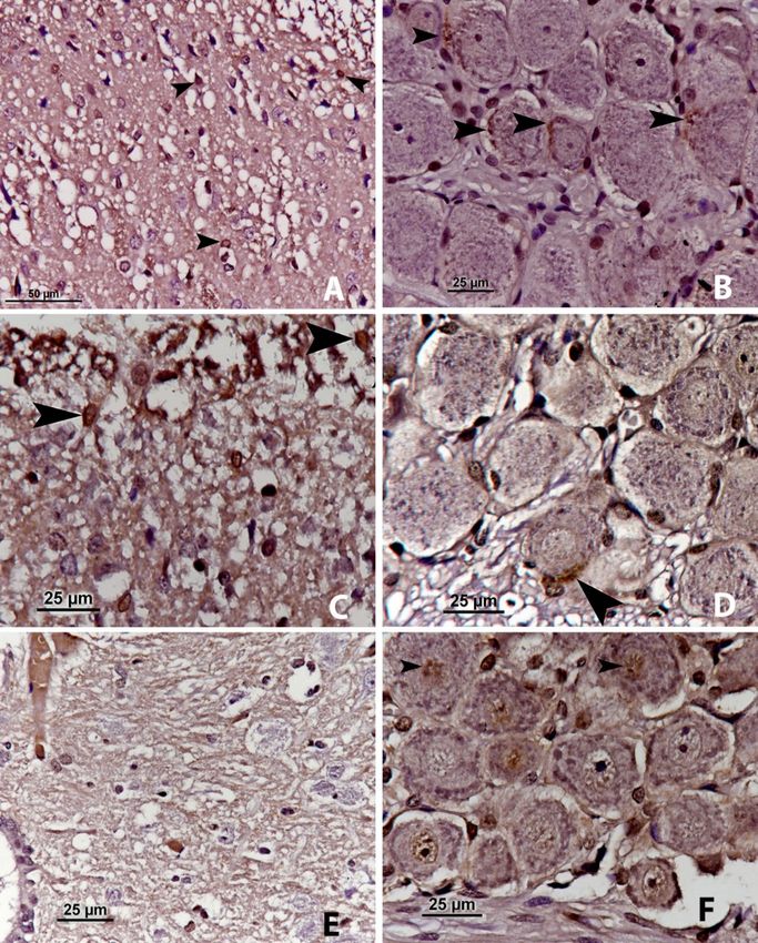
446                                      O. Yılmaz et al./J Vet Res/64 (2020) 445-453

and tissue damage ensue in many vital organs with                   ganglion of rats exposed to ionising gamma radiation of
resulting molecular destruction, and pain is the                    the liver.
consequent response (13, 23).
      Nitric oxide (NO) is a gas derived from the amino
acid L-arginine by the action of the enzyme nitric                  Material and Methods
oxidase. NO is a very reactive molecule, and therefore
has a very short half-life. It is an unusual inhibitory                   Experimental animals. A total of 32 rats aged
neurotransmitter because it does not bind to membrane               4–5 weeks, with a body weight of 190–210 g were used
receptors. NO is a stimulating molecule produced by                 in the study. The animals were housed at a standard
endothelial cells and neurons in the brain. It may have             temperature (22 ± 1°C), and in a 12-h light–dark cycle
a function in memory creation, because it is located in             environment. They were given drinking water and
the hippocampal formation. In addition, NO is also                  standard pelleted rat food ad libitum. At the end of the
found in the olfactory system, cerebellum, striatum,                three-day period of the experiment, the study
cerebral cortex, hypothalamus, mesencephalon, and                   was terminated. Then, the rats were anaesthetised
spinal cord (24, 37) and may have a role in the                     with a combination of ketamine (50 mg/kg, intra
development of ependymal cells (29). NO plays a role                peritoneally) and xylazine (10 mg/kg, intra
in epileptic activity, nociception (as an important                 peritoneally) and euthanised. Thoracic spinal cord
determinant (27)), learning and memory, anxiety,                    segments (th 7–13) and the nodose ganglia were
depression, stress, eating and drinking behaviour,                  removed and fixed by immersion for 2 h in 10%
sexual behaviour, neuroendocrine functions, the                     buffered formalin.
modulation of neurotransmitter release, the continuity                    Exposure to radiation. The rats were randomly
of the blood–brain barrier, the cardiac rhythm, the                 classified into four groups of eight per group. The
regulation of the sleep–wake cycle, apoptosis,                      radiation groups (B, C, and D) received 10 Gy of
differentiation, development, and synaptic plasticity. It           ionising gamma radiation. The irradiations were
is also responsible for the neurotoxicity observed in               performed using a cobalt-60 teletherapy device. The
diseases such as Alzheimer’s, Huntington’s, and                     beam concentration was placed at the appropriate depth
cerebral ischaemia (4).                                             for the liver region in the ionising radiation groups. The
      It is well known that NO is produced by isoforms              scheme was as follows:
of nitric oxide synthase (neuronal NOS, inducible                         1) Group A: The control group, to which no
NOS, endothelial NOS, and bacterial NOS). In general,               ionising gamma radiation was applied;
nNOS is a neuronal and essential form, iNOS is a form                     2) Group B: 10 Gray (Gy) of ionising gamma
that is distributed throughout the body after suitable              radiation was applied and the experiment was
stimulation or induction, and eNOS is found                         terminated on the first day;
in the vascular endothelium (6) located in endothelial                    3) Group C: 10 Gy of ionising gamma radiation
cells (2). Recently, bNOS activity has been determined              was applied and the experiment was terminated on the
in several bacterial species, including notorious                   second day;
pathogens such as Staphylococcus aureus and Bacillus                      4) Group D: 10 Gy of ionising gamma radiation
anthracis (12).                                                     was applied and the experiment was terminated on the
      The liver is associated with all of the stimulation           third day.
in the body, both humoral and neural. The brain is                        Light microscopic examination. At the end of
potentially stimulated to respond to the sensory                    the dissection and fixation, tissue samples (liver,
information associated with the metabolic, immune,                  thoracic spinal cord segments (th 7–13), and the nodose
and other functions of the liver (5, 11, 22). It can alter          ganglia) were embedded in paraffin blocks and serial
these functions through hormonal and neural pathways                sections of 4 μm were taken with a microtome and
(31). The neural relationship between the liver and the             prepared on slides. All the sections were stained with
central nervous system is both vagal sensory                        haematoxylin-eosin. Then, the liver sections were
(parasympathetic) and spinal sensory (sympathetic).                 examined histopathologically by light microscopy
Most of the stimuli transmitted by the vagus nerve are              (Nikon Eclipse 80i-DS-RI2, Japan). In the next phase,
delivered to the nucleus of the solitary tract via the left         immunohistochemical staining for nNOS, iNOS, and
nodose ganglion and a small part via the right nodose               eNOS was performed in the remaining tissue sections
ganglion. Spinal sensory stimuli are transmitted                    using the immunoperoxidase method. Commercial
through spinal nerves 7–13 to the thoracic spinal cord.             antibodies were visualised on 4-μm-thick paraffin
Vagal sensory nerve cell bodies are located in the                  sections using an indirect streptavidin/biotin
nodose ganglion (NG), and spinal sensory nerve cells                immunoperoxidase kit (Histostain Plus Bulk Kit,
are found in the dorsal root ganglion (DRG) (5).                    Zymed, USA). The thoracic spinal cord segments
      In this study, we aimed to investigate the                    (th 7–13) and nodose ganglion sections were incubated
NOS isoforms (nNOS, eNOS, and iNOS)                                 with primary antibodies (nNOS, ab1376; iNOS,
immunohistochemically in the spinal cord and nodose                 ab15323; eNOS, ab76198; all at 1/100 dilution; all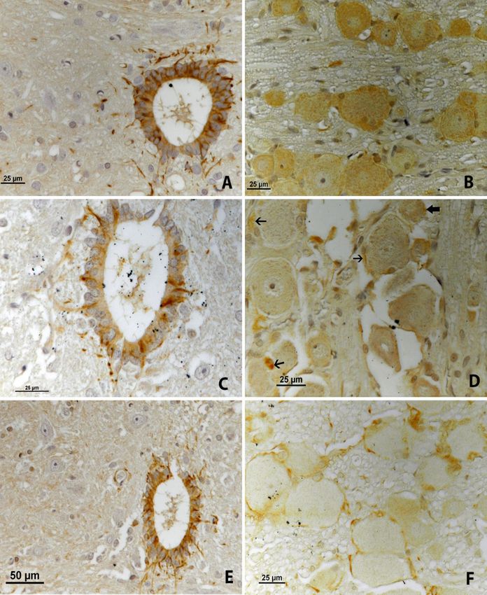
O. Yılmaz et al./J Vet Res/64 (2020) 445-453 447
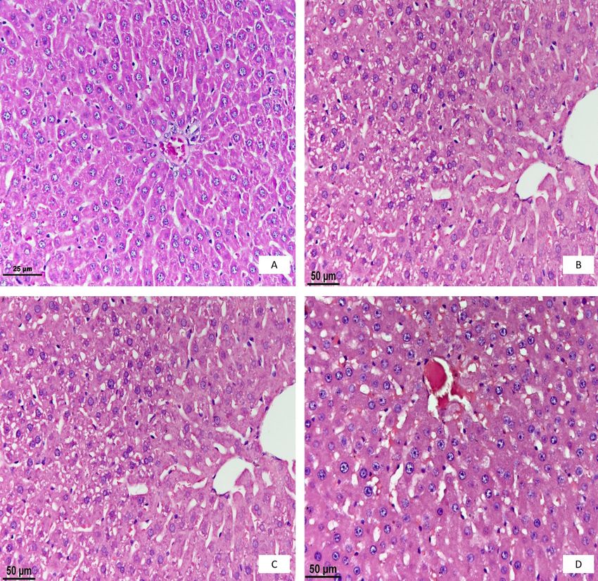
Abcam, UK) overnight at 4°C. Finally, to visualise the On the second day, it was found that these changes
reactions, the sections were reacted for 5–15 min with had also taken place in the nuclei of the hepatocytes in
diaminobenzidine. The sections were observed and all regions of the liver lobules, and in addition, partial
photographed through the light microscope. shrinkage had occurred in these nuclei, and hydropic
degeneration was seen in their cytoplasm (Fig. 1C).
On the third day, the second-day changes were
Results less prominent in two cases, but in the remaining cases
the nucleus and cytoplasm of hepatocytes had a normal
Histopathological examination. In the control appearance (Fig. 1D). This shows that the regeneration
group, a normal histological appearance of the liver of hepatocytes occurred on the third and later days after
was determined (Fig. 1A). On the first day after the rats’ irradiation.
irradiation, the vesicular structure in the nuclei of Immunohistochemical examination. The
hepatocytes was replaced by a grey homogenous mass. presence and distribution of all forms of NOS were
Furthermore, it was noted that the nuclei of hepatocytes investigated in the spinal cord and nodose ganglion by
were smaller and marginally hyperchromatic, while the immunohistochemical staining on the first, second, and
cytoplasm was paler (Fig. 1B). third days. Rexed lamination was used to identify the
locations of NOS-positive cells in the spinal cord (25).
Fig. 1. Histopathological examination of the liver. A – the control group; B – the first day; C – the second day; D – the third day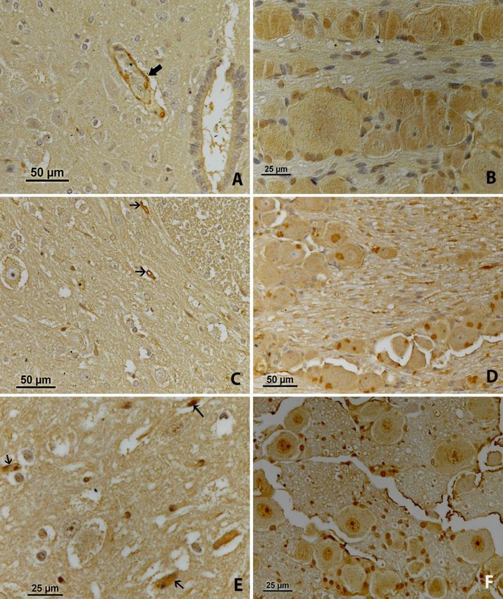
448                                             O. Yılmaz et al./J Vet Res/64 (2020) 445-453

     n-NOS immunoreactivity. On the first day,                              nNOS in nodose ganglia also decreased and was seen
nNOS-positive cells were observed in the superficial                        perineurally in one cell (Fig. 2D, arrow).
lamina (lamina I–II) of the spinal cord of the rats which                        On the third day, a few nNOS-positive cells were
received ionising radiation to the liver. These cells                       observed in lamina X of the spinal cord with no
were also found to a lesser extent in lamina III–IV                         remarkable staining in the nodose ganglia (Fig. 2E, F),
(Fig. 2A, arrows). In the nodose ganglia, nNOS                              and nNOS-positivity was detected very mildly in the
immunoreactivity was seen as mild perineural staining                       nucleus of the neurons in the nodose ganglia (Fig. 2F).
in several cells (Fig. 2B, arrows).                                         The immunoreactivity of nNOS, which was present in
     On the second day, it was seen that nNOS                               both tissues on the first day, lessened on the second
immunoreactivity decreased in lamina I–II to one or                         day, and disappeared on the third day.
two cells (Fig. 2C, arrows). The immunoreactivity of

Fig. 2. nNOS immunoreactivity. A – the first day, in the spinal cord; B – the first day, in the nodose ganglion; C – the second day, in the spinal
cord; D – the second day, in the nodose ganglion; E – the third day, in the spinal cord; F – the third day, in the nodose ganglion
O. Yılmaz et al./J Vet Res/64 (2020) 445-453 449
iNOS immunoreactivity. On the first day, (Fig. 3D, thick arrows) in the cytoplasm of neurons,
intensive iNOS immunoreactivity was observed in the and more prominently in some neuroglial cells
ependymal cells around the central canal of the spinal (Fig. 3D, arrows).
cord. In addition, lamina X had intense, long axonal On the third day, the immunoreactivity of iNOS
staining (Fig. 3A). In the nodose ganglia, iNOS was determined to be less intense than on the second
staining was observed commonly as fine granules in the day in the ependymal cells of the central canal of the
cytoplasm of neurons (Fig. 3B). spinal cord. The axonal staining in lamina X was
On the second day, the immunoreactivity of iNOS weaker than on the second day (Fig. 3E). This three-
was evident with less intensity than on the first day in day pattern in the spinal cord shows a gradual reduction
the ependymal cells of the central canal of the spinal in iNOS-expression. In the nodose ganglia, iNOS
cord. Also, lamina X had weaker and shorter axonal staining was noticed as thin granules in the periphery of
staining than on the first day (Fig. 3C). In the nodose the cytoplasm of neurons, and in the neuroglial cells
ganglia, iNOS staining was seen as thin granules (Fig. 3F).
Fig. 3. iNOS immunoreactivity. A – the first day, in the spinal cord; B – the first day, in the nodose ganglion; C – the second day, in the spinal
cord; D – the second day, in the nodose ganglion; E – the third day, in the spinal cord; F – the third day, in the nodose ganglion450 O. Yılmaz et al./J Vet Res/64 (2020) 445-453
e-NOS immunoreactivity. On the first day, NOS- diffuse cytoplasmic and intense local staining were
positive staining was found in the capillary endothelial determined in the neurons (Fig. 4D).
cells of the spinal cord (Fig. 4A, arrow). In the nodose On the third day, intense eNOS-positive staining
ganglia, light diffuse cytoplasmic staining was was visible in several endothelial cells of the spinal
observed in the neurons (Fig. 4B). cord (Fig. 4E, arrows). A gradual slight increase in
On the second day, light eNOS-positive staining eNOS expression in the spinal cord was seen. However,
was observed in one or two endothelial cells of the light nuclear staining was observed in the neurons of
spinal cord (Fig. 4C, arrows). In the nodose ganglion, the nodose ganglia (Fig. 4F).
Fig. 4. eNOS immunoreactivity. A – the first day, in the spinal cord; B – the first day, in the nodose ganglion; C – the second day, in the spinal
cord; D – the second day, in the nodose ganglion; E – the third day, in the spinal cord; F – the third day, in the nodose ganglionO. Yılmaz et al./J Vet Res/64 (2020) 445-453 451
Discussion these laminae, it suggests that the presence of nNOS in
the spinal cord can be interpreted as being provided
The basic mechanism of action of ionising through the sensory pathways. We believe that
radiation results in tissue and various levels of cell a definitive elucidation could be made with calcitonin
damage in many vital organs, such as the liver, and gene-related peptide (CGRP)-nNOS double staining.
nociceptive pain may be experienced (13, 23). NO is Indeed, c-Fos-nNOS positive double-labelled cells in
an important neurotransmitter substance in nociceptive these laminae were detected as a result of noxious
events of the body (27). The sensory hepatic nerves of stimuli (28). In the nodose ganglion, nNOS
the liver – the vagal and spinal afferent innervation of immunoreactivity is seen as mild perineural staining in
the liver – are well known anatomically, which guides several cells. The presence of nNOS in both the spinal
the investigation of the effects of damage to the liver on cord and nodose ganglion suggests that the application
the nervous system (5). of ionising radiation causes noxious stimuli in the liver.
It has long been known that the mechanism The fact that the presence of nNOS in both tissues is
underlying the sense of pain is the release of NO from high at the beginning, then diminishes, and is not seen
the spinal cord (20, 27). As an example of the at the end, is consistent with the time-dependent
mechanism in effect, nociceptive behaviours have been decrease reported in the literature (10, 20, 26, 29, 30).
observed to manifest less in most animal models after Inducible NOS was intensely stained in the
intrathecal administration of NOS inhibitors (17, 18, ependymal cells of the spinal cord from the first day,
27). It has been reported that NO is activated as a result and this staining weakened day by day. Ependymal
of pain following ionising radiation applied to the liver cells are considered a type of neuroglia cell.
region (19, 32). This information supports the data Considering that among the functions of neuroglia cells
found in our study that demonstrate the is protection of nerve cells (an immune defence
immunoreactivity of the NOS isoforms in the spinal function) (35), it can be interpreted that changes in the
cord and the nodose ganglion. However, the present liver caused by ionising radiation affect the central
study is the first report that expresses the nervous system, and these radiation stimuli cause
immunoreactivity of the NOS isoforms in the spinal increased defence activities in the glial cells. Based on
cord and the nodose ganglion after ionising radiation this hypothesis, the dense presence of iNOS in the
applied to the liver region where the pain might have ependymal cells on the first day and sparser presence
occurred. on the following days suggests that in the central
Although the three forms of NOS (nNOS, iNOS, nervous system in the early days after ionising
and eNOS) are all found in the nervous system (1), radiation, NO may have a defensive role in these cells.
nNOS predominates in terms of the source of NO in Dense, granular, focal cytoplasmic staining of iNOS in
neurons and their localisation in the synaptic area the nodose ganglia was observed, and the presence of
following NO synthase-interacting protein application iNOS in these cells is shown for the first time in this
(9). Normally, nNOS immunoreactivity was confirmed study. The synthesis of iNOS results from the
in the dorsal root ganglia (26, 36, 38), the neurons of stimulation of microbial endotoxins and cytokines (3,
the superficial layer of the dorsal horn of the spinal 21). In this regard, it is necessary to investigate whether
cord, around the central canal, and the neurons of the the iNOS-positive neuron cells in the nodose ganglion
intermediolateral column cells following noxious are activated by the vagal pathway or by the blood
peripheral stimulation (10, 30, 34, 36). In addition, circulation.
nNOS immunoreactivity was observed in some motor Endothelial NOS could not be detected in
neurons of the ventral horn of the spinal cord of rats significant presence in the spinal cord. However, from
following induction of chronic arthritis (36). The the first day, it was stained in several endothelial cells.
release of nNOS in the superficial layer of the dorsal In contrast, in the nodose ganglion, mild diffuse
horn does not occur during the first few days after cytoplasmic staining in neurons on the first day, intense
birth, and begins to develop in the second week (16, 30, local cytoplasmic staining on the second day, and
36). In the postnatal development phase, its activity nuclear staining on the third day were determined. This
was determined in the ependymal cells of the central eNOS pathway in the spinal cord suggests that NO
canal (29). In the present study, the findings for nNOS- activated by eNOS may be involved in nuclear
immunoreactivities supported those in the cited regeneration. However, the presence of eNOS in the
literature. neurons of the nodose ganglia is observed for the first
In the present study, nNOS immunoreactivity was time. As there are not enough studies on this subject,
found in the superficial lamina (lamina I–II) of the further studies should be undertaken to clarify what
spinal cord of the rats with ionising radiation insult to kind of nerve cells the eNOS-positive cells are, or
the liver, and to a lesser extent in lamina III–IV. This whether these cells are the same as or different from
laminar distribution is consistent with the places where those synthesised by the other isoforms.
afferent nerve cells activated by noxious visceral In general, the results of this study are consistent
stimuli terminate in the spinal cord (7, 8, 33). Given with the anatomy of the nerves innervating the liver.
that nNOS is more likely to be associated with cells in They support the data representing the innervation of452 O. Yılmaz et al./J Vet Res/64 (2020) 445-453
the liver by the spinal and vagal nerves (5). In addition, communication: a visceral chemosensory pathway. Auton
consideration should be given to the possibility that the Neurosci 2000, 85, 49–59.
12. Gusarov I., Starodubtseva M., Wang Z.Q., McQuade L.,
immunoreactivity of the irradiation-induced NOS Lippard S.J., Stuehr D.J., Nudler E.: Bacterial nitric-oxide
isoforms in the spinal cord is via the agency of sensory synthases operate without a dedicated redox partner. J Biol
neurons, due to damage to the skin where the radiation Chem 2008, 283, 13140–13147, doi: 10.1074/jbc.M710178200.
was applied. We think that differentiation could be 13. Halliwell B.: Free radicals and antioxidants: A personal view. Nutr
achieved with CGRP double-labelling in future studies. Rev 1994, 52, 253–265.
14. Hendee W.R., Edwards F.M.: Health effects of exposure to low-
To conclude, it was observed that three NOS level ionizing radiation. Acta Radiol 1998, 39, 453–454.
isoforms (nNOS, iNOS, and eNOS) were activated in 15. Karslioğlu I., Ertekin M.V., Taysi S., Koçer I., Sezen O.,
the spinal cord and nodose ganglion of rats treated with Gepdiremen A., Koç M., Bakan N.: Radioprotective effects of
ionising radiation to the liver, and that this occurred in melatonin on radiation-induced cataract. J Radiat Res 2005, 46,
different cells (neuronal cells, neuroglia cells, and 277–282.
16. Liuzzi F.J., Wu W., Scoville S.A., Schinco F.P.: Development of
vascular endothelial cells) of the nervous system. nitric oxide synthase expression in the superficial dorsal horn of
the rat spinal cord. Exp Neurol 1993, 121, 275–278.
Conflict of Interests Statement: The authors declare 17. Mabuchi T., Matsumura S., Okuda-Ashitaka E., Kitano T.,
that there is no conflict of interests regarding the Kojima H., Nagano T., Minami T., Ito S.: Attenuation of
publication of this article. neuropathic pain by the nociceptin/orphanin FQ antagonist JTC-
801 is mediated by inhibition of nitric oxide production. Eur J
Neurosci 2003, 17, 1384–1392.
Financial Disclosure Statement: This project was 18. Malmberg A.B., Yaksh T.L.: Spinal nitric oxide synthesis
supported by the Van Yüzüncü Yıl University inhibition blocks NMDA-induced thermal hyperalgesia and
Scientific Research Projects Coordination Unit (project produces antinociception in the formalin test in rats. Pain 1993,
code TSA-2016-5402). 54, 291–300.
19. Mansour H.H., Hafez H.F., Fahmy N.M., Hanafi N.: Protective
effect of N-acetylcysteine against radiation induced DNA
Animal Rights Statements: The study was approved damage and hepatic toxicity in rats. Biochem Pharmacol 2008,
by the Van Yüzüncü Yıl University Local Ethics 75, 773–780, doi: 10.1016/j.bcp.2007.09.018.
Committee (VAN YUHADYEK, decision no 2016/06). 20. Meller S.T., Gebhart G.F.: Nitric oxide (NO) and nociceptive
processing in the spinal cord. Pain 1993, 52, 127–136.
21. Nathan C., Xie Q.: Regulation of biosynthesis of nitric oxide.
J Biol Chem 1994, 269, 13725–13728.
References 22. Obici S., Rossetti L.: Minireview: nutrient sensing and the
regulation of insulin action and energy balance. Endocrinology
1. Alderton W.K., Cooper C.E., Knowles R.G.: Nitric oxide 2003, 144, 5172–5178.
synthases: structure, function and inhibition. Biochem J 2001, 23. Ofluoğlu F.E.: Effects of caffeine on brain L-arginine
357, 593–615. metabolism in rats. Gazi University, Institute of Health Sciences,
2. Alexander B.: The role of nitric oxide in hepatic metabolism. PhD Thesis 2007, Ankara, Turkey.
Nutrition 1988, 14, 376–390. 24. Patestas M.A., Gartner L.P.: A Textbook of Neuroanatomy:
3. Alexandrova R., Mileva M., Zvetkova E.: Nitric oxide and Neurotransmitter Substances. Blackwell Publishing, Malden,
cancer. Exp Pathol Parasitol 2001, 4, 13–18. 2006, pp. 44–53.
4. Aricioglu F.: Nitrik oksit ve santral sinir sistemi. Türk 25. Rexed B.: The cytoarchitectonic organization of the spinal cord
Farmakoloji Derneği, Farmakoloji Eğitim Seminerleri Proğramı, in the cat. J Comp Neurol 1952, 96, 415–495.
Nitrik Oksitin Farmakolojisi, Seminar Abstracts 2005, pp. 26. Ruda M.A., Besse D., Inagaki S., DeLeón M., Ren K.: Nitric
19–21. oxide expression and regulation in the dorsal root ganglion and
5. Berthoud H.R.: Anatomy and function of sensory hepatic nerves. spinal cord. Ann NY Acad Sci 1994, 738, 181–190.
Anat Rec A Discov Mol Cell Evol Biol 2004, 280, 827–835. 27. Schmidtko A., Gao W., König P., Heine S., Motterlini R.,
6. Burette A., Zabel U., Weinberg R.J., Schmidt H.H.H.W., Ruth P., Schlossmann J., Koesling D., Niederberger E.,
Valtschanoff J.G.: Synaptic localization of nitric oxide synthase Tegeder I., Friebe A., Geisslinger G.: cGMP produced by NO-
and soluble guanylyl cyclase in the hippocampus. J Neurosci sensitive guanylyl cyclase essentially contributes to
2002, 22, 8961–8970. inflammatory and neuropathic pain by using targets different
7. Chinapen S., Swann J.M., Steinmann J.L., Komisaruk B.R.: from cGMP-dependent protein kinase I. J Neurosci 2008, 28,
Expression of c-Fos protein in lumbosacral spinal cord in 8568–8576.
response to vaginocervical stimulation in rats. Neurosci Lett 28. Soygüder Z.: Mechanisms that are involved in the induction of
1992, 145, 93–96. c-Fos by mustard oil in the rat spinal cord. University of
8. DeLeo J.A., Cooms D.W., McCarthy L.E.: Differential c-fos-like Liverpool PhD Thesis 1996, Liverpool, UK.
protein expression in mechanical versus chemically induced 29. Soygüder Z., Karadağ H., Nazli M.: Neuronal nitric oxide
visceral nociception. Mol Brain Res 1991, 11, 167–170. synthase immunoreactivity in ependymal cells during early
9. Dreyer J., Schleicher M., Tappe A., Schilling K., Kuner T., postnatal development. J Chem Neuroanat 2004, 27, 3–6.
Kusumawidijaja G., Muller-Esterl W., Oess S., Kuner R.: Nitric 30. Soygüder Z., Schmidt H.H.H.W., Morris R.: Postnatal
oxide synthase (NOS)-interacting protein interacts with neuronal development of nitric oxide synthase type 1 expression in the
NOS and regulates its distribution and activity. J Neurosci 2004, lumbar spinal cord of the rat: a comparison with the induction of
24, 10454–10465. c-fos in response to peripheral application of mustard oil.
10. Dun N.J., Dun S.L., Wu S.Y., Forstmann U., Schmidt H.H.H.W., Neurosci Lett 1994, 180, 188–192.
Tseng L.F.: Nitric oxide synthase immunoreactivity in the rat, 31. Stumpel F., Scholtka B., Jungermann K.: Stimulation by portal
mouse, cat, and squirrel monkey spinal cord. Neuroscience 1993, insulin of intestinal glucose absorption via hepatoenteral nerves
54, 845–857. and prostaglandin E2 in the isolated, jointly perfused small
11. Goehler L.E., Gaykema R.P., Hansen M.K., Anderson K., intestine and liver of the rat. Ann NY Acad Sci 2000, 915,
Maier S.F., Watkins L.R.: Vagal immune-to-brain 111–116.O. Yılmaz et al./J Vet Res/64 (2020) 445-453 453
32. Taysi S., Koc M., Büyükokuroğlu M.E., Altinkaynak K., 1175, edited by A. Verkhratsky, M. Ho, R. Zorec, V.
Sahin Y.N.: Melatonin reduces lipid peroxidation and nitric Parpura. Springer Nature, Singapore, pp 15–44, 2019, doi:
oxide during irradiation-induced oxidative injury in the rat liver. 10.1007/978-981-13-9913-8_2.
J Pineal Res 2003, 34, 173–177. doi: 10.1034/j.1600- 36. Wu J., Lin Q., Lu Y., Willis W.D., Westlund K.N.: Changes in
079x.2003.00024.x. nitric oxide synthase isoforms in the spinal cord of rat following
33. Traub R.J., Pechman P., Iadarola M., Gebhard G.F.: Fos-like induction of chronic arthritis. Exp Brain Res 1998, 118,
proteins in the lumbar spinal cord following noxious and non- 457–465.
noxious colorectal distention in the rat. Pain 1992, 49, 393–403. 37. Yilmaz O., Soygüder Z.: Neurotransmitter substances and
34. Valtschanoff J.G., Weinberg R.J., Rustioni A., Schmidt anatomical localizations. Van Vet J 2017, 28, 177–182.
H.H.H.W.: Nitric oxide synthase and GABA colocalize in 38. Zhang X., Verge V., Wiesenfeld-Hallin Z., Ju G., Bredt D.,
lamina II of rat spinal cord. Neurosci Lett 1992, 148, 6–10. Synder S.H., Hökfelt T.: Nitric oxide synthase-like
35. Verkhratsky A., Ho M.S., Parpura V.: Evolution of immunoreactivity in lumbar dorsal root ganglia and spinal cord
Neuroglia. In: Neuroglia in Neurodegenerative Diseases, of rat and monkey and effect of peripheral axotomy. J Comp
Advances in Experimental Medicine and Biology volume Neurol 1993, 335, 563–575.You can also read