SNAIL expression correlates with the translocation of syndecan 1 intracellular domain into the nucleus in prostate cancer cell lines
←
→
Page content transcription
If your browser does not render page correctly, please read the page content below
INTERNATIONAL JOURNAL OF MOlecular medicine 45: 1073-1080, 2020

 SNAIL expression correlates with the translocation of syndecan‑1
 intracellular domain into the nucleus in prostate cancer cell lines
                  NANCY FARFÁN1, OCTAVIO ORELLANA‑SERRADELL1, DANIELA HERRERA1,
                    DOMINIQUE CHRZANOWSKY1, PAULINA CUBILLOS1, GABRIEL MARÍN1,
            ANTONIO GARCÍA DE HERREROS2,3, ENRIQUE A. CASTELLÓN1 and HÉCTOR R. CONTRERAS1

       1
           Department of Basic and Clinical Oncology, Faculty of Medicine, University of Chile, Santiago 8380453, Chile;
                 2
                  Program of Research in Cancer, Institute of Medical Research Hospital of Mar, 08003 Barcelona;
             3
              Department of Experimental Sciences and Health, University of Pompeu Fabra, 08002 Barcelona, Spain

                                     Received September 16, 2019; Accepted January 21, 2020

                                                     DOI: 10.3892/ijmm.2020.4488

Abstract. Zinc finger protein SNAI1 (SNAIL) and zinc                   present study demonstrated that nuclear ID‑SDC‑1 localiza-
finger protein SNAI2 (SLUG) transcription factors promote              tion was associated with SNAIL expression in PCa cell lines.
epithelial‑mesenchymal transition, a process through which
epithelial cells acquire a mesenchymal phenotype, increasing           Introduction
their migratory and invasive properties. In prostate cancer
(PCa) progression, increased expression levels of SNAIL and            Prostate cancer (PCa) is the second most commonly diag-
SLUG have been described. In advanced PCa, a decrease                  nosed cancer in men and the fifth most common cause of
in the cell surface proteoglycan syndecan‑1 (SDC‑1), which             cancer‑associated mortality worldwide (1). PCa progression
has a role in cell‑to‑extracellular matrix adhesion, has been          involves transformation of the prostate gland structure. During
observed. Notably, SDC‑1 nuclear location has been observed            this process, which is known as epithelial‑mesenchymal tran-
in mesenchymal cancers. The present study aimed to deter-              sition (EMT), epithelial cells lose their characteristics, such
mine if SNAIL and SLUG may be associated with the nuclear              as cell‑to‑extracellular matrix (ECM) adhesion, and increase
location of SDC‑1 in PCa. To determine the location of SDC‑1,          their migratory and invasive properties, acquiring a mesen-
antibodies against its intracellular domain (ID) or extracellular      chymal phenotype (2,3). This process has been associated with
domain (ED) were used in benign prostatic hyperplasia (BPH)            an increase in EMT transcription factors, including the zinc
and PCa samples with high Gleason scores. Only ID‑SDC‑1                finger protein SNAI1 (SNAIL), Twist‑related protein (TWIST)
was located in the cell nuclei in advanced PCa samples, but            and zinc finger E‑box‑binding (ZEB) families, which repress
not in the BPH samples. ED‑SDC‑1 was located in the cell               epithelial markers expression (4).
membrane and cytoplasm, exhibiting decreased levels in PCa                 PCa progression has been associated with increases in
in comparison with those in BPH. Furthermore, LNCaP and                the levels of SNAIL and SLUG, which are SNAIL family
PC3 PCa cell lines with ectopic SNAIL expression exhibited             members, and TWIST transcription factors (5), while the
nuclear ID‑SDC‑1. No change was observed in the ED‑SDC‑1               levels of epithelial cadherin (E‑cadherin) and other epithelial
levels, and maintained its location in the cell membrane and           markers such as syndecan‑1 (SDC‑1) decrease following PCa
cytoplasm. SLUG induced no change in ID‑SDC‑1 location. At             progression (5‑7). In this context, ectopic SDC‑1 expression
the protein level, an association between SNAIL and nuclear            has been associated with decreased rates of tumor growth in
ID‑SDC‑1 was observed. In conclusion, the results of the               myeloma (8), breast cancer (9) and PCa (10).
                                                                           SDC‑1 is a transmembrane proteoglycan primarily expressed
                                                                       in epithelial cells, with a role in cell‑to‑ECM adhesion, motility
                                                                       and intracellular signalling of other receptors, such as integrins.
                                                                       The extracellular domain of SDC‑1 (ED‑SDC‑1) is a large
Correspondence to: Dr Héctor R. Contreras or Dr Enrique                fragment with glycosaminoglycans [heparan sulfate (HS) and
A. Castellón, Department of Basic and Clinical Oncology, Faculty of
                                                                       chondroitin sulfate], which binds extracellular ligands. The
Medicine, University of Chile, Independencia 1027, Santiago 8380453,
                                                                       transmembrane domain is connected to the intracellular domain
Chile
E‑mail: hcontrer@med.uchile.cl                                         of SDC‑1 (ID‑SDC‑1), which has a smaller extension (11).
E‑mail: ecastell@med.uchile.cl                                             Although SDC‑1 has a cellular membrane location,
                                                                       previous studies have described nuclear SDC‑1 location in
Key words: prostate cancer, syndecan‑1 intracellular domain,           malignant mesothelioma cells (12), myeloma cells (13,14)
nuclear location, zinc finger protein SNAI1, zinc finger protein       and mesenchymal tumors (15,16). Also, shed ED‑SDC‑1 has
SNAI2                                                                  been identified in the nucleus of bone marrow‑derived stromal
                                                                       cells (17). In these articles, HS has an important role in nuclear
                                                                       traffic (13,15,17‑19).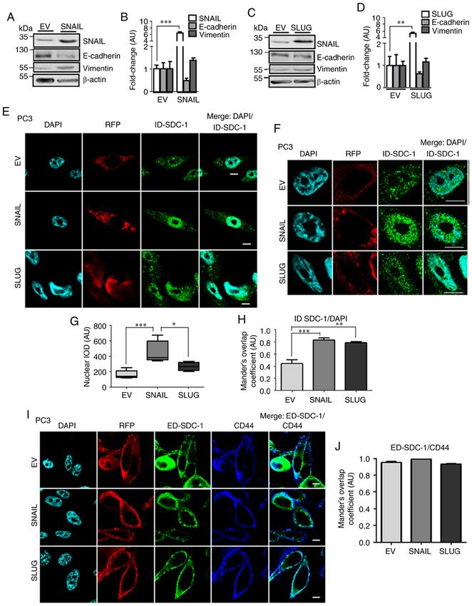
1074        FARFÁN et al: NUCLEAR TRANSLOCATION OF SDC-1 INTRACELLULAR DOMAIN IN PROSTATE CANCER CELLS

    The function of nuclear SDC‑1 is not clear; however, histone     Alexa Fluor 488 and Alexa Fluor 405 (cat. nos. A‑11008 and
acetyltransferase (HAT) inhibition, leading to chromatin             A‑31553, respectively; both from Thermo Fisher Scientific,
compaction (13), cell cycle control, decreases in proliferation,     Inc.; 1:200). The mounted coverslips were observed under a
transcriptional machinery regulation and protein transport           confocal microscope (LSM‑410 Axiovert 100 + Axio Imager;
to the nucleus (19), have been suggested. Additionally, our          Carl Zeiss AG; magnification, x600). Positive RFP expression
previous study demonstrated that SDC‑1 expression was                was used as the marker of successful transduction. In total,
repressed by ZEB1 in prostate cell lines (20). However, an           50 cells were quantified for each marker. To determine only
association between SNAIL family transcription factors and           nuclear ID‑SDC‑1, Adobe Photoshop CS6 Software (2012,
nuclear SDC‑1 location has not been demonstrated yet.                version 13.0; Adobe Systems, Inc.) was utilized to delete the
    Based on these data, the present study aimed to investigate      nuclei from the DAPI images, which were overlapped with the
if SNAIL or SLUG may be associated with the nuclear loca-            ID‑SDC‑1 images. Quantification and the Menders' overlap
tion of SDC‑1 in PCa.                                                coefficient were determined using ImageJ v.1.52f software
                                                                     (NIH).
Materials and methods
                                                                     Total, cytoplasmic and nuclear protein extraction. Cells were
Specimens. Samples of benign prostatic hyperplasia (BPH) (n=3)       seeded in a 100‑mm dish (3x10⁶ or 2.2x10⁶ for LNCaP or PC3
and those with high Gleason Score PCa (8 and 9) (n=3), were          cells, respectively). Total protein extraction was performed as
obtained from biopsy archives of the Anatomy and Pathology           previously described (20). For cytoplasmic and nuclear protein
Service, Clinical Hospital of the University of Chile (CHUCh).       extraction, cells were harvested, treated with 300 µl buffer 1
All protocols and authorization for biopsy use were approved         [50 mM Tris, 0.5% Triton X‑100, 137 mM NaCl, 10% glycerol
by the Faculty of Medicine and CHUCh ethics committees               and protease inhibitors (Roche Diagnostics)] and incubated for
(approval no. 135‑2015). These protocols included written            15 min on ice. The extracts were centrifuged at 500 x g for
informed consent of the patients in order to use part of the tumor   15 min at 4˚C; these supernatants contained the cytoplasmic
samples for research purposes. All protocols and handling of         proteins. The pellet was then resuspended in 150 µl buffer 1
hazardous materials were approved by the Faculty of Medicine         (50 mM Tris pH 7.5, 0,5% Triton X‑100, 137 mM NaCl, 10%
of the University of Chile Risk and Biosecurity Unit.                glycerol + protease and phosphatase inhibitors) with 0.5%
                                                                     SDS, and then passed through a tuberculin syringe (27.5 G
Immunohistochemistry. The immunohistochemical proce-                 x 1/2"; Plastipak™; BD Biosciences), sonicated at 20 kHz
dures and digitalization of the images (magnification, x20)          for 10 sec and centrifuged at 17,000 x g for 15 min at 4˚C.
were performed as described previously (20). The primary             Following this step, the supernatant now contained the nuclear
antibodies were as follows: Anti‑SNAIL (1:100; cat. no. 3879;        proteins. A BCA kit (Thermo Fisher Scientific, Inc.) was used
Cell Signaling Technology, Inc.); anti‑SLUG (1:50; cat.              for protein quantification.
no. sc‑15391; Santa Cruz Biotechnology, Inc.); anti‑ED‑SDC‑1
(1:100; cat. no. sc‑5632; Santa Cruz Biotechnology, Inc.); and       Western blot analysis. SDS‑PAGE analysis was performed
anti‑ID‑SDC‑1 (1:100; cat. no. 362900, Invitrogen; Thermo            following loading of 50 µg cytoplasmic or total protein
Fisher Scientific, Inc.). ImageJ v.1.52f software [National          and 10 µg nuclear protein into each lane. The gels use were
Institutes of Health (NIH)] was used to quantify the images.         6‑12%. The proteins were then transferred to a nitrocellu-
For each immunodetection, 50 images were included and                lose membrane and blocked with 5% milk in 1X TBS/0.1%
quantified.                                                          Tween‑20 at room temperature for 1 h. The membranes were
                                                                     incubated with anti‑ED‑SDC‑1 (1:500; cat. no. sc‑5632; Santa
Cell culture. The human PCa LNCaP (CRL‑1740TM) and PC3               Cruz Biotechnology, Inc.), ID‑SDC‑1 (1:250; cat. no. sc‑7099;
(CRL‑1435™) cell lines were obtained from the American Type          Santa Cruz Biotechnology, Inc.), lamin‑B1 (1:1,000; cat.
Culture Collection and cultured as previously described (20).        no. sc‑374015; Santa Cruz Biotechnology, Inc.), β ‑actin
                                                                     (1:1,000; cat. no. sc‑81178; Santa Cruz Biotechnology, Inc.),
Lentiviral transduction. Transduction was performed as               SNAIL (1:1,000; cat. no. C15D3; Cell Signaling Technology,
described in a previous study (20), with lentiviral particles        Inc.) and SLUG (1:1,000; cat. no. C19G7; Cell Signaling
purchased from GenTarget Inc. and the lentiviral plasmid             Technology, Inc.), vimentin (1:500; cat. no. ab8978; Abcam)
pLenti suCMV (target sequence)‑Rsv red fluorescent protein           and E‑cadherin (1:1,000; cat. no. 610181; BD Transduction
(RFP)‑Puro (GenTarget Inc.), in which the target sequences           Laboratories; BD Biosciences) primary antibodies overnight
were SNAIL (NM_005985.3) or SLUG (NM_003068.4), or                   at 4˚C. The membranes were then incubated with the following
without a target sequence as the empty vector (EV) control.          horseradish peroxidase (HRP)‑conjugated secondary anti-
                                                                     bodies for 1 h at room temperature: Peroxidase AffiniPure
Immunofluorescence. A total of 5x10⁴ cells were seeded on            Goat Anti‑Mouse IgG (H+L) (cat. no. 115‑035‑003),
coverslips in 24‑well plates. The procedure was performed as         Peroxidase AffiniPure Goat Anti‑Rabbit IgG (H+L) (catalog
previously described (21). The primary antibodies dilutions          no. 111‑035‑003) and Peroxidase AffiniPure Rabbit Anti‑Goat
were: 1:50 for anti‑ID‑SDC‑1 (cat. no. 362900; Invitrogen;           IgG (H+L) (catalog no. 305‑035‑045), all purchased from
Thermo Fisher Scientific, Inc.); 1:100 for anti‑ED‑SDC‑1             Jackson ImmunoResearch Laboratories, Inc. and all used at
(cat. no. sc‑5632; Santa Cruz Biotechnology, Inc.); and 1:100        1:10,000. The membranes were developed using the PierceTM
for anti‑CD44 antigen (CD44; cat. no. ab6124; Abcam). The            Enhanced chemiluminescence Western Blotting Detection kit
fluorophores conjugated to the secondary antibodies were             for HRP (cat. no. 32209; Thermo Fisher Scientific, Inc.) in an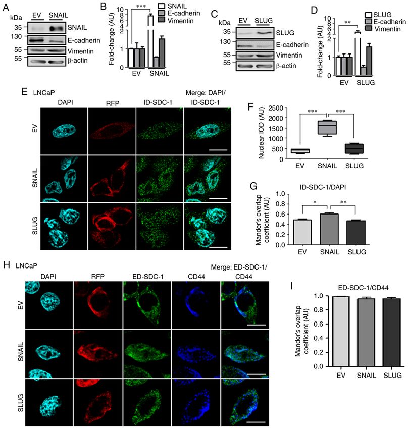
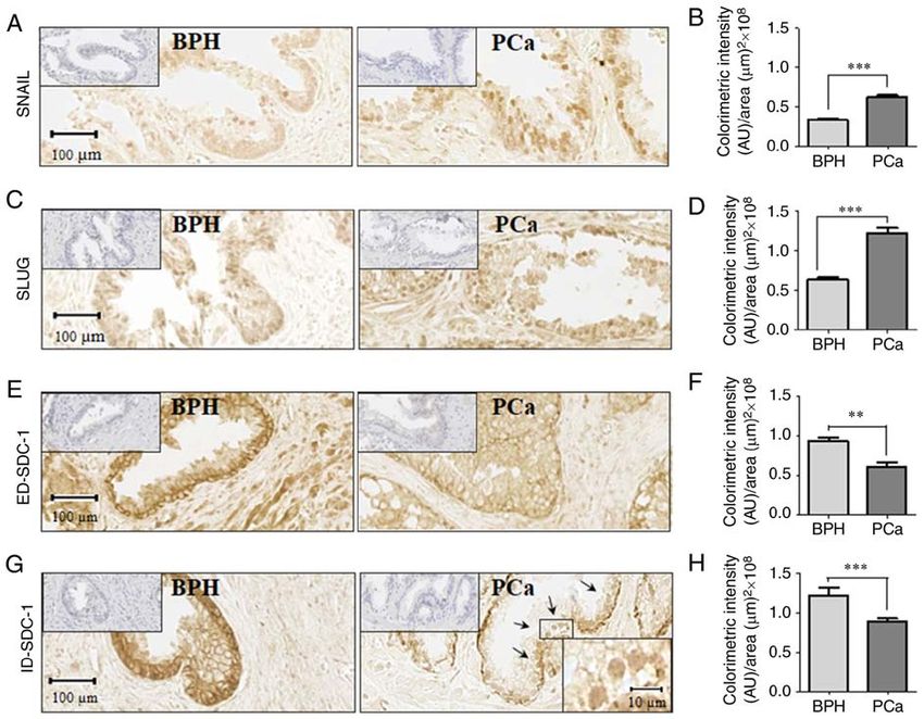
INTERNATIONAL JOURNAL OF MOlecular medicine 45: 1073-1080, 2020                                                      1075

Figure 1. Immunohistochemistry in benign prostatic hyperplasia and prostate cancer samples. Localization of (A) SNAIL, (C) SLUG, (E) ED‑SDC‑1 and
(G) ID‑SDC‑1. (G) Nuclear ID‑SDC‑1 (black arrows) and magnification (rectangle in the center of the image) are included in the lower right corner. Hematoxylin
staining (negative control) is presented in the upper left corner. (B) SNAIL (P= 0.0011), (D) SLUG (P= 0.0004), (F) ED‑SDC‑1 (P= 0.0011) and (H) ID‑SDC‑1
(P= 0.0004) protein levels were quantified. The data represent the average of 3 independent experiments, and the data are presented as the mean ± standard
error of the mean. Data were analyzed using a Student's t‑test. **P
1076          FARFÁN et al: NUCLEAR TRANSLOCATION OF SDC-1 INTRACELLULAR DOMAIN IN PROSTATE CANCER CELLS

Figure 2. ID‑SDC‑1 and ED‑SDC‑1 location in LNCaP cells with ectopic SNAIL or SLUG expression. (A and C) Western blot analysis of SNAIL, SLUG,
vimentin and E‑cadherin protein levels. (B and D) Quantification of the western blot analysis data. Data were analyzed using a Student's t‑test. (E) Confocal
microscopy of DAPI (nuclei), RFP (transduction control) and ID‑SDC‑1 (green) in EV, SNAIL or SLUG‑transduced cells. (F) Nuclear ID‑SDC‑1 quantifica-
tion (integrated optical density per area, arbitrary units). Data were analyzed using ANOVA followed by a Tukey post hoc test. (G) Colocalization of ID‑SDC‑1
with DAPI was assessed using Manders' overlap coefficient. Data were analyzed using analysis of variance followed by a Tukey post hoc test. (H) Confocal
microscopy of DAPI (nuclei), RFP (transduction control), ED‑SDC‑1 (green) and CD44 (blue) in EV, SNAIL or SLUG‑transduced cells. Scale bar=10 µm.
(I) Colocalization of ED‑SDC‑1 with CD44 was assessed using Manders' overlap coefficient. Data were analyzed using ANOVA followed by a Tukey post
hoc test. The data represent the average of 3 independent experiments, and the data are presented as the mean ± standard error of the mean. *P
INTERNATIONAL JOURNAL OF MOlecular medicine 45: 1073-1080, 2020                                                     1077

Figure 3. ID‑SDC‑1 and ED‑SDC‑1 location in PC3 cells with ectopic SNAIL or SLUG expression. (A and C) Western blot analysis of SNAIL, SLUG,
vimentin and E‑cadherin protein levels. (B and D) Quantification of the western blot analysis data. Data were analyzed using a Student's t‑test. (E) Confocal
microscopy of DAPI (nuclei), RFP (transduction control) and ID‑SDC‑1 (green) in EV, SNAIL or SLUG cells. Scale bar=10 µm. (F) Nuclear region magnifica-
tion. Scale bar=10 µm. (G) Nuclear ID‑SDC‑1 quantification (integrated optical density per area, arbitrary units). Data were analyzed using ANOVA followed
by a Tukey post hoc test. (H) ID‑SDC‑1 with DAPI Manders' overlap coefficient. Data were analyzed using ANOVA followed by a Tukey post hoc test.
(I) Confocal microscopy of DAPI (nuclei), RFP (transduction control), ED‑SDC‑1 (green) and CD44 (blue) in the EV, SNAIL or SLUG cells. Scale bar=10 µm.
(J) ED‑SDC‑1 with CD44 Manders' overlap coefficient. Data were analyzed using ANOVA followed by a Tukey post hoc test. The data represent the average
of three independent experiments and are presented as the mean ± standard error of the mean. *P
1078          FARFÁN et al: NUCLEAR TRANSLOCATION OF SDC-1 INTRACELLULAR DOMAIN IN PROSTATE CANCER CELLS

Figure 4. ID‑SDC‑1 and ED‑SDC‑1 protein levels in the cytoplasm and nucleus of LNCaP and PC3 cells with ectopic SNAIL or SLUG expression. Nuclear
and cytoplasmic ID‑SDC‑1 protein levels of (A and B) LNCaP and (C and D) PC3 cells with ectopic EV, SNAIL or SLUG expression. Total ED‑SDC‑1 protein
levels in (E and F) LNCaP or (G and H) PC3 cells. The levels were noramlized to those of lamin B1 (nuclear proteins) and β‑actin (cytoplasmic and total
proteins). The fold‑change (arbitrary units) was normalized to (B and F) EV LNCaP and (D and H) PC3 protein levels. Data were analyzed using ANOVA
followed by a Tukey post hoc test. The data represent the average of 3 independent experiment, and the data are presented as the mean ± standard error of the
mean. *P
INTERNATIONAL JOURNAL OF MOlecular medicine 45: 1073-1080, 2020                                     1079

activity (13), suggesting that nuclear SDC‑1 location could         Authors' contributions
be associated with chromatin compaction. According to the
ID‑SDC‑1/DAPI co‑localization results from the present              NF, AGdH, EAC and HRC conceived and designed the study.
study, the majority of ID‑SDC‑1 was located in the compacted        NF, OOS, PC, GM, DC and DH conducted the experiments
chromatin area. In addition, SNAIL has been demonstrated            and analyzed the data. NF, EAC and HRC wrote and revised
to act as a regulator of heterochromatin domains, through           the manuscript. All the authors read and approved the final
the co‑repressor Lysyl Oxidase Like 2, in mouse embryonic           manuscript.
fibroblast pericentromeric domains (25). Therefore, SNAIL
overexpression may be associated with high heterochromatin          Ethics approval and consent to participate
stabilization and may favor an increased probability of nuclear
ID‑SDC1 with DAPI co‑localization. However, more detailed           The protocol used for tissue collection was approved by
studies of co‑localization of ID‑SDC1 with heterochromatin          Faculty of Medicine and CHUCh Ethics Committees. All
markers such as histone H3 lysine 9‑methylation or co‑immu-         patients provided written informed consent. All protocols and
noprecipitation of heterochromatin sequences with ID‑SDC‑1          handling of hazardous materials were approved by the Faculty
are required.                                                       of Medicine of the University of Chile Risk and Biosecurity
    SNAIL‑overexpressing cells exhibited increased nuclear          Unit.
ID‑SDC‑1 protein levels compared with cytoplasmic levels.
This could be associated with an alternative translation            Patient consent for publication
initiation, like that described for HER2 intracellular domains,
located in the cytoplasm and nucleus (23).                          All patients provided written informed consent.
    Although EMT has been associated with the nuclear
location of other proteins such as E‑cadherin in other cancer       Competing interests
types (26,27), at present, the association between EMT factors
and ID‑SDC‑1 location has not been described. In conclusion,        The authors declare that they have no competing interests.
the results of the present study demonstrated an association
between SNAIL expression and nuclear ID‑SDC‑1 location in           References
PCa cell lines.
    The primary limitation of the present study is the low           1. Bray F, Ferlay J, Soerjomataram I, Siegel RL, Torre LA and
                                                                        Jemal A: Global cancer statistics 2018: GLOBOCAN estimates
number of samples used for immunohistochemistry analyses (3             of incidence and mortality worldwide for 36 cancers in 185
in each group). However, the statistical significance observed          countries. CA Cancer J Clin 68: 394‑424, 2018.
supports the conclusions concerning the expression and loca-         2. Nieto MA, Huang RY, Jackson RA and Thiery JP: EMT: 2016.
                                                                        Cell 166: 21‑45, 2016.
tion of SNAIL, SLUG and ED‑SDC‑1. Nevertheless, a more               3. Micalizzi DS, Farabaugh SM and Ford HL: Epithelial-
extensive study is necessary for the clinical validation of these       mesenchymal transition in cancer: Parallels between normal
changes in the progression of PCa.                                      development and tumor progression. J Mammary Gland Biol
                                                                        Neoplasia 15: 117‑134, 2010.
                                                                     4. Puisieux A, Brabletz T and Caramel J: Oncogenic roles of
Acknowledgements                                                        EMT‑inducing transcription factors. Nat Cell Biol 16: 488‑494,
                                                                        2014.
The authors would like to thank to Mrs. Graciela Caroca              5. Poblete CE, Fulla J, Gallardo M, Muñoz V, Castellón EA,
                                                                        Gallegos I and Contreras HR: Increased SNAIL expression and
(Department of Basic and Clinical Oncology, Faculty of                  low syndecan levels are associated with high Gleason grade in
Medicine, University of Chile, Santiago, Chile) for their tech-         prostate cancer. Int J Oncol 44: 647‑654, 2014.
nical assistance. The authors would also like to thank Dr María      6. Contreras HR, Ledezma RA, Vergara J, Cifuentes F, Barra C,
                                                                        Cabello P, Gallegos I, Morales B, Huidobro C and Castellón EA:
Julieta González, Dr Isabel Castro and Dr María José Barrera,           The expression of syndecan‑1 and ‑2 is associated with Gleason
from Biomedical Sciences Institute, University of Chile, for            score and epithelial‑mesenchymal transition markers, E‑cadherin
the confocal microscope use.                                            and beta‑catenin, in prostate cancer. Urol Oncol 28: 534‑540,
                                                                        2010.
                                                                     7. Ledezma R, Cifuentes F, Gallegos I, Fullá J, Ossandon E,
Funding                                                                 Castellon EA and Contreras HR: Altered expression patterns
                                                                        of syndecan‑1 and ‑2 predict biochemical recurrence in prostate
The present study was supported by grants from FONDECYT                 cancer. Asian J Androl 13: 476‑480, 2011.
                                                                     8. Dhodapkar MV, Abe E, Theus A, Lacy M, Langford JK,
awarded to HRC (grant nos. 1110269 and 1151214) and to                  Barlogie B and Sanderson RD: Syndecan‑1 is a multifunc-
EAC (grant no. 1140417), Grants from U‑APOYA ENLACE,                    tional regulator of myeloma pathobiology: Control of tumor
University of Chile (grant nos. ENL‑22/19 and ENL 23/19), State         cell survival, growth, and bone cell differentiation. Blood 91:
                                                                        2679‑2688, 1998.
Research Agency and the European Regional Development                9. Leppä S, Mali M, Miettinen HM and Jalkanen M: Syndecan
Fund (grant no. SAF2016‑76461‑R) awarded to AGH and the                 expression regulates cell morphology and growth of mouse
CONICYT (National Commission of Science and Technology)                 mammary epithelial tumor cells. Proc Natl Acad Sci USA 89:
                                                                        932‑936, 1992.
scholarship (grant no. 21140772) awarded to NF.                     10. Hu Y, Sun H, Owens RT, Gu Z, Wu J, Chen YQ, O'Flaherty JT and
                                                                        Edwards IJ: Syndecan‑1‑dependent suppression of PDK1/Akt/bad
Availability of data and materials                                      signaling by docosahexaenoic acid induces apoptosis in prostate
                                                                        cancer. Neoplasia 12: 826‑836, 2010.
                                                                    11. Tumova S, Woods A and Couchman JR: Heparan sulfate proteo-
All data generated and analyzed during the current study are            glycans on the cell surface: versatile coordinators of cellular
available from the corresponding author on reasonable request.          functions. Int J Biochem Cell Biol 32: 269‑288, 2000.
1080 FARFÁN et al: NUCLEAR TRANSLOCATION OF SDC-1 INTRACELLULAR DOMAIN IN PROSTATE CANCER CELLS
12. Brockstedt U, Dobra K, Nurminen M and Hjerpe A: 21. Herrera D, Orellana‑Serradell O, Villar P, Torres MJ, Paciucci R,
Immunoreactivity to cell surface syndecans in cytoplasm and Castellón EA and Contreras HR: Silencing of the transcriptional
nucleus: Tubulin‑dependent rearrangements. Exp Cell Res 274: factor ZEB1 alters the steroidogenic pathway, and increases the
235‑245, 2002. concentration of testosterone and DHT in DU145 cells. Oncol
13. Purushothaman A, Hurst DR, Pisano C, Mizumoto S, Sugahara K Rep 41: 1275‑1283, 2019.
and Sanderson RD: Heparanase‑mediated loss of nuclear 22. Fortini ME: Gamma‑secretase‑mediated proteolysis in
syndecan‑1 enhances histone acetyltransferase (HAT) activity cell‑surface‑receptor signalling. Nat Rev Mol Cell Biol 3:
to promote expression of genes that drive an aggressive tumor 673‑684, 2002.
phenotype. J Biol Chem 286: 30377‑30383, 2011. 23. Anido J, Scaltriti M, Bech Serra JJ, Santiago Josefat B, Todo FR,
14. Chen L and Sanderson RD: Heparanase regulates levels of Baselga J and Arribas J: Biosynthesis of tumorigenic HER2
syndecan‑1 in the nucleus. PLoS One 4: e4947, 2009. C‑terminal fragments by alternative initiation of translation.
15. Zong F, Fthenou E, Wolmer N, Hollósi P, Kovalszky I, Szilák L, EMBO J 25: 3234‑3244, 2006.
Mogler C, Nilsonne G, Tzanakakis G and Dobra K: Syndecan‑1 24. Solovei I, Thanisch K and Feodorova Y: How to rule the nucleus:
and FGF‑2, but not FGF receptor‑1, share a common transport Divide et impera. Curr Opin Cell Biol 40: 47‑59, 2016.
route and co‑localize with heparanase in the nuclei of mesen- 25. Millanes‑Romero A, Herranz N, Perrera V, Iturbide A,
chymal tumor cells. PLoS One 4: e7346, 2009. Loubat‑Casanovas J, Gil J, Jenuwein T, García de Herreros A
and Peiró S: Regulation of heterochromatin transcription by
16. Szatmári T and Dobra K: The role of syndecan‑1 in cellular Snail1/LOXL2 during epithelial‑to‑mesenchymal transition. Mol
signaling and its effects on heparan sulfate biosynthesis in Cell 52: 746‑757, 2013.
mesenchymal tumors. Front Oncol 3: 310, 2013. 26. Céspedes MV, Larriba MJ, Pavón MA, Alamo P, Casanova I,
17. Stewart MD, Ramani VC and Sanderson RD: Shed syndecan‑1 Parreño M, Feliu A, Sancho FJ, Muñoz A and Mangues R:
translocates to the nucleus of cells delivering growth factors and Site‑dependent E‑cadherin cleavage and nuclear translocation
inhibiting histone acetylation: A novel mechanism of tumor‑host in a metastatic colorectal cancer model. Am J Pathol 177:
cross‑talk. J Biol Chem 290: 941‑949, 2015. 2067‑2079, 2010.
18. Bernfield M, Götte M, Park PW, Reizes O, Fitzgerald ML, 27. Chetty R, Serra S and Asa SL: Loss of membrane localization and
Lincecum J and Zako M: Functions of cell surface heparan aberrant nuclear E‑cadherin expression correlates with invasion in
sulfate proteoglycans. Annu Rev Biochem 68: 729‑777, 1999. pancreatic endocrine tumors. Am J Surg Pathol 32: 413‑419, 2008.
19. Kovalszky I, Hjerpe A and Dobra K: Nuclear translocation of
heparan sulfate proteoglycans and their functional significance.
Biochim Biophys Acta 1840: 2491‑2497, 2014. This work is licensed under a Creative Commons
20. Farfán N, Ocarez N, Castellón EA, Mejía N, de Herreros AG Attribution-NonCommercial-NoDerivatives 4.0
and Contreras HR: The transcriptional factor ZEB1 represses International (CC BY-NC-ND 4.0) License.
Syndecan 1 expression in prostate cancer. Sci Rep 8: 11467, 2018.You can also read