ADDRESS: A Database of Disease-associated Human Variants Incorporating Protein Structure and Folding Stabilities
←
→
Page content transcription
If your browser does not render page correctly, please read the page content below
Database

                             ADDRESS: A Database of Disease-
                             associated Human Variants
                             Incorporating Protein Structure and
                             Folding Stabilities

Jaie Woodard 1, Chengxin Zhang 1 and Yang Zhang 1,2⇑

1 - Department of Computational Medicine and Bioinformatics, University of Michigan, Ann Arbor, MI 48109, USA
2 - Department of Biological Chemistry, University of Michigan, Ann Arbor, MI 48109, USA

Correspondence to Yang Zhang:⇑Department of Computational Medicine and Bioinformatics, University of
Michigan, Ann Arbor, MI 48109, USA. zhng@umich.edu (Y. Zhang)
https://doi.org/10.1016/j.jmb.2021.166840
Edited by Michael Sternberg

Abstract
Numerous human diseases are caused by mutations in genomic sequences. Since amino acid changes
affect protein function through mechanisms often predictable from protein structure, the integration of
structural and sequence data enables us to estimate with greater accuracy whether and how a given
mutation will lead to disease. Publicly available annotated databases enable hypothesis assessment
and benchmarking of prediction tools. However, the results are often presented as summary statistics
or black box predictors, without providing full descriptive information. We developed a new semi-
manually curated human variant database presenting information on the protein contact-map,
sequence-to-structure mapping, amino acid identity change, and stability prediction for the popular Uni-
Prot database. We found that the profiles of pathogenic and benign missense polymorphisms can be
effectively deduced using decision trees and comparative analyses based on the presented dataset.
The database is made publicly available through https://zhanglab.ccmb.med.umich.edu/ADDRESS.
                                                                               Ó 2021 Elsevier Ltd. All rights reserved.

Introduction                                                 tion, including binding with proteins and other mole-
                                                             cules in its interaction network.
   Mendelian human disease is often the result of a             Pathogenicity prediction tools often utilize protein
change in a single amino acid within a protein. With         evolutionary information, identifying homologous
the ever-increasing wealth of genomic and                    sequences and determining whether mutated
structural data, it is possible to quantitatively            residues are well or poorly conserved.7–9 Such pre-
assess, on a genome-wide scale, why some                     dictors may also utilize information on changes in
missense mutations result in disease, while others           amino acid sequence identity10–14; for instance, a
are benign. Early studies on few proteins revealed           change from a hydrophobic to a charged residue
changes in protein stability as a causative factor,          may be expected to have a high likelihood of dis-
with mutations in buried residues being more                 ease association. In recent programs, the structural
common among pathogenic variants.1 Later studies             neighborhood of the mutation may also be taken
focused also on the important role of protein and            into account. Examples include DAMpred,15 which
ligand binding interactions, as well as active sites         includes many structural contact-related features
involved in enzymatic function.2–4 The overall con-          and builds models of unknown structures,
clusion is that there are multiple routes by which a         RAPSODY,16,17 which takes into account structural
protein’s function can be deterred within the cellular       dynamics, and Missense3D,18 which considers a
environment, in which it must maintain a sizable             variety of potentially disruptive structural changes
folded fraction5,6 and be able to carry out its func-        to evaluate whether a given variant is pathogenic.
0022-2836/Ó 2021 Elsevier Ltd. All rights reserved.                             Journal of Molecular Biology 433 (2021) 166840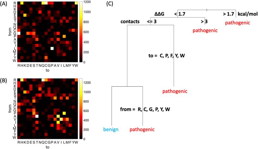
J. Woodard, C. Zhang and Y. Zhang                                   Journal of Molecular Biology 433 (2021) 166840

Likewise, predictions of mutation-induced free-              leucine to proline, arginine to cysteine, and
energy change (DDG) and changes in binding affin-            arginine to tryptophan, representing dramatic
ity to functional partners could be expected to carry        changes in terms of physicochemical properties
useful information relevant to potential loss of pro-        and/or torsional preference. Common benign
tein function, as explored in previous database              mutations appear overall more conservative:
annotations19 Efforts to integrate structural informa-       alanine to threonine, isoleucine to valine, valine to
tion with mutation data in database format include           isoleucine, alanine to valine, and proline to leucine.
COSMIC,20 which, however, only contains data on              Other mutations, such as cysteine to tyrosine are
somatic mutations in cancer. Other related                   found much more often in pathogenic cases (or
resources include Swissvar and MSV3d,21 although             benign, as for threonine to alanine), although they
many servers lack full annotation details and com-           are overall rare. Since the dataset represents a
parison with computational predictions22,23 or are           broad range of residue changes, such data can be
no longer maintained.                                        informative towards predicting pathogenicity. While
   The UniProt Humsavar database (https://www.               the heatmaps are largely symmetric, we find
uniprot.org/docs/humsavar) contains information              statistical significance for several asymmetries,
on pathogenicity of more than 70,000 human                   including glycine to arginine being more frequent,
variants and is often used to benchmark tools                in the pathogenic case, than arginine to glycine. A
developed to predict pathogenicity of missense               full statistical treatment of amino acid changes
single nucleotide polymorphisms. The majority of             upon mutation is presented in the Supplemental
variants are annotated either as neutral                     Information (Text S1 and Tables S1–S3).
“Polymorphism” or disease-associated variants                  Next, we develop a rule-based approach using
(39.4% and 50.0%, respectively), with a small                data on pathogenicity. The decision tree in
number of unclassified entries (10.6%). While the            Figure 1(C) shows a data-informed process based
Humsavar file includes UniProt accession, it does            on the ADDRESS dataset, generated by rpart in
not link directly to experimental structures or              R, using only four features: the type of amino acid
provide overall insight into the relationship                before and after mutation, the stability change
between structure and disease. Integration with              predicted by FoldX, and the number of contacts
structural information and stability predictions             formed by the mutated residue. Here, we chose
could be useful in assessing which factors are               the decision tree over more powerful supervised
most important to maintaining or disrupting protein          machine learning algorithms mainly considering
functions, as well as facilitating new prediction            the better interpretability of the decision tree
tools to aid researchers in developing results               results. Nonetheless, the simple decision tree
relevant to clinical practice.                               method still achieves an appreciable Mathews
   In this study, we present a new semi-manually             Correlation Coefficient (MCC) of 0.27 in the binary
curated      database,      ADDRESS       (Annotated         classification of pathogenic versus benign
Database of Disease-RElated Structures and                   mutations.
Sequences), mapping mutation sites annotated in                The first branching of the decision tree is based
UniProt Humsavar to residue numbers in example               on the predicted DDG of the mutation, such that
structural files from the Protein Data Bank (PDB).           changes that lead to a sufficiently large decrease
Next to each structure, we provide the number of             in stability (large positive values) predict
contacts in which the mutated residue participates           pathogenic consequences. However, the value of
and the predicted DDG of the mutation, according             1.7 kcal/mol is still relatively small in comparison
to the EvoEF empirical force field24,25 and FoldX            to common folding stability values, such that if
results.29 The database may be searched by the               considering equilibrium thermodynamics alone, in
type of variant, by the starting/ending amino acid           most cases the majority of the protein would still
type, the number of contacts, or predicted DDG.              be in the folded state. A similar observation was
Our database presents an exploratory interface to            made previously, where selection for kinetics in a
pathogenicity data, as well as a useful starting point       crowded environment was proposed to be the
for advanced statistical and machine-learning                cause of such a low DDG value, for a small
based method developments.                                   number of mutations in a single protein.26 In the
                                                             decision tree, when the stability change is small or
Results                                                      negative, the mutation is still predicted to be patho-
                                                             genic in the case of larger numbers of contacts or
Descriptive analysis of residue identities                   mutations from or to a set of relatively “extreme”
                                                             residues, including mutation from cysteine or trypto-
  By generating heatmaps displaying the frequency            phan. Such a decision tree supports intuition
of each possible amino acid change, it is clear that         regarding which changes are likely to be patho-
pathogenic and benign mutations have distinct                genic, while providing additional insights, such as
profiles (Figure 1(A) and (B), respectively). The            support for the selection for kinetic stability hypoth-
arginine to glutamine or histidine mutations are             esis on the scale of thousands of mutations. A deci-
common in both pathogenic and benign cases.                  sion tree excluding FoldX information and therefore
Also common in disease are glycine to arginine,
                                                         2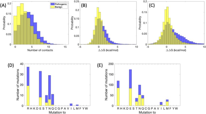
J. Woodard, C. Zhang and Y. Zhang                                  Journal of Molecular Biology 433 (2021) 166840

  Figure 1. Summary of sequence and pathogenicity information from the ADDRESS database. (A–B) Amino acid
identity change represented in a two-dimensional histogram, for pathogenic (A) and benign (B) variants. Brighter
colors indicate higher frequencies. (2) A simple decision tree predicting whether a variant is pathogenic or benign.
Stability change DDG is the FoldX predicted value.

emphasizing EvoEF predictions achieves a similar             pathogenic mutations (mean value 0.72 kcal/mol)
accuracy (MCC = 0.26, Figure S1) but with number             than benign ones (mean value 0.06 kcal/mol) (Fig-
of contacts prioritized and DDG value deprioritized          ure 2(B)). Similarly, the DDG predicted by another
within the tree.                                             widely used predictor, FoldX,29 also shows a signif-
                                                             icant p-value for the difference in pathogenic and
Residue contact information and predicted                    benign distributions of 3.5  10232 (Figure 2(C)).
stability change                                             The overall correlation between EvoEF and FoldX
                                                             DDG values was 0.67.
   Pathogenic and benign variants show distinct                 As a case study, we examine in Figure 2(D) and
distributions of the number of contacts                      (E) the frequency of benign and pathogenic
surrounding the mutated residue (Figure 2(A)), in            variants occurring on Lysine which is an amino
line with the observation that local packing density         acid with both hydrophobic properties and positive
correlates strongly with surface exposure27 and that         charge. Here, we consider differences in the
solvent exposure in turn is strongly correlated with         frequency of mutations to different types of
the site specific rate of mutation.28 For benign vari-       residues and their benign or pathogenic state,
ants, results are more skewed towards smaller                when the number of contacts in the crystal
numbers, indicating that residues tend to be more            structure of the original protein is small vs. when it
buried in the case of disease-causing variants, with         is large. As shown in the plots, mutation to a
a p-value = 1.4  10299 in Student’s t-test. This is        charged or polar residue is more often benign
consistent with previous results on a smaller set of         rather than pathogenic when the number of
proteins showing that pathogenic mutations tend              contacts with the mutated residue is small.
to be located in the protein interior more often than
benign ones4 and with depth and contact informa-
                                                             Online database setting
tion from another former investigation.15 Overall,
the number of contacts for the specified cutoff val-            The webserver of ADDRESS mainly consists of
ues peaks around four contacts in the case of                three parts: a top banner for view switching, a
pathogenic mutations and two contacts in the case            JSmol30 applet to display the PDB structure, and a
of benign mutations. The mean is 2.7 contacts for            main table that lists the database entries (Figure 3).
benign variants and 3.9 for pathogenic; the median           First, the view switching banner allows the user to
values are 2 and 4, respectively. Likewise, the              select one of the five views: “Browse by structure”
change in protein stability, DDG, predicted by               lists one PDB chain per row in the main table;
EvoEF was substantially higher (more positive, with          “Browse by mutations on structure” lists one muta-
a p-value = 1.9  10211 in Student’s t-test) for            tion mapped to a PDB chain row in the table;
                                                         3
J. Woodard, C. Zhang and Y. Zhang Journal of Molecular Biology 433 (2021) 166840
Figure 2. Local structural information from missense SNPs. (A) Histogram of number of contacts (at least six atom–
atom contacts less than five angstroms), for benign and pathogenic variants, with the mutated residue within a known
crystal structure. (B) DDG in kcal/mol, predicted by EvoEF. (C) DDG in kcal/mol, predicted by FoldX. (D-E) Stacked
bar plots indicating the number of benign and pathogenic variants mutated from lysine to the specified residue for (D)
number of contacts greater than or equal to five, (E) number of contacts less than five.
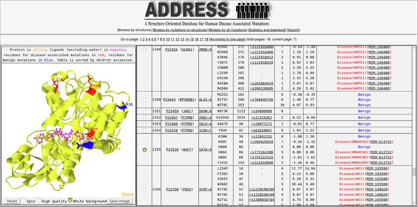
Figure 3. A screenshot of the ADDRESS online database interface. The interface contains three parts. The view
switching banner is the top under the database logo, switched to “Browse by structure” page 1 in this screenshot. A
JSmol structure applet is at lower left, displaying PDB structure 1a7a Chain A for human Adenosylhomocysteinase
protein AHCY (yellow cartoon) in complex with the NAD and ADC ligands (magenta sticks) and mutation sites (red
and blue lines of pathogenic and benign mutations, respectively). The main table at the lower right displays all entries
sorted by PDB IDs, where 1a7a Chain A for AHCY is currently selected. At the upper right corner of the database
interface is the “Search” button, with which users can search database entries by protein names, PDB IDs, mutations,
diseases, and structural features (e.g., contact numbers and free energy changes).
4J. Woodard, C. Zhang and Y. Zhang Journal of Molecular Biology 433 (2021) 166840
“Browse all mutations” displays all mutations, sequence and structure data, ADDRESS is in
regardless of whether each can be mapped to a active development. Currently, we are working on
structure. Since it is difficult to load all data in a extending our database to combine literature
web-browser due to cache size limit, all online search and other primary human variance
tables are split into multiple pages to facilitate brow- datasets such as the Clinvar.34 We also plan to pro-
ser rendering. If a user would like to view all data vide additional features that are highly discrimina-
contained within ADDRESS, an Excel spreadsheet tory, such as protein–protein and protein–ligand
can be downloaded at the bottom of the “Statistics interaction information, as well as 3D structure mod-
and download” page, which also includes the data els from the start-of-the-art modeling pipelines.35,36
analysis figures (Figures 1 and 2) for general statis- We plan to later add information from the ProTherm
tics of ADDRESS. Finally, the “Search” page per- experimental database on protein stability, for
forms database search using PDB IDs, gene mutants that map to the Humsavar database. How-
names, UniProt accessions, diseases, amino acid ever, upon preliminary consideration, we believe
types, or range of contact numbers and free- that such a task will require substantial additional
energy change. The database contains information effort beyond the scope of the initial database (in-
on both the residue number in UniProt, in the col- cluding some manual effort due to labeling errors
umn “Mutation on UniProt sequence,” and the resi- and inconsistencies in the ProTherm database)
due number mapped to in the PDB structure, under and will also cover only a small fraction (about 80)
“Residue index in PDB,” which was obtained of the proteins in the ADDRESS database. We
through sequence alignment as described in the believe that a high-quality and up-to-date human
Methods section. variance database featured with enriched structural
The JSmol applet displays the 3D structure of the bioinformatics data will have critical importance in
PDB chains together with any non-water ligands facilitating investigations relevant to early diagnosis
and the mutation sites, as selected by the first and treatment of human genetic diseases.
column of the main table. The main table also
includes columns for PDB ID (linked to the RCSB
PDB website), UniProt accession (linked to the
UniProt database) and Gene name (linked to the
Methods
neXtProt31), mutation amino acid types and residue Data collection and alignment
index on the UniProt sequence and on the PDB
structure, a link to dbSNP database, number of con- The UniProt Humsavar database (version
tacts per residue, EvoEF estimated free energy 2020_04 as of this manuscript) was downloaded
change upon mutation, and disease association of from Humsavar, where labeled benign
the mutation. For disease-associated mutations, “Polymorphism” and pathogenic “Disease”
the disease symbol and disease ID in the OMIM variants were considered. Protein sequences were
database32,33 are displayed in the last column of downloaded from UniProt. For each mutation, the
the table. experimental structure containing this mutation in
its residue range which had the greatest overall
sequence coverage was chosen as a
Conclusion representative structure. The UniProt sequence
was aligned with the string of residues contained
A better understanding of which mutations lead to in the protein structure, using NW-align,37 with
disease can be useful in prioritizing experimental adjusted gap penalties to determine the position of
study of variants and understanding protein the mutated residue in the experimental structure.
evolution from a theoretical perspective. We have Cases where the mutated residue aligned with a
introduced a new database of human variants with gap before or after the sequence were discarded.
each entry enriched with various types of This procedure results in 13,624 pathogenic vari-
structural bioinformatics information including ants and 7,633 benign variants that are mapped to
residue mutation identity, numbers of contacts, 3,583 PDB chains.
and predicted change in protein stability. As an
illustration of an application, we have approached
data analyses from a descriptive and exploratory
Feature extraction
perspective, gaining insight into why mutations
may be pathogenic or benign. Our results provide Initial and mutated residue identities were
data relevant to future prediction tools and permit extracted from the Humsavar data file. To remove
a variety of comparisons on this sizable dataset. redundant coordinates from a PDB structure, only
We anticipate our database to be more useful in atoms with the alternative location indicator ‘A’ or ‘
generating aggregate statistics and comparisons, ’ (space) were kept. For the entries with multi-
rather than predicting results for individual model NMR structures, only the first model was
proteins, for which further modeling and predictors selected. Residues were considered in contact
with more features may be necessary. With the with the residue to be mutated if there were six or
rapid accumulation and availability of various more pairwise atomic contacts within five A.
5J. Woodard, C. Zhang and Y. Zhang Journal of Molecular Biology 433 (2021) 166840
Folding stability change calculation Appendix A. Supplementary Data
The mutation-induced folding stability change,
Supplementary data to this article can be found
DDG, is estimated by EvoEF,24 which is an empiri-
online at https://doi.org/10.1016/j.jmb.2021.
cal force field and has been shown to have a strong
166840.
correlation with the experimental DDG measure-
ments.25 Preceding EvoEF calculation, the amino
acid types in the PDB are first standardized by Received 1 October 2020;
removing non-standard amino acids and by map- Accepted 20 January 2021;
ping selenomethionine (MSE) to methionine Available online 2 February 2021
(MET), as MSE is commonly engineered to replace
Keywords:
MET to facilitate X-ray structure determination. In
database;
the case of inconsistent amino acid type between
Single-nucleotide polymorphism;
UniProt sequence and PDB structure, the EvoEF
disease variant;
BuildMutant subroutine is used to make convert
pathogenicity prediction
amino acid on a PDB structure to that of the UniProt
sequence. EvoEF RepairStructure function is
applied to fill in missing atoms such as hydrogens,
and EvoEF BuildMutant is performed used to build
mutant structure. The folding free energies of wild- References
type and mutant structures (DG wt and DG mut ,
respectively) are estimated by EvoEF ComputeSta- 1. Wang, Z., Moult, J., (2001). SNPs, protein structure, and
bility function. The stability change is therefore disease. Hum Mutat., 17, 263–270.
DDG ¼ DG mut DG wt , in the units of kcal/mol. 2. Steward, R.E., MacArthur, M.W., Laskowski, R.A.,
As FoldX and EvoEF use almost identical function Thornton, J.M., (2003). Molecular basis of inherited
names, ADDRESS also predicts DDG by FoldX diseases: a structural perspective. Trends Genet., 19,
following essentially the same protocol as above. 505–513.
The folding free energies of wild-type and mutant 3. Sunyaev, S., Ramensky, V., Bork, P., (2000). Towards a
structures (DG wt and DG mut , respectively) are structural basis of human non-synonymous single
estimated by FoldX Stability function. nucleotide polymorphisms. Trends Genet., 16, 198–200.
4. Gao, M., Zhou, H., Skolnick, J., (2015). Insights into
disease-associated mutations in the human proteome
through protein structural analysis. Structure, 23, 1362–
1369.
DECLARATION OF COMPETING INTEREST
5. Serohijos, A.W., Shakhnovich, E.I., (2014). Merging
The authors declare that they have no known molecular mechanism and evolution: theory and
competing financial interests or personal relationships computation at the interface of biophysics and
that could have appeared to influence the work evolutionary population genetics. Curr. Opin. Struct. Biol.,
reported in this paper. 26, 84–91.
6. Goldstein, R.A., (2011). The evolution and evolutionary
consequences of marginal thermostability in proteins.
Proteins, 79, 1396–1407.
7. Kumar, P., Henikoff, S., Ng, P.C., (2009). Predicting the
effects of coding non-synonymous variants on protein
function using the SIFT algorithm. Nature Protoc., 4,
Acknowledgements 1073–1081.
8. Tang, H., Thomas, P.D., (2016). PANTHER-PSEP:
We thank Dr. Jeffrey Brender for helpful
predicting disease-causing genetic variants using
discussions. This work is supported in part by the
position-specific evolutionary preservation. Bioinformatics,
National Institute of General Medical Sciences
32, 2230–2232.
(GM136422, S10OD026825), the National 9. Reva, B., Antipin, Y., Sander, C., (2011). Predicting the
Institute of Allergy and Infectious Diseases functional impact of protein mutations: application to cancer
(AI134678), and the National Science Foundation genomics. Nucleic Acids Res., 39, e118
(IIS1901191, DBI2030790, MTM2025426). 10. Adzhubei, I.A., Schmidt, S., Peshkin, L., Ramensky, V.E.,
Gerasimova, A., Bork, P., et al., (2010). A method and
server for predicting damaging missense mutations. Nature
Methods, 7, 248–249.
Author contributions 11. Hecht, M., Bromberg, Y., Rost, B., (2015). Better prediction
of functional effects for sequence variants. BMC
Y.Z. conceived and designed the research. J.W. Genomics, 16 (Suppl 8), S1.
developed the database and performed the 12. Ioannidis, N.M., Rothstein, J.H., Pejaver, V., Middha, S.,
analyses. C.Z. constructed the online server. J.W. McDonnell, S.K., Baheti, S., et al., (2016). REVEL: an
drafted the manuscript. All authors edited and ensemble method for predicting the pathogenicity of rare
approved the manuscript. missense variants. Am. J. Hum. Genet., 99, 877–885.
6J. Woodard, C. Zhang and Y. Zhang Journal of Molecular Biology 433 (2021) 166840
13. Calabrese, R., Capriotti, E., Fariselli, P., Martelli, P.L., 25. Huang, X., Pearce, R., Zhang, Y., (2020). EvoEF2:
Casadio, R., (2009). Functional annotations improve the accurate and fast energy function for computational
predictive score of human disease-related mutations in protein design. Bioinformatics, 36, 1135–1142.
proteins. Hum. Mutat., 30, 1237–1244. 26. Godoy-Ruiz, R., Ariza, F., Rodriguez-Larrea, D., Perez-
14. Li, B., Krishnan, V.G., Mort, M.E., Xin, F., Kamati, K.K., Jimenez, R., Ibarra-Molero, B., Sanchez-Ruiz, J.M.,
Cooper, D.N., et al., (2009). Automated inference of (2006). Natural selection for kinetic stability is a likely
molecular mechanisms of disease from amino acid origin of correlations between mutational effects on protein
substitutions. Bioinformatics, 25, 2744–2750. energetics and frequencies of amino acid occurrences in
15. Quan, L., Wu, H., Lyu, Q., Zhang, Y., (2019). DAMpred: sequence alignments. J. Mol. Biol., 362, 966–978.
recognizing disease-associated nsSNPs through Bayes- 27. Yeh, S.W., Liu, J.W., Yu, S.H., Shih, C.H., Hwang, J.K.,
guided neural-network model built on low-resolution Echave, J., (2014). Site-specific structural constraints on
structure prediction of proteins and protein-protein protein sequence evolutionary divergence: local packing
interactions. J. Mol. Biol., 431, 2449–2459. density versus solvent exposure. Mol. Biol. Evol., 31, 135–
16. Ponzoni, L., Bahar, I., (2018). Structural dynamics is 139.
a determinant of the functional significance of 28. Franzosa, E.A., Xia, Y., (2009). Structural determinants of
missense variants. Proc. Natl. Acad. Sci. USA, 115, protein evolution are context-sensitive at the residue level.
4164–4169. Mol. Biol. Evol., 26, 2387–2395.
17. Ponzoni, L., Penaherrera, D.A., Oltvai, Z.N., Bahar, I., 29. Guerois, R., Nielsen, J.E., Serrano, L., (2002). Predicting
(2020). Rhapsody: predicting the pathogenicity of human changes in the stability of proteins and protein complexes:
missense variants. Bioinformatics, 36, 3084–3092. A study of more than 1000 mutations. J. Mol. Biol., 320,
18. Ittisoponpisan, S., Islam, S.A., Khanna, T., Alhuzimi, E., 369–387.
David, A., Sternberg, M.J.E., (2019). Can predicted protein 30. Hanson, R.M., Prilusky, J., Renjian, Z., Nakane, T.,
3D structures provide reliable insights into whether Sussman, J.L., (2013). JSmol and the next-generation
missense variants are disease associated?. J. Mol. Biol., web-based representation of 3D molecular structure as
431, 2197–2212. applied to Proteopedia. Israel J. Chem., 53, 207–216.
19. Karchin, R., Diekhans, M., Kelly, L., Thomas, D.J., Pieper, 31. Zahn-Zabal, M., Michel, P.A., Gateau, A., Nikitin, F.,
U., Eswar, N., et al., (2005). LS-SNP: large-scale Schaeffer, M., Audot, E., et al., (2020). The neXtProt
annotation of coding non-synonymous SNPs based on knowledgebase in 2020: data, tools and usability
multiple information sources. Bioinformatics, 21, 2814– improvements. Nucleic Acids Res., 48, D328–D334.
2820. 32. Amberger, J.S., Bocchini, C.A., Schiettecatte, F., Scott, A.
20. Tate, J.G., Bamford, S., Jubb, H.C., Sondka, Z., Beare, D. F., Hamosh, A., (2015). OMIM.org: Online Mendelian
M., Bindal, N., et al., (2019). COSMIC: the catalogue of Inheritance in Man (OMIMÒ), an online catalog of human
somatic mutations in cancer. Nucleic Acids Res., 47, genes and genetic disorders. Nucleic Acids Res., 43,
D941–D947. D789–D798.
21. Luu, T.D., Rusu, A.M., Walter, V., Ripp, R., Moulinier, L., 33. McKusick, V.A., (1998). Mendelian Inheritance in Man. A
Muller, J., et al., (2012). MSV3d: database of human Catalog of Human Genes and Genetic Disorders. Johns
MisSense Variants mapped to 3D protein structure. Hopkins University Press, Baltimore.
Database (Oxford),. bas018. 34. Landrum, M.J., Lee, J.M., Benson, M., Brown, G.R., Chao,
22. Stephenson, J.D., Laskowski, R.A., Nightingale, A., Hurles, C., Chitipiralla, S., et al., (2018). ClinVar: improving access
M.E., Thornton, J.M., (2019). VarMap: a web tool for to variant interpretations and supporting evidence. Nucleic
mapping genomic coordinates to protein sequence and Acids Res., 46, D1062–D1067.
structure and retrieving protein structural annotations. 35. Yang, J., Yan, R., Roy, A., Xu, D., Poisson, J., Zhang, Y.,
Bioinformatics, 35, 4854–4856. (2015). The I-TASSER Suite: protein structure and function
23. Radusky, L., Modenutti, C., Delgado, J., Bustamante, J.P., prediction. Nature Methods, 12, 7–8.
Vishnopolska, S., Kiel, C., et al., (2018). VarQ: a tool for the 36. Zheng, W., Li, Y., Zhang, C., Pearce, R., Mortuza, S.M.,
structural and functional analysis of human protein Zhang, Y., (2019). Deep-learning contact-map guided
variants. Front Genet., 9, 620. protein structure prediction in CASP13. Proteins, 87,
24. Pearce, R., Huang, X., Setiawan, D., Zhang, Y., (2019). 1149–1164.
EvoDesign: designing protein-protein binding interactions 37. Yan, R., Xu, D., Yang, J., Walker, S., Zhang, Y., (2013). A
using evolutionary interface profiles in conjunction with an comparative assessment and analysis of 20 representative
optimized physical energy function. J. Mol. Biol., 431, sequence alignment methods for protein structure
2467–2476. prediction. Sci. Rep., 3, 2619.
7You can also read