Site-Specific Protein Labeling with Fluorophores as a Tool To Monitor Protein Turnover - mediaTUM
←
→
Page content transcription
If your browser does not render page correctly, please read the page content below
Full Papers
ChemBioChem                                             doi.org/10.1002/cbic.201900651

  Site-Specific Protein Labeling with Fluorophores as a Tool
  To Monitor Protein Turnover
  Yonatan G. Mideksa,[a] Maximilian Fottner,[a] Sebastian Braus,[a, c] Caroline A. M. Weiß,[a]
  Tuan-Anh Nguyen,[a] Susanne Meier,[a] Kathrin Lang,[a, b] and Matthias J. Feige*[a, b]

  Proteins that terminally fail to acquire their native structure are                      substrates and shown that our approach preserves normal cel-
  detected and degraded by cellular quality control systems. In-                           lular quality control, assembly processes, and protein function-
  sights into cellular protein quality control are key to a better                         ality and works for different proteins and fluorophores. We
  understanding of how cells establish and maintain the integrity                          have further extended our approach to a pulse-chase type of
  of their proteome and of how failures in these processes cause                           assay that can provide kinetic insights into cellular protein be-
  human disease. Here we have used genetic code expansion                                  havior. Taken together, this study establishes a minimally inva-
  and fast bio-orthogonal reactions to monitor protein turnover                            sive method to investigate protein turnover in cells as a key
  in mammalian cells through a fluorescence-based assay. We                                determinant of cellular homeostasis.
  have used immune signaling molecules (interleukins) as model

  Introduction

  In order to become biologically active, most proteins have to                               In a mammalian cell, the endoplasmic reticulum (ER) is speci-
  adopt defined three-dimensional structures: that is, to fold.                            alized in the production and folding of membrane and secret-
  Protein folding is among the most fundamental processes of                               ed proteins.[4] These proteins mediate cellular communication,
  self-organization in living systems and at the same time is                              transport processes, and coordinated development, and in
  highly complex: in a correctly structured protein, thousands of                          total make up more than one third of a typical mammalian
  atoms each adopt a well-defined position in space.[1] Accord-                            proteome.[5] As such, the ER can be considered a bona fide
  ingly, cells have developed a comprehensive machinery of pro-                            protein-folding factory in the cell, which also sets the stand-
  tein folding helpers, molecular chaperones, to aid protein fold-                         ards that determine whether a protein shall be transported to
  ing—but also to scrutinize its success and to degrade proteins                           the cell surface or shall be degraded if it fails to fold.[6] This
  that do not achieve their native structure.[2] Failures in these                         process is termed ER quality control (ERQC). Generally, proteins
  processes give rise to protein misfolding disorders such as Par-                         that fail to acquire their native structures in the ER are elimi-
  kinson’s or Alzheimer’s diseases.[3]                                                     nated in a process called ER-associated degradation (ERAD).[7]
                                                                                           ERAD includes recognition of misfolded proteins in the ER,
                                                                                           their retro-translocation back to the cytosol, ubiquitination,
                                                                                           and subsequent degradation by the major proteolytic machi-
  [a] Y. G. Mideksa, M. Fottner, S. Braus, C. A. M. Weiß, T.-A. Nguyen, Dr. S. Meier,
      Prof. Dr. K. Lang, Prof. Dr. M. J. Feige                                             nery of the cell, the proteasome.[7, 8] Its fundamental role in cell
      Center for Integrated Protein Science Munich (CIPSM) at the Department of            biology, and its immediate involvement in human disease,
      Chemistry                                                                            have spurred great interest in understanding the molecular
      Technical University of Munich                                                       basis of ERAD,[9] as well as in developing techniques to monitor
      Lichtenbergstrasse 4, 85748 Garching (Germany)
      E-mail: matthias.feige@tum.de                                                        this process.[10–12]
  [b] Prof. Dr. K. Lang, Prof. Dr. M. J. Feige                                                The site-specific incorporation of unnatural amino acids
      Institute for Advanced Study, Technical University of Munich                         (UAAs) through genetic code expansion and their subsequent
      Lichtenbergstr.2a, 85748 Garching (Germany)                                          chemoselective labeling with fluorescent probes might present
  [c] S. Braus                                                                             an ideal tool with which to monitor ERAD and protein turnover
      Current address: Institute of Molecular Biology and Biophysics
                                                                                           in general.[13] Recent years have seen tremendous progress in
      ETH Zerich
      8093 Zerich(Switzerland)                                                             the development of rapid and selective bio-orthogonal reac-
     Supporting information and the ORCID identification numbers for the                   tions such as inverse-electron-demand Diels–Alder cycloaddi-
     authors of this article can be found under https://doi.org/10.1002/                   tion (iEDDAC) between strained dienophiles and tetra-
     cbic.201900651.                                                                       zines.[14–17] Unnatural amino acids bearing strained alkene or
      T 2020 The Authors. Published by Wiley-VCH Verlag GmbH & Co. KGaA.                   alkyne motifs, such as bicyclo[6.1.0]nonyne (BCNK)[16, 18] and
      This is an open access article under the terms of the Creative Commons
                                                                                           trans-cyclooctene (TCOK),[16, 17] have been incorporated site-spe-
      Attribution Non-Commercial License, which permits use, distribution and
      reproduction in any medium, provided the original work is properly                   cifically into proteins in Escherichia coli and in mammalian cells
      cited and is not used for commercial purposes.                                       with the aid of specific and orthogonal pyrrolysyl-tRNA synthe-

  ChemBioChem 2020, 21, 1861 – 1867                                                     1861   T 2020 The Authors. Published by Wiley-VCH Verlag GmbH & Co. KGaA, Weinheim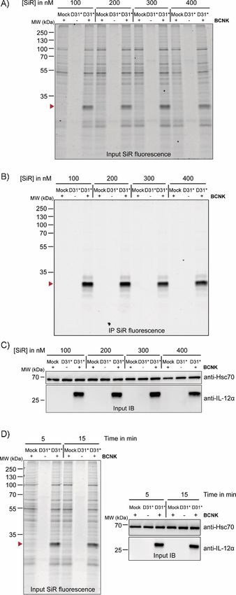
Full Papers
ChemBioChem                                   doi.org/10.1002/cbic.201900651

  tase (PylRS/tRNACUA) pairs. Rapid iEDDAC with tetrazine fluoro-           On the basis of these findings, we next assessed wild-type
  phore conjugates allows the site-specific and selective labeling       (wt)-like behavior of the BCNK-bearing IL-12a variants in terms
  of proteins in living cells with a diverse range of fluoro-            of ER quality control. IL-12a is retained in cells in isolation and
  phores,[16, 17] and has been used for imaging of cell-surface and      forms incorrect disulfide bonds. Only the co-expression of IL-
  intracellular proteins,[19–23] as well as to control enzyme activi-    12b induces correct folding.[30] IL-12a D31BCNK showed wt be-
  ties in cells.[24]                                                     havior and was retained in cells in isolation and only secreted
     Here we have made use of this potential and developed a             efficiently upon co-expression of IL-12b, including modification
  fluorescence-based assay for monitoring protein turnover in            of its glycan residues in the Golgi,[30] thus implying that ERQC
  mammalian cells. We have site-specifically incorporated                works properly on the modified protein (Figure 1 D). The same
  BCNK[16, 18] into low-abundance mammalian proteins by using            behavior was observed for IL-12a Q57BCNK, thus corroborat-
  an efficient PylRS variant, which allows for fast and selective        ing the minimally invasive character of BCNK incorporation
  protein labeling with tetrazine-fluorophore conjugates. This ex-       (Figure 1 E). By using interleukin-responsive reporter cell lines
  tends the arsenal of methods that have been developed in               we were furthermore able to show that IL-12 containing BCNK
  recent years to monitor protein turnover in cells[25–27] by a fluo-    was also biologically active, even when fluorophore was
  rescence-based approach that relies on rapid bio-orthogonal            added, thus implying proper folding in the presence of BCNK
  reactions. As model substrates, we have used members of the            and fluorophore (Figure S1 C).
  human interleukin 12 (IL-12) family.[28, 29] Interleukins are key         Lastly, by extending our approach to IL-23, another IL-12
  signaling molecules in our immune system and, as such, un-             family member prominently involved in autoimmune reactions
  derstanding of their biogenesis and degradation is of great in-        and tumor development,[29, 35, 36] we found that BCNK could also
  terest. Like other secreted proteins, interleukins are produced        be incorporated site-specifically into this protein with preserva-
  in the ER, each IL-12 family member being a heterodimeric              tion of normal ERQC31 and biological activity (Figure S1 D–G).
  protein made up of an a and a b subunit.[28, 29] It has recently
  been shown that both IL-12a and IL-23a fail to fold in the ab-
                                                                         Rapid bio-orthogonal fluorescence labeling of interleukin
  sence of their b subunits and are thus rapidly degraded,[30–32]
                                                                         subunits
  providing native clients of ER quality control. Our data show
  that incorporation of BCNK and site-specific fluorophore label-        Having established efficient incorporation of BCNK into IL-12a
  ing of IL-12a and IL-23a preserves their normal biogenesis pro-        and IL-23a with preservation of wt-like behavior, we proceed-
  cesses and provides an ideal tool with which to monitor pro-           ed to fluorescent labeling of BCNK-modified proteins. We fo-
  tein turnover in mammalian cells.                                      cused on IL-12a for a detailed characterization. Our first goal
                                                                         was to establish optimal labeling conditions. By using either
                                                                         tetramethylrhodamine-tetrazine (TAMRA-tetrazine, Figure S2A–
                                                                         F) or silicon rhodamine-tetrazine (SiR-tetrazine, Figure 2 A–D)
  Results and Discussion                                                 we could efficiently label IL-12a containing BCNK in living cells
                                                                         at fluorophore concentrations from 100–400 nm (Figure 2 A–C)
  Unnatural amino acids can be incorporated into interleukin
                                                                         even after as little as 5 min (Figure 2 D), with increasing label-
  subunits while maintaining their wild-type behavior
                                                                         ing efficiencies being observed after longer labeling times in
  A major advantage of genetic code expansion is that it offers          the case of TAMRA-tetrazine (Figure S2D-F) and very rapid la-
  the capability to label any position within a polypeptide se-          beling in that of SiR-tetrazine (Figure S2, G and H).
  quence with only minimal modifications to the protein under
  scrutiny. Here we have used a selective and highly efficient
                                                                         Bio-orthogonal fluorescence labeling allows protein turn-
  PylRS variant (Y271G, C313V, Methanosarcina barkeri number-
                                                                         over to be monitored
  ing, dubbed BCNKRS, Figure S1 A in the Supporting Informa-
  tion)[33] for expression of low-abundance interleukins bearing         Taken together, these characteristics (wt-like behavior com-
  BCNK at defined positions. Beginning with the a subunit of IL-         bined with rapid and efficient labeling) provided us with an ex-
  12 (IL-12a, Figure 1 A), we searched for a suitable position at        cellent tool with which to monitor protein turnover. To this
  which to introduce BCNK (Figure 1 B). Criteria were complete           end, we first labeled IL-12a containing BCNK at position 31 or
  solvent-exposure in the native heterodimeric IL-12 structure as        57 with SiR-tetrazine or BODIPY-tetramethylrodamine-tetrazine
  well as no predicted interference with IL-12b heterodimeriza-          (B-TMR-tetrazine), respectively, for 15 min. Subsequently, we
  tion.[34] Asp31 (D31) and Gln57 (Q57) fulfilled these criteria (Fig-   washed out the fluorophore and added an excess of free BCNK
  ure 1 A). By using BCNKRS and BCNK we could achieve signifi-           to quench all tetrazine fluorophore and at the same time main-
  cant expression for the IL-12a constructs in which D31 or Q57          tain translation of the target protein IL-12a. Hence, this ap-
  were replaced by an amber stop codon (D31* or Q57*, respec-            proach should allow for observation of selective degradation
  tively). Expression did not vary significantly between 0.1 and         of the fluorescently labeled protein pool without disturbing
  1 mm BCNK (Figure S1B). At 0.25 mm BCNK, expression levels             global translation in the cell (schematic in Figure 3 A, B). Be-
  of IL-12a D31BCNK were still (34 : 4) % (mean : SEM) and for           cause an UAA was used instead of radioactive amino acids to
  IL-12a Q57BCNK (54 : 6) % (mean : SEM) of those observed for           follow protein degradation in classical pulse-chase experi-
  the wild-type (wt) protein (Figure 1 C).                               ments, we termed this approach uChase. As expected, we ob-

  ChemBioChem 2020, 21, 1861 – 1867    www.chembiochem.org           1862   T 2020 The Authors. Published by Wiley-VCH Verlag GmbH & Co. KGaA, Weinheim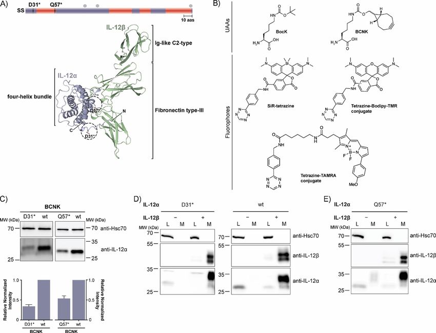
Full Papers
ChemBioChem                                         doi.org/10.1002/cbic.201900651

  Figure 1. Noncanonical amino acid incorporation into IL-12 in mammalian cells. A) Top: Schematic representation of the IL-12a secondary structure (SS = sig-
  nal sequence, red = a-helices, mauve = loops, hexagons = predicted glycosylation sites, 10 aas = 10 amino acids). Positions of amber codons (D31 and Q57)
  are indicated. Bottom: PDB structure of heterodimeric IL-12 (PDB ID: 3HMX. Missing residues were modeled); N and C termini are indicated for IL-12a. The po-
  sitions selected for UAA incorporation into IL-12a are marked with dashed circles. B) Unnatural amino acids (UAAs) and fluorophores used in this study.
  C) Top: Immunoblots of HEK-293T cells expressing the BCNKRS/PylT pair and IL-12aD31TAG or IL-12aQ57TAG (abbreviated as D31* or Q57*, respectively) in the pres-
  ence of BCNK. Bottom: Expression of IL-12a D31BCNK and IL-12a Q57BCNK relative to wild-type IL-12a was quantified from N = 6 (D31BCNK) or N = 3
  (Q57BCNK) immunoblots (mean : SEM), respectively. Samples were normalized for Hsc70 levels. D) IL-12a D31BCNK was tested for assembly-induced secretion
  upon co-expression of IL-12b. HEK293T cells were co-transfected with the indicated constructs in the presence of BCNK and samples were analyzed by immu-
  noblotting. An increase in molecular weight upon secretion can be attributed to modification of IL-12a glycans in the Golgi and IL-12b populates two differ-
  ent glycospecies.[30] L = lysate, M = medium, wt = wild-type control. E) As in (D) but for IL-12a Q57BCNK.

  served decreasing IL-12a fluorescence for D31BCNK and                            of BCNK during these steps and thus lead to degradation of a
  Q57BCNK in uChase experiments (Figure 3 C, D top). Notably,                      small amount of IL-12a before the BCNK excess is added and
  this analysis was based on whole-cell lysates, and thus does                     translation from Amber codon-containing transcripts can
  not depend on any downstream enrichment steps and also                           resume. Consistent with previous studies,[30, 32] the half-life for
  works for interleukins as proteins expressed at low levels (Fig-                 IL-12a degradation determined by our new uChase assay was
  ure S3 A–D). Furthermore, the assay was compatible with use                      (2.5 : 0.3) h (with use of D31* and SiR) and (2.2 : 0.2) h (with
  either of SiR-tetrazine (Figure 3 C) or of B-TMR-tetrazine (Fig-                 use of Q57* and B-TMR; Figure 3 C, D, bottom), thus showing
  ure 3 D) as fluorophore-tetrazine conjugates to label IL-12a.                    that labeling position and choice of fluorophore did not signifi-
  Starting from steady-state, immunoblots revealed that the                        cantly affect the outcome of our experiment. Furthermore, nei-
  overall level of IL-12a essentially remained constant over the                   ther BCNK incorporation nor SiR labeling (Figure S4 A and B)
  time of the chase, as initially hypothesized (Figure 3 C, D,                     significantly changed the half-life of IL-12a in cycloheximide
  middle). The slight increase in protein levels can likely be at-                 (CHX) chases, underscoring the minimally invasive character of
  tributed to the necessary washing steps before labeling, which                   our approach and establishing uChase as a way to monitor
  were performed in the absence of BCNK. This will deplete cells                   protein degradation. Two assays were performed to confirm

  ChemBioChem 2020, 21, 1861 – 1867         www.chembiochem.org               1863     T 2020 The Authors. Published by Wiley-VCH Verlag GmbH & Co. KGaA, Weinheim
Full Papers
ChemBioChem doi.org/10.1002/cbic.201900651
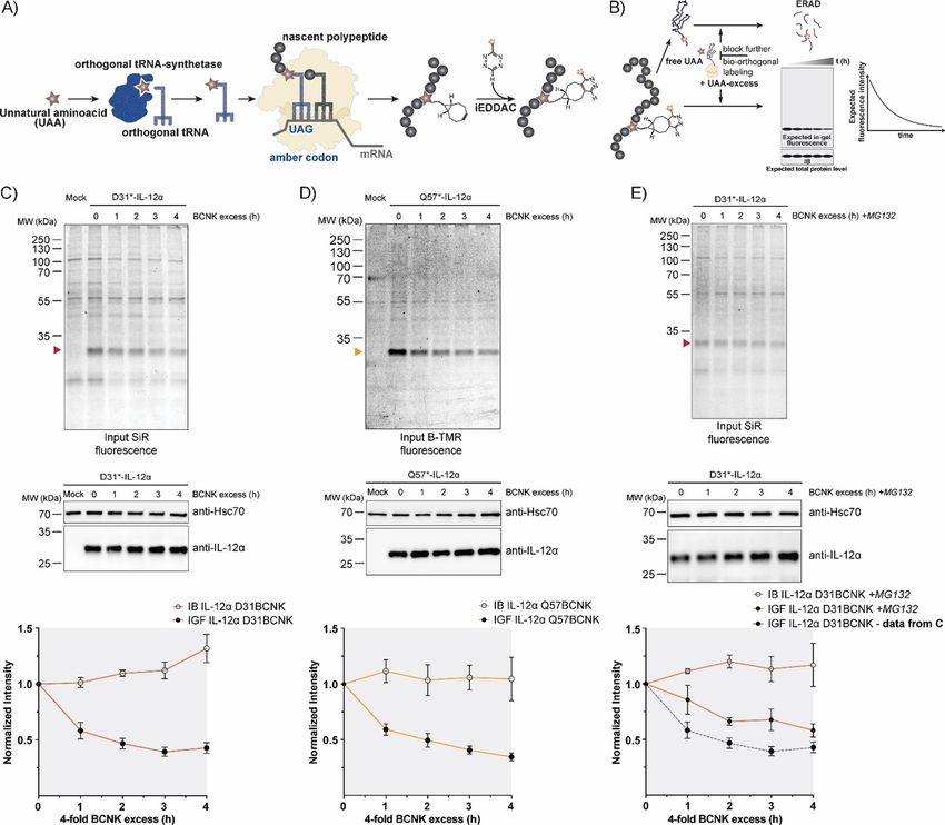
Figure 2. Bio-orthogonal fluorescent labeling of BCNK-modified IL-12a is fast
and efficient. A)–C) Cells expressing IL-12a D31* constructs at 0.25 mm
BCNK were treated with the indicated concentrations of SiR-tetrazine fluoro-
phore for 15 min and analyzed by A) in-gel fluorescence of lysates (IL-12a
D31BCNK is marked with a red arrowhead), B) after immunoprecipitation
(IP), and C) by immunoblotting (IB). The + and @ lanes refer to the presence
or absence of BCNK. SiR-tetrazine fluorophore was present in all samples.
D) Left: Representative gel for a reaction time course between 400 nm SiR-
tetrazine and IL-12a D31BCNK analyzed by in-gel fluorescence. Right: Cell ly-
sates were additionally analyzed by immunoblotting.
(Figure 3 E). As a further test we used a dominant negative
mutant of the ERAD E3 ubiquitin ligase Hrd1 (Hrd1C[291]S).[37]
This mutant decelerated degradation of IL-12a in CHX chases
(Figure S4 C) and to a similar extent degradation of B-TMR-la-
beled IL-12a Q57BCNK in uChase assays (Figure S4 D and E),
thus further validating our assay as a tool with which to moni-
tor ERAD.
To demonstrate a more general applicability of our approach
we additionally analyzed IL-23a L150BCNK. For this protein,
degradation occurred with a half-life of less than 2 h, and was
again not significantly affected by incorporation of BCNK (Fig-
ure S4 F). When IL-23a L150BCNK was labeled with B-TMR, our
uChase approach again yielded a half-life similar to those de-
termined by CHX chases[31] (Figure S4 F and G), thus showing
that our approach can also be extended to other proteins with
more rapid cellular turnover.
Development of a fluorescence-based pulse-chase assay
So far, our approach was similar to classical CHX chases be-
cause we started from a steady-state pool of proteins, and
then monitored their degradation, yet without globally inhibit-
ing translation. Ideally, as possible with radioactive labeling[10]
and recently developed techniques for cytoplasmic proteins,[13]
one would like to label the protein of interest produced within
a defined time interval, and then monitor its cellular fate. This,
for example, would allow analysis of transport processes and
post-translational modifications such as changes in glycosyla-
tion with temporal resolution.[38] To assess if such an approach
(Figure 4 A) was in reach with our setup, we first analyzed how
long BCNK needed to be added to cells in order for expression
of IL-12a Q57BCNK to be observed. Even after as little as
30 min we observed sufficient expression of IL-12a Q57BCNK
for detection by immunoblotting (Figure 4 B, C). Addition of
BCNK for a 1 h pulse, labeling with B-TMR for 15 min, and sub-
sequent addition of a BCNK excess (chase) allowed us to moni-
tor protein degradation of IL-12a Q57BCNK produced within
this 1 h time window (Figure 4 A, D, E).
In these experiments, a half-life of (1.1 : 0.1) h (from input
samples) was observed for IL-12a Q57BCNK. This might sug-
gest that on a molecular level an IL-12a pool produced within
only a 1 h pulse period differs to some extent from the steady-
state pool. It should be noted that IL-12a forms different redox
that our assay monitored ERAD. Degradation of IL-12a in our species in cells, including homodimers.[30] Their formation has
uChase assay was mediated by the proteasome as expected for not been kinetically analyzed yet but might impact degrada-
an ERAD substrate, because it could be inhibited by MG132 tion and give rise to this behavior. This further highlights the
ChemBioChem 2020, 21, 1861 – 1867 www.chembiochem.org 1864 T 2020 The Authors. Published by Wiley-VCH Verlag GmbH & Co. KGaA, Weinheim
Full Papers
ChemBioChem                                         doi.org/10.1002/cbic.201900651

  Figure 3. Protein turnover monitored by the uChase approach. A) Workflow of in cellulo bio-orthogonal labeling. During translation, an in-frame TAG stop
  codon is suppressed by an evolved orthogonal tRNA-synthetase/tRNA pair carrying the UAA. Next, the UAA can be labeled chemoselectively with a suitable
  probe, such as a fluorophore. B) uChase as a tool for monitoring protein degradation. To monitor protein removal rates (e.g., through ERAD), free UAA is
  washed out before the labeling reaction. Then, cells are treated with an excess of free UAA for a desired time interval to block any further bio-orthogonal la-
  beling of newly synthesized proteins without altering the protein biogenesis machinery. The protein half-life can be measured from intensity plots quantified
  from in-gel fluorescence (IGF) intensities of cell lysates. Total protein levels, as measured by immunoblotting (IB), are expected to remain constant. C) Top:
  Time course of fluorescence decrease for labeled IL-12a D31BCNK; 0.25 mm BCNK and 400 nm SiR-tetrazine fluorophore (15 min labeling) were used, followed
  by incubation with a fourfold excess of BCNK for the indicated time points and analyzed by in-gel fluorescence. Middle: Total protein levels of IL-12a
  D31BCNK were analyzed by immunoblotting. Bottom: The graph shows quantifications from in-gel fluorescence and immunoblotting data, N = 5 (mean :
  SEM). The intensity of the 0 h time point was set to 1. IB: immunoblotting. IGF: in-gel fluorescence. D) The same analyses as shown in (C), but for the IL-12a
  Q57BCNK variant labeled with B-TMR [N = 4 (mean : SEM)]. E) IL-12a D31BCNK degradation was inhibited by the proteasomal inhibitor MG132 and the effect
  was analyzed by use of the uChase assay as in (C), N = 4 (mean : SEM). A dotted gray line (bottom panel) shows IGF data from (C), (bottom panel) as a refer-
  ence.

  importance of pulse-chase types of approaches. Because we                        the synthesis of new, nonfluorescent IL-12a Q57BCNK mole-
  were starting from a small pool of IL-12a Q57BCNK (Figure 4 B,                   cules (Figure 4 F). Taken together, these data show that a
  C), the decrease in the pool of fluorescently labeled protein                    pulse-chase approach is also possible with uChase, which
  (Figure 4 D, E) was accompanied by an increase in overall IL-                    opens up further future fluorescence-based applications.
  12a Q57BCNK levels during the 4 h chase. This is due to the
  fact that BCNK was present during the chase that allows for

  ChemBioChem 2020, 21, 1861 – 1867         www.chembiochem.org               1865     T 2020 The Authors. Published by Wiley-VCH Verlag GmbH & Co. KGaA, Weinheim
Full Papers
ChemBioChem doi.org/10.1002/cbic.201900651
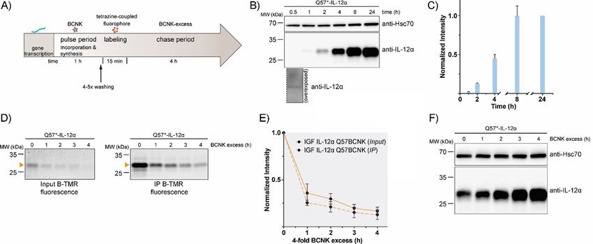
Figure 4. Establishment of a uChase-based pulse-chase assay. A) Illustration of a uPulse-chase experiment. BCNK addition for a defined time interval (pulse
period) allows protein synthesis from mRNA present to occur. After washing out of free BCNK and brief labeling with a tetrazine-coupled fluorophore, a chase
period follows (as described Figure 3 B). B) Representative immunoblots showing expression levels of IL-12a Q57BCNK after different time intervals of the
0.25 mm BCNK pulse. An overexposure for the 30 min time point is shown below the blot. C) The graph shows a quantification from immunoblots; N = 4
(mean : SEM). Samples were normalized for Hsc70 levels. The intensity of the 24 h time point was set to 1. D)–F) Following the schematic in (A), IL-12a
Q57BCNK degradation was followed by a uPulse-chase experiment. D) Either whole-cell lysates (Input, left) or immunoprecipitations to increase signal/noise
(IP, right) are shown. E) Quantifications of (D); N = 4 (mean : SEM). F) Immunoblots of IL-12a Q57BCNK expression after different time points of the BCNK-
excess-induced chase. The same samples as in (D), Input, were used, showing a decrease in the fluorescently labeled IL-12a Q57BCNK pool and an increase in
non-labeled protein levels.
Conclusions important factors become limiting. We show that the tech-
nique we have established also provides the basis for more
In this study, we have established a minimally invasive ap- complex fluorescence-based pulse-chase assays. Furthermore,
proach to monitor protein turnover in cells. To this end we in- because we were able to confirm receptor binding of our
corporated the tetrazine-reactive amino acid BCNK into differ- modified interleukins, fluorescently modified interleukins might
ent positions of the two human interleukins IL-12 and IL-23. provide a valuable tool for assessing the characteristics of their
BCNK incorporation was completely compatible with wt-like receptor engagement and/or to include other chemically reac-
folding, assembly, cellular quality control, and function of the tive probes to modulate their functions selectively and thus go
interleukins. These characteristics allowed us to establish beyond being a tool for measuring protein turnover.
uChase as a new assay for monitoring protein degradation in
mammalian cells. Our approach does not depend on any tag
or other significant modifications of the protein of interest. It is
Experimental Section
furthermore compatible with different proteins even at very
low expression levels, allows rapid degradation processes to See the Supporting Information for details on the materials and ex-
be monitored, and does not depend on any downstream en- perimental procedures used, synthesis of small-molecule com-
pounds, and additional figures.
richment processes (e.g., immunoprecipitation, in which a spe-
cific antibody is needed). Furthermore, no global inhibition of
translation is needed as in CHX chases; this can have pleiotrop-
ic effects and lead, for example, to the unwanted degradation Acknowledgements
of ERAD factors relevant for the protein under scrutiny. It
should be noted that due to the low expression levels of inter- Y.G.M. gratefully acknowledges funding from a DAAD PhD schol-
leukins, background labeling is present; for proteins expressed arship. K.L. and M.J.F. are Rudolf Mçßbauer Tenure Track Profes-
at higher levels this was mostly absent (Figure S3). Although sors and as such gratefully acknowledge funding through the
we monitor protein turnover through in-gel fluorescence, we Marie Curie COFUND program and the Technical University of
envision that our approach might be amenable to future mi- Munich Institute for Advanced Study, funded by the German Ex-
croscopy/FACS-based live-cell assays to monitor protein degra- cellence Initiative and the European Union Seventh Framework
dation directly in living cells when expression is sufficiently Program under Grant Agreement 291 763. This work was per-
high. This would be a major advantage over currently available formed within the framework of SFB 1035 (German Research
approaches. Use of overexpressed proteins, however, of course Foundation DFG, Sonderforschungsbereich 1035, projects B10
always comes with the caveat of possibly altered turnover if and B11).
ChemBioChem 2020, 21, 1861 – 1867 www.chembiochem.org 1866 T 2020 The Authors. Published by Wiley-VCH Verlag GmbH & Co. KGaA, WeinheimFull Papers
ChemBioChem doi.org/10.1002/cbic.201900651
Conflict of Interest [22] T. Schvartz, N. Aloush, I. Goliand, I. Segal, D. Nachmias, E. Arbely, N. Elia,
Mol. Biol. Cell 2017, 28, 2747 – 2756.
[23] R. Serfling, L. Seidel, A. Bock, M. J. Lohse, P. Annibale, I. Coin, ACS Chem.
The authors declare no conflict of interest. Biol. 2019, 14, 1141 – 1149.
[24] Y. H. Tsai, S. Essig, J. R. James, K. Lang, J. W. Chin, Nat. Chem. 2015, 7,
554 – 561.
Keywords: bio-orthogonal reactions · fluorescent probes · [25] D. C. Dieterich, A. J. Link, J. Graumann, D. A. Tirrell, E. M. Schuman, Proc.
genetic code expansion · interleukins · protein folding Natl. Acad. Sci. USA 2006, 103, 9482 – 9487.
[26] P. Wu, M.-X. Lu, X.-t. Cui, H.-Q. Yang, S.-l. Yu, J.-b. Zhu, X.-l. Sun, B. Lu,
[1] A. R. Fersht, V. Daggett, Cell 2002, 108, 573 – 582. Acta Pharmacol. Sin. 2016, 37, 1307.
[2] F. U. Hartl, M. Hayer-Hartl, Nat. Struct. Mol. Biol. 2009, 16, 574 – 581. [27] I. Fierro-Monti, J. Racle, C. Hernandez, P. Waridel, V. Hatzimanikatis, M.
[3] L. M. Luheshi, D. C. Crowther, C. M. Dobson, Curr. Opin. Chem. Biol. Quadroni, PLoS One 2013, 8, e80423.
2008, 12, 25 – 31. [28] D. A. Vignali, V. K. Kuchroo, Nat. Immunol. 2012, 13, 722 – 728.
[4] B. M. Adams, M. E. Oster, D. N. Hebert, Protein J. 2019, 38, 317 – 329. [29] E. D. Tait Wojno, C. A. Hunter, J. S. Stumhofer, Immunity 2019, 50, 851 –
[5] L. Ellgaard, N. McCaul, A. Chatsisvili, I. Braakman, Traffic 2016, 17, 615 – 870.
638. [30] S. Reitberger, P. Haimerl, I. Aschenbrenner, J. Esser von Bieren, M. J.
[6] I. Braakman, N. J. Bulleid, Annu. Rev. Biochem. 2011, 80, 71 – 99. Feige, J. Biol. Chem. 2017, 292, 8073 – 8081.
[7] S. S. Vembar, J. L. Brodsky, Nat. Rev. Mol. Cell Biol. 2008, 9, 944 – 957. [31] S. Meier, S. Bohnacker, C. J. Klose, A. Lopez, C. A. Choe, P. W. N. Schmid,
[8] M. H. Smith, H. L. Ploegh, J. S. Weissman, Science 2011, 334, 1086 – 1090. N. Bloemeke, F. Ruhrnossl, M. Haslbeck, J. E. Bieren, M. Sattler, P. S.
[9] C. J. Guerriero, J. L. Brodsky, Physiol. Rev. 2012, 92, 537 – 576. Huang, M. J. Feige, Nat. Commun. 2019, 10, 4121.
[10] E. Simon, D. Kornitzer, Methods Enzymol. 2014, 536, 65 – 75. [32] R. Jalah, M. Rosati, B. Ganneru, G. R. Pilkington, A. Valentin, V. Kulkarni,
[11] A. Khmelinskii, M. Meurer, C. T. Ho, B. Besenbeck, J. Fuller, M. K. Lem- C. Bergamaschi, B. Chowdhury, G. M. Zhang, R. K. Beach, C. Alicea, K. E.
berg, B. Bukau, A. Mogk, M. Knop, Mol. Biol. Cell 2016, 27, 360 – 370. Broderick, N. Y. Sardesai, G. N. Pavlakis, B. K. Felber, J. Biol. Chem. 2013,
[12] A. B. Alber, E. R. Paquet, M. Biserni, F. Naef, D. M. Suter, Mol. Cell 2018, 288, 6763 – 6776.
71, 1079 – 1091. [33] S. V. Mayer, A. Murnauer, M.-K. von Wrisberg, M.-L. Jokisch, K. Lang,
[13] N. Schneider, C. Gabelein, J. Wiener, T. Georgiev, N. Gobet, W. Weber, M. Angew. Chem. Int. Ed. 2019, 58, 15876 – 15882; Angew. Chem. 2019, 131,
Meier, ACS Chem. Biol. 2018, 13, 3049 – 3053. 16023 – 16029.
[14] S. Mayer, K. Lang, Synthesis 2017, 49, 830 – 848. [34] C. Yoon, S. C. Johnston, J. Tang, M. Stahl, J. F. Tobin, W. S. Somers, EMBO
[15] B. L. Oliveira, Z. Guo, G. J. L. Bernardes, Chem. Soc. Rev. 2017, 46, 4895 – J. 2000, 19, 3530 – 3541.
4950. [35] B. Oppmann, R. Lesley, B. Blom, J. C. Timans, Y. Xu, B. Hunte, F. Vega, N.
[16] K. Lang, L. Davis, S. Wallace, M. Mahesh, D. J. Cox, M. L. Blackman, J. M. Yu, J. Wang, K. Singh, F. Zonin, E. Vaisberg, T. Churakova, M. Liu, D.
Fox, J. W. Chin, J. Am. Chem. Soc. 2012, 134, 10317 – 10320. Gorman, J. Wagner, S. Zurawski, Y. Liu, J. S. Abrams, K. W. Moore, D. Ren-
[17] T. Plass, S. Milles, C. Koehler, J. Szymanski, R. Mueller, M. Wiessler, C. nick, R. de Waal-Malefyt, C. Hannum, J. F. Bazan, R. A. Kastelein, Immuni-
Schultz, E. A. Lemke, Angew. Chem. Int. Ed. 2012, 51, 4166 – 4170; ty 2000, 13, 715 – 725.
Angew. Chem. 2012, 124, 4242 – 4246. [36] P. J. Lupardus, K. C. Garcia, J. Mol. Biol. 2008, 382, 931 – 941.
[18] A. Borrmann, S. Milles, T. Plass, J. Dommerholt, J. M. Verkade, M. Wiess- [37] E. Nadav, A. Shmueli, H. Barr, H. Gonen, A. Ciechanover, Y. Reiss, Bio-
ler, C. Schultz, J. C. van Hest, F. L. van Delft, E. A. Lemke, ChemBioChem chem. Biophys. Res. Commun. 2003, 303, 91 – 97.
2012, 13, 2094 – 2099. [38] I. Braakman, L. Lamriben, G. van Zadelhoff, D. N. Hebert, Curr. Protoc.
[19] I. Nikic, T. Plass, O. Schraidt, J. Szymanski, J. A. Briggs, C. Schultz, E. A. Protein Sci. 2017, 90, 14.1.1 – 14.1.21.
Lemke, Angew. Chem. Int. Ed. 2014, 53, 2245 – 2249; Angew. Chem. 2014,
126, 2278 – 2282.
Manuscript received: October 27, 2019
[20] C. Uttamapinant, J. D. Howe, K. Lang, V. Beranek, L. Davis, M. Mahesh,
Revised manuscript received: January 28, 2020
N. P. Barry, J. W. Chin, J. Am. Chem. Soc. 2015, 137, 4602 – 4605.
[21] N. Aloush, T. Schvartz, A. I. Konig, S. Cohen, E. Brozgol, B. Tam, D. Nach- Accepted manuscript online: February 3, 2020
mias, O. Ben-David, Y. Garini, N. Elia, E. Arbely, Sci. Rep. 2018, 8, 14527. Version of record online: March 9, 2020
ChemBioChem 2020, 21, 1861 – 1867 www.chembiochem.org 1867 T 2020 The Authors. Published by Wiley-VCH Verlag GmbH & Co. KGaA, WeinheimYou can also read